A Dissertation on
TO EVALUATE THE DIAGNOSTIC EFFICACY OF
CORE NEEDLE BIOPSY IN DISTINGUISHING
PHYLLODES TUMOUR AND FIBROADENOMA IN
FIBROEPITHELIAL BREAST LESIONS
Dissertation Submitted to
THE TAMILNADU Dr.M.G.R. MEDICAL UNIVERSITY CHENNAI - 600 032
with partial fulfilment of the regulations for the award of the degree of M.S. GENERAL SURGERY
(BRANCH I))
COIMBATORE MEDICAL COLLEGE, COIMBATORE
DECLARATION
The dissertation titled “TO EVALUATE THE DIAGNOSTIC EFFICACY OF CORE NEEDLE BIOPSY IN DISTINGUISHING PHYLLODES TUMOUR AND FIBROADENOMA IN FIBROEPITHELIAL BREAST LESIONS” is being submitted by me to "The Tamil Nadu Dr. M.G.R. Medical University in partial fulfilment
of the regulation for the completion of the M.S. General Surgery Degree
Examination to be held in 2019.
This work has been carried out in the Department of General
Surgery, Coimbatore Medical College and Hospital, Coimbatore, under
the guidance of Dr.T.SRINIVASAN.MS, Professor of General Surgery, Coimbatore Medical College and Hospital, Coimbatore.
Date : Dr.Raja.R
CERTIFICATE
I, hereby declare that the dissertation entitled “TO EVALUATE THE DIAGNOSTIC EFFICACY OF CORE NEEDLE BIOPSY IN
DISTINGUISHING PHYLLODES TUMOUR AND
FIBROADENOMA IN FIBROEPITHELIAL BREAST LESIONS” is the bonafide research work done by Dr.R. RAJA and submitted in partial fulfilment of the requirement of the degree of Master of Surgery in General
Surgery, Coimbatore Medical College and Hospital, Coimbatore.
Date : Unit Chief
Date : Professor &HoD
Department of General Surgery
Date : Dean
CERTIFICATE-II
This is to certify that dissertation entitled “TO EVALUATE THE DIAGNOSTIC EFFICACY OF CORE NEEDLE BIOPSY IN
DISTINGUISHING PHYLLODES TUMOUR AND
FIBROADENOMA IN FIBROEPITHELIAL BREAST LESIONS” of the candidate Dr.R. RAJA with registration Number 221611311 for the ward of M.S in the Branch of General Surgery, I personally verified the
urkund.com website for the purpose of plagiarism Check. I found that the
uploaded thesis file contains 86 pages from introduction to conclusion and the
result shows 2% (Two) percentage of plagiarism in the dissertation.
ACKNOWLEDGEMENT
My sincere thanks to Prof.Dr.Asokan.MS.,MCH, DEAN,
Coimbatore medical and hospital for allowing me to conduct this study in
the Department of General Surgery, Coimbatore Medical and Hospital.
I am extremely grateful to Dr.P.Elango.MS, Professor and Head of the Department of General Surgery, Coimbatore Medical College and
Hospital for his encouragement and permission in granting unrestricted
access to utilizing the resources of the department.
I thank my mentor and guide Dr.T.Srinivasan.Ms, Professor of General Surgery, Coimbatore Medical College and Hospital for his
valuable guidance during the tenure of my course.
I thank my professors Dr.Nirmala.Ms, Dr.Ganesh Babu.Ms, Dr.Lakshmi Narayani. Ms for their immense guidance.
I thank my professor Dr.Lalitha, Head of the Department of Pathology. Dr.Muralidharan, Head of the Department of Radiology for their support and guidance.
I also acknowledge my assistant professor and co-author
I also acknowledge my assistant professor Dr.A.M.Umashankar DMRD,MS, Dr.T.Gunalasuresh.Ms for her valuable support and timely help rendered to complete this study.
I thank my colleagues and friends, who helped me throughout my
study.
I would like to thank the entire medical and paramedical staff of
the Department of General Surgery, Department Of Pathology,
Department Of Radiology.
My utmost thanks to all my patients who co-operated to complete
my dissertation, without their help it would have been impossible for me
to complete this study.
I thank Dr.Nivethitha for her immense help and support.
ABBREVIATIONS
FNAC - Fine needle aspiration cytology
USG - Ultrasonography
CSP - Cystosarcomaphyllodes
FCD - Fibro cystic disease
CNB - Core needle biopsy
BIRADS - Breast imaging reporting and data system
PT - Phyllodes tumour
TABLE OF CONTENTS
SL.NO TITLES PAGE.NO
1 INTRODUCTION 1
2 AIMS AND OBJECTIVES 3
3 REVIEW OF LITERATURE 4
4 METHODOLOGY 60
5 RESULTS 62
6 CONCLUSION 86
7 BIBLIOGRAPHY 87
8 ANNEXURES
PROFORMA 91
CONSENT FORM 92
LIST OF TABLES
SL.NO TABLES PAGE NO
1 SITE OF OCCURRENCE OF TUMOUR 63
2 DURATION OF THE PRESENTATION OF LUMP
64
3 AGE OF PRESENTATION 65
4 SIZE OF THE LESION 66
5 MAMMOGRAPHIC FINDINGS 68
6 USG FINDINGS 70
7 FNAC FINDINGS 72
8 CORE NEEDLE BIOPSY FINDINGS 74
9 EPITHELIAL FRAGMENTS 75
10 CELLULARITY 76
11 SPINDLE CELLS 77
12 MITOTIC FIGURES 78
1
INTRODUCTION
Fibro epithelial breast lesions are the most common breast abnormality in adolescent females referred to as fetal fibroadenoma, cellular fibroadenoma, juvenile fibroadenoma, giant fibroadenoma, fibroadenoma variant, tubular adenoma, hamartoma , phyllodes tumour.
The aetiology of the fibroepithelial lesions is found to be unclear. These are the benign conditions of the breast but some of them have the potential to turning into malignant.
Among the breast lump benign disease of breast constitutes of 80%.of these 7% are fibroadenoma and phyllodes tumour contributes to less than 1%. Even though the incidence is low, the importance of phyllodes tumour is that it is further divided by WHO into benign borderline and malignant, based on the stromal cellularity of atypical, stromal overgrowth, mitotic figures. So compared with fibroadenoma ,phyllodes tumour has more potential for turning to malignancy.
So the fibroadenoma requires a simple enucleation but for the phyllodes tumour, since it has more malignant potential, a wide excision is needed with a clearance of at least 1 cm.
2
operatively the histopathological examination of the specimen done. If the reports were found to be phyllodes tumour then we have to readmit the patient and post the patient for re-operation, which is the removal of excision biopsy site with clearance of about 1cm.
This increase the morbidity to the patient and period of hospital stay of the patient is also increased. Secondly if the patient has lost follow up, then there is a high chance of reoccurrence of tumour and the patient will again present with breast lump over the excision biopsy site.
To prevent the above mentioned difficulties, we need an investigation, which can pre-operatively distinguish a fibroadenoma and phyllodes tumour.
Many studies have been undergone to differentiate the fibroadenoma and phyllodes tumour based on the clinical features, FNAC features, radiological features of mammography and USG.
3
AIMS AND OBJECTIVES
1. To determine the incidence of fibroepithelial lesion in relation to
age.
2. To analyse and interpret the histological features in core needle
biopsy to diagnose phyllodes tumour.
3. To prevent re-operation and recurrence of tumour.
4. To analyse and interpret core needle biopsy findings with the
findings of clinical examination, mammography,
ultrasonography, fine needle aspiration cytology in diagnosing
4
REVIEW OF LITERATURE
DEVELOPMENT OF BREAST
The breast develops from the mammary ridge or milk line or line of
Schultz, which is an ectodermal thickening.
It develops at around 4th week of intrauterine life.
The milk line extends from the axilla to groin, but in human beings
most of them disappears except in the pectoral region.
The stroma is mesodermal in origin.
From the persisting parts, mammary pit develops; the floor of the
mammary pit gives rise to secondary bud which divides further to form
the lobes of the gland.
The entire system is solid which later gets canalized.
5 ANATOMY OF BREAST
Breast is one of the modified sweat gland.
More precisely apocrine gland.
It is present in the pectoral region. Both male and female found to
have breast, but it is rudimentary in male, and well developed in female.
In the female reproductive system it forms an accessory organ. In
the newborn it provides nutrition in the form of milk. Shape of female
breast is due to the fat contained within the fibrous septa.
Breast lies within the skin and pectoral fascia to which it is loosely
attached.
6
a prolongation of parenchymatous tissue, axillary tail runs upwards
between the pectoralis major and latissimus dorsi muscles to blend with
fat of axilla.
The glandular tissue consists of 15 to 20 lobules (clusters of milk
forming glands, also called as acini) which enters into branching and
interconnected ducts.
Just beneath the nipple the ducts enlarges as lactiferous sinus and
then empty via the nipple opening. The secondary unit is a group of
saccular alveoli draining into duct.
The ducts of the alveoli are lined by single layer of epithelial cells.
The ducts are surrounded by myoepithelial cells which were not found
7
The myoepithelial cells are contractile and helps to more secretion
along the duct system.
BLOOD SUPPLY OF BREAST
ARTERIAL SUPPLY
Breast is one of the extremely vascular organs.
The branches of following arteries supply the mammary gland:
1. Perforating branches of internal thoracic artery which is a branch of
subclavian artery.
2. Perforating branches of posterior intercostals arteries.
3. Lateral thoracic, superior thoracic, acromio thoracic branches of
8 VENOUS SUPPLY OF BREAST:
1. The veins follow the arteries.
2. All veins converge towards the base of the nipple and form an
anastomotic venous circle.
3. From where veins run in superficial and deep veins.
4. Superficial vein drains into internal thoracic vein.
5. Deep vein drains into posterior intercostals, internal thoracic and
axillary vein.
Nerve supply of breast
Mammary gland is supplied by anterior and lateral cutaneous
9
These nerve fibres are autonomic to blood vessels and smooth
muscles and sensory fibres to skin.
Milk secretion is not controlled by these nerves.
Lymphatic of breast drains into following lymph nodes.
Axillary lymph node which includes
Anterior (pectoral group)
Posterior group
Lateral group
Central group
10
1. Internal mammary (para sternal) nodes lies along the internal
thoracic vessels
2. Some of the lymphatics also reach the supra clavicular nodes,
cephalic nodes, posterior intercoastal nodes.
3. The lymphatic s of the breast divided into superficial and deep
lymphatics.
4. Skin of the breast except nipple and areola drains into superficial
lymphatics.
5. Parenchymal of breast, nipple and areola drain into deep
11 HISTOLOGY OF NORMAL BREAST:
1. Breast has
- 2 types of epithelial cells
- 2 types of stroma
- 2 main structures
Epithelial cells:
Luminal cells
Myoepithelialcells
Stroma:
-Interlobular stroma
-Intralobar stroma
Structure:
Large ducts
12 NIPPLE:
Covered by squamous epithelium (pigmented).
Near the nipple orifice majority of nipple have TOKER cells.
Lactiferous sinus have serrated contour admixed with smooth
muscles, collagen and elastic fibers. Basement membrane of skin
is continuous with basement membrane of duct.
AREOLA;
Pilosebaceous unit and hair present only at the periphery.
13 SKIN APPENDAGES:
These are
- Montgomrey tubercles.
- Eccrine sweat glands and ducts.
- Apocrine sweat gland and ducts.
MAMMARIAN STROMA:
Divided into Interlobular stroma and Intralobular stroma.
PHYSIOLOGY OF BREAST
At puberty growth of the mammary gland is caused by oestrogens.
Secondary alveoli are stimulated by the progesterone and prolactin
hormone from hypophysis cerebri
14
2. Congenital and developmental abnormalities
3. Amastia- total lack of breast tissue
4. Athlelia- absence of nipple
5. Polythelia–supernumerary nipples
6. Polymastia – supernumerary breast
7. When polymastia is present in the women, the additional breast
tissue can secrete milk when nipple is present.
GYNAECOMASTIA
Gynecomastia is the growth is glandular tissue in male breast. It is a
benign condition account s for more than 65% of male breast
abnormalities. It is usually unilateral and occurs in Young man. bilateral
gynecomastia is due to systemic cause.
CAUSES OF GYNECOMASTIA
Physiological causes
Neonatal gynecomastia
Pubertal gynecomastia
15 Pathological causes
Anorchia
Klinefelter's syndrome
Bilateral cryptorchidism
Mumps
Irradiation
Hypopituitarism
Isolated gonadotropin deficiency
Endocrine tumours
Alcoholic cirrhosis
16 Treatment of gynecomastia:
For physiological causes reassurance is all what is needed
Stop drugs causing gynecomastia.
Subcutaneous mastectomy in trouble some cases.(Webster’s
procedure)
Liposuction- assisted mastectomy
DUCT ECTASIA
- It is a widening of a ducts
- It is more common in women’s In Their 40s and 50s.
- The most common symptom is nipple discharge which is usually
gray to green in colour.
- Tenderness and redness of nipple and surrounding breast tissue
may also be present.
- Microscopically – the peri ductal elastic tissue is destroyed and
the surrounding tissue is infiltrated with lymphocytes and plasma
17 Treatment
Small volume discharge managed conservatively.
Socially embarrassing discharge is treated by major duct excision.
Galactocele:
Cystically dilated terminal ductules that are filled with milk and
lined by double layer of breast epithelium and myoepithelium.
Classically appears as a painless lump weeks to months after cessation of
breast feeding. It is probably formed by obstruction to a duct in the
purpaerium. The milk retained proximal to the obstruction eventually
18
The common complications of this type of swelling are infection.
The treatment is by surgical excision
Intraductal papilloma
This benign lesions of the lactiferous duct wall occurs centrally
beneath the areola in 75% of cases
They most commonly produce a bloody nipple discharge,
something associated with pain.
19
Intra ductal papilloma s should be treated by excision of a duct as a
wedge resection.
FIBROADENOMA
Fibroadenomas are benign tumours composed of stromal and epithelial
elements. The tumours are commonly seen
In young women.
Fibroadenoma is a common well - circumscribed lesion of the breast &
develop in the breast prior to menopause.
Pericanalicular tumors usually being found below the age of 30 &intracanalicular tumors there after.
Either breast may be affected and multiple & successive tumors may
20
• The preicanalicular tumor forms a firm discrete mass, which is
freely mobile in the breast tissue, hence the name (BREAST MOUSE )
• The intracanalicular tumors tend to be softer and may grow to
such size that there is necrosis of the overlying skin. To such a
condition the terms serocystic disease of bordie OR cystosarcoma phylloides OR Giant fibroadenoma have been given. However despite the implication of malignancy in the later term, the
21 Pathophysiology:
• Fibroadenomas are benign tumours that represent a hyper plastic or
proliferative process in a single terminal ductal unit; their
development is considered to be an aberration of normal
development. The cause is unknown. Approximately 10% of
fibroadenomas disappear each year, and most stop growing after
they are 2-3 cm in size.
• Fibroadenomas may involutes in postmenopausal women, and
coarse calcifications may develop.
• Conversely, the tumours may grow rapidly during pregnancy,
during hormone replacement therapy, or during
22
• Fibroadenoma variants include juvenile fibroadenoma, which
occurs in female adolescents.
• This swelling has been variously regarded as a simple hyperplasia
of epithelial and / or connective tissue elements or as a
composite neoplasm of the breast in which the epithelial &
23 CLINICAL FEATURES:
• On clinical examination, fibroadenomas may be nonpalpable or
palpable, oval, freely mobile, rubbery masses. Their size varies from smaller than 1 cm in diameter to as large as 15 cm in diameter
in the giant forms.
• Most commonly, the tumours are removed surgically when they
are 2-4 cm in diameter. In young women, the tumours are usually
palpable. In older women, the tumours typically appear as a mass
on mammograms, and the tumour may be palpable or nonpalpable.
• The size of fibroadenomas also can vary during the menstrual cycle
and during pregnancy.
• In the postmenopausal period, tumours regress and often develop
24 Types
- Solitary
- Few (< 5 / breast )
- Multiple (> 5 / breast )
- Giant (> 4 / 5 cms) & Juvenile
Natural history
- Majority remain small & static 50% involutes spontaneously - No future risk of malignancy
Investigation
- Breast, fibroadenoma Sonogram. Demonstrates a hypo echoic
mass with smooth partially lobulated margins that is typical of a
fibroadenoma.
- Breast, fibroadenoma, Craniocaudal mammograms obtained 1
year apart demonstrates a newly developing mass in the outer part
25 TREATMENT
Reassurance of the patient Excisional biopsy
The natural history of these lesions has recently been elucidated
and has resulted in a change in management policy.
Over a 2 year period approximately 20% slowly increase in size,
26
With knowledge of this natural history a conservative management
policy can often be adopted.
In those <35 years and with a triple assessment supporting the
diagnosis then observation with regular review is acceptable.
In those > 35 years and in younger patients requesting it, excision
biopsy should be considered.
CYSTOSARCOMA PHYLLODES (CSP)
This is one of the rare conditions, found predominantly as benign
tumour that occurs almost only in the female breast. The name
cystosarcoma phyllodes were derived from the Greek words sarcoma, which means fleshy tumour, and phyllo, means leaf.
Gross appearance shows characteristics of a large, malignant
sarcoma, that takes a leaf like appearance when cross sectioned, and it
shows epithelial cyst like spaces when seen histologically (hence the
name phyllodes).
The most common tumours is benign, so the name is misleading.
27 PATHOPHYSIOLOGY OF CSP
Pathophysiology:
Phyllodes tumour is the common nonepithelial neoplasm that
28
Clinically presents as a sharply demarcated, smooth texture and
freely movable mass. It presents as large tumour, and the average size is
more than 5 cm. Some lesions are also more than 30 cm in size that have
been reported.
TREATMENT: Surgical Care:
The preferred surgery is wide local excision with a rim of normal
tissue of about 1cm of clearance.
If the tumour to the breast ratio is sufficiently high to provide a
satisfactory cosmetic appearance by segmental excision or total
mastectomy, followed by with or without flap reconstruction, is an
alternative.
Radical procedures are usually not warranted. If clinically suspicious
nodes present then lymph node dissection is to be done. Mostly all of
these nodes are reactive and they do not have malignant cells in them.
FIBROCYSTIC DISEASE
- Commonest lesions of female breast are fibrocystic disease.
- Two common descriptions are Cystic lobular hyperplasia &
29
- Cystic hyperplasia is nothing but a variant of normal changes of
the breast that occur with menstruation.
- Usually presents bilaterally.
- It is most painful in the premenstrual period
- Benign condition of breast has varying Incidence, in relation to
age.
- Of which 20% are Menstruating age group.
- Of the premenopausal age group it constitutes 30-50%.
- The other names are mammary dysplasia, Cystic disease, Cyclic
Mastopathy, Cystic Hyperplasia.
Path physiology of fibrocystic disease:
- The cause is mostly unknown Hormonal basis.
- Of the hormones Oestrogen , Progesterone and Prolactin
- Thyroid Methylexanthiones
- The causative was considered to be Oestrogen predominance over
progesterone.
30
- Progesterone levels are reduced to 1/3 normal. So the females
having progesterone deficiency are at a fivefold high risk of
premenopausal breast cancer.
- 70% due to Corpus Luteal Deficiency and Anovulation.
- Pre Menstrual Tension syndrome is risk factors.
Increased in about 1/3 of women most probably due to the
dominance of Oestrogen in pituitary.
Thyroid –
Low levels sensitize epithelium of mammary gland to Prolactin.
Methylexanthiones-High intake of coffee, tea, cold drinks chocolate is a high risk.
Path morphology
Proliferation of the connective and epithelial tissues are stimulated
31
The polymorphism of fibrocystic disease is:
- Fibrosis
- Formation of cyst, proliferation of epithelial cells,
- Atrophy of the lobular-alveoli
- Course of fibrocystic disease
- It represents a clinical problem presents in approximate 30% of
patients.
Predominantly found in,
- Abnormal menstrual cycle
- Nulliparous women
- Spontaneous abortions history
- oral contraceptives nonusers
- early menarche
- Late menopause.
32 There are three phases clinically,
Phase I - Stromal fibrosis which is moderate, just started hardness in the breast tissue and tenderness in premenstrual
breast
Phase II – Increased fibrosis causing increased hardening and tenderness, formation of cyst, nodularity
33
DIAGNOSIS OF FIBROCYSTIC DISEASE
Triple Assessment
Symptoms and Signs
-- Many months to several years of history.
- Ovulating women, multiparous women, and patients using oral
contraceptives rarely presents with fibrocystic disease.
- Tenderness of breast is observed in most of the patients.
- Irregular menses, dysmenorrhoea, menometrorrhagia, or ovarian
cysts are found to be associated in 40% to 60%
Nipple secretion-
Spontaneous discharge or secretion can be expelled from the
nipple. Cytological features are amorphous material (fat, proteins), ductal
cells, erythrocytes, and / or foam cells. Straw yellow, greenish, or bluish
34
TREATMENT OF FIBROCYSTIC DISEASE
MEDICAL Goal-
- Progression should be stopped
- For relieving pain
- To Soften breast tissue
Indicated when-
- FDB not increasing in size
- Nipple discharge not present
- Psychological effect ont present
Surgical-
Indicated
when-- Size is progressively increasing
- Discharges that are Serous / Serosanguineous / bloody.
- disturbed psychology
MEDICAL- Hormones OC pills-
- They are protected from FBD
35 Progestogens-
- Have to be started in the luteal phase for a period of 9-12
months
- Of which 80% get relief and have to restart the therapy in 40%
Danazol-
- One of the most effective therapy
- Basis are the ovarian suppression
- 200-600mg/day is the usual dosage.
Medical-
- This is one of the ineffective modalities
- Diet therapy- Restriction of caffeine
- Diuretics
- Agents that containing iodine.
- Thyroid hormone ,Evening Primrose oil Vitamin E & B6
Dihydroergotamine and Antiprolactin drugs
Surgical treatment-
36
CLINICAL EXAMINATION
The patient is examined in the following posture:
1. Arms by the side of body and patient sitting
2. Arms raised above head
3. Hands over hips alternatively contraction and releasing.
4. Patient leaning forward
37 INSPECTION:
Inspection of the patient in sitting posture arms by the side of her body:
1. Breast
- Position compared with opposite Brest
- Site
- Shape
- Any mass
- Ulcer
2. Skin over the breast
- Dilated veins
- Dimple
- Puckering
- Retraction
- Peau'dOrange
- Nodules
- Ulceration
- Fumigation
3. Nipple
- Presence
- Position
38 - Size
- Shape
- Discharge
4. Areola
- Colour
- Size
- Surface
5. Arm and forearm
- Edema
- Nodules
6. Axilla
7. Supraclavicular fossa
- Inspection of the patient with arms raised above the head:
- Look for
- Peau d' Orange
- Fixity
- Retraction of nipple
- Inspection on leaning forward:
- Fixity to chest wall
- Inspection on Contraction and relaxing pectoralis major:
39 PALPATION:
Palpate systemically from areola, concentrically outwards.
1. Local temperature and tenderness
2. Swelling :
- Number
- Site
- Size
- Shape
- Margins
- Consistency
- Fluctuations
- Tenderness
3. Fixity to skin
4. Fixity to chest wall
5. Intrinsic mobility
Examination of nipple:
1. retraction
2. Crackles
3. Discharge
Examination of axillary lymph node:
40
MAMMOGRAPHY
- Low voltage, high ampere x-rays
- 300 mA and 40ky exposed
- This is one technique of taking x-rays known as seems strahlung
type x-rays
- Delivers radiation of 0 1cGy per study by comparison chest x ray
delivers only 25% of this dose
- Sensitivity increases with the age as the breast tissue becomes less
dense.(used in age more than 40 years)
Two views:
- Craniofacial
41 INDICATION
- Age greater than 50 years
- Age greater than 40 years with risk factors
- Already operated one side
- If we plan for can conservative surgery, then we have to rule out
multimodal involvement.
42
Rounded borders with lobulated appearance in mammography are
more in favour of the phyllodes tumour
43 ULTRASOUND
- It is an adjuvant to mammography
- Many interventional procedures in breast lesions can be done under
its guidance
- It differentiate solid masses from cystic masses
- A linear 7mhz array transducer is used, but transducer up to 10 to
13 MHz can be used most preferably.
Benign Lesions Look Like
- hyper or hypo echogenic
- Shape of ellipsoid
- Well circumscribed margins
44 - Margins are irregular
- Hypoechoic to surrounding tissue
- Posterior acoustic shadow present
45
FINE NEEDLE ASPIRATION CYTOLOGY
INTRODUCTION
FNAC is a technique where a thin bore needle is used to obtain
cells from a lesion and smears done, for cytopathological diagnosis.
This basis of this technique is that the tumor cells are less cohesive
and can be aspirated easily.
Using this technique we can diagnose the breast lumps, thyroid
nodules, liver disease, subcutaneous soft tissue mass, salivary gland
diseases and oral diseases.
This is very helpful in the lesions of oral cavity which is very
vascular, and an open biopsy can cause bleeding which becomes difficult
to control.
FNAC solves these problems, by using a 10 ml Syringe with which
adequate material can easily be obtained, from an intraoral or extra oral
site without any discomfort to the patient. There will be no risk of
bleeding.
A subsequent surgery may not needed in some cases and patient
46
Reports of FNAC can be prepared within 24 hours of sampling. It
gives early, rapid information to the surgeon about the lesion the surgeon
dealing with.
HISTORICAL PERSPECTIVE
Martin and Ellis introduced this technique in 1930 in the United
States, but it never became widespread.
Since the 1950s it has been used extensively in Scandinavia and in
Holland.
Fine Needle for aspiration were first introduced in 1950 in Europe
by Lopez-Cardozo in the Netherlands and Soderstrom in Sweden
Publication of the Zajicek from Karolinska Hospital present in
Stockholm that brought aspiration cytology to the level of international
47 ADVANTAGE
- Simple outpatient technique
- fast diagnosis
- cost effective
- multiple sites can be sampled in the one sitting
- diagnostic accuracy is very high
Many other test such as bacterial culture, immunocytochemistry,
flow cytometry, cytogenetics, polymerase chain reaction,etc. are possible
from FNAC material.
LIMITATIONS
- Tissue architecture is lost.
- We cannot detect Capsular invasion and lymphovascular
invasions.
- In situ versus invasive carcinoma is difficult to differentiate.
48 IN CLINICAL INVESTIGATION
It was first used as a mean for confirming a clinical suspicion of
local recurrence or distant metastasis of known cancer, so that further
surgical intervention may not be needed.
It is also used in Inflammation, infection, degenerative
conditions, in diagnosis and monitoring of graft rejection
transplantation surgery
This can be used as an Alternative to frozen section
Per operative cytology can be done.
Successful reporting of FNAC depends on four fundamental requirements:
- Representative samples from the lesion should be obtained.
- Adequate sample size in terms of cells and other tissue
components should be obtained.
- Correct smearing and processing of sample should be done.
- With correct clinical/radiological information biopsy should be
49 EQUIPMENTS NEEDED:
1. NEEDLES: 22-23 gauge needle.
2. Syringes: 20 ml syringe
3. Pistol handle- as shown below for negative suction:
4. Sterile container: Physiological saline or Hank’s balanced solution as preservative.
5. Slides: clean, dry & free of dirt and grease.
6. Haemocytometer cover slip of 0.4mm gives better control on smearing pressure and a proper spread.
50
8. Stains- to differentiate various structures.
9. Microscopes –for viewing the slides
TECHNIQUE OF FINE NEEDLE ASPIRATION:
There are 2 techniques:
1. FNAC with aspiration
52 Steps:
-Biopsy site should be cleaned by spirit and betadine.
-The swelling is gently palpated and needle is introduced and is
moved in a to and fro fashion. When this act is performed a gentle
negative suction is also created by withdrawing the piston.
-on a slide, the aspirated material is expelled
-The smearing is done, by gently pressure by pressing the upper
slide on the lower one.
-Following this, air is taken into the syringe and needle hub is
reattached to the syringe.
53
- Zajdela introduced this technique in 1987.
- He devised based on the fact that the capillary pressure that has
been generated in a fine needle is sufficient enough to keep the
separated cells inside the lumen of the needle.
CAUSES OF FAILURE TO OBTAIN A REPRESENTATIVE SAMPLE:
- The target tissue has been missed by the needle while taking the
samples.
- Needle has entered the lesion where cystic/necrotic/hemorrhagic
area present which are devoid of diagnostic cells.
- Needle may have entered a dominant benign mass and have
missed a small nearby malignant lesions.
- Fibrotic/desmoplastic tissue yield a scant amount of cells.
SAMPLE PROCESSING:
A clean & dry microscope slide are used in which the sample are
54 SMEARING
DIRECT –
DRY
Dry smearing can be done if the sample is creamy in consistency
and consists of large amount of cells suspended in the background of
small amount of tissue fluid.
WET
Wet smearing is done when there are smaller number of cells which are suspended in fluid or blood.
- Indirect smearing is done: samples obtained are processed by centrifugation.
- Other alternative technique are Millipore nucleopore filtration and
Thin prep technique
FIXATION & STAINING
FNAC uses two techniques:
- Simple Air drying which is followed by staining with a haematological stain such as MAY GRUNWALD- GIEMSA
55
- Alcohol fixation and then followed by staining according to PAP or with H&E.
COMPLICATIONS
- Usually there are no complications
- Some of the complications are Bleedings, hematoma, emphysema
(in lung).
- One of the Rare complication is anaphylactic reaction due to
56
CORE NEEDLE BIOPSY
It is a procedure in which a chunk of tissue is removed using
specialised device, in contrast to fine needle aspiration cytology in which
the individual cells are aspirated with syringe.
- More reliable than cytology.
- Less invasive than surgical biopsy.
- Allows planning of therapy.
Disadvantages are:
- More expensive than cytology.
- More invasive than cytology.
Indication:
1. As a first approach :
- Large lesions clearly malignant at imaging (when future treatment
is neo-adjuvant therapy.
- When there is no palpable mass but micro calcifications are
present.
2. After fine needle aspiration cytology:
57
- When the findings are different between clinical ,radiological and
cytology
- Stereotactic - guided vacuum assisted core biopsy.
- Conventional true –cut biopsy.
Choice of needle:
Core needle biopsy is usually done with 14G needle but
ultrasonograpy guided core needle biopsy can be done with smaller
needle of size 16G/18G.
Advantages of smaller needle:
- They are sharper and can penetrate more easily through firm dense
breast tissue.
- Bleeding risk is minimal.
58 PROCEDURE:
Under aseptic precaution patient in supine position parts painted
and draped, the swelling of interest is palpated and fixed, a small nick is
made using 11 size blade. Using a conventional or self retained gun
59 Processing:
- At least 3 hematocilin and eosin stained sections are cut attained
interval of 50mirons.
- Specimens are radio graphed to look for micro calcification.
- Fibroepithelial lesions are categorised under B3. In UK BSP
category.
- ER ,PR and HER 2 status can be accessed in core needle biopsy.
- Core needle biopsy accurately identifies of more than 90% cases of
60
METHODOLOGY
STUDY DESIGN: Prospective co-hort study.
SAMPLE SIZE: n= 40
INCLUSION CRITERIA: Patient’s who are satisfying the PADDINGTON’S clinico-pathological suspicion score in department of
surgery, CMCH during the period of JAN 2017 to DEC 2017 are
included in to the study.
PADDINGTON’S CLINICOPATHOLOGICAL SCORE
61 EXCLUSION CRITERIA:
- Invasive malignant breast tumour.
- Immunocompromised state.
- Coagulation disorders.
OUT COME:
Phyllodes tumour is diagnosed to pre –operatively using core
needle biopsy.
62
RESULTS
The collected data were analysed with IBM.SPSS statistics
software 23.0 Version .To describe about the data descriptive statistics
frequency analysis, percentage analysis were used for categorical
variables and the mean & S.D were used for continuous variables. The
Receiver Operator Characteristic (ROC) curve analysis was used to find
the Sensitivity, Specificity ,PPV and NPV on comparison of efficacy of
the tools with Histopathology .To find the significance in categorical data
the Fisher's Exact was used. In all the above statistical tools the
63
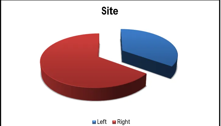
[image:74.595.135.498.453.660.2]SITE OF OCCURRENCE OF TUMOUR:
TABLE 1:SITE OF OCCURRENCE OF TUMOUR:
Site Frequency Percent
Left 14 35.0
Right 26 65.0
Total 40 100.0
From the above tabular column, the frequency of lesion occurring over
right is found to be on the higher side. Of the 40 patients studied, 26
presented with lesion over right side and 14 over the left side. The
percentage of occurrence over right is 65% and of the left is 35%.
Site
64
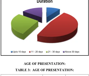
[image:75.595.108.535.521.676.2]DURATION OF THE PRESENTATION OF LUMP: TABLE 2:DURATION OF THE PRESENTATION OF LUMP:
Duration Frequency Percent
Upto 10 days 4 10.0
11 - 20 days 19 47.5
21 - 30 days 11 27.5
Above 30 days 6 15.0
Total 40 100.0
From the above tabular column, most of patients have the duration of the
lesion between 11 to 20 days, of the percentage of about 47.5%, which
constitutes about 19 patients. About 11 patients have the duration
between 21 to 30 days, constitutes a percentage of 27.5%.very less
number of patients presents less than 10 days and more than 30 days.
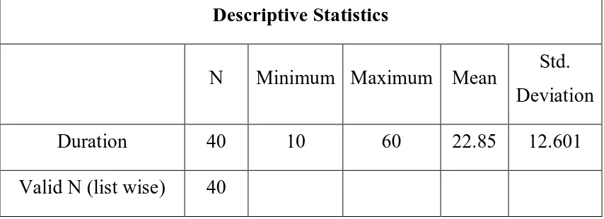
Descriptive Statistics
N Minimum Maximum Mean Std.
Deviation
Duration 40 10 60 22.85 12.601
Valid N (list wise) 40
The mean duration of presentation among the 40 patients included in
65
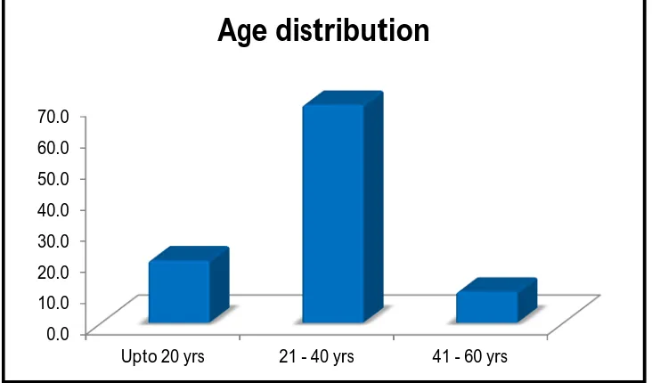
[image:76.595.132.498.86.400.2]AGE OF PRESENTATION:
TABLE 3: AGE OF PRESENTATION:
AGE RANGE Frequency Percent
Up to 20 yrs 8 20.0
21 - 40 yrs 28 70.0
41 - 60 yrs 4 10.0
Total 40 100.0
From the above tabular column, the frequency of occurrence of
fibroepithelial lesions are more in 21 to 40 years of age, which accounts
for about 70% of patients. Up to 20 years and more than 40 years the
percentage of occurrence is 20% and 10% respectively.
Duration
66
The following graph shows the age distribution of fibroepithelial lesions,
[image:77.595.134.500.148.363.2]SIZE OF THE LESION:
TABLE 4:SIZE OF THE LESION:
Size Frequency Percent
2 3 7.5
3 9 22.5
4 7 17.5
5 15 37.5
6 6 15.0
Total 40 100.0
0.0 10.0 20.0 30.0 40.0 50.0 60.0 70.0
Upto 20 yrs 21 - 40 yrs 41 - 60 yrs
67
From the above tabular column, most of the lesions have 5 cm of
diameter, which accounts for 15 patients, of percentage of 37.5%. About
9 patients have a size of 3cm which constitutes about 22.5%. 7 cases have
a size of 4cm, of a percentage of 17.5%.6 cases have a size of 6cm, of a
percentage of 15%.
Size Percent
2
3
4
5
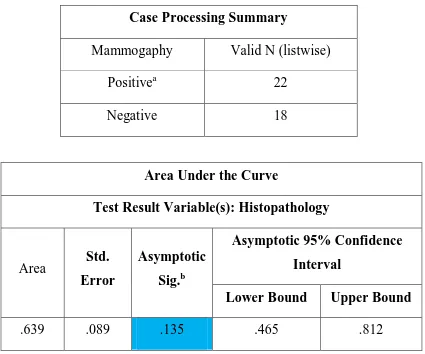
68 MAMMOGRAPHIC FINDINGS:
Table 5 : MAMMOGRAPHIC FINDINGS:
Mammography findings Frequency Percent
Yes 22 55.0
No 18 45.0
Total 40 100.0
From the above tabular column, mammographic findings of suspicion of
phyllodes tumour is found in 22 patients which constitutes about 55%.
Case Processing Summary
Mammogaphy Valid N (listwise)
Positivea 22
Negative 18
Area Under the Curve
Test Result Variable(s): Histopathology
Area Std.
Error
Asymptotic Sig.b
Asymptotic 95% Confidence Interval
Lower Bound Upper Bound
.639 .089 .135 .465 .812
69 The area under the curve is 0.639.
- Sensitivity- 73.33
- Specificity-56.00
- PPV-50.00
- NPV-77.78
- Accuracy -64.67
Chi-Square Tests Value df Asymp. Sig.
(2-sided) Exact Sig. (2-sided) Exact Sig. (1-sided) Pearson
Chi-Square 3.259
a
1 .071
Continuity
Correctionb 2.182 1 .140 Likelihood Ratio 3.357 1 .067
Fisher's Exact
Test .104 .069
Linear-by-Linear
Association 3.178 1 .075 N of Valid Cases 40
From the above tabular column, the p value of the mammographic
findings is more than .05 which shows there no significant co- relation of
70
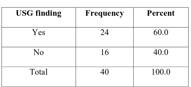
[image:81.595.160.473.112.256.2]ULTRASONO GRAPHY: TABLE 6:USG FINDINGS:
USG finding Frequency Percent
Yes 24 60.0
No 16 40.0
Total 40 100.0
From the above table USG findings suggestive of phyllodes tumour is
found in 24 cases, constitutes a percentage of 60%.
Case Processing Summary
USG Valid N (listwise)
Positive 24
Negative 16
Area Under the Curve
Test Result Variable(s): Histopathology
Area Std. Errora
Asymptotic Sig.b
Asymptotic 95% Confidence Interval
Lower Bound Upper Bound
71
On comparing with the results of histopathology of excised specimen.
- Area under the curve- 0.708
- Sensitivity- 86.67
- Specificity- 56.00
- PPV-54.17
- NPV-87.50
- Accuracy -71.33
Chi-Square Tests Value df Asymp. Sig.
(2-sided) Exact Sig. (2-sided) Exact Sig. (1-sided) Pearson
Chi-Square 7.111
a
1 .008
Continuity
Correctionb 5.444 1 .020
Likelihood Ratio 7.764 1 .005
Fisher's Exact Test .010 .008
Linear-by-Linear
Association 6.933 1 .008
N of Valid Cases 40
From the above tabular column the p value of USG Findings is equal to
0.01,which shows that there is a significant correlation of USG findings
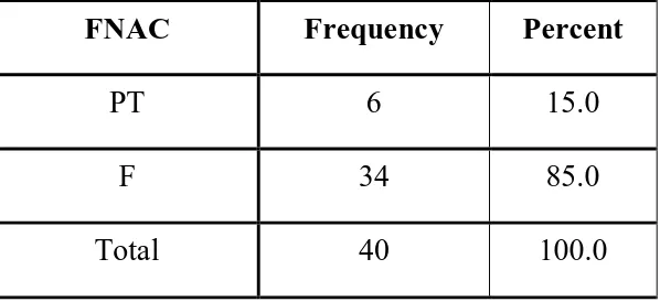
72 FNAC:
TABLE 7:FNAC FINDINGS:
FNAC Frequency Percent
PT 6 15.0
F 34 85.0
Total 40 100.0
From the above tabular column, of the 40 patients 34 have FNAC
findings in favour of fibroadenoma, 6 to phyllode tumour which
contributes to 85% and 15% respectively.
Case Processing Summary
FNAC Valid N (list wise)
Positivea 6
Negative 34
Area Under the Curve
Test Result Variable(s): Histopathology
Area Std. Errora Asymptotic Sig.b
Asymptotic 95% Confidence Interval
Lower Bound Upper Bound
73
On comparing with the results of histopathology of excised specimen.
- Area under the curve-0.868
- Sensitivity-40.00
- Specificity-100.00
- PPV-100.00
- NPV-73.53
- Accuracy -70.00
Chi-Square Tests Value df Asymp. Sig.
(2-sided) Exact Sig. (2-sided) Exact Sig. (1-sided) Pearson
Chi-Square 11.765
a
1 .001
Continuity
Correctionb 8.837 1 .003
Likelihood Ratio 13.626 1 .000 Fisher's Exact
Test .001 .001
Linear-by-Linear
Association 11.471 1 .001
N of Valid Cases 40
From the above tabular column the p value of FNAC is less than 0.01
which is highly statistically significant and there is a significant
74 CORE NEEDLE BIOPSY:
TABLE 8: CORE NEEDLE BIOPSY FINDINGS:
Core needle biopsy Frequency Percent
PT 16 40.0
F 24 60.0
Total 40 100.0
From the above tabular column, out of the 40 patients core needle biopsy
diagnosed 16 cases of phyllodes tumour, constitutes a percentage of 40%.
The rest 60% (i.e) 24 patients were diagnosed as fibroadenoma.
The diagnosis by core needle biopsy is based 4 features,
- epithelial fragments
- stormal fragments
- mitotic figures
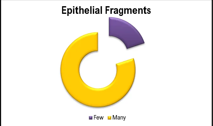
75
TABLE 9: EPITHELIAL FRAGMENTS:
Epithelial Fragments Frequency Percent
Few 8 20.0
Many 32 80.0
Total 40 100.0
From the above table, 80% of patients have many epithelial fragment and
20% have few fragments, of about 32 and 8 patients respectively.
Epithelial Fragments
76
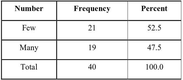
Number Frequency Percent
Few 21 52.5
Many 19 47.5
Total 40 100.0
From the above table, 47.5%of patients have many stormal fragment and
[image:87.595.158.474.71.209.2]52.5% have few fragments, of about 19 and 21 patients respectively.
TABLE 10: CELLULARITY:
Cellularity Frequency Percent
1+ 17 42.5
2+ 13 32.5
3+ 10 25.0
Total 40 100.0
From the above table, 42.5% of patients have 1+ cellularity that comprise
of 17 patients, 32.5% have 2+ and 25% have 3+celularity. This
77
TABLE 11: SPINDLE CELLS:
Spindle Cells Frequency Percent
Negative 22 55.0
Positive 18 45.0
Total 40 100.0
From the above table, 45% of patients have spindle cells that constitutes
18 patients and rest 55% spindle cells are absent which comprise of 22
patients.
Cellularity
78
TABLE 12: MITOTIC FIGURES:
Mitotic Figures Frequency Percent
High 10 25.0
Low 30 75.0
Total 40 100.0
From the above table, 75% of patients have low mitotic figures that
constitutes 30 patients and the rest 25% have high mitotic figures that
constitutes 10 patients.
Spindle Cells
79
Case Processing Summary
core needle biopsy Valid N (listwise)
Positivea 16
Negative 24
Area Under the Curve
Test Result Variable(s): Histopathology
Area Std. Errora
Asymptotic Sig.b
Asymptotic 95% Confidence Interval
Lower Bound Upper Bound
.917 .054 .0005 .810 1.000
Mitotic Figures
80
On comparing with the results of histopathology of excised specimen
- Area under the cuve-0.917
- Sensitivity-93.33
- Specificity-92.00
- PPV-87.5
- NPV-95.83
81
Chi-Square Tests Value df Asymp. Sig.
(2-sided) Exact Sig. (2-sided) Exact Sig. (1-sided) Pearson
Chi-Square 28.444 a
1 .000
Continuity
Correctionb 25.000 1 .000
Likelihood Ratio 32.555 1 .000
Fisher's Exact Test .0005 .000
Linear-by-Linear
Association 27.733 1 .000
N of Valid Cases 40
From the above tabular column the P value of the core needle biopsy is
less 0.01 which shows that core needle biopsy is highly significant in
diagnosing phyllodes tumour.
On comparing the detection percentage of fibroadenoma and phyllodes
tumour by various tool the following tabular column is obtained,
Mammography USG FNAC Core needle
biopsy Histopathology
PT 55.0 60.0 15.0 40.0 37.5
82
[image:93.595.108.525.114.361.2]The same is explained in the graph
TABLE 13:PROCEDURE DONE:
Procedure Done Frequency Percent
Enucleation 24 60.0
Wide Excision 16 40.0
Total 40 100.0
From the tabular column, based on the pre operative diagnosis 60% of
cases have undergone enucleation and rest 40% have undergone wide
excision, which constitutes 24 and 16 patients respectively.
0% 20% 40% 60% 80% 100%
Outcomes of the various tools
83 The following explains the above
So to detect the diagnostic efficacy of core needle biopsy,we
compare the ROC curve of mammographic findings, usg findings,FNAC
findings with the standard gold standard results of histopathology of
excised specimen.
Case Processing Summary
Histopathology Valid N (listwise)
Positivea 15
Negative 25
Procedure Done
84
Area Under the Curve
Test Result
Variable(s) Area
Std. Errora
Asymptotic Sig.b
Asymptotic 95% Confidence Interval Lower
Bound Upper Bound
Mammography .647 .090 .124 .470 .823
USG .713 .083 .025 .551 .876
FNAC .700 .093 .036 .517 .883
Core needle
biopsy .927 .049 .0005 .830 1.000
P - Value ** Highly Significant at P ≤ .01
P - Value * Significant at 0.01 < P ≤ .050
85
So on comparing the area under the curve of mammographic
findings,USG findings,FNAC findings and core needle biopsy findings
,the core needle biopsy is found to be the most significant tool in
detecting the phyllodes tumour. The area under the curve of
mammographic findings, USG findings , FNAC findings and core needle
biopsy findings are 0.647,0.713,0.700,0.927 respectively, in which core
needle biopsy has highest area under curve. The p-value also 0.0005 for
86
CONCLUSION
To conclude, though the incidence of phyllodes tumour is very less, it has a potential for turning into malignant. So pre-operative diagnosis of phyllodes tumour is essential as it decides the line of management, which is a simple enucleation for fibroadenoma and wide excision for phyllodes tumour. Fibroadenoma is a benign condition which has clinical findings, radiological findings, FNAC findings similar to that of phyllodes tumour so the differentiation between fibroadenoma and phyllodes tumour becomes difficult, so we use core needle biopsy of the lesion and we analyse 4 features which are the epithelial fragments, stormal fragments, spindle cell and mitotic figures to differentiate the phyllodes tumour and fibroadenoma.
87
BIBLIOGRAPHY
1. Lakhani SR; International Agency for Research on Cancer; World
Health Organization: WHO Classification of Tumours of the
Breast. Geneva, WHO, 2012, pp 141-147.
2. Foxcroft LM, Evans EB, Porter AJ: Difficulties in the
pre-operative diagnosis of phyllodes tumours of the breast: a study of
84 cases. Breast 2007;16:27-37.
3. Simi U, Moretti D, Iacconi P, Arganini M, Roncella M, Miccoli P,
et al: Fine needle aspiration cytopathology of phyllodes tumor:
differential diagnosis with fibroadenoma. Acta Cytol
1988;32:63-66.
4. Stanley MW, Tani EM, Rutqvist LE, Skoog L: Cystosarcoma
phyllodes of the breast: a cytologic and clinicopathologic study of
23 cases. Diagn Cytopathol 1989;5:29-34.
5. Dusenbery D, Frable WJ: Fine needle aspiration cytology of
phyllodes tumor: potential diagnostic pitfalls. Acta Cytol
88
6. Rao CR, Narasimhamurthy NK, Jaganathan K, Mukherjee G,
Hazarika D: Cystosarcoma phyllodes:diagnosis by fine needle
aspiration cytology. Acta Cytol 1992;36:203-207.
7. Shimizu K, Masawa N, Yamada T, Okamoto K, Kanda K:
Cytologic evaluation of phyllodes tumors as compared to
fibroadenomas of the breast. Acta Cytol 1994;38:891-897.
8. Shabb NS: Phyllodes tumor. Fine needle aspiration cytology of
eight cases. Acta Cytol 1997;41:321-326.
9. Deen SA, McKee GT, Kissin MW: Differential cytologic features
of fibroepithelial lesions of the breast. Diagn Cytopathol
1999;20:53-56.
10. Bhattarai S, Kapila K, Verma K. Phyllodes tumor of the breast: a
cytohistologic study of 80 cases. Acta Cytol 2000;44:790-796.
11. Krishnamurthy S, Ashfaq R, Shin HJ, Sneige N: Distinction of
phyllodes tumor from fibroadenoma: a reappraisal of an old
problem. Cancer 2000;90:342-349.
12. Scolyer RA, Mckenzie PR, Achmed D, Soon Lee C: Can phyllodes
tumours of the breast be distinguished from fibroadenomas using
89
13. Veneti S, Manek S: Benign phyllodes tumour vs fibroadenoma:
FNA cytological differentiation. Cytopathology 2001;12:321-328.
14. Jayaram G, Sthaneshwar P: Fine-needle aspiration cytology of
phyllodes tumors. Diagn Cytopathol 2002;26:222-227.
15. Shimizu K, Korematsu M: Phyllodes tumor of the breast: a
cytomorphologic approach based on evaluation of epithelial cluster
architecture. Acta Cytol 2002;46:332-336.
16. Bandyopadhyay R, Nag D, Mondal SK, Mukhopadhyay S, Roy S,
Sinha SK: Distinction of phyllodes tumor from fibroadenoma:
cytologists' perspective. J Cytol 2010;27:59-62.
17. El Hag IA, Aodah A, Kollur SM, Attallah A, Mohamed AAE,
Al-Hussaini H: Cytological clues in the distinction between phyllodes
tumor and fibroadenoma. Cancer Cytopathol 2010;118:33-40.
18. Shabalova IP, Chemeris GJ, Ermilova VD, Rodionova LM,
Pavlikova NA, Syrjänen KJ: Phyllodes tumour: cytologic and
histologic presentation of 22 cases, and immunohistochemical
90
19. Komenaka IK, El-Tamer M, Pile-Spellman E, Hibshoosh H: Core
needle biopsy as a diagnostic tool to differentiate phyllodes tumor
from fibroadenoma. Arch Surg 2003;138:987-990.
20. Jacklin RK, Ridgway PF, Ziprin P, Healy V, Hadjiminas D, Darzi
A: Optimising preoperative diagnosis in phyllodes tumour of the
breast. J Clin Pathol 2006;59:454-459.
21. Adesoye T, Neuman HB, Wilke LG, Schumacher JR, Steiman J,
Greenberg CC: Current trends in the management of phyllodes
tumors of the breast. Ann Surg Oncol 2016;23:3199-3205.
22. American College of Radiology . Illustrated breast imaging
reporting and data system (BI-RADS) 4th ed. Reston: American
91
PROFORMA
NAME:
AGE:
SEX:
IP.No:
CLINICAL FINDINGS:
MAMMOGRAPY FINDINGS:
ULTRASONOGRAPY FINDINGS:
FINE NEEDLE ASPIRATION CYTOLOGY REPORT:
CORE NEEDLE BIOPSY REPORT:
PRE-OPERATIVE DIAGNOSIS:
PROCEDURE DONE:
92
xg;g[jy; gotk;
nehahspapd; bgah;
:njjp
:taJ
/ghypdk;
:cs;nehahsp vz;
:neha;f;Fwp
:---
Mfpa ehd;
ehsJ njjpapy; nfhit kUj;Jtf; fy;Y}hp kUj;Jtkidapy; khh;gf
fl;o mWit rpfpr;irf;fhf mDkjpf;fg;gl;Ls;nsd;.
khh;gf fl;oapd; jd;ik Fwpj;J Muha rij ghpnrhjid
vLf;fg;gLk; vd;Wk;/ mjd; Kot[f;F Vw;g mWitapd; jd;ik
Vw;gLk; vd;gJk; kUj;Jtuhy; bjhptpf;fg;gl;lJ.
,j;jpR
ghpnrhjid
vdJ
rpfpr;irapd;
juj;ij
cah;j;Jtjw;fhfnt vd;gija[k;/
,jd; fhuzkhf vdJ rpfpr;irapy; fhyjhkjk; Vw;glhJ
vd;gija[k;/
Ma;t[ Kot[fspd; ,ufrpak; fhf;fg;gLk; vd;gJk;/
ghpnrhjid Kot[fs; Ma;t[f;F cl;gLj;jg;gLk; vd;gJk;/
ehd; tpUk;gpdhy; vg;bghGJ ntz;LkhdhYk; Ma;tpypUe;J
tpyfpf; bfhs;syhk; vd;gija[k; kUj;Jth; \yk; bjhpe;J
bfhz;nld;.
,tw;iw KGikahfg; g[hpe;J bfhz;L KGkdJlDk;/ Raepidt[lDk;/
vt;tpj eph;ge;jKk; ,d;wp ghpnrhjidf;Fk;/ rpfpr;irf;Fk;/ Ma;t[f;Fk;
xj;JiHg;g[ ju rk;kjpf;fpnwd;.
,lk;
:njjp
:93
MASTER CHART
S.No Name
A
ge In Patient
Number D u rat ion S it e S iz e M am m ogap h y F in d in gs U S G F in d in gs F N A C
CORE NEEDLE BIOPSY
P re O p er at ive D iagn os is P roc ed u re D on e Histopathology of Excised Specimen Epithelial Fragments Stormal Fragments S p in d le C el ls M it ot ic F igu re s
Number Cellularity
1 Meena 24 125530 10 R 5 N N F Many Few 2+ - Low Fibroadenoma Enucleation Fibroadenoma
2 Soundariya 17 127592 14 R 2 Y N F Few Many 1+ - Low Fibroadenoma Enucleation Fibroadenoma
3 Pushpharani 40 119054 50 L 5 Y Y PT Many Many 2+ + High Phyllodes Tumour Wide Excision Phyllodes Tumour
4 Gunavathivel 34 128100 14 R 3 N Y F Many Few 1+ + Low Fibroadenoma Enucleation Fibroadenoma
5 Pechiyammal 35 120322 60 R 4 N Y F Many Many 3+ + Low Phyllodes Tumour Wide Excision Phyllodes Tumour
6 Saraswathy 45 120359 20 L 3 N Y F Many Many 2+ + Low Phyllodes Tumour Wide Excision Phyllodes Tumour
7 Rajeswari 60 127569 30 R 3 Y Y F Many Many 1+ - Low Fibroadenoma Enucleation Fibroadenoma
8 Lavanya 30 127890 15 R 4 N N F Few Few 2+ - Low Fibroadenoma Enucleation Fibroadenoma
9 Geetha 38 123036 30 R 5 Y Y F Many Few 1+ + Low Fibroadenoma Enucleation Phyllodes Tumour
10 Aarthi 19 128537 40 R 5 Y N F Few Few 1+ - Low Fibroadenoma Enucleation Fibroadenoma
11 Sumathi 33 123653 15 R 6 Y Y PT Many Many 3+ + High Phyllodes Tumour Wide Excision Phyllodes Tumour
12 Vaneeshwari 25 129654 45 R 5 N Y F Many Few 2+ - Low Fibroadenoma Enucleation Fibroadenoma
13 Chithra 35 135453 10 L 6 N N F Many Many 2+ + High Phyllodes Tumour Wide Excision Fibroadenoma
14 Ayyammal 36 131041 50 R 3 Y Y F Many Few 2+ - Low Fibroadenoma Enucleation Fibroadenoma
15 Parameswari 34 119295 25 L 5 Y Y PT Many Many 3+ + High Phyllodes Tumour Wide Excision Phyllodes Tumour
16 Brindha 20 131765 14 R 6 N N F Many Few 1+ - Low Fibroadenoma Enucleation Fibroadenoma
17 Shamala 34 131489 14 R 3 Y N F Few Few 1+ + Low Fibroadenoma Enucleation Fibroadenoma
18 Jomiveimma 20 125036 20 R 4 Y Y PT Many Many 3+ + High Phyllodes Tumour Wide Excision Phyllodes Tumour
19 Pushphalani 40 123536 21 R 4 N Y F Many Many 3+ + Low Phyllodes Tumour Wide Excision Phyllodes Tumour
94
21 Pavithra 20 43151 18 R 2 Y Y F Many Few 2+ - Low Fibroadenoma Enucleation Fibroadenoma
22 Prabhavathy 25 126476 28 L 3 Y Y F Many Many 3+ + Low Phyllodes Tumour Wide Excision Phyllodes Tumour
23 Vasanthi 40 106478 21 R 4 Y N F Many Many 2+ - High Phyllodes Tumour Wide Excision Phyllodes Tumour
24 Saradha 40 105486 14 L 5 N Y F Many Many 2+ + Low Phyllodes Tumour Wide Excision Phyllodes Tumour
25 Rajeswari 29 100676 10 L 6 N N F Few Few 1+ - Low Fibroadenoma Enucleation Fibroadenoma
26 Krishnaveni 35 102036 15 L 4 Y Y PT Many Many 3+ + High Phyllodes Tumour Wide Excision Phyllodes Tumour
27 Vinmathi 23 10784 21 R 5 Y N F Many Few 2+ - Low Fibroadenoma Enucleation Fibroadenoma
28 Indirarani 29 121996 14 L 2 N Y F Few Many 1+ - Low Fibroadenoma Enucleation Fibroadenoma
29 Kowsalya 17 122263 14 R 5 N N F Many Many 2+ + High Phyllodes Tumour Wide Excision Fibroadenoma
30 Minorani 28 121999 30 R 3 Y Y F Many Few 1+ - Low Fibroadenoma Enucleation Fibroadenoma
31 Kamala 45 101100 14 R 4 Y N F Many Many 3+ + Low Phyllodes Tumour Wide Excision Phyllodes Tumour
32 Nirmala 28 1260684 14 R 6 N N F Many Few 1+ - Low Fibroadenoma Enucleation Fibroadenoma
33 Ramya 22 114266 10 L