JOURNALOFVIROLOGY, May1984, p. 309-315 Vol.50, No. 2
0022-538X/84/050309-07$02.00/0
Copyright © 1984, American SocietyforMicrobiology
Characterization of
the
DNA-Protein
Complex
at
the Termini of the
Bacteriophage
PRD1
Genome
DENNIS H.
BAMFORD'
AND LEONARDMINDICH2*
Department ofMicrobiology, The Public Health Research Institute ofTheCity ofNew York, Inc., New York,New York 10016,2 and Department ofGenetics, University of Helsinki, Helsinki,
Finland'
Received 14November1983/Accepted 18January 1984
DNAofbacteriophagePRD1 hasprotein P8atits termini. Extracts of infected cells are abletoderivatize
P8invitrowith labeled dGTP. Twoearlyproteins,P1 andP8, productsofgenes I and VIII,respectively,are
theonly phage proteins necessary forthe formationof the
protein
P8-dGMPcomplex.Thiswas shownbycomplementation of extracts from cells infected with mutants and by use of extracts from cells carrying cloned genes I and VIII. With Escherichia colimutantsthataretemperature sensitive for DNAsynthesis,it was possible to show that the formation ofthe
protein
P8-dGMPcomplex
wasdependent
upon the hostreplicationapparatus. Theanalysis of the purified proteinP8-dGMPcomplex by hydrolysisandenzymatic
digestionshowed that there isacovalent phosphodiester bond between tyrosine and 5'-dGMP.
Bacteriophage PRD1 is a broad-host-range
lipid-contain-ing bacteriophagewhich caninfectgram-negativehosts such
asEscherichia coli and Salmonella typhimurium harboring
an appropriate plasmid. Fora more detailed description of
this phage system, see Bamford et al. (4), Bamford and
Mindich(3),McGraw etal. (15), andMindichetal. (16, 17).
We have shown that there is a terminal protein strongly
bound to the DNAof
bacteriophage
PRD1 (2). This protein is an early phage-specific protein designated P8. It is aproduct of gene VIII and is necessary for phage DNA
replication. Infected-cellextractscatalyzetheformation ofa
complexbetween protein P8and
[a-32P]dGTP
(2).The relationship oftheterminalproteintoDNA synthesis
appears to be analogous to that found in the cases of
adenovirus (8, 12, 13, 27) and bacteriophage 429(10, 22).
Inthisstudy we have refined theconditions for theprotein
P8-dGMP reaction and have shown thatextracts fromcells
containing cloned PRD1 genes I and VIII form theprotein
P8-dGMPcomplex much moreeffectively than do
infected-cell extracts, that the terminal 5'-deoxyguanosine is
cova-lently linked to tyrosine inprotein P8 via aphosphodiester
bond, and that the host cellreplication apparatus is needed
fortheformation ofthiscomplex.
MATERIALSANDMETHODS
Phages and bacteria. The bacterial strains used in this
study are listed in Table 1. Plasmid pLM2 (18), which is
necessary for phage adsorption, was transferred to E. coli JW1073 and RS116 by cross-streaking overnight broth cul-tures with cultures of S. typhimurium LT2(pLM2) and
selecting for kanamycinand tetracycline resistance. For E.
coli JW302 the donor was Pseudomonas phaseolicola
HB10Y(pLM2), which is not capable of growing above ca.
34°C, and selection was for kanamycin resistance at 37°C.
Plasmidclone 3,which has phage genes I and VIII (19) and is
now designated pLM3, was introduced into E. coli
JW1O1,
JW107, JW110, JW111, JW112, JW176, JW177, JW180,
JW267, and JW284 by transformation as previously de-scribed (19). Plasmid clone 141 is now designated pLM141. The transformants were expressed in L broth overnight at
* Correspondingauthor.
26°C and selected for resistance totetracyclineand strepto-mycin over 2 nights at 29°C. The transformants obtained were purified and tested.
The phages used in this study are listed in Table 2. Nonsense mutants of PRD1 are designated with a mutant
isolation number and a roman numeralthat denotesthe gene
affected by the mutation. This number refers also to the
nameofthe protein product of thecorrespondinggene; e.g.,
sus2 contains(I) a mutation of gene I andis missing protein
P1 in cells that do not suppress amber mutations. Phages
PRD1, PR3, PR4, PR5, L17, PR722,and 12-1 were grown in
LT2(pLM2), AP50 was propagatedin nonpathogenic
Bacil-lus anthracis CN18-74, and
429
waspropagated in Bacillus subtilis IS230. Thebacteriain Lbroth at37°C were infectedat amultiplicityofinfection of10 at acelldensity of ca. 5 x
108/ml. The phageconcentrationandpurification weredone
as described by Bamford and Mindich (3), except that the
final phage pellets were suspended into 50 mM
Tris-hydro-chloride(pH 7.5)-2.5 mM MgCI2.The nonsense mutants of PRD1 were grown onappropriate suppressor hosts as indi-cated in Table 2. Purified single-stranded bacteriophage fd DNA was obtained from Loren A. Day, and bacteriophage
4~X174
DNA was obtainedfrom OpinderS. Bhanot. Preparation of cell extracts. Infected-cell extracts were made as described previously (2). Cells containing cloned PRD1 genes were grown in L broth containing 20 ,ug oftetracycline perml to adensity of ca. 5 x
108/ml
at 37°C or,inthe case of temperature-sensitive (ts) mutants, at 29°C and then processed as infected cells were.
Standard conditions for forming the protein P8-dGMP complex. Reaction mixtures (10 ,ul) contained 25 mM Tris-hydrochloride (pH 7.5), 20 mM MgCl2, 1 mM ATP, 1 mM
dithiothreitol, 1.5 x
1011
purified phage particles disruptedby heatingthem to100°C for 20 s, 1 ,uM cold dGTP, 0.15 ,uCi
of
[a-32P]dGTP
(400Ci/mmol; Amersham Corp.), and 4,ulof cell extractcontaining20mg ofprotein per ml. The standardconditions were 30°C for 75 s. The reactions were stopped,
and thesamples were prepared for protein gels as previously described (2). For reaction optimization,
MgC92,
ATP,phage, dGTP, and extract concentrations, as well as
tem-perature and time, were varied as described below. When extractsfromts cells were used, the low-temperature reac-tion was run for 1 min at 30°C. The high-temperature
309
on November 10, 2019 by guest
http://jvi.asm.org/
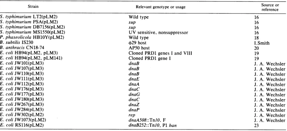
TABLE 1. Bacterial strains used in this study
Strain Relevantgenotypeorusage Source or
reference
S.typhimurium LT2(pLM2) Wild type 16
S.typhimurium PSA(pLM2) sup 16
S.typhimurium DB7156(pLM2) sup 16
S.typhimurium MS1550(pLM2) UV sensitive, nonsuppressor 16
P. phaseolicola HB1OY(pLM2) Wild type 18
B. subtilis IS230 429host I.Smith
B.anthracis CN18-74 AP50host 20
E.coliHB94(pLM2, pLM3) Cloned PRD1genes I and VIII 19
E. coliHB94(pLM2, pLM141) Cloned PRD1gene I 19
E. coliJW101(pLM3) dnaB J. A. Wechsler
E. coliJW107(pLM3) dnaB J. A. Wechsler
E. coli JW110(pLM3) dnaB J. A. Wechsler
E. coli JW111(pLM3) dnaE J. A. Wechsler
E. coli JW112(pLM3) dnaA J. A. Wechsler
E. coli JW176(pLM3) dnaC J. A. Wechsler
E. coliJW177(pLM3) dnaG J. A. Wechsler
E.coliJW180(pLM3) dnaC J. A. Wechsler
E.coliJW267(pLM3) dnaZ J. A. Wechsler
E.coliJW284(pLM3) dnaP J. A. Wechsler
E.coliJW302(pLM2) rep J. A. Wechsler
E.coli JW1073(pLM2) dnaA508::TnlO, F J. A. Wechsler
E. coli RS116(pLM2) dnaB252::TnIO,P1ban 23
treatment was donebyincubating theextractsfor20 minat
37°C before runningthereactionat 30°C for 1 min.
Purificationandanalysis oftheprotein P8-dGMP complex. The standard reaction was run with 35 times the normal volume(350
pl).
Fifty microliters of lOx nuclease buffer (0.5M sodium borate [pH 8.75], 5 mM CaC12) and 150 U of
nuclease (Staphylococcus aureus nuclease; Boehringer
Mannheim Biochemicals) were added to this mixture, and the mixture was incubated for 2 h at 37°C. The
nuclease-digestedmixture was runthrough a SephadexG-25 column
equilibratedwith0.1% sodium
dodecyl
sulfate(SDS)-50
mMsodiumacetate, pH7.5.The columnfractionswerecounted
andanalyzedin
protein
gels.Theprotein
P8-dGMPcomplexpeak containing SDS was acetone
precipitated
and used indigestion studies. Theacid
hydrolysis
was performedin 5.6MHClat 110°C for90, 120, and 135 min. After
hydrolysis,
theHCl wasevaporated
overnight
under reduced pressure,and the samples were suspended in water. Ten microliters
eachof
phosphoserine,
phosphothreonine,
and [image:2.612.68.308.562.721.2]phosphotyr-osine(1 mg/ml)wasadded, and thesamples weredried and
TABLE 2. Phage strains usedinthis study
Relevant
Phage genotypeor Host Reference or
usage
PRD1 Wildtype LT2(pLM2) 16
PRD1 sus2(I) PSA(pLM2) 16
PRD1 sus38(I) PSA(pLM2) 16
PRD1 sus233(VIII) DB7156(pLM2) 16 PRD1 sus247(VIII) DB7156(pLM2) 16
PR3 Wild type LT2(pLM2) 4
PR4 Wild type LT2(pLM2) 4
PR5 Wild type LT2(pLM2) 4
L17 Wild type LT2(pLM2) 4
PR722 Wildtype LT2(pLM2) 5
N Wild type LT2(pLM2) J. N. Coetzee
12-1 Wild type LT2(pLM2) J. N. Coetzee
AP50 Clearplaque CN18-74 20
,29 Wild type IS230 1
analyzed by two-dimensional thin-layer electrophoresis as described by Collett et al. (6). The electrophoresis plates were stained with0.5% ninhydrin in acetone and developed for 5minat 110°C. Autoradiograms were prepared from the stained plates.
Enzymatic digestions of the acetone-precipitated purified
protein P8-dGMP complex were performed as follows. To ca. 10,000 Cerenkov cpm ofdried complex, enzymes and buffers were added to obtain a 20-,ul final volume. The conditions for different enzymes were as follows: 2 ,ug of pronasein30 mMTris-hydrochloride (pH8)-10 mM MgCl2; 0.03 U of phosphodiesterase I (from snake venom; Sigma
type VIII) in 30 mM Tris-hydrochloride (pH
8)-10
mMMgCl2;0.035 UofphosphodiesteraseII(frombovine spleen;
SigmatypeI) in 50 mMsodiumacetate,pH5.2; and 2.5 Uof
alkalinephosphatase(frombovineintestine;Sigma type VII-L) in 30 mM Tris-hydrochloride (pH
8)-i
mM MgCl2-0.01mM ZnCl2. The mixtures were incubated for 100 min at
37°C. A 3x protein gel sample buffer was added, and the
samples wereboiled for 2 min andanalyzed in protein gels.
Theproteingels were15.5%polyacrylamide, and the system
was as previously described (16). The gels were dried, and
autoradiogramswereprepared.Visible bandswerescanned,
and the peak areaswere calculated. RESULTS
Formingthe protein P8-dGMPcomplex withphage geneI and VIIIproducts.Byuseofinfected-cellextracts,disrupted
phage particles,and[ao-32P]dGTP,it waspossibleto incorpo-rate 32p into protein P8 (2). This reaction was dependent upon added disrupted phage. Uninfected extracts did not have anyactivity. dGTPwastheonlynucleotide whichwas
incorporated.Thelowactivityof this reaction limited further
characterization of theproducts.When theextract wasmade from cells containing cloned genes for PRD1 replication
proteins P1 and P8 (pLM3 [15]), theefficiency of the dGTP derivatization of protein P8 increased at least 100 times.
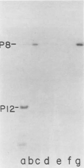
Figure 1 shows that protein P8 was labeled by radioactive
dGTP (Fig. lb and g) and that an extract ofa plasmid-free
on November 10, 2019 by guest
http://jvi.asm.org/
DNA TERMINAL PROTEIN 311
uninfected culture did not incorporate any nucleotide,
al-though all
nucleotides
were labeled intheassay (Fig.lf).
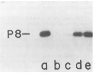
Phage mutants sus2 and sus38 in class I and mutants sus233 and sus247 in class VIII complement each other in a plaque assay. Extractsfrom LT2(pLM2) cellsinfectedwith these mutants were tested for their ability to form theprotein P8-dGMP complex. The comparison was made with the
activity achieved in a reaction with an extract of
wild-type-infected cells (Fig. 2a). None ofthe mutants were able to
form this complex alone (Fig. 2b, c, d, and e); however,
extracts made from mutants sus2 (I) and sus233 (VIII)
clearly complemented each other (Fig. 2f). Very weak
complementationwas seenwith mutants sus2 (I) and sus247
(VIII) (Fig.2g). Noothercomplementation was observed. Optimization of the protein P8-dGMP reaction. An
investi-gation of reaction conditions revealed that the maximum
amountofproduct wasobtainedafter a 75-s reaction time at 30°C. Thepreviously used30-minreaction time resulted in a
reducedamountofproduct (Fig. 3A). The reaction
tempera-ture did not seem to be very critical, but a slight peak
appeared around 30°C (Fig. 3B). Because of the short
P84_
0w
a
bc
defgh
FIG. 2. Complementationassayfor the P8-dGMPreaction with extracts from cells infected with nonsense mutants froa classesI and VIII. Standard reaction with extract from cells infected with wild-type PRD1 (a). Reactions with extracts fromcellsinfectedwith
PRD1 mutants:(b) sus2(I);(c)sus38(1);(d) sus233 (VIII);(e)sus247 (VIII). Reactionswith twoextractsfrom cellsinfected withmutants: (f)sus2(I) andsus233(VIII);(g)sus2(I) and sus247 (VIII); (h) sus38 (I)and sus233(VIII);(i)sus38(I) and sus247(VIII);(j) sus2 (I) and sus38(I); (k)sus233(VIII) and sus247 (VIII).
reaction time, the reaction components were prewarmed
shortly beforemixing.The magnesium concentration forthe
maximum yield started at ca. 10 mM, but even four times
thatconcentration did not have an inhibitory effect (Fig. 3E). The reaction proceeded without the addition of ATP, buta
threefold stimulation was achievedat aconcentration ofca.
1 mM (Fig. 3D). The addition of unlabeled dGTP up to a
concentrationof ca. 1 ,uM increased theyield (Fig. 3C). Table 3 shows further reaction characteristics. The
reac-
P8-PI2p
a, 7) C5-a,
abc
d
e
fg
FIG. 1. Polyacrylamide gelelectrophoresis of reaction products formed withextractsfromcellscontaining cloned PRD1genesI and
VIII (pLM3). Samples were digested and analyzed on a 15.5% polyacrylamide gel. (a)[35S]methionine-labeledearly PRD1 proteins in UV-sensitive MS1550(pLM2) cells; (b and g) standardreaction with [a-32P]dGTP; (c) [ox-32P]dCTP; (d) [a-32P]dATP; (e)
[a-32P]dTTP;
(f) [C-_32P]dGTP, [a-32P]dATP, [a-32P]dCTP, and [ox-32P]dTTP in a reaction mixture with extracts from cells without plasmid pLM3.FIG. 3. Optimization of the protein P8-dGMP reaction. The reactionwas run asdescribed in the text. "Relative yield" on the ordinate refers to therelative intensity oftheautoradiogramband for32P-labeled P8. (A)Timing of the reaction at 30°C. Prewarmed reactioncomponentsweremixed, and the reaction was stopped by boiling. The Mg2",ATP, andunlabeleddGTP concentrationswere asinstandardmixtures.(B)Effect oftemperature ontheformation of the P8-dGMP complex. (C) Effect ofthe addition ofunlabeled dGTP. (D) Effect ofthe ATPconcentration. (E) Effect of theMgCl2 concentration.
VOL. 50, 1984
1.1,. I,;c
I.j"--,j -.,
9 O'.
I
on November 10, 2019 by guest
http://jvi.asm.org/
[image:3.612.319.544.72.229.2] [image:3.612.92.255.309.636.2] [image:3.612.313.552.443.632.2]312 BAMFORD AND MINDICH
TABLE 3. Protein P8-dGMP complex formation
Relative
Standard reaction yield
(%S)a
With[c-32P]GTP ... 0
Minus phage ...0...O One-fifth the amount of phage ... ... 52
Minusextract... 0
Extractwithout pLM3 but with pBR322... ... 0
Extractfrom cells with pLM141
...
... 0One-third theamountofpLM3 extract... 31
Phagetreated with 60p.gof trypsin permlb... ... 0
Phagetreated with 300U of nuclease perml... 8
Extracttreated with 300 Uof nuclease permid.28 With 50
,ug
ofactinomycinD perml... 32With 20
,ug
ofrifampin permle... 100With 30 ,ug of Triton X-100 per ml ... ... 104
With 450p.g of Triton X-100 per ml ... ... 79
With 3000 igof Triton X-100 per ml ... ... 18
With 10%glycerol... 108
With 25%glycerol... 95
With phages PR3, PR4, PR5, L17, PR722, N, and I21...75-100
I2-lf
.. ... ... . .... ... . .... 7-0 With phage AP50 or+29f... ... 0a The reaction sample was digestedas described in the text and applied to a15.5% polyacrylamide gel. The extent of the reaction wasmeasured by densitometry. One hundred percent is about 3,000 cpm,which is equivalentto 0.1 pmol ofdeoxyguanosine. Sincethe amountof templateis about0.5pmolof ends, the maximum yield is about 20% of the amount of template.
b Theheat-disrupted phagewasincubated withtrypsin for30min at 37°C, followed by the addition of a 10-fold excess oftrypsin inhibitor for 10 minat37°C before addition tothereaction mixture. Trypsin inhibitortreatment alone did not have any effect on the reaction.
'Theheat-disrupted phage wasincubated with micrococcal
nu-cleasefor 30 min at32°C, followed by5minat65°C before addition
tothe reaction mixture.
d Thecell extract with cloned geneswastreated with nucleaseas in footnotec.
eTherifampinwasdissolvedasdescribedby Sinclairetal.(25).
fThese phages, instead of PRD1, were tested one by one. The phagedisruption was doneas inthe caseofPRD1.
tion was
specific
fordGTP,
since no detectable reactionoccurred when
GTP
was used.Lowering
bothphage
andextract concentrations reduced the
yield,
and the reactionwas dependent on these components. Extracts from cells
having onlythepBR322vectordidnotsupport any
reaction,
nordidextracts of cells
containing
pLM141, aplasmid
that carries gene I but not gene VIII(15).
Treatment ofheat-disrupted phage with trypsin followed
by
trypsin
inhibitortreatment before the addition ofthe phage to the reaction
mixture led to a loss ofactivity. Nuclease treatment ofthe
phage alsoled to a loss ofactivity.Theaddition of
actinomy-cin D led to reducedactivity,but
rifampin
didnothaveanyeffect. Low amounts of Triton X-100 and
glycerol
hadslightly stimulatory effects, whereas
higher
concentrationsreduced theactivity. Closelyrelatedphages
PR3, PR4,
PR5,
L17,
PR722,
N, and 12-1 allcatalyzed
theincorporation
of[a-32P]dGTP
and noothernucleotide intoprotein
P8. DNAfrom morphologically related lipid-containing phage AP50
infectingagram-positivehostdid not stimulate the
incorpo-ration ofanynucleotide intoproteinP8. Thesame was true
for phage
4.29,
which actively forms a DNA-terminalpro-tein-dAMPcomplex inB. subtilis(22).
When single-stranded
DNAs
from phages4X174
and fdwere usedinthe assay, instead of disrupted phages,a very weakincorporation ofall four deoxynucleotides to protein P8was seen(datanot shown).
Characterization of the bond between protein P8 and dGMP. The protein P8-dGMP complex was prepared with standard conditions and treated with nuclease. The
nucle-ase-digested reaction mixturewas passed through a
Sepha-dex G-25 column. When acetate bufferwith 0.1% SDSwas used asan eluate, ashoulderof radioactivitywasobserved
before the bulk radioactivity. Gel electrophoresis of the
fractions showed the protein P8-dGMP complex in a peak of
four fractions, with a slight tailing in subsequent fractions
(data not shown). The early partofthe peak showed very
little low-molecular-weight radioactivity in the gel, whereas later fractions had stronglow-molecular-weight radioactiv-ity. No detectable protein P8-dGMP complex could be eluted from the column in the absence of SDS. Theprotein
P8-dGMP complex from the early portion of the peak was
concentrated,and theSDS was removedbyacetone
precipi-tation. The complex was hydrolyzed with HCl and analyzed for labeled phosphoamino acids by two-dimensional
thin-layer electrophoresis. Figure 4 shows that theonly labeled
phosphoamino acid liberated from the P8-dGMP complex
was phosphotyrosine. An unidentified labeled hydrolysis product was observed between the originand
phosphotyro-q_Pi
~'
Pser
~, P
.>>w)Pthr
I')
.Sx
pH
1.9--FIG. 4. Two-dimensional thin-layerelectrophoresis of the acid-hydrolyzed (5.6MHCl,110°C,135min)P8-[a-32P]dGMPcomplex.
The dotted circles correspond to the ninhydrin-stained unlabeled phosphoamino acidstandardsontheautoradiogramof thethin-layer plate.
J. VIROL.
"In-lw
,..AWO',
M ;"..w
Pt
y
r
on November 10, 2019 by guest
http://jvi.asm.org/
[image:4.612.329.563.358.672.2]DNA TERMINAL PROTEIN 313
sine. Thisspot was strongafter 90 min of hydrolysis but was
diminished after120and 135 min, whereas the
phosphotyro-sine spot became more intense (data not shown). This
indicated that this spot is a partial hydrolysis product of P8-dGMP containing labeledphosphotyrosine.
Theacetone-precipitatedP8-dGMP complex was sensitive
to pronase(Fig. 5b) and to snake venom phosphodiesterase I
(Fig.5c), whereas it was resistant to spleen
phosphodiester-aseII and to alkaline phosphatase (Fig. Sd and e).
Phospho-diesterase I recognizes afree 3'-OH group and cleaves the
diesterasebond between the phosphorus and the next
nucle-otideorother group, whereas phosphodiesterase II needs a
free 5'-OH group to make the diesterase bond cleavage. The resistance to alkaline phosphatase indicated that the labeled phosphate groupis not free.
To study the possibility ofa phosphoramidate, the
P8-dGMPcomplex was incubated with 0.1 M HCl at 37°C for 2 h,atreatmenthydrolyzing phosphoramidates (9). Compared
with the untreated control, 96% of the p8-dGMP complex
radioactivity, was left after thistreatment.
Involvementof hostreplicationfunctions in P8-dGMP com-plexformationand inphagereplication.Totestthenecessity
of host replication functions for the formation of the
P8-dGMP complex, pLM3 containing early phage genes I and
VIII with transferred to E. coli strains thermosensitive for
replication genes dnaB, dnaE, dnaA, dnaC, dnaG, dnaZ,
and dnaP. Withextractsfrom these transformedcells,itwas
possibletoformtheP8-dGMPcomplexat alow temperature
with all the mutants except dnaZ (Fig. 6j, top). When extracts were
pretreated
at37°C
for 20 min before beingassayed at
30°C,
all the other reactions behaved as thoughthey were thermosensitive with the
exception
of thatcon-taining the primase
mutation,
dnaG (Fig. 6g). There weredifferences amongdifferentmutantsin thedegreeof
sensitiv-ity in the same gene, as seen in the case of dnaB mutants
(Fig. 6b, c,andd) and dnaCmutants
(Fig.
6h andi).
PRD1 replication was tested in mutants defective in the
helicase protein (rep) (24), in a strain in which the dnaA function was replaced by an integrated F-factor, and in a
dnaB mutant which is
dependent
on P1 banreplication
protein (23). Plasmid pLM2 was transferred to strains
JW302, JW1073, and RS116 (Table 1). The phage
plating
efficiencywasmeasured in thesestrainsandwasfoundtobe
0.8 for rep
[JW302(pLM2)],
0.7 for dnaA replaced by F-replication factor [JW1073(pLM2)], and 0.7 for dnaBre-P8-
UP
_
a
bc
de
FIG. 5. Polyacrylamide gelelectrophoresis after enzymatic hy-drolysis of the P8-5'-[a-32P]dGMP complex purified by column chromatography. Untreated (a).Digestionwith: (b) 2 ,ugof pronase; (c) 0.03 U of snake venom phosphodiesterase I; (d) 0.035 U of bovine spleen phosphodiesterase II;(e) 2.5 U of bacterial alkaline phosphatase.
P8-
qP8-
Amab c
de
f
g
h
ii@
k
FIG. 6. Formationof the P8-dGMPcomplex withextractsfrom cellscontaining cloned PRD1 genes Iand VIII and various dnats
mutations. Extracts werepreincubatedat30 (top)or37°C (bottom) for 20 min before being assayed at 30°C. Polyacrylamide gel electrophoresis of reaction products withextractsfrom cellsbearing
ts mutations is as follows: (a) wild-type cells; (b, c, and d) dnaB mutants; (e)dnaE;(f) dnaA;(g)dnaG; (handi) dnaC; (j)dnaZ; (k) dnaP.
placed by P1 ban [RS116(pLM2)]. Plating of PRD1 was also testedinS. typhimurium strainscarrying mutations insupX (21). Theefficiencyofplatingwasnormalin several different point and deletion mutant strains, indicating that topoisom-erase Icoded for supXis not necessaryfor PRD1 replication.
DISCUSSION
Early PRD1 genes I and VIII must be functional to form
the initial replication complex between protein P8 and
dGMP, as shown by mutant extract complementation data
(Fig. 2). When cell extracts were prepared from cells
con-taining these two genes on a plasmid (pLM3) (15), it was
possibletoincreasetheP8-dGMPcomplexformationby two
orders of magnitude compared with infected-cell extracts.
The increase in activity was probably due to the relatively
largeamountof underivatized P8 in cells containing plasmid
pLM3, since there is no template available to catalyze the derivatization of P8. In infected cells, the P8 that is formed is
probably used immediately for phage DNA synthesis. The
plasmid-bearingextracts made it possible to further
charac-terize this reaction and the structure of the complex. The
reaction is dependent on template DNA with the terminal
protein.The only phage proteins needed areP1and P8 itself.
The, albeit, weak reaction with all nucleotides and protein
P8when single-stranded
4XX174
and fd DNAs are used as atemplateindicates that protein P8 is capable of accepting any
nucleotide and that specificity in selecting the nucleotide is not aproperty ofthis protein. This is similar to the finding made with adenovirus terminal protein (11). The system, however, is specific for deoxynucleotides, since GTP did not show anyactivity. Seven other closely related phages direct-ed only dGMP incorporation, indicating that the 5'-terminal nucleotide is conserved in these phages. It is known that in
VOL. 50,1984
on November 10, 2019 by guest
http://jvi.asm.org/
[image:5.612.311.551.73.275.2] [image:5.612.96.250.550.670.2]the PR4 system there is an early protein migrating at the same rate as P8 in PRD1 (protein 5A of Davis and Cronan [7]). It seems likely that P8 and 5A are homologous. The PRD1 system was unable to incorporate any nucleotide when DNA of B. subtilis phage 4)29 was used as a template in thissystem,despite the fact that this DNA also has proteins atits termini.
Hydrolysis and enzymatic digestion of the purified P8-dGMP complex were used to show that tyrosine is the amino acid inP8 that accepts the deoxyguanosine nucleotide (Fig. 4). The label was removed by phosphodiesterase I, which cleaves the diester bond between the 5'-phosphate and tyrosine. Phosphodiesterase II and alkaline phosphatase had no effect. Hydrolysis resultsalso excluded the possibility of phosphoramidates. We, therefore, conclude that protein P8 iscovalently linked to the PRD1 terminal nucleotide through aphosphodiester bondbetween tyrosine and 5'-dGMP.
In the experiments with host replication mutants, it was evident that all the mutant classes except dnaG were tem-peraturesensitive forthederivatizationofP8.Thisindicates that almost the entire replication complex may be necessary for this initiation reaction. However, interpretation of tem-perature sensitivity of DNA synthesis must await further biochemical characterization. Neither the adenovirus (26) nor 4)29 (28) system appears to require host polymerase for complex formation. The rep geneproduct (ahelicase) is not important in PRD1 replication, noristopoisomerase I. Davis and Cronan (7) reported that PR4DNA synthesis is normal in the presence of nalidixic acid, which indicates that DNA gyrase (topoisomerase II) is also not necessary for PRD1 DNAreplication.
The comparison with other DNA terminal proteins (ade-novirus and 4)29) shows thatalthough each system is specif-ic, there is not an overall restriction to a single phospho-amino acid or to a purine or pyrimidine base when the replication initiationcomplex ismade. Thus, the adenovirus terminilink deoxycytidine to serine,4)29 links deoxyadeno-sine to serine, and PRD1 links deoxyguanosine to tyrosine. The principle of a terminal DNA bound protein and an auxiliary early viral protein necessary formaking the initial complex seems to occur in adenovirus (27), 4)29 (14), and bacteriophage PRD1 (this paper).
ACKNOWLEDGMENTS
We thank Martin Romantschuk for his help in the column chromatography, JamesWechslerfor the E.coli replicationmutants and helpfuldiscussion concerningthem, andAnastasiaGregoriades for her help in the thin-layer electrophoresis. PRD1-related phages Nand 12-1(not yetdescribedinprint) weregenerously providedby J. N. Coetzee. We alsothankAnnabel Howard for the preparation of this manuscript.
Thisinvestigation wassupported inpart by Public HealthService grant GM-31709 from the National Institute of General Medical Sciences awarded to L.M. and a fellowship from the European MolecularBiologyOrganizat,ion awardedto D.H.B. for his work at The PublicHealthResearch Instituteofthe City of New York, Inc.
LITERATURE CITED
1. Anderson, D. L., and B. E. Reilly. 1974. Analysis of bacterio-phage
429
gene function: protein synthesis in suppressor-sensitive mutantinfection ofBacillus subtilis.J. Virol. 13:211-221.2. Bamford,D.,T. McGraw,G.Mackenzie,and L. Mindich.1983.
Identificationof aproteinbound to theterminiofbacteriophage
PRD1 DNA. J. Virol. 47:311-316.
3. Bamford, D., and L. Mindich. 1982. Structure of the lipid-containing bacteriophage PRD1: disruption of wild-type and
nonsensemutantphage particles withguanidinehydrochloride. J. Virol. 44:1031-1038.
4. Bamford, D. H., L. Rouhiainen, K. Takkinen, and H. Soderlund.
1981. Comparisonof thelipid-containing bacteriophages PRD1, PR3, PR4, PR5 and L17. J. Gen. Virol. 57:365-373.
5. Coetzee, W. F., and P. J. Bekker. 1979. Pilus-specific, lipid-containingbacteriophages PR4 and PR772: comparison of phys-icalcharacteristics ofgenomes. J. Gen. Virol. 45:195-200. 6. Collett, M. S., A. F. Purchio, and R. L. Erikson. 1980. Avian
sarcoma virus-transforming protein, pp6Osrc shows protein ki-nase activity specific for tyrosine. Nature (London) 285:167-169.
7. Davis, T. N., and J. E. Cronan, Jr. 1983. Nonsense mutantsof the lipid-containing bacteriophage PR4. Virology 126:600-613. 8. Enomoto, T., J. H. Lichy, J.-E. Ikeda, and J. Hurwitz. 1981.
Adenovirus DNAreplicationin vitro: purification of the termi-nalprotein inafunctional form. Proc.Natl. Acad. Sci. U.S.A. 78:6779-6783.
9. Gumport, R. I., andI. R. Lehman. 1971.Structure of the DNA ligase-adenylate intermediate: lysine (e-amino)-linked adenosine monophosphoramidate. Proc.Natl.Acad.Sci. U.S.A. 68:2559-2563.
10. Hermoso, J. M., and M. Salas. 1980. Protein p3 islinked to the DNAof phage 429 through a phosphoester bond between serine and 5'-dAMP. Proc. Natl. Acad. Sci. U.S.A. 77:6425-6428. 11. Ikeda, J.-E., T. Enomoto, and J. Hurwitz. 1982. Adenoviral
protein-primed initiation of DNA chains in vitro. Proc. Natl. Acad. Sci. U.S.A. 79:2442-2446.
12. Lichy, J. H., J. Field, M. S. Horwitz, and J. Hurwitz. 1982. Separation ofthe adenovirus terminal protein precursor fromits associated DNA polymerase: role of both proteins in the initiation ofadenovirus DNAreplication. Proc. Natl. Acad. Sci. U.S.A. 79:5225-5229.
13. Lichy, J. H., M. S. Horwitz,and J.Hurwitz. 1981.Formation of a covalent complex between the 80,000-dalton adenovirus ter-minal protein and 5'-dCMP in vitro. Proc. NatI. Acad. Sci. U.S.A. 78:2678-2682.
14. Matsumoto, K., T. Saito, and H. Hirokawa. 1983. In vitro initiation of bacteriophage429and M2 DNA replication: genes required for formation of a complex between the terminal protein and 5' dAMP. Mol. Gen. Genet. 191:26-30.
15. McGraw, T., H.-L.Yang, andL. Mindich. 1983. Establishment of a physical and genetic map for bacteriophage PRD1. Mol. Gen. Genet. 190:237-244.
16. Mindich, L., D. Bamford, C. Goldthwaite, M. Laverty, and G. Mackenzie. 1982. Isolation of nonsense mutants of lipid-contain-ing bacteriophage PRD1. J. Virol. 44:1013-1020.
17. Mindich, L., D. Bamford, T. McGraw, and G. Mackenzie. 1982. Assemblyofbacteriophage PRD1:particle formation with wild-type and mutant viruses. J. Virol. 44:1021-1030.
18. Mindich, L., J. Cohen, and M. Weisburd. 1976. Isolation of nonsense suppressor mutants in Pseudomonas. J. Bacteriol. 126:177-182.
19. Mindich, L., and T. McGraw. 1983. Molecular cloning of bacteriophage PRD1 genomic fragments. Mol. Gen. Genet. 190:233-236.
20. Nagy, E.,0. Herczegh, andG. Ivanovics. 1982.Lipid-containing anthrax phage AP50: structural proteinsand lifecycle. J. Gen. Virol. 62:323-329.
21. Overbye, K. M., S. K. Basu, and P. Margolin. 1983. Loss of DNAtopoisomerase Iactivity alters many cellular functions in Salmonella typhimurium. Cold Spring Harbor Symp. Quant. Biol.47:785-791.
22. Penalva, M. A., and M. Salas. 1982. Initiation of phage 429 DNA replication in vitro: formation of a covalent complex between the terminal protein, p3, and 5'-dAMP. Proc. Natl. Acad. Sci. U.S.A. 79:5522-5526.
23. Sclafani, R. A., and J. A. Wechsler. 1981. Growth ofphages X and 4X174 under Plban control in the absence of host dnaB function. Virology 113:314-322.
24. Scott, J. F., and A. Kornberg. 1978. Purification of the rep protein ofEscherichia coli, an ATPase which separatesduplex DNA strands in advance of replication. J. Biol. Chem.
on November 10, 2019 by guest
http://jvi.asm.org/
DNA TERMINAL PROTEIN 315
253:3292-3297.
25. Sinclair, J. F., J. Cohen, andL.Mindich.1976.The isolation of suppressible nonsense mutants ofbacteriophage +6. Virology
75:198-208.
26. Stillman, B. W.1983. The replication of adenovirus DNA with purified proteins. Cell 35:7-9.
27. Stillman, B. W., F. Tamanoi, and M. B. Mathews. 1982. Purifica-tion ofanadenovirus-coded DNA polymerase that is required
for initiation of DNAreplication. Cell 31:613-623.
28. Watabe, K., M.-F. Shih, and J. Ito. 1983. Protein-primed initiation of phage +29 DNA replication. Proc. Natl. Acad. Sci. U.S.A. 80:4248-4252.
VOL. 50, 1984
![FIG. 4.Thehydrolyzedphosphoamino Two-dimensional thin-layer electrophoresis of the acid- (5.6 M HCl, 110°C, 135 min) P8-[a-32P]dGMP complex](https://thumb-us.123doks.com/thumbv2/123dok_us/1428886.95495/4.612.329.563.358.672/fig-thehydrolyzedphosphoamino-dimensional-layer-electrophoresis-acid-hcl-complex.webp)