JOURNAL OFVIROLOGY, Sept.1988, p. 3092-3102 Vol.62, No. 9 0022-538X/88/093092-11$02.00/0
Copyright © 1988,American SocietyforMicrobiology
Interaction
between the
Octamer-Binding
Protein
Nuclear
Factor
III
and
the Adenovirus Origin of DNA Replication
GER J. M. PRUIJN, ROB T. VANMILTENBURG, JOHNNY A. J. CLAESSENS, AND PETER C. VAN DERVLIET* Laboratory forPhysiologicalChemistry, State University ofUtrecht, Vondellaan24a, 3521 GG Utrecht, The Netherlands
Received29 February1988/Accepted16May1988
Nuclear factor III (NFIII) is a HeLa sequence-specific DNA-binding protein that stimulates initiation of adenovirusDNAreplicationinvitro and may be involvedinregulation oftranscriptionof several cellular and viral genes.Wehavestudied the interaction betweenNFIIIandthebindingsiteintheadenovirus type 2(Ad2) origin in detail by methidiumpropyl-EDTA iron(II) and hydroxyl radical footprinting and by alkylation interference experiments. Our resultsindicatethat(i) the core oftherecognition sequenceis 5'-TATGATAAT-3'; (ii) both major and minor groove base contactsaredetected, and allbasepairs inthe core areinvolvedin binding; (iii) many backbonecontacts areobserved divided into alargedomaincoincidingwiththe core and asmalldomain; (iv)contactpointsare notconfinedto oneside ofthe DNAhelixin contrast tothe nuclearfactor I(NFI)-bindingsite;(v)thebinding site overlaps theNFI-binding site foratleast onenucleotide.Anumber of Ad2 mutants as well asrelated binding sites in the origins of other adenovirus serotypeswere systematically comparedforbinding with NFIII. The resultsarein good agreement withthe contactpoint studies andshow that atleast one ATbase pair is commonlyrequired by NFI and NFIIIfor optimal binding. The strongest binding site, which contains the octamer/decanucleotide motif (ATGCAAAT[NA]), was found in the Ad4 origin, which lacksanNFI-binding site. Stimulation ofinvitro DNA replicationofAd2, Ad4, and Adl2by NFIII showed that the maximal levelof stimulation is dependent on theaffinity ofNFIIIfor the origin.
Besides virus-encoded proteins, optimal replication of
adenovirus
DNA in vitro requires several proteins isolatedfrom uninfected HeLa cell nuclei. Two of these, nuclear
factors I and III (NFI and NFIII), are sequence-specific
DNA-binding proteinsthatstimulate the initiation of
adeno-virus DNA replication several times (27, 30, 36). A third, NFII, atype I topoisomerase, is required for elongation of
full-length adenovirus DNA chains (28). NFI and NFIII
recognize specific sequences in the origin ofDNA
replica-tion, which islocatedintheinverted terminalrepeat(ITR)of
the adenovirus genome. Binding ofNFI and NFIII to their
recognition sequences is essential for the stimulation of
replication in vitro (9, 14, 25, 30, 32, 36). TheNFI-binding
site (consensus sequence
TGGA/CN5GCCAA)
has been studied in great detail by mutagenesis of the recognition sequence, DNase Ifootprinting, and contact pointanalysis (9, 10, 13, 25, 32, 40, 53).NFIIIbindsto asequenceadjacent to the NFI recognition sequence encompassing 5'-TATGATAATGA-3' inthe
ade-novirus type 2 (Ad2) origin (30, 36). Sequences highly
homologous to both the NFI- and the NFIII-binding sites
within the Ad2 origin have been detected in a number of
transcription regulatory elements (5, 17, 31, 42). Recently,
we demonstrated that NFIIIinteracts with these promoter/
enhancer moduleswhich share the conserved octamer/deca-nucleotide ATGCAAAT(NA) (31).
The binding of NFl and NFIII to these (cellular)
se-quences suggests that these proteins might function as trans-acting factors in the regulation of transcription. This notion is supported by the observation that purified NFI is
indistinguishable from a purified CCAAT box-binding
tran-scription factor and that a protein with binding properties
verysimilar to those of NFIII is required for transcription of
histone H2B genes(22, 44).
Aprevious contactpoint analysis of the NFI recognition
* Correspondingauthor.
sequence revealed symmetrical binding to one side ofthe
DNAhelix, presumably as adimer,and NFI displays only
majorgroove contacts (10) which suggesta helix-turn-helix
binding mode (1). To understand the interaction of NFIII
withitsbinding site in more detail, we determined contacts with NFIII within the Ad2binding site by using
methidium-propyl-EDTA iron(II)
[MPE.
Fe(II)] footprinting,hy-droxyl radical footprinting, and alkylation interference
as-says. The results show that, in contrast to NFI, NFIII contacts arepresentallaround the DNAhelixand that both
majorandminor groovecontacts areinvolvedin binding.
Furthermore, we compared the NFIII binding sites of
various adenovirus serotypes and Ad2 mutants, usinga gel
retardation competition assay. The results provide
addi-tionalinformationonthe bordersoftheAd2-bindingsite and
reveal differences in affinity forthe binding sitein different adenovirus serotypes. The level of stimulation of in vitro
DNA replication by NFIII appeared to be related to the
affinity of theproteinfor theorigin.
MATERIALS ANDMETHODS
DNApreparations. PlasmidpHRIwas agenerousgift ofR.
Hay (15) and contains the Ad2 ITR. Forfootprinting and
contact point analyses, the 331-base-pair (bp) NdeI-XbaI
fragment of pHRI,
32P-labeled
at either the 5'-endof the XbaI sitebypolynucleotidekinaseorthe3'-end of the XbaI site by DNA polymerase I (Klenow fragment), was used. Plasmidsp4A85A(containingthe terminal 78bp of the Ad4 ITR), pHRA40, pHRA41, and pHRA43 (containing nucleotides 40 to 106, 41 to 106, and 43 to 106 of Ad2, respectively) were alsoobtained from R. Hay (15, 16). For
competition experiments, PvuII-PvuII fragments ofpHRI,
p4A85A,
pHRAv40, pHRA41, and pHRA43 were isolated. Thesefragmentsconsistedof409, 384, 370, 369, and 367bp,respectively. The simian adenovirus type 7 (SA7)
origin-containing 428-bp competitor fragment was isolated from
plasmid pSA7-1-565 (kindly provided byH. vanOrmondt),
3092
on November 10, 2019 by guest
http://jvi.asm.org/
NUCLEAR FACTOR III RECOGNITION SITE 3093
which containsthe terminal 565-bpEcoRIfragment of SA7P
(cloned in pAT153; 8), by digestion with BgIl andSspI. The
Adl2400-bp EcoRI-PvuII fragment was isolated from
plas-midpAdl2-RICI (kindly provided by J. L. Bos; 6, 50). The
Ad4O and Ad4l competitors consisted of the 396-bp
Sall-DraI fragment and the 362-bp SalI-BamHI fragment from
pAd4OClaB and pAd41ClaD(52),respectively.
Footprinting reactions. Each footprinting reaction
con-tained about1 ngoftheend-labeled DNAfragment, which
was incubated with NFIII in the presence of 1 ,ug of
poly(dI-dC) poly(dI-dC-1 ,ugof bovineserumalbuminin a
total volume of 50,ulcontaining 10 mMTris hydrochloride
(pH 7.5),1mMEDTA,1 mMdithiothreitol, 0.025% Nonidet
P-40, 50 mM NaCl for 30 min at room temperature. For
MPE- Fe(II) footprinting, 5 ,u ofa pKB67-88
DNA-cellu-losefraction(see NFIIIpurification)wasused.Afterbinding
occurred, dithiothreitolwasaddedtothereaction mixtureto
afinal concentration of 5mMfollowedby6,ul ofa100 ,uM
MPE -200
jiM
ferrous ammonium sulfate solution (18, 37).The mixturewas incubated for2 min at30°C, and cleavage
was stoppedby the addition ofa mixtureofEDTA(50mM
final concentration) and ammonium acetate (0.5 M final
concentration).
For hydroxyl radical footprinting (49), NFIII was
incu-bated with end-labeledDNA asdescribedabove, after which
9
RI
of a mixture of 0.13 mM EDTA, 0.07 mM ferrousammoniumsulfate, 2% H202,6.7 mMsodium ascorbatewas
added, andthemixture was incubated atroomtemperature
for 4 min. The cleavage reaction was stopped by adding
thioureato afinal concentration of67 mM.
Afterphenol-chloroform extractionandethanol
precipita-tion, samples
wereanalyzedonan8%denaturing
polyacryl-amide gel.
Alkylation interference experiments. For
ethylation
inter-ference
experiments,
labeled DNAfragments
wereincu-bated with
1-ethyl-1-nitrosourea
by the method of Siebenlistand
Gilbert
(41). After ethanolprecipitation,
theprobe waspurified
on a 5% polyacrylamide gel. The ethylated probewas eluted from thegel and ethanol precipitated.
Approxi-mately 30,000 cpm (Cerenkov) (about 2 ng) of ethylated
DNA wasused perincubation.Formethylationinterference
experiments, labeled DNAfragmentsweremethylated with
dimethylsulfate
as described previously (41). The methyl-atedDNA wasethanol precipitated twice. Atotal of 15,000cpm (Cerenkov) of methylated DNA wasused per
incuba-tion.
Binding
reactionswere performed underthe samecondi-tions as those described for the footprinting experiments,
using
thepKB67-88 DNA-cellulose NFIIIfraction.Protein-boundDNA wasseparated from freeDNAby filtrationover
a nitrocellulose filter
(HAWP;
Millipore). DNAretainedonthe filter was eluted in 0.5 M ammonium acetate-0.1%
sodium
dodecyl
sulfate(SDS)-1
mM EDTA. Both thefilter-bound and free DNA fractions were
phenol-chloroform
extracted,
ethanolprecipitated,
and analyzed on an 8% denaturing polyacrylamide gel.Gel retardation analysis.
Competition experiments
forbinding
of NFIII to the Ad4 origin were performed aspreviously
describedby usingthegel retardationassay(31). DNAreplicationin vitro. For DNAreplicationassays,weusedpurifiedAd5DNA-binding protein andanAd5
precur-sor terminal protein-DNA polymerase complex isolated
from recombinant vaccinia virus-infected cells (46) and
purified
by phosphocellulose and denatured calf thymus DNA-cellulose chromatography followed by glycerolgradi-ent centrifugation (33). These proteins were preincubated
with either 20 ng of an EcoRI-AvaIl digest of pHRI or p4A85A or 32 ng of an EcoRI-Aval digest of pAdl2-RIC1
in atotal volume of7 ,ulcontaining 200 jiM aphidicolin,25
mM HEPES (N-2-hydroxyethylpiperazine-N'-2-ethanesul-fonic acid)-KOH(pH 7.5), 4 mM MgCl2, 1 mM dithiothrei-tol,and 0.1 mgof bovineserumalbuminper mlfor 30 minat
30°C. After preincubation, ATP, deoxynucleoside
triphos-phates,andvarious amounts of NFIII were added and DNA
replication was allowed toproceed for 90 min at 30°C in a
totalreaction volume of15jilcontaining1.7 mMATP,5 mM
creatine phosphate, 5 jigof creatine kinase perml, 40 jiM
each ofdATP, dGTP, and dTTP, 2.5 jiM [ot-32P]dCTP (0.3
iCi), 25 mMHEPES-KOH (pH 7.5), 4 mM MgCl2, 1 mM
dithiothreitol, 0.1 mg of bovine serum albumin perml, 100
,uMaphidicolin, andtheindicatedamountsofNFIII
(pKB-67-88 DNA-cellulose fraction; 0.05 mg ofprotein per ml). Reactionproductswereanalyzed by electrophoresisina1% agarosegelin the presenceof0.1%SDS andby autoradiog-raphy of the dried gel.
NFIII purification. NFIII was
purified
from a HeLa cellnuclearextract. Extraction of the nuclei andthefirst
purifi-cationsteps (DEAE-celluloseandphosphocellulose column
chromatography) were
performed
aspreviously
described(30).
NFIII-containing
fractions were furtherpurified
bysequence-specific DNAaffinity chromatographyon
pKB67-88DNA-celluloseasdescribed
by
Rosenfeld andKelly
(35).Afterthe columnwaswashed with bufferB (30)
containing
0.2 M
NaCl,
NFIIIwaseluted with0.5 M NaCl in buffer B.Finally, the active fractions were chromatographed on a
denaturedcalfthymus DNA-cellulose column.This column was washed with 0.1 M NaCl followed by step elutions at
0.35MNaCl and1.0 MNaCl.NFIIIeluted withthe0.35M
NaClstep.
During
the entirepurification procedure,
NFIIIwas monitored by its stimulatory activity of adenovirus
DNA
replication
in vitro. A 95-kilodalton (kDa)protein
co-eluted with the
replication-stimulatory
andDNA-binding
activities and was shown,
by
renaturation afterSDS-poly-acrylamide gel
electrophoresis,
to beresponsible
for DNAbinding (G.
J. M.Pruijn
and P. C.vanderVliet,
manuscript
in
preparation).
Densitometry and computer graphics. The results of
pro-tection and interference
experiments
werequantitated
bydensitometric
scanning
oftheautoradiograms
with achro-matogram spectrophotometer (model KM-3;
Zeiss).
Levels ofprotection
were determinedby using
the formula(band
intensity
in the presence ofNFIII)/(band intensity
in the absence ofNFIII)
normalized forregions
outside the pro-tectedarea.Ethylation interference levels were calculated with the
formula 100% x
[(%
unboundexperimental
- % unboundaverage)/(100%
- % unboundaverage)].Methylation
inter-ference levels were calculatedby taking
the average of(100%
- %bound)
and%unbound.Three-dimensional
representations
of theNFIII-andNFI-binding
sites of Ad2 weregenerated
on apicture
system(PS300;Evans&Sutherland) by usingthe MOGLI program.
The
bp
31to54or20to54of Ad2werevisualizedaccording
to the coordinates of idealized B-DNA. Van der Waals
surfaces ofphosphate and base contact
points
weredis-played
as dottedspheres.
RESULTS
BordersofNFIII-bindingsite inAd2origin. The DNase I
footprint
of NFIII on the Ad2origin partially overlaps
the NFIfootprint
(30, 36). Inordertodetermine the bordersofVOL.62, 1988
on November 10, 2019 by guest
http://jvi.asm.org/
3094 PRUIJN ET AL.
top
-40
-48
J. VIROL.
bottom
'iS....
X
dia
ii-ssw
...
...*
4.
...
...4 -;*4l :9#a
top
-
4
8M
I
39
-47
1
2
3
4
5
6
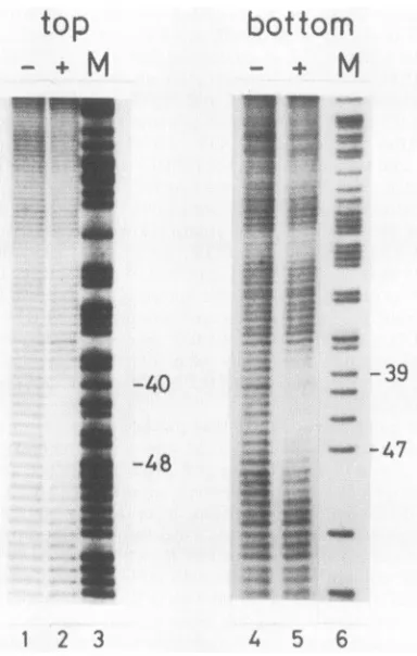
FIG. 1. MPE Fe(II)footprints ofNFIII onthe Ad2origin.The 331-bpNdeI-XbaIfragment of pHRI, labeledateither the 3'(top)or
the 5' (bottom) end of the XbaI site, was briefly digested with MPE Fe(II) in the absence (-)orpresence(+) ofNFIII (pKB67-88 DNA-cellulosefraction). Products were electrophoresedon an 8%polyacrylamide gel in parallel withanA+Gsequencing reaction (M). Numbersindicateposition in the Ad2ITR.
the NFIII-binding site more accurately, we performed
MPE Fe(II)
footprinting
with NFIII. The use ofMPE Fe(II) instead ofDNase I as
cleaving
agent has twomajor advantages. (i) In contrast to DNase I,
cleavage by
MPE Fe(II) occurs much more
randomly,
and (ii)MPE Fe(II) is smaller than DNase I and
consequently
artifacts caused by steric hindrance are less
likely
to occur.Thus, MPE Fe(II) footprinting delimits the
binding
sitemore accuratelythan DNase I
footprinting
(18, 37).The end-labeled331-bp Ad2
origin-containing
NdeI-XbaIfragment of pHRI wasincubated with NFIII, cleaved with
MPE. Fe(II), and analyzedon a
denaturing polyacrylamide
gel (Fig. 1). Densitometer
scanning
of theautoradiogram
revealed agradual increase ofprotection at the borders of
thebinding site. NFIII protects 11 to 13nucleotides in both
strands more than 50% against
cleavage
by
MPE Fe(II) (see Fig. 5A), while 20 phosphate bonds were protectedagainst cleavage by DNase I (30). Ifwe define thebinding
site as the region protected more than 50% against
MPE Fe(II)cleavage, the
NFIII-binding
site extends fromposition 37 to position 51. This agrees well with results
obtainedbytheanalysisof deletionmutantsspanningthe left border(see Table 1).
NFIII contact points within Ad2 origin-binding site. To
identify contact points of NFIII with its recognition
se-quence, we employed hydroxyl radical footprinting and
chemical modification of DNAby alkylatingreagents.
ff--f..
..f.
*W..
oqw
ON
bottom
-
4
8
M
-40
q s. _ ,
*ebh...m
U.4pm a~~~~~~~~~~~~~~~~~~~~~~~~~~~~~~~~~~~~~~~~~~~~~~
-48'w
1
2
3
456
_
.s1-M
.W.
W...
-39
-47
1 am
...o
0_
[image:3.612.332.535.70.465.2]7
8
FIG. 2. Hydroxyl radicalfootprintsof NFIII onthe Ad2origin. Labeled DNAfragments,asdescribed in thelegendtoFig. 1,were digestedbyhydroxylradicals for 4 min after incubation with 4or8 ,ul orwithout (-) NFIII(pKB67-88 DNA-cellulosefraction). Prod-ucts wereanalyzedon an8%polyacrylamide gelinparallelwithan A+G sequencing reaction (M). Numbers indicate position in the Ad2 ITR.
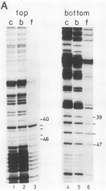
Hydroxyl radical footprinting. MPE. Fe(II) strand scis-sion is probably mediated by the production of hydroxyl radicals which induce oxidative degradationof the deoxyri-bosering.Recently,anotherfootprinting techniquehas been developedonthe basis of thesamereactionamonghydroxyl radicals andDNA, designated hydroxylradicalfootprinting (49).With thistechnique,hydroxylradicalsgeneratedbythe reductionofhydrogen peroxideby iron(II)cleavethe DNA strand unless theprotein is in close contactwith the sugar ring in the DNA backbone. Figure 2 shows the hydroxyl
radicalfootprintof NFIII onthe Ad2 origin. Densitometric scanning revealed that two regions on both strands were partially protected by NFIII. The most strongly protected region coincided with the center of the MPE Fe(II) foot-print (see Fig. 5A), whereas a smaller, weakly protected regionwas detectedaroundposition 38.
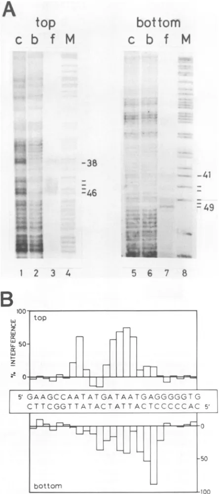
Ethylation interference. Todetectphosphatecontacts be-tween NFIII and the DNA backbone, an end-labeled Ad2
op
iw-k,
P.
0
k
0
on November 10, 2019 by guest
http://jvi.asm.org/
[image:3.612.79.271.71.373.2]NUCLEAR FACTOR III RECOGNITION SITE 3095
origin-containing DNA fragment was partially ethylated by
1-ethyl-1-nitrosourea under conditions in which less than
onephosphategroup per 200 bpismodified. After bindingto
NFIII and separation of protein-bound and free DNA by
filtration over nitrocellulose, the DNA was cleaved at the
sites of ethylation andanalyzed on adenaturing
polyacryl-amide gel (Fig. 3). Bands that were relatively weak in the filter-bound DNA pattern and predominant in the filtrate
DNA pattern represent positions of ethylated phosphate
groups that interfere with binding of NFIII (Fig. 3A). Thepercentage of interferenceis plottedin ahistogram (Fig.
3B) which illustrates that ethylation of some phosphates
almost completely prevents binding ofNFIII while
ethyla-tion at other positions only partially interferes. More than
50% interferencewasobservedafter ethylation ofthe
phos-phate 5'from position 39, 44, 45, 46, or 47inthe top strand
and 5' fromposition 48 or 49inthe bottom strand. A 25 to
50% level of interference was observed after ethylation of
thephosphate5'from position41, 44, 45, or 47 in thebottom
strand. The positions of phosphate contacts are shown in
Fig. SA.
The differences in interference might be explained by
differential effectson NFIIIbindingof ethylation at either of
the two free oxygen atoms of the phosphate or by variable
binding modes between thephosphate groupand the amino
acid side chains involvedin backbone binding.
Methylation interference. Dimethyl sulfate methylates
gua-nine residues at the N7 position in the major groove and
adenine residues at the N3 position in the minorgroove of
the B-DNA helix. The DNA can be specifically cleaved at
methylated residues by chemicaltreatment. This procedure
provides an approach to identify G and A contacts of a
(sequence-specific) DNA-binding protein since methylation
of certain residues might interfere with binding. Partially
methylated Ad2origin-containing DNAfragments were in-cubated with NFIII, and protein-bound DNA fragments were separated from free DNA byfiltrationover
nitrocellu-lose filters. After cleavage at the modified residues,
filter-bound(protein-bound) and filtrate (free)DNA wereanalyzed
on asequencing gel (Fig.4).Methylation oftheGatposition
42 andofthe A's atpositions 43, 45, and46in thetop strand
and at positions 44 and 47 in the bottom strand shows
relatively strong interference (30 to 60%) with binding of
NFIII.Thehistogram displayingthe percentagesof interfer-ence shows that about 20% interference is observed for
methylation of the A residues at positions 39, 40, and 41.
Methylation ofallotherpurines didnotsignificantly interfere
with NFIII binding. Since the probe was not completely
saturated with protein, a small percentage of controlDNA
bands is present in the f lanes. These
methylation-interfer-enceexperiments indicate that both major and minorgroove
contacts areinvolved in theinteraction betweenNFIIIand
the Ad2 origin in contrast to the NFI-Ad2 interaction for
which only majorgroove contacts were observed (10).
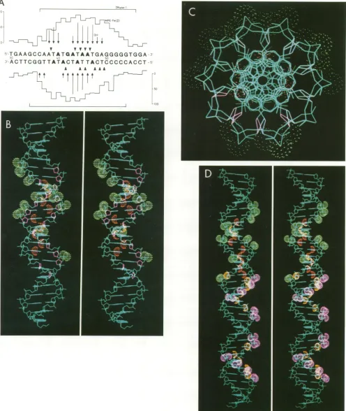
NFIII-bindingsite in Ad2origin.Theresults of all
protec-tion and interference experiments are summarized in Fig.
5A. For the top strand, hydroxyl radical footprinting de-tectedcontacts at all positions in which phosphate contacts
werefound,while in the bottom strand additional phosphate
contacts were observedatpositions48and 49.
Thespatial
pattern
of the contacts identified in theNFIII-binding site on theAd2 origin is shown in
computer-gener-atedstereographics (Fig.
SB).
Contactpoints are indicatedasorange (N7ofGresidue,N3ofAresidue)and green(Oof
phosphate) spheres and as purple deoxyribose rings. This
pattern
illustrates that NFIII contact points are scatteredA
top
c
b f M
bot
tom
c
b f
M
-38
-41 -46
- 49
iii
12: 31 2 3 4
z LJ
L.J
z
0
5.
li
i.
=s 1
%M 4-x
5 6 7 8
top
top 1
F----H
GAAGCCAATATGATAATGAGGGGGTG
CT TCGGTTATACTAT T ACTCCCCCAC 5'
0
FIG. 3. Ethylation interference analysis of the NFIII-binding site.(A)Partially ethylatedDNAfragments (describedin thelegend to Fig. 1) were incubated with NFIIIand separated in a protein-bound (lane b) and a free (lane f) fraction by filtration over
nitrocellulose. Aftercleavageatethylatedpositions, bothfractions were analyzed on an 8% polyacrylamide gel in parallel with the cleaved input DNA fragment (lane c) and an A+G sequencing marker(M). Positionsinterferingformorethan25%whenethylated areindicated. Numberscorrespondtopositionin the Ad2 ITR.(B) Quantitation ofethylationinterference. Levelsofinterferencewere
determined afterdensitometric scanningoftheautoradiogram and
areplottedinahistogramfor the Ad2 sequence betweenpositions31 and56.
VOL.62, 1988
-1-1 -
LLJ
on November 10, 2019 by guest
http://jvi.asm.org/
[image:4.612.328.550.90.591.2]3096 PRUIJN ET AL.
A
top
c
b
f
f..
- -I4
-bottom
c
b f
cb_
UT.:<.
-B
LJ
v z
Li
LA-z top
50
Ii
nfh -11
FH
JjI11I
5'GAAGCCAATATGATAATGAGGGGGTG
CTCGGTTATACTATTCCT T C;ACTATTACTCC CC C ACCCCC 5'
-- TI r r 1 T T-lr!
"mu
50
*ILa
-40 _ - -39
-46
2 3
Oman -47
[image:5.612.91.276.72.405.2]4 5 6
FIG. 4. Methylation interferenceanalysis ofthe NFIII-binding site. (A) Partially methylated DNA fragments (described in the legend to Fig. 1) were incubated with NFIII and separated in a
protein-bound (lane b)andafree(lanef)fractionby filtrationover
nitrocellulose. Aftercleavage at methylated positions, these frac-tionswereanalyzedon an8%polyacrylamide gelinparallelwith the
cleavedinputDNAfragment (lane c).Residues thatinterferedmore
than 10% when methylatedare indicated. Numberscorrespond to
position in the Ad2 ITR. (B) Quantitationofmethylation
interfer-ence. Levels of interference were determined by densitometric
scanningof theautoradiogramandareplottedinahistogramforthe
purinesbetweenpositions31 and56 in the Ad2sequence.
overtherecognition sequenceand notconfinedtoone side
of thehelix, which isevenmoreevident from aviewdown
theaxis of the helix (Fig. 5C). Previously, the NFI-binding site on the Ad2 origin was analyzed in a similar way (10).
Since thebinding sites for NFI and NFIIIontheAd2origin are so closely spaced, we also generated a stereograph in
which all known contact points for both NFI (phosphate contacts are purple, base contacts are yellow) and NFIII
(phosphate contacts are green, base contacts are orange)
were highlighted (Fig. 5D). This picture emphasizes the
slight overlap of the binding sites for thesetwoproteins on
theAd2origin. Remarkably, thetwobases ofbp 39 and the twophosphate groups ineach strand nextto bp 39contact both proteins. Also (weak) deoxyribose contacts forNFIII
were detected very close to NFI phosphate contacts at positions 36 and 37. It should be stressedthat thesecontact point analyses were performed for both proteins
indepen-dently and thus donotexclude the possibility that changes
occurwhen bothproteinsarebound simultaneously.
Interaction of NFIII with origin of other adenovirus
se-rotypes. We compared the NFIII-binding sites ofa number
of different adenovirus serotypes containing small
differ-enceswithin theNFIII-binding region. Gel retardation
com-petition experiments were carried out with an end-labeled
85-bp EcoRI-XbaI fragment of p4A85A containing the Ad4 origin asprobe. Without specific competitor DNA, asingle
retarded band is observed after incubation with NFIII, which represents a specific NFIII-DNA complex (31) (Fig.
6). Origin-containing fragmentsof about 400bp, eitherfrom
the human serotypes Ad2, Ad4, Adl2, Ad4O, or Ad4l or
[image:5.612.324.516.73.233.2]from the simian serotype SA7, were used as competitors.
Figure 6 shows the results for the Ad2, Ad4, and Adl2 competitors. Relative bindingaffinitieswerecalculatedfrom
themolar concentration ofcompetitor DNA that resultedin 50% competition (Table 1). In agreement with previous results(31), the affinity for the Ad4 origin is higher thanthat for the Ad2 origin. NFIII also binds stronger to the SA7-binding site, which contains only one point mutation
com-pared with the Ad2-binding site (23), whereas the affinityfor Adl2 is lower. The NFIII-binding sequence of Ad4O and Ad4l deviates from Ad2 only at the right border of the binding site. Similar binding affinities were observed for
these origins. The relative binding affinitiesfortwo control DNA(pUC) fragments wereless than 0.2%(31).
Interaction of NFIII with mutated Ad2-binding sites. The
very similar affinities of NFIII for Ad2, Ad4M, and Ad4l suggeststhatnucleotidesbeyond position50of Ad2arenot important forNFIIIbinding, thereby definingarightborder
tothebinding site.Thepresenceof several deletionmutants deletingtheNFIII-bindingsequencefrom the left enabledus
toobtaininformation abouttheleft border. Fragmentswere
isolated from deletion mutants pHRA40, pHRA41, and pHRA43, which containthe Ad2 ITRstartingatposition 40, 41, and 43, respectively. The results in Table 1 show that mutation ofnucleotides before position40 of Ad2 reduced the binding affinity more than threefold. Mutation of one
additionalnucleotide oftherecognitionsequence(pHRA41)
resultedinafurthertwofold decrease inaffinity. Surprisingly
H
bottom
J. VIROL.
on November 10, 2019 by guest
http://jvi.asm.org/
A
Lr--l
1--.
L
.-I.
-.- I
TGAAGCCAATATGATAATGAGGGGGTGGA:
--ACTTCGGTTATACTATTACTCCCCCACCT--A AA AAA
[image:6.612.63.559.36.624.2]titlist
FIG. 5. (A) Summaryoffootprintingandalkylation interferenceassaysof theNFIII-bindingsite in the Ad2ITR.Results fortop-strand analysesareshown above thesequence; results for bottom-strandanalysesareshown below. DNase Ifootprints (30)areindicatedby large brackets. Levels ofprotection (Oto1009o)bybound NFIIIfromcleavagebyMPE Fe(II)areindicatedbythehistogramsilhouette.Bases whose deoxyribose is protected from attack by hydroxyl radicalsareindicated byarrows;the length of the arrowindicates the level of protectionbyNFIII. Arrowheads indicatephosphategroupswhoseethylationinterferes morethan25% withNFIIIbinding. Baseswhose methylation interferes more than 10o with NFIII binding are in bold type. (B) Computer-generated stereographic side view of the NFIII-binding site.The Van derWaals radii ofG-N7,A-N3 (both orange)andphosphate-free oxygen(green)atomsincontactwith NFIII areshown asspheres; protected deoxyribose rings (hydroxyl radicalfootprinting)areshownaspurplepentagons. InpanelC,thehelixis tilted 900toobtainaviewdownthe axisof the helix. (D)Stereographic side view of both the NFI- (phosphate contacts,purple; basecontacts, yellow) and theNFIII(phosphatecontacts, green;basecontacts,orange)-binding sites.InpanelsBandD,bp54ofAd2isthe top basepair, and in panelC, bp31is the frontbasepair.
3097
LL-i
on November 10, 2019 by guest
http://jvi.asm.org/
3098 PRUIJN ET AL.
Ad2
Ad4
Ad12
[image:7.612.61.297.73.264.2] [image:7.612.62.558.557.702.2]-. tCJ 7) -0
...
to ...1 2 3 4 5 6 7 8 9 10 11 12 13
FIG. 6. Gel retardation competition assay for Ad2, Ad4, and
Adl2. An end-labeled 85-bp EcoRI-XbaIfragment containing the Ad4originwasincubated withNFIIIin thepresenceof increasing
amountsofunlabeled competitorDNA,and the resultingcomplexes
wereanalyzedon anondenaturingpolyacrylamide gel. Lanes:1,no
competitor; 2 to 5, 16, 32, 64, and 128 ng ofa 409-bpfragment containing theAd2 origin; 6to9, 4, 8, 16,and 32 ngofa384-bp
fragmentcontaining theAd4origin;10to13,32, 64,128,and 256ng
ofa400-bp fragment containing the Adl2 origin. BandFindicate the positions ofthe DNA-protein complex and ofthe free DNA fragmentinthe gel,respectively.Thearrowmarks thestartpointof migration.
however, pHRA43 binds somewhat strongerthan pHRA41. This might be explained by the restoration of the original
sequencefrom position 37to39by the ligated EcoRI linker. Inthisregion,backbonecontactpoints with the proteinwere
detected (Fig. 5A). Moreover, the presenceoftwo
consec-utiveAresiduesin frontof T-39coincided with high binding affinities when cellular NFIII-binding sites were compared
(31).
Finally, the relative binding affinity for the point mutant Xpm46Gwas 10-fold reduced. This A- G transversionwas
previously shownto preventstimulation of DNA replication by NFIII(30).
Stimulation ofDNAreplication ofAd2, Ad4, and Adl2 by NFIH. The previously reported differences in the level of
stimulation of DNA replication of Ad2 and Ad4 by NFIII (31) and the results obtained for the point mutant Xpm46G (30) suggested that stimulation of adenovirus DNA replica-tion by a fixed amount of NFIII is directly related to the affinity of NFIII for the adenovirus origin. In order to investigate this more systematically, we determined the
levelsof stimulation of Ad2, Ad4, and Adl2 DNAreplication in vitro by increasing amounts of NFIII. Using terminal protein-free plasmid DNA as template, initiation in vitro
only takes place when the origin isexposedatthe end ofa
DNA molecule (48, 50). Therefore, plasmids pHRI and p4A85A were digested with EcoRI and Avall and
pAdl2-RIC1 was digested with EcoRI and AvaI to yield origin-containing fragments of1,528, 1,503, and 1,642 bp,
respec-tively. Equimolar amounts of origin-containing fragments
were incubated with the purified viral replication factors
(Ad5 DNA-binding protein, Ad5precursorterminal
protein-polymerase complex) in the presence of deoxynucleoside
triphosphates. NFI was left out of the reaction mixtures since the Ad4 origin lacks an NFI-binding site. Previous
results showed that NFIII alone can stimulate Ad2 DNA
replication (31). Replication products were visualized by
electrophoresis of the reaction mixture inanSDS-containing agarosegel followed byautoradiography. Duetothe reduced mobility of primary replication products which are
cova-lently attached to the primer pTP, a separation from input
DNA fragments was obtained. The results in Fig. 7 show
that the maximal levelof stimulationby NFIII isdependent
ontheaffinity of NFIII for the adenovirusorigin. A saturat-ingamountof NFIII stimulated Ad2 replication 5-fold, Ad4 replication 10-fold, and Adl2 replication 2-fold. With the Ad4template, afaster-migrating bandwas observed, which
presumably represents single-stranded DNA originating from multiple rounds of replication, thus indicating a high
level ofreplication (19, 51).
DISCUSSION
Our detailed study oftheNFIII-binding site on the Ad2
genomehas defined thebinding bordersaswell asmanyof
thepossible contactsthat NFIIImakes with itsrecognition
sequence. MPE. Fe(II) footprinting delimited the binding site between positions 37 and 51 in the Ad2 ITR. The previously derived consensus sequence for optimal NFIII binding, 5'-TATGCAAAT-3' (31),correspondstothecenter of the MPE. Fe(II) footprint, taking into account the two TABLE 1. Adenovirus NFIII-binding site sequencesandtheir relative binding affinities
Virusormutant NFIIIrecognitionsequencea
Relative
affinitybinding()40C 50c
Ad2 C C A A T A T G A T A A T G A G G G G 100
Ad4 T T A A T A T G c A A A T A A G G c G 250
SA7 C C A A T A T G c T A A T G A G G T G 200
Adl2 C C A A T A T T A A A A T G A A G T G 55
Ad40 C C A A T A T G A T A A T G A G G G A 83
Ad4l C C A A T A T G A T A A T G A G T G A 83
pHRA&40 A T T c c A T G A T A A T G A G G G G 30
pHRA&41 A A T T c c T G A T A A T G A G G G G 15
pHRA43 G G A A T T c c A T A A T G A G G G G 25
Xpm46G C C A A T A T G A T A G T G A G G G G 10
a Nucleotidesdiffering fromthe Ad2sequenceareindicatedin smallcapitals. b Calculated fromthe molarratio ofcompetitorDNA toprobeat50% competition. cNumberscorrespondtopositionin the Ad2 ITR.
J. VIROL.
on November 10, 2019 by guest
http://jvi.asm.org/
NUCLEAR FACTOR III RECOGNITION SITE 3099
Ad2
AdL4
Adl2
I _ LO o 0o
) AD CN
tCn
- 5a d
-_ 4,
MD_04
4* MD _
- ',N -N
-Apr~~w _
1
2
3
4
5
6
7
8
9
10
1112
13
1415 16 17
18FIG. 7. Stimulation ofAd2, Ad4, and Adl2 DNAreplication in vitroby NFIII. Ad2, Ad4, and Adl2 origin-containing plasmidswere
digestedwithrestrictionenzymestoyield origin-containingfragments (o)of1,528, 1,503,and1,642bp, respectively, whichwereincubated
inareconstituted DNAreplication system(see Materialsand Methods) in the absence (lanes1, 7,and13)or presenceof31nl(lanes 2, 8,
and 14), 62nl (lanes3,9,and15), 125nl (lanes 4, 10, and 16), 250 nl (lanes 5, 11, and 17),or500 nl (lanes 6, 12, and 18)of NFIII (pKB67-88
DNA-cellulose fraction).Duetotheprotein-priming mechanism of adenovirus DNA replication, replication productsarecovalentlyattached
topTP,leadingtoareducedelectrophoretic mobility (arrows). The faster-migratingreplicationproduct with Ad4 (s) presumablyrepresents
single-strandedDNAoriginating from multiplerounds ofreplication.
mutations (CA -* AT) that have occurred in the Ad2
sequence compared with the optimal binding sequence
presentinAd4.
Both at the left- and right-hand side of this consensus sequence,basepairs modulate the relative binding affinityas
observedfrom the mutant studies described here and else-where (31). At theleft-hand side,twoconsecutive Aresidues improve binding, as indicated by the affinity for
immuno-globulin heavy and light chainpromoters (31)aswellasfor
deletionmutantpHRA43. At the right-hand side, 4 additional bp are strongly protected against MPE- Fe(II) cleavage. It
should be noted thatoneof these isanAT basepair(position
49), which appears tobehighly conserved among
immuno-globulinpromoters,a reasonwhy the conserved box in this
case was preferably indicated as a decanucleotide (11)
instead of an octanucleotide. Recent competition
experi-mentswithsynthetic oligonucleotides indicated thata muta-tion of this particular base pair slightly lowers the binding affinity of NFIII (26a). The affinity of Ad4l suggests that position 51canbemutated without alteration of theaffinity.
Contactpoint analysis revealedmany basecontacts. Inter-ference with NFIIIbinding inalkylation interference
exper-iments is most likely due to steric hindrance by the alkyl
groups or to alteration of the charge distribution on the alkylatedpartof the DNA.Therefore, acloseinteraction of
the protein with the site of alkylation is required for inter-ference; consequently, these sites are denoted contact points. Strikingly, allAand G residuescorrespondingtothe previously derivedconsensus sequence (positions 39to 47) appeared to interfere to some extent with NFIII binding when methylated, albeit to various extents. Some of these contacts had been detected previously by methylation
pro-tection, i.e., the G residueatposition42 in the topstrandand the A residues at positions 44 and 47 in the bottom strand (30, 31). The results of the contact point analysis
summa-rized in the computer-generated stereographic representa-tions shown in Fig. 5clearly demonstrate thatcontactpoints
wereidentifiedatall sides of the DNA helix and indicate that NFIII penetrates both the major and minorgroove of the
DNA helix.
The hydroxyl radical footprinting andethylation
interfer-enceexperiments confirm that the rightpartoftheconsensus sequence (positions 42 to 47) represents the core of the binding site. Nevertheless, additional contacts outside the
core were identified. Around position 49, contacted phos-phates whose ethylation strongly interfered with the binding of NFIIIwere observed. Aroundposition 38,asecondarea
of backbone contacts was observed which confirm the
importance of this region.
Closely spaced binding sites for NFI andNFIII. The sum-mary of NFI and NFIII contacts (Fig. SD) indicates, in addition to the pronounced difference between the two binding modes, thatNFI and NFIII bindveryclosetoeach other. In particular, at position 39, a T-base contact (C5, major groove) with NFI is observed (10), while at this positionnotonlyanA-basecontact(complementary strand; N3, minorgroove) but also a phosphate backbone contact with NFIIIwasfound. The importance of this AT-base pair
both for NFI and NFIII binding is also apparent from deletion studies. A mutantcontaining onlythefirst38bpis defective for NFI binding (25), whereas bp 39 is also important foroptimalNFIIIbindingasshownbyanalysis of
pHRA40 andpHRA41 (Table 1). Despite thisclosespacing, NFI andNFIII areabletobind simultaneouslytothe same
DNAmoleculeanddonotexclude eachother(30).The close spacing raises the possibility of protein-protein interactions which could improve the binding by a cooperative effect.
Thus farwehaveobtainednoindications for strong
cooper-ativity by comparing the binding affinity of NFI in the
presence and absence of NFIII and vice versa, but more
NF1Em
'
_
°
I C4 LO o C
M ADLD"
_L £O
ni
-0
0
.d.A. Adowsk
Pr
VOL.62,1988
on November 10, 2019 by guest
http://jvi.asm.org/
[image:8.612.101.530.67.290.2]3100 PRUIJN ET AL.
stringent analyses with higher concentrations of purified proteins may be required to establish this point. Presently,
we are investigating whether alteration of the position and
orientationofthe NFIII-binding site will lead to any change
inbindingor, more importantly, to the functioning of NFIII
in DNA replication. It should be stressed that in
transcrip-tion regulatory elements, NFIII sites are present in both
orientations and at various positions relative to the
transcrip-tion initiation site.
Comparison with other octamer-binding proteins. Several
reports describe the identification of
octamer/decanucleo-tide-binding activities in crude nuclear extracts of various
cell types(2-4, 7, 20, 21, 24, 26, 34, 39, 43-45). Recently, the
purification of four octamer/decanucleotide-binding
tran-scription factorshas been described (12, 29, 38, 47). OTF-1
and OBP100 were purified from HeLa nuclear extracts,
whereasthe B-cell-specific factor OTF-2 was purified from
Namalwa cell nuclear extracts. OTF-1, which stimulates
transcriptionof a human histone H2B gene, was identified as
a 90-kDa protein; OBP100 was identified as a 100-kDa
protein. Thesimilar molecular sizes (NFIII was identified as
a92-kDa protein(29) or as a 95-kDa protein; Pruijn and van
derVliet, in preparation) and DNA-binding specificities raise
thepossibility that NFIII, OTF-1, and OBP100 are identical
proteins. A strong indication that at the least they share
identicalDNA-binding domains comes from a comparison of
methylation interference experiments and effects of
muta-tions within binding sites. The methylation interference
results for NF-A1, NF-A2,
oct-Bl,
oct-B2, OTF-1, OTF-2,andOBP100 mostly coincide with the methylation
interfer-ence/protection results for NFIII (30, 31, 34, 38, 47).
Fur-thermore, the reduction of binding affinity by a number of
mutations within the binding sites of these proteins reflects
theobserved effects of similar mutations on NFIII binding
(3, 4, 34, 44, 45). For instance, a double point mutant
(oct.
a) of an H2B promoter octamer, containing twotrans-versions comparable to those of Xpm46G and
Adl2
to-gether, lost the ability to compete for binding of OTF-1
(crude extract) to the wild-type H2B promoter (44).
The results of competition experiments performed by
Rosalesetal. (34) for binding of HeLa
oct-Bl
to the octamermotifof the SV40enhancer, however, only very marginally
correspond toour results (31). The high binding affinity for
the Ad2 NFIII-binding site compared with sequences
con-taining a complete match with our consensus sequence is
especially surprising. At present, the only plausible
expla-nation for these discrepancies is the assumption that crude
extracts and(partially)purified proteins behave differently in
competition assays. Recent competition experiments
com-paring the binding to the octamer/decanucleotide- and
ACCC-containing
motifs of a K light chain promoter offactors present in a crude Raji nuclear extract with the
bindingof apartiallypurified NFIII preparation and a highly
purified NFIIIpreparationstrengthen the existence of such a
phenomenon. In these studies, an ACCC-containing
oligo-nucleotide competed for octamer/decanucleotide binding of
apartially purified NFIII preparation which was shown to
contain a protein that interacts with the ACCC-containing
sequence byfootprinting techniques. The same
oligonucle-otide,however, didnot compete for octamer/decanucleotide
binding ofhighly purified NFIII (26a).
Stimulation of adenovirus DNA replication by NFIII. The
replication assays for Ad2, Ad4, andAdl2 showed that the
level ofstimulation by NFIII was dependent on the affinity ofNFIII for the origin. Even with high concentrations of NFIII,stimulationof Ad2and Adl2 replication did not reach
the level of
Ad4,
suggesting that NFIII stimulation is not merely related to occupation ofthe binding site but also to the strength ofthe interaction with the binding site. Alter-natively, sequencedifferencesoutsidetheNFIII-binding sitemight affect theactivity ofNFIIIorgive rise to
qualitatively
different interactions among NFIII and other
replication
proteins. In agreement with the formerhypothesis, the lack
of stimulation of replication of the Ad2
point
mutantXpm46G (30) coincides with a low relative bindingaffinity.
The results obtained with the in vitro DNA
replication
system do not necessarily mean that similardifferences exist
in vivo. However, since the origins of adenovirus serotypes
containing a strong NFIII-binding site lack an
NFI-binding
site (e.g.,
Ad4),
it is very attractive to assume that these adenoviruses require a strong NFIII-binding site and conse-quently that their DNA replicationis stimulated stronglyby NFIII.At present, the mechanism of action of NFIII in DNA replication is unclear. The protein might change the DNA structure either at its binding site or at the replication initiation site in a manner that favors the initiation reaction. Alternatively, NFIII might interact (when bound to the DNA) with other replication factors by direct protein-protein interaction, thus promoting the formation of an initiation complex.
ACKNOWLEDGMENTS
We thank M. M. G. Koning and R. Kaptein of the Department of Organic Chemistry (State University of Utrecht) for help with computer graphics; R. Hay, H. van Ormondt, J. L. Bos, A. E.van Loon, E. de Vries, P. J. Rosenfeld, and T. J. Kelly for making the various plasmids available; P. B. Dervan for a gift of MPE; and E. de Vries for critical reading of the manuscript.
This work was supported in part by The Netherlands Foundation for Chemical Research, with financial aid from The Netherlands Organization for Scientific Research.
LITERATURE CITED
1. Anderson, J. E., M. Ptashne, and S. C. Harrison. 1985. A phage repressor-operator comples at 7 Aresolution. Nature (London) 316:596-601.
2. Augereau, P., and P. Chambon. 1986. The mouse immunoglob-ulin heavy chain enhancer: effect on transcription in vitro and binding of proteins present in HeLa and lymphoid B cell extracts. EMBO J. 5:1791-1797.
3. Ballard, D. W., and A. Bothwell. 1986. Mutational analysis of the immunoglobulin heavy chain promoter region. Proc. Natl. Acad. Sci. USA 83:9626-9630.
4. Bohmann, D., W. Keller, T. Dale, H. R. Scholer, G. Tebb, and
I.
Mattaj. 1987.A transcription factor which binds to the enhanc-ers of SV40, immunoglobulin heavy chain and U2 snRNA genes. Nature (London) 325:268-272.5. Borgmeyer, U., J. Nowock, and A. E. Sippel. 1984. The TGGCA binding protein: a eukaryotic nuclear protein recognizing a symmetrical sequence on double-stranded linear DNA. Nucleic Acids Res. 12:4295-4311.
6. Bos, J. L., L. J.Polder,R. Bernards, P. I. Schrier, P. J. van den Elsen,A. J. van der Eb, and H. van Ormondt. 1981. The 2.2 kb E1B mRNA of human
Adl2
andAdS
codes for two tumor antigens starting at different AUG triplets. Cell 27:121-131. 7. Davidson, I., C. Fromental, P. Augereau, A. Wildeman, M.Zenke,and P. Chambon. 1986. Cell-type specific protein binding to the enhancer of simian virus 40 in nuclease extracts. Nature (London) 323:544-548.
8. Dekker, B. M. M., D. A. M. Konings, T. S. Denisova, R. A. Gibadulin, and H. van Ormondt. 1984. The nucleotide sequence of the leftmost
XhoI
fragment(6%) of simian adenovirus SA7P. J. Gen. Virol. 65:1699-1708.9. De Vries, E., W. van Driel, M. Tromp, J. van Boom, and P. C. J. VIROL.
on November 10, 2019 by guest
http://jvi.asm.org/
NUCLEAR FACTOR III RECOGNITION SITE 3101 van der Vliet. 1985. Adenovirus DNA replication in vitro:
site-directed mutagenesis of the nuclear factor I binding site of the Ad2 origin. Nucleic Acids Res. 13:4935-4952.
10. De Vries, E., W. van Driel, S. J. L. van den Heuvel, and P. C. van der Vliet. 1987. Contactpoint analysis of the HeLa nuclear factor I recognition site reveals symmetrical binding at one side of the DNA helix. EMBO J. 6:161-168.
11. Falkner, F. G., and H. G. Zachau. 1984.Correct transcription of an immunoglobulin K gene requires an upstream fragment containing conserved sequence elements. Nature (London) 310: 71-74.
12. Fletcher, C., N. Heintz, and R. G. Roeder. 1987. Purification and characterization of OTF-1, a transcription factor regulating cell cycle expression of a human histone H2b gene. Cell 51:773-781. 13. Gronostajski, R. M. 1987. Site-specific DNA binding ofnuclear factor I: effect of the spacer region. Nucleic AcidsRes. 15:5545-5559.
14. Guggenheimer, R. A., B. W. Stillman, K. Nagata, F. Tamanoi, and J. Hurwitz. 1984. DNA sequences required for in vitro replication of adenovirus DNA. Proc. Natl. Acad. Sci. USA 81: 3069-3073.
15. Hay, R. T. 1985. The origin of adenovirus DNA replication: minimal DNA sequence requirement in vivo. EMBO J. 4:421-426.
16. Hay, R. T. 1985. Origin of adenovirus DNA replication: role of the nuclear factor I binding site in vivo. J. Mol. Biol. 186:129-136.
17. Henninghausen, L., U. Siebenlist, D. Danner, P. Leder, D. Rawlins, P. Rosenfeld, and T. Kelly. 1985. High-affinity binding site for a specific nuclear proteinin thehumanIgMgene. Nature (London) 314:289-292.
18. Herzberg, R. P., and P. B. Dervan.1984.CleavageofDNA with methidiumpropyl-EDTA-Iron(II): reactionconditionsand prod-uct analyses. Biochemistry 23:3934-3945.
19. Horwitz, M. S., and H. Ariga. 1981. Multiplerounds of adeno-virus DNA synthesisin vitro. Proc. Natl. Acad. Sci. USA 78: 1476-1480.
20. Hromas, R., and B. Van Ness. 1986. Nuclear factors bind to regulatory regions of the mouse kappa immunoglobulin gene. Nucleic Acids Res. 14:4837-4848.
21. Janson, L., C. Bark, and U. Petterson. 1987. Identification of proteins interacting with the enhancer of human U2 small nuclear RNA genes. Nucleic AcidsRes. 15:4997-5016. 22. Jones, K. A., J. T. Kadonaga, P.J.Rosenfeld, T. J.Kelly, and R.
Tjian. 1987. A cellular DNA binding protein that activates eukaryotic transcription andDNA replication. Cell 48:79-89. 23. Kelly, T. J. 1984. AdenovirusDNAreplication, p. 271-308. In
H. S. Ginsberg (ed.), The adenoviruses. Plenum Publishing Corp., New York.
24. Landolfi, N. F., J. D. Capra, and P. W. Tucker. 1986. Interac-tion of cell-type-specific nuclear proteins withimmunoglobulin VHpromoterregion sequences. Nature(London) 323:548-551. 25. Leegwater, P. A.J., W. van Driel, and P. C. van derVliet.1985. Recognition site of nuclearfactor I, asequence-specific DNA-binding protein from HeLa cells that stimulates adenovirus DNA replication. EMBO J. 4:1515-1521.
26. Mocikat, R., F. G.Falkner,R.Mertz, and H. G. Zachau.1986. Upstream regulatory sequences ofimmunoglobulin genes are recognized by nuclearproteins whichalso bind to other gene regions. NucleicAcids Res. 14:8829-8844.
26a.Mocikat, R., G. J. M. Pruijn, P. C. van der Vliet, and H. G. Zachau. 1988. AnACCC-containingprotein-bindingsequence in the neighbourhood of thedecanucleotiderecognition site of the immunoglobulin gene promoter. Nucleic Acids Res. 16:3693-3704.
27. Nagata,K., R. A.Guggenheimer, T.Enomoto,J.H. Lichy, and J. Hurwitz. 1982. Adenovirus DNA replication in vitro: identi-fication ofa host factor thatstimulates synthesis of the preter-minalprotein-dCMP complex. Proc. Natl. Acad. Sci. USA 79: 6438-6442.
28. Nagata, K., R. A.Guggenheimer, andJ. Hurwitz. 1983. Adeno-virus DNA replication in vitro: synthesis of full-length DNA with purified proteins. Proc. Natl. Acad. Sci. USA
80:4266-4270.
29. O'Neill, E. A., and T.J. Kelly. 1988. Purification and charac-terization ofnuclearfactor III (originrecognitionproteinC),a
sequence-specific DNA binding protein required for efficient initiation of adenovirus DNA replication. J. Biol. Chem. 263: 931-937.
30. Pruin, G. J. M., W. van Driel, and P. C. van der Vliet. 1986. Nuclear factor III, a novel sequence-specific DNA-binding protein from HeLa cells stimulating adenovirus DNA replica-tion. Nature (London) 322:656-659.
31. Pruijn, G. J.M., W.vanDriel,R. T. vanMiltenburg,and P. C. van derVlet.1987. Promoter and enhancer elementscontaining aconserved sequence motifare recognized by nuclear factor III,aproteinstimulatingadenovirusDNAreplication. EMBO J. 6:3771-3778.
32. Rawlins, D. R., P. J. Rosenfeld, R. J.Wides, M. D.Challberg, and T. J. Kelly. 1984. Structure andfunction of the adenovirus origin of replication. Cell37:309-319.
33. Rjnders,A. W. M., B. G. M. van Bergen, P. C. van derVliet, and J. S.Sussenbach. 1983. Specificbinding of the adenovirus terminal protein precursor-DNA polymerase complex to the origin of DNAreplication. Nucleic AcidsRes.11:8777-8789. 34. Rosales, R., M. Vigneron, M. Macchi, I.Davidson, J. H.Xiao,
and P. Chambon. 1987. In vitro binding of cell-specific and ubiquitous nuclear proteins to the octamermotif of the SV40 enhancer and related motifs present in other promoters and enhancers. EMBO J. 6:3015-3025.
35. Rosenfeld, P. J., and T. J. Kelly. 1986. Purification of nuclear factor I by DNA recognition site affinity chromatography. J. Biol. Chem. 261:1398-1408.
36. Rosenfeld, P. J., E. A. O'Neill, R. J. Wides, and T. J. Kelly. 1987. Sequence-specific interactions between cellular DNA-binding proteins and theadenovirusorigin of DNAreplication. Mol. Cell. Biol. 7:875-886.
37. Sawadogo, M., and R. G. Roeder. 1985. Interaction ofa gene-specific transcription factor with the adenovirus major late promoter upstreamofthe TATA boxregion. Cell 43:165-175. 38. Scheidereit, C., A. Heguy, and R. G. Roeder.1987. Identification
andpurificationof a humanlymphoid-specific octamer-binding protein (OTF-2) that activatestranscription ofan immunoglob-ulin promoterinvitro. Cell51:783-793.
39. Schlokat, U., D. Bohmann, H. Scholer, and P. Gruss. 1986. Nuclear factorsbindingspecific sequences withinthe immuno-globulin enhancer interact differentially with other enhancer elements. EMBO J. 5:3251-3258.
40. Schneider, R., I. Gander, U. Muller, R. Mertz, and E. L. Winnacker. 1986. Asensitive and rapid gel retentionassayfor nuclear factor I and other DNA-binding proteins in crude nuclear extracts. Nucleic Acids Res. 14:1303-1317.
41. Siebenlist, U., and W. Gilbert. 1980. Contacts between Esche-richia coli RNApolymerase andanearlypromoterofphageT7. Proc. Natl. Acad. Sci. USA77:122-126.
42. Siebenlist, U., L.Henninghausen, J. Battey, and P. Leder. 1984. Chromatin structure andproteinbindingin theputative regula-toryregion of the c-myc genein Burkittlymphoma. Cell 37:381-391.
43. Singh, H., R. Sen, D. Baltimore,and P.Sharp. 1986. A nuclear factor that binds to a conserved sequence motif in transcrip-tional control elements of immunoglobulin genes. Nature (London)319:154-158.
44. Sive, H. L., and R. G. Roeder. 1986. Interaction ofa common factor with conserved promoter and enhancer sequences in histone H2B, immunoglobulin, and U2 small nuclear RNA (snRNA) genes. Proc. Natl. Acad. Sci. USA83:6382-6386. 45. Staudt, L. M., H. Singh, R. Sen, T. Wirth, P. A.Sharp, and D.
Baltimore. 1986. A lymphoid-specific protein binding to the octamer motif ofimmunoglobulingenes. Nature (London) 323: 640-643.
46. Stunnenberg, H. G., H. Lange, L. Philipson, R. T. van Milten-burg, and P. C. van der Vliet. 1988. High expression of func-tional adenovirus DNA polymerase and precursor terminal protein using recombinant vaccinia virus. Nucleic Acids Res. 16:2431-2444.
VOL.62, 1988
on November 10, 2019 by guest
http://jvi.asm.org/
3102 PRUIJN ET AL.
47. Sturm, R., T. Baumruker, B. R. Franza,and W. Herr.1987. A 100-kDHeLacell octamerbinding protein(OBP100)interacts differently withtwo separateoctamer-relatedsequenceswithin theSV40enhancer.Genes Dev. 1:1147-1160.
48. Tamanoi, F., and B. W. Stillman. 1982.Function ofadenovirus terminalprotein in the initiation ofDNAreplication.Proc.Natl. Acad. Sci.USA 79:2221-2225.
49. Tullius, T. D., and B. A. Dombroski. 1986. Hydroxyl radical "footprinting":high-resolution information about DNA-protein
contacts andapplication tolambdarepressorand Cro protein.
Proc. Natl. Acad. Sci. USA 83:5469-5473.
50. Van Bergen, B. G. M., P. A.vander Ley, W.vanDriel,A. D. M. vanMansfeld, and P.C.vanderVliet. 1983.Replicationoforigin
containing adenovirus DNA fragments that do not carry the terminal protein.NucleicAcidsRes. 11:1975-1989.
51. Van Bergen, B. G. M., and P. C.vanderViiet.1983. Temper-ature-sensitive initiation and elongation of adenovirus DNA replicationin vitrowith nuclearextractsfrom H5ts36-,H5ts149-, andHSts125-infectedHeLa cells.J. Virol.46:642-648. 52. Van Loon, A. E., R.Maas, R. T. M. J. Vaessen, A. M. C. B.
Reemst, J. S. Sussenbach, and T. H.Rozin.1985. Cell transfor-mation bytheleft terminalregionsof theadenovirus 40 and 41
genomes.Virology147:227-230.
53. Wides, R.J.,M. D.Challberg, D. R.Rawlins,and T.J. Kelly. 1987. Adenovirusorigin ofDNAreplication: sequence
require-mentsforreplicationin vitro.Mol. Cell. Biol. 7:864-874. J. VIROL.