Vol. 63, No. 1 JOURNAL OFVIROLOGY,Jan. 1989, p. 148-158
0022-538X/89/010148-11$02.00/0
Copyright© 1989,American Society for Microbiology
Isolation of
a
Pathogenic Clone of Mouse
Mammary Tumor Virus
DAVID W.
MORRIS,'*
HARVEY D. BRADSHAW, JR.,2 HELMUTH T. BILLY,1 ROBERT J. MUNN,'AND ROBERT D. CARDIFF1
DepartmentofPathology, SchoolofMedicine, University of California, Davis, California 95616,1 andDepartmentof Biochemistry, Universityof Washington, Seattle, Washington 981952
Received5July 1988/Accepted26September 1988
Exogenousmouse mammarytumorvirus(MMTV)wascloned fromaGRmammarytumor.Clone XGRT39
containedafull-length integrated MMTV(GR) provirus and both 5' and 3' hostflanking DNA. The XGRT39 provirus had noapparentstructural changes associated withcloningand retained theexogenousMMTVgag
genepoisonsequence. Whenintroduced intoratmammaryadenocarcinoma LA7cells, theXGRT39provirus
was fully expressed. XGRT39-transfected LA7 cells made MMTV RNA, had gp52 SU protein on the cell
surface, and produced B-type retrovirus particles characteristic of MMTV. Mammary tumorsdeveloped in
hormone-stimulated BALB/c females injected with MMTV from XGRT39-transfected LA7 cells [MMTV (XGRT39)]. The tumors had new, clonally integrated copies of the MMTV(XGRT39) provirus and were expressingMMTVantigen.Thesedataindicate that the XGRT39provirusisbiologicallyactiveandpathogenic.
Mouse mammary tumor virus (MMTV) is a
replication-competent, slow-transforming retrovirus that causes
mam-mary adenocarcinomas in female mice (41). In addition,
MMTV causes avariety ofpremalignant mammary lesions andis useful for studiesofmultistepneoplastic development
(27). MMTVexpression is hormone regulated (32), and there
isastrongendocrine influenceontheinduction of mammary
tumors by MMTV (18). Pathogenesis is due in part to
mutation of host genesby MMTVprovirusinsertion (9,29). Geneticanalysisof MMTV and its role in mammarytumor
development has been hampered for many yearsby the lack of infectious and pathogenic DNA clones of exogenous MMTV proviruses. The failuretogeneratesuch clones was
attributedto an MMTV sequencelocalized to the 5' end of
the gag gene,calledthepoisonsequence(5, 10, 17, 24, 39).
This sequence could not beclonedintact inplasmidvectors
and was not recovered from lambda libraries screened by
standard techniques.
Inthisreport,wepresentdataindicatingthat(i)full-length exogenous MMTVprovirusDNAfrom the GRmousestrain
[MMTV(GR)]
canbeclonedandpropagatedin thebacterio-phage lambda vector XL47 by using modifications of
stan-dard screening andamplification protocols, (ii) cloned pro-virus DNA can direct the production of recombinant DNA-derived MMTV(GR) in cultured ratmammary
adeno-carcinoma cells, and (iii) recombinant DNA-derived
MMTV(GR) is tumorigenic. These experiments form the basis for studies, using MMTV(GR) mutants produced in vitro, to address basic problems pertaining to the regulation of MMTVexpression, MMTV infectivity, and the molecular mechanisms ofMMTVpathogenesis.
MATERIALS ANDMETHODS
Mice. The GRSA mouse strain (referred to here as GR)
was obtained from the Netherlands Cancer Institute, Am-sterdam (28), and theBALB/c/Crgl mouse strain (referred to here as BALB/c) was obtained from the Cancer Research
Laboratory, University of California, Berkeley. Mammary
tumorswereidentified by weekly palpation of female mice. Whentumors were 0.5 to 1.5 cmin the longest dimension,
* Correspondingauthor.
theywerecollectedand frozenat -70°C.BALB/c mice used in the tumorigenesis experiment were hormone stimulated
with pituitary isografts by implantation of two pituitaries fromsyngeneicdonors intotheleft no. 4mammaryfatpad.
DNA cloning. A genomic library was constructed in bac-teriophage lambda of MboI partials. Cellular DNA from a
GR mammary tumor was partially digested with MboI to
give restriction fragmentswithamodalsizedistribution of15
to 20 kilobases (kb). This size fractionwaspurified on5 to
20%potassiumacetategradients(25). ThefractionatedDNA was ligated to BamHI-cleaved bacteriophage XL47 (22) arms,and theligation productswerepackaged in vitro,using
strains from Sternberg et al. (37) and standard
packaging
protocols (19). A total of 1.5 x 10⁶ PFU was plated on
Escherichia coli KH802(42)andscreenedby plaque hybrid-ization (2), using MMTV(C3H) probe pCP4.2 (see Fig. 1). Positive cloneswere pickedand plaque purified.
Minipreps of phage DNAs for mapping and subcloning
were prepared from nuclease-treated plate lysates. Plate
lysateswerepreparedasdescribed elsewhere(25),using1 x
105to2 x 105 PFU/100-mm plate.DNase Iand RNase Iwere
addedto1
pLg/ml,
and thesampleswereincubatedfor30minat roomtemperature. Phageparticleswerepelletedat90,000
x g for2h at4°C. Pelleted phage was suspended in 10 mM Tris (pH 7.5)-10 mM NaCI-10 mM trisodium EDTA-1% sodiumdodecyl sulfate
(SDS)-100
,ugofproteinaseK perml and extracted with buffer-equilibrated phenol. Phage DNAwas ethanolprecipitated andsuspendedin 10 mM Tris (pH
7.5-10mMNaCl-0.1 mMtrisodium EDTA.
Restrictionfragments from recombinantlambda clones for
use as probes were subcloned in pUC12 (40). Insert and vector DNAsfor subcloning were purified by electrophore-sis in 5% polyacrylamide gels and electroelution of the
appropriate fragments. Ligations were performed at 15°C
overnight in 50 mM Tris (pH 7.5)-20 mM NaCI-10 mM
MgCl2-1
mM dithiothreitol-1 mM ATP-1 U of T4 DNAligase per ,ul at aDNAconcentration of 10 ,g/ml and a 1:3 molarratio ofvector toinsert. Ligated DNAs were used to transform E. coli JM83 (40) by standard techniques (25).
Miniprep DNA samples (25) from Lac- clones were
screenedby restriction mapping.
Cellular DNA and RNA extraction. Cellular DNAs were
148
on November 10, 2019 by guest
http://jvi.asm.org/
MMTV(GR)
-PSSP SP P S
_:
I1
P P S S
5'LTR gag pol env 3' LTR
C G E F
II
A E H F
r 3
pCP1.4
pDM5
B
D
D
C
pCP4.2 pCP1.8
I I I I I I I I I I I I I I
0 1 2 3 4 5 6 7 8 9 10 11 12 13 14 kb
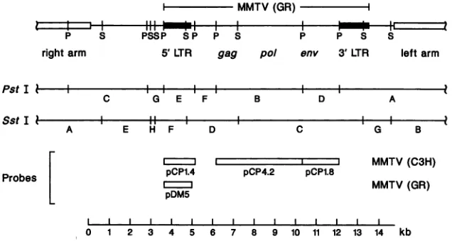
FIG. 1. RestrictionmapofXGRT39 and location of the probes used in this study. The XGRT39PstI(P) and SstI(S) restrictionmaps are
shown. XGRT39 containsanentire MMTV(GR) proviralgenome aswellas5'and 3' host flankingDNAon a14.7-kbMboIpartial.The SstI
Cfragment isuniquetoMMTV(GR) andwasusedasamarkerformousecellsinfected with MMTV(GR)orMMTV(XGRT39).The PstI F and SstI Dfragments contain theexogenousMMTVpoisonsequence.
isolated by homogenizing minced tissue or cells scraped
fromtissue culture plates withaDounce homogenizer in10 mM Tris (pH 7.5)-10 mM NaCI-10 mM trisodium EDTA. The suspensions werelysed by adding SDStoa
concentra-tion of0.5% and then extractedoncewithbuffer-equilibrated
phenol, once with buffer equilibrated
phenol-chloroform-isoamyl alcohol (25:24: 1), andoncewithchloroform-isoamyl
alcohol(24:1). Extractsweredialyzed for 48 hat4°C against
several changes of10 mM Tris (pH 7.5)-10 mM NaCl-0.1
mM trisodium EDTA to remove residual organic solvents.
DNA concentrationswere determined by measuringA26.
Total cellular RNAs were prepared by standard
tech-niques (25). Guanidine isothiocyanate lysates of tissue
cul-ture cells were pelleted through a 5.7 M CsCl cushion to
removeDNAandprotein. The RNA pelletswere extracted with chloroform-1-butanol (4:1) and then suspended in wa-ter.
Southern blot analysis. Restriction enzymes were pur-chased from Bethesda Research Laboratories, Inc. (Gai-thersburg, Md.). CellularDNAs(5 to 10 ,ug)were digested withathreefold excessofenzymefor 3 h under buffer and temperatureconditions recommended by the manufacturer. Reactions were stopped by adding trisodium EDTA to a
concentration of 50 mM. Digested DNAs were
electropho-resed in horizontal 1%agarose gelscontaining 50 mMTris base, 20mM sodium acetate, 2 mM tetrasodium EDTA, 22
mM NaCl, and 30 mM acetic acid. After electrophoresis, gels were stained with ethidium bromide and checked by transilluminationat260nmtoverify complete digestion.The
restriction fragments were transferred to nitrocellulose by
themethodof Southern(35)in3.0 MNaCl-0.3 M trisodium citrate (38). Hybridizations wereperformed in 2 ml of 45%
formamide, 0.45 M NaCl, 0.045 M trisodium citrate, 3x
Denhardtsolution,100,ug ofyeastRNAperml,and 50,ugof base-sheared salmon spermDNApermlat37°C for 3days
with aprobe concentration of5 x 106 cpm/ml. Blots were washed once in 0.3 MNaCl-0.03 Mtrisodium citrate-0.1% SDS atroomtemperaturefor 10min,twice in 0.03M NaCl-0.003 M trisodium citrate-0.1% SDSatroomtemperature for 10min, andonce in0.3 M NaCl-0.03 M trisodium
citrate-0.1%SDSat65°Cfor 30min. Filterswere wrappedinSaran
Wrap and autoradiographed for 2to 7 days, using XAR-5 film(Eastman KodakCo.,Rochester, N.Y.) with aCronex
Lightning-Plus intensifying screen (E. I. du Pont de Ne-mours &Co., Inc., Wilmington, Del.) at -70°C.
RNA dot blot analysis. RNAs samples for dot blotanalysis
were bound to nitrocellulose by manifold filtration in
satu-rated Nal (3). Residual DNA and rRNA does not bind efficiently to nitrocellulose in high-molarity Nal solutions. Hybridizations were performed as described for Southern
blotanalysis with the following exceptions: the formamide concentration of thehybridization solutionwasincreasedto
50%, and 1% SDSwas added.
Probes. The probes used in this study are diagramed in
Fig. 1. pBR322 clones of three of the four internal PstI restriction fragments from unintegrated exogenous MMTV(C3H) provirus were provided by Majors and
Varmus (24). The gag-pol, env, and long terminal repeat
(LTR) clones were subcloned into pUC12 and named
pCP4.2, pCP1.8, and pCP1.4, respectively. An equal-mass mixture of the inserts from these three plasmids is referredto asrepprobe inthis report. pDM5 isa 1.3-kb PstI-to-PvuII fragment from XGRT39 containing the MMTV(GR) 5' LTR cloned inPstI- and SmaI-cleavedpUC12.
Preparation of radiolabeled DNAs. 32P-labeled MMTV
(C3H) cDNA was prepared by reverse transcription of random-primedviralRNA. A mixture of500ngof viralRNA and200 ,ug ofbase-sheared calfthymus DNAprimers was
heatedto100°Cfor1minand allowedtoannealfor 20minat
37°C in 25 mM KCl.Thereversetranscription reactionwas
carried out with 100 ,uCi of [a-32P]dATP (3,000 Ci/mmol;
Dupont,NENResearchProducts,Boston, Mass.)ina50-pul reaction mixture containing 50 mM Tris (pH 8.1), 10 mM
MgCl2, 5 mMKCl,2mM dithiothreitol, 25 ,ugof actinomy-cin D per ml, 0.2 mM each dCTP, dGTP, and dTTP, 0.03
mMdATP, 10,gofMMTV(C3H)viral RNA perml, and1 U of avian myeloblastosis virus reversetranscriptase (Life
Sciences, Inc., St. Petersburg, Fla.) per ml for 30 min at
37°C.The viral RNAtemplatewasbasehydrolyzedafterthe reaction for 3 h in 0.3 M NaOH-50 mM trisodium EDTA-0.5% SDS. Unincorporated [a_-32P]dATP was removed by gelfiltration of the hydrolyzedreaction producton a
Seph-adex G-50 column.
32P-labeledplasmidDNAwaspreparedbynicktranslation ofgel-purified insert DNA. Insert DNAs from various
plas-mids were purified by gel electrophoresis of restriction
P S
rightarm
PstI b-SstI
s-Probes [
leftarm
A
l
G B
MMTV(C3H)
MMTV(GR)
on November 10, 2019 by guest
http://jvi.asm.org/
[image:2.612.138.456.77.247.2]150 MORRIS ET AL.
enzyme digests in 5% polyacrylamide gels, followed by
electroelution of the desiredfragments.Purified insert DNAs
(200 ng) were nick translated with 100 ,uCi of[c-32P]dCTP
(3,000 Ci/mmol; ICN Biomedicals Inc., Irvine, Calif.) in a
20-pIreaction mixture containing 50 mM Tris (pH 7.5), 10 mM MgCl2, 1 mM dithiothreitol, 50 pug of bovine serum
albuminperml,0.1 mM eachdATP, dGTP,anddTTP, 10pg
of DNase I (SigmaChemical Co., St. Louis, Mo.) per ml,
and0.5 U of DNApolymerase I (BethesdaResearch
Labo-ratories)per
[Li
at 15°C for 0.5 to 2 h (31). Unincorporated [ao-32P]dCTP was removed by gel filtration ofthe reactionproducton aSephadexG-50 column. Probesroutinelyhada
specific activity of 2 x 10⁸to4 x
10'
cpm/pg.Transfections. The LA7 cell line selected for the gene
transferexperimentswasderived froma dimethylbenz(a)an-thracene-induced rat mammary adenocarcinoma (1, 11).
CellsweremaintainedonDulbecco modifiedEagle medium
supplemented with 5% calf serum and 5 pug of dexame-thasone perml. LA7 cells were seeded at a densityof5 x
105/30-mmplate48 hbefore transfection. XGRT39DNAwas cotransfected with pSV-1/neofrom P. A. Luciw (a pBR328
derivative ofpSV2neo) carrying the TnS neo gene, which confers G-418 resistance to mammalian cells (36). Approxi-mately60,ugof XGRT39orwild-type ADNAwasmixed with 20 pug of pSV-1/neo DNA, ethanol precipitated, and
sus-pended in 2 ml of Hanks buffered saline solution with a
carefully adjusted pHof 7.10. Then 0.2 mlof 1.25MCaCl2
wasaddeddropwisewhile the solutionwasgentlymixedby bubblingairthroughthesample.Theprecipitatewasallowed toformfor 20minatroomtemperatureand then diluted with 2 ml of Hanks balanced salt solution. A 0.5-ml aliquot of
precipitatesolutionwasaddedperplateandadsorbed for 20
min at room temperature before the addition of 5 ml of
growth medium. After incubation at37°C for 4 h, the cells were washed and glycerol shocked for 3 min in Hanks balanced salt solution containing 15% glycerol (23). Selec-tion forgene transferwascarriedoutbythe addition of 400
pLgofG-418permltothe growth mediumbeginningat24 h after transfection. More than 100 foci per plate were ob-served. Individual foci were picked and expanded into cell linesforanalysisofXGRT39-directedexpressionof MMTV.
Immunoperoxidase staining. Mammary tissues were fixed in70%ethanol-aceticacid-formaldehyde (20:1:1)foratleast 24 h (21). Fixed tissues were embedded in paraffin and sectioned. The sections were immunoperoxidase stained withpolyclonalrabbit anti-MMTV antisera andaVectastain ABC kit (Vector Laboratories, Burlingame, Calif.)
accord-ing to the instructions of the manufacturer and counter-stained withhematoxylin. A1:1,000dilutionof anti-MMTV antiserum wasused. This antiserum was prepared by using
MMTV(C3H), purifiedfromaBALB/cfC3H tumorprimary culture, as an immunogen (7).
Indirectimmunofluorescent staining.Live cellswere
ana-lyzedforcell surface expressionof the MMTV SU protein (20) gp52 by indirectimmunofluorescentstaining, using the
anti-gp52 rat monoclonal antibody 72H3 from A. Sonnen-berg. Approximately 104cells were passagedonto ethanol-washedglasscover slips in24-well tissueculture plates and
maintained in 0.25 ml ofgrowth medium containing5 pLg of dexamethasoneperml.Then 60
RI
ofprimary antibody 72H3 was added directly into the growth medium, and the cellswere incubated at 37°C for 30 min. The cells were washed threetimes toremovetheunboundprimary antibody, and 20
,u1 offluorescein isothiocyanate-labeled goatanti-rat immu-noglobulin G antiserum in 1 ml of growth medium was added. After incubation at 37°C for 30 min, the cells were
washed three times, fixed in 1 ml of 2% paraformaldehyde, covered with cover slips, and photographed with a fluores-cence microscope.
Electron microscopy. Tissueculture cells for transmission electron microscopy were fixed in 2.5%glutaraldehyde-0.1 M sodium cacodylate (pH 7.2) for 2 h at roomtemperature and then overnight at 4°C. The fixed cells were washed twice with 0.1 M sodium cacodylate (pH 7.2), postfixed with 1% Os04-0.1 M sodium cacodylate (pH 7.2) for 1 h at 4°C, washedfor 30 s in distilled water, andstainedfor 2 h with 2% aqueousuranylacetate. Inamodification of thetechnique of Brinkleyetal. (4), the cellswereembedded insitu, using an
epon-aralditeresin. Thin sections of selectedareaswerecut with adiamond knife, mounted on 200-mesh grids, stained with uranyl acetate and lead citrate, and examined in a
PhilipsEM-400electron microscope.
MMTV infections ofBALB/c neonates. MMTV for injec-tion into BALB/cneonates waspreparedfrom the
XGRT39-transfected LA7 cell line L16-9B10 or the GR mouse
mam-marytumorcell line GR3A. Cell culturesweresupplemented with 5pLg ofdexamethasone permltoincrease virus expres-sion. Cells were scraped from the culture surface with a rubberpoliceman, and both cells and mediumwere homog-enizedgentlyinaDouncehomogenizer.Thesuspensionwas
clarifiedbylow-speed centrifugation in a clinicalcentrifuge for 5 min. Additional debriswas removeby centrifugation at 15,000 x g for 30 min in a refrigerated centrifuge. Virus
particles were pelleted from the supernatant at 4°C by centrifugationat150,000 x gfor 30min inanultracentrifuge. Virus pellets were suspended in approximately 200 ,ul of Hanksbalanced salt solution per confluent T75 culture flask.
One-day-old BALB/c pups were injected intraperitoneally with 100
[LI
of virus suspension.RESULTS
Cloning ofa full-length integratedexogenous MMTV(GR) provirus in bacteriophage lambda. A full-length integrated
exogenous provirus was cloned from mouse mammary tu-mor sample GRTu7, which was collected from our GR
mousestrainbreeding colony. Thistumorhadasinglenew,
clonally integrated MMTV(GR) exogenous provirus. A
ge-nomic library was constructed in bacteriophage lambda vectorAL47byusinghigh-molecular-weight tumor DNA and screened with radiolabeled probe pCP4.2 DNA from the
gag-pol region of MMTV(C3H) (Fig. 1). A total of 39 positive plaques were observed, and 24 were successfully
plaquepurified. Phageminiprep DNAswere prepared from theserecombinantsandcompared bySouthernblotanalysis
of SstI digests, using probe pCP1.4. Aclone containingthe
full-length
exogenous GRTu7provirus, XGRT39, wasiden-tified bythe presence of(i)the6.1-kb MMTV(GR)-specific
SstI restriction fragment (13) and (ii) low-molecular-weight
host-virus junction restriction fragments that comigrated with novel exogenous proviral restriction fragments in the
original tumor DNA. XGRT39 contained the entire
MMTV(GR) provirusandboth 5' and 3' host flanking DNA
(Fig. 1). The provirus was integrated upstreamof the int-2 gene(D. W. Morris, P. A. Barry, H. D. Bradshaw, Jr., and
R. D. Cardiff, manuscript in preparation).
WhenXGRT39 was amplifiedto preparephage DNA for molecularanalysis, a deletion mutantappeared (apparently
by homologous recombination between the two LTRs) that removed the exogenous provirus poison sequence. Normal XGRT39 plaques were small and had a relatively low titer. The deletion mutantsformedlarge, high-titer plaques. Dur-J. VlIROL.
on November 10, 2019 by guest
http://jvi.asm.org/
ing successive amplifications the originalXGRT39 clone was rapidly lost, particularly if the clone was amplified by using liquid culture techniques (25).
The deletion problem was circumvented by starting with early picks ofXGRT39, amplifying the clone by plate lysate methods, and limiting the number of sequential amplifica-tions. Secondary picks ofXGRT39 were identified by plaque hybridizations of low-density platings of the primary pick. Individual secondary picks were amplified as 100-mm plate lysates, and DNA was purified as described above. DNA from each amplified secondary pick was checked for deletion mutant DNA by digestion with HindIll, which cleaves MMTV(GR) once near the middle of the genome. Amplifi-cations that had large plaques or restriction pattern evidence of the deletion mutant were discarded. The remaining DNA preparations were pooled. The XGRT39 DNA yield from each individual amplified secondary pick was approximately 1 to 2
jig.
With this procedure, sufficient DNA for molecularanalysis and gene transfer experiments could be obtained.
Exogenous MMTV poison sequence in XGRT39. The re-striction map of the XGRT39 provirus was the same as previously reported maps of exogenous MMTV(GR) (13, 14, 34). Figure 1 shows the XGRT39 restriction map for the enzymes relevant to this report,PstI and SstI. To verify that XGRT39 contains the exogenous MMTV poison sequence, the MMTV(GR) restriction patterns of XGRT39 and a GR mammary tumor cell line, GR3A, were compared (Fig. 2). TheGR3A cell line has a high copy number of the exogenous MMTV(GR) provirus (13). An MMTV viral RNA cDNA
probe was used because it recognizes the entire MMTV
genome, including the poison sequence. All internal MMTV(GR) restriction fragments in the GR3A cell line were also present inXGRT39(Fig. 2). In particular, the1.1-kb PstI fragment that originally localized the exogenous MMTV poison sequence (24) was present inXGRT39(Fig. 2, arrow). No differences in MMTV restriction fragment mobilities between XGRT39 and GR3A DNAs were detected that would suggest that major structural changes had occurred during cloning or amplification. Although these data did not conclusively demonstrate that XGRT39 had an intact, un-rearranged provirus, they were sufficient to justify testing the biological activity of the clone.
Expression of theXGRT39 provirus after transfection into rat mammary adenocarcinoma cells. To test the biological activity ofXGRT39,the clone was introduced into LA7 cells. The LA7 cell line was derived from achemically induced rat mammary adenocarcinoma (1, 11). This cell line was
se-lected for two reasons. First, MMTV expression is tissue
specific; therefore, a mammary cell line was used to maxi-mize the chances of efficient production ofXGRT39-encoded
retrovirus particles. Second, all mouse mammary cell lines have endogenous MMTV proviruseswhich could
hypothet-ically be induced by the experimental manipulations in-volved in these experiments; therefore, a cell line from a closely related species was used.
XGRT39wascotransfectedintoLA7cellswith a pSV2neo
derivative, and G-418-resistanttransformants were selected.
Twenty-one cell lines were established from individual
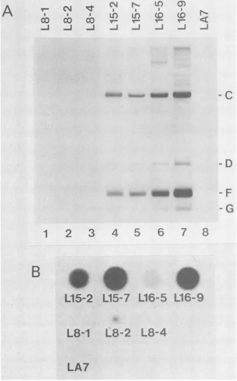
G-418-resistant foci. Southern blotanalysis with MMTV(C3H)
rep probe showed that 76% (16 of 21) ofthe cell lines had stably integrated the unselected XGRT39DNA. Four of the
hGRT39-transfected cell lines (L15-2, L15-7, L16-5, and L16-9) and three control cell linestransfected with wild-type
X DNA(L8-1, L8-2, and L8-4) were arbitrarily selected for
further analysis. Figure 3A shows a Southern blot ofDNAs from these cell lines digested with SstI and hybridized with
cv,
Pst I
z
0
*: .:e
..t.#
.k
.-A %f
[image:4.612.322.537.75.332.2]i1i
ad"|
c)
<:
Sst I
2
z
kb
;
--
~~~3.1
-9.4
__C- 6.6
D - 2.3
D
E
1
2
-
2.0
dIIW
FG - 1.4
1.1
1
2
FIG. 2. Southern blot comparison of the XGRT39 provirus and exogenous MMTV(GR) proviruses-in a GR mammary tumor cell
lide. ThePstIand SstI restriction patterns ofXGRT39 (lanes 2) and the MMTV(GR)-containing GR3A mammary tumor cell line (lanes 1) were compared by Southern blot analysis, using 32P-labeled MMTV(C3H) cDNA as a probe. Letters in the margins refer to specific restriction fragments diagramed in Fig. 1. GR3A cells contain >50 copies of exogenous MMTV(GR). Consequently, the prominent GR3A bands are from internal restriction fragments of
the high-copy-number exogenous provirus. XGRT39 DNA was
spiked into DNA from an MMTV-free mouse line (8) so that the
XGRT39 and GR3A lanes containedidentical amountsof total DNA. AllMMTV(GR)PstI and SstI internal restriction fragments, includ-ing the 1.1-kb PstI poison sequence fragment (24), were presentin
XGRT39, and these fragments all precisely comigrated with their counterparts in the GR3A cell line. The 1.1-kb PstI poison sequence is labeled with an arrow. Fragment A' is the XGRT39 PstI A fragment (Fig. 1) annealed via the cos site to the first small PstlI fragment from the left arm of XL47. The restriction patterns of
HindIII-cleaved X DNA and HaeIII-cleaved 4X174 DNA markers are also shown.
32P-labeled
MMTV(C3H) rep probe. AllXGRT39-trans-fected samples had the SstI C, D, F, and G fragments from the XGRT39 provirus. In addition, several faint fragments were detected that could have been from rearrangements during transfection or reintegration of reverse-transcribed virus transcripts. On long exposures, several MMTV-related
rat restriction fragments were detected that could be par-tially eliminated by increasing the stringency of the
hybrid-izationand wash stepsoftheSouthern blotprocedure. None of the rat fragments comigrated with the internal SstI C fragment diagnostic for MMTV(GR) and MMTV(XGRT39). In addition, there was no evidence of expression of the rat
sequences in the wild-type X-transfected controls by RNA dot blot analysis, immunofluorescent staining, or electron microscopy (see below).
Expression of the XGRT39 provirus was first observed in
the XGRT39-transfected celllines by RNAdotblot analysis (Fig. 3B). RNA wasbound to nitrocellulose membranes by manifold filtration in the presence of saturated Nal (3) and
on November 10, 2019 by guest
http://jvi.asm.org/
152 MORRIS ET AL.
A
NIl N qq I
~~L
bcbc r
-i -j -J
1 2 3 4
BeS
L15-2
L15-7
L8-1
L8-2
LA7
FIG. 3. Expression of the XGRI adenocarcinoma cells.(A)Southern
fected LA7cells. LA7 cells(lane8) (lanes 5 through 7) ortransfected v
through 3)wereanalyzed bySouther
weredigested with SstIandprobed Letters in the margins refer to spe gramedinFig. 1.(B)RNA dot blotz
LA7 cells. Totalcellular RNAs from Awereappliedtonitrocellulose by Nal andhybridizedwithMMTV(C31 detectedonlyin celllinesthatweret
probed with 32P-labeled MMT%
transcriptsweredetected in allfc
lines, although the level ofexpi was low. EquivalentRNA loadi the RNA in situ on the nitrocell
blue(25). Asanalysisof thesecc determined thattheoriginalstoc
inated with mycoplasma virus.
knownto affect the levelofexpr(
culture(26), no attemptswere m
of MMTV RNA expression in t: the levels of expression with XGRT39 DNA in each line. On
RNAdata, L16-9was selected f
0)
eG,To determine whether the MMTV RNA expressed in L16-9 cells was
capable
ofdirecting
expression
of MMTV<
protein,
live cellswereanalyzed
for cell surfaceexpression
_J _J -i _J of MMTV SU
protein gp52.
The ratanti-gp52
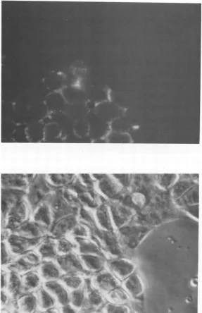
monoclonal antibody 72H3 was used in an indirect immunofluorescent staining procedure on L16-9, GR3A, and L8-2 cells. Patches of cells in the L16-9 culture werepositive forgp52antigen(Fig. 4). The level ofexpressionwascomparable to that of cells from the positive control GR3A cell line. No viral protein was detected in control L8-2 cultures.
Nonexpress-C
ing cells in the L16-9 culturedid notcontain XGRT39DNA (see below).Production of MMTV(XGRT39) retrovirus particles by
XGRT39-transfected rat mammary adenocarcinoma
cells.
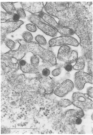
From the data presented above, it appeared that XGRT39 was biologically active. To determine whether retrovirus particles were being produced, L16-9 and control cultures - D were examined by transmission electron microscopy. L16-9 cells were found to produce B-type retrovirus particles characteristic of MMTV (Fig. 5). Budding, immnature, and
mature B particles as well as intracytoplasmic A particles G were observed. The mature 13 particles were characterized by electron-dense, eccentric nucleoids and the presence of spikes on the virus membrane. The particles observed in
5 6 7 8 L16-9 cultures could not be morphologically distinguished from B particles observed in mammary tumor cultures by
using the same techniques. No retrovirus particles were
detected in thenegative control culture L8-2. On the basis of these
data,
we concluded thatXGRT39
wasfully expressed
S
in LA7 cells and that these cells were making MMTVL16r
5 LI 6-9(AGRT39)
encodedby
theXGRT39
provirus.
L16- 5 L16- 9
Induction of mammary tumors in BALB/c mice byMMTV(XGRT39)fromXGRT39-transfected LA7 cells. The B particles observed by electron microscopy in
XGRT39-trans-L8-4
fected LA7 cells were tested in vivo fortumorigenicity
by
injection into hormone-stimulated females from the exoge-nous virus-free BALB/c mouse strain. This strain was
se-lected becauseof its sensitivitytocarcinogenesis by
exoge-nousMMTVandits very low(<1%)spontaneous mammary
tumorincidence(18).
T39 provirus in rat mammary A 100%immunofluorescence-positive, mycoplasma virus-blotanalysisof XGRT39-trans- free subline of L16-9,L16-9B10,wasestablishedforuse as a transfected with XGRT39 DNA source ofMMTV(XGRT39). Ten sublines were cloned from
vith wild-typeX DNA (lanes 1 L16-9 by endpoint dilution. DNAs from five lines were
rn blotanalysis.Cell line DNAs analyzed by Southern blot analysis with 32P-labeled probe [with MMTV(C3H)rep
probe.
pDM5. Threelines did nothave detectableXGRT39
DNA.clyfic
restroctton
fragments
dia-
Two lines had MMTV restrictionpatterns
identicaltothat of thecell lines analyzed in panel the parentalL16-9
cell line, which suggested that the originalmanifold filtrationin saturated L16-9 focus was picked before stable integration of the
H) rep probe.MMTVRNA was unselected XGRT39 DNA and that horizontal spread of
transfectedwithXGRT39 DNA. MMTV(XGRT39) does not occur in LA7 cultures. The
L16-9B10 sublinewastreatedfor mycoplasma virus
contatn-1(C3H)
rep probe. MMTV ination with BM-Cycline (Boehringer MannheimBiochemi-urAGRT39-transfectedcell cals, Indianapolis, Ind.) accordingtotheinstructions ofthe ression in L15-2 and L16-5
manufacturer
and then recharacterized to verify that theing was verified by staining cells were still producing MMTV antigen and B particles. lulose filter with methylene Virus particles from L16-9B10 cultures were injected ,ll lineswasproceeding,we intraperitoneally into BALB/c neonates. Control neonates kof LA7 cellswascontam- wereinjected with similar preparations from theL8-2 nega-Since mycoplasma virus is tive control cell line or the GR3A positive control cell line.
essionofretroviruses in cell Neonates were used to avoid animmune responseto the rat iade toquantitatethe levels proteins present in the virus preparations. Female mice were he cell lines or tocorrelate stimulated by pituitary isografts at 1 month of age, and male the amount of transfected micewere removed from the experiment.
the basis of the DNA and Table 1 shows the tumorincidence in these groups at 10
orfurther analysis. months. Onlyfemales that survived to maturity are included.
J. VIROL.
YXi.>>"
!
on November 10, 2019 by guest
http://jvi.asm.org/
[image:5.612.70.314.81.472.2]FIG. 4. Expressionof viral envelopeSUprotein gp52onthe surfaceof XGRT39-transfected LA7 cells. Cell line L16-9wasanalyzedfor cellsurfaceexpressionofgp52 byindirectimmunofluorescentstainingof live cells,usingtheanti-gp52monoclonalantibody72H3. Apatch
offluorescentgp52-positivecells is shown in thetoppanel.Thenegativecells didnotcontainXGRT39 DNA(see text)andserve as aninternal
negativecontrolforbackgroundfluorescence. Aphasephotomicrographof thesameregion is shown in thebottompanel.
Tumorsbegantoappearat9monthspostinjection, and
one-third ofthe experimentalgrouphadtumorsby10 monthsof age. At this time, we cannot project what the final tumor
incidence will be with this induction
protocol,
although
weanticipate
more tumors. The animals will be held for 2years.
Expression of viral antigens in MMTV(XGRT39)-induced BALB/cmammarytumors.
Immunohistochemical
analysis of the first mammary adenocarcinoma toappear
in theMMTV(XGRT39)-injected
animals is shown in Fig. 6. Aparaffin section of the tumor was treated with polyclonal rabbit anti-MMTV antiserum (7) and immunoperoxidase stained. The tumor is a mammary adenocarcinoma, the
predominant histopathological type of mammary tumor
as-sociated with MMTV infection. Concentrations ofMMTV
antigenwerereadily detectable inthetumor,particularlyon
the apical surfaces of alveolar structures. The other three
tumors in mice injected with L16-9B10 MMTV(XGRT39)
and the two tumors in mice injected with GR3A MMTV (GR) were also mammary adenocarcinomas and expressed MMTVantigen.
New copies of clonally integrated exogenous MMTV (AGRT39) proviruses in MMTV(XGRT39)-induced BALB/c mammary tumors. If the mammary adenocarcinomas
ap-pearing in the BALB/c mice injected with L16-9B10 virus
preparations were induced by MMTV(XGRT39), then two
predictions could bemade.First,the tumors should havethe SstI C restriction fragment diagnostic for exogenous
pro-virus fromthe GR mouse strain(Fig. 1). Second,thetumors
should have newlyintegrated copiesofexogenous provirus
on November 10, 2019 by guest
http://jvi.asm.org/
[image:6.612.162.449.76.520.2]J. VIROL. 154 MORRIS ET AL.
t
*5
.0'a
Q4m , 'j
-jV~ ~
t
;4.
..
;t
;
w f u
&
<
s
vs
c
Aw
rA
'K* .4
w~~~~~~~~~w
. ;'t.l4 @4S -k
O5.m
54.
Wo.' .. x' > '; s *w '~~~~~~~~~2
FIG. 5. Detection of B-type retrovirusparticlesinXGRT39-transfectedLA7cellsbyelectronmicroscopy.Budding, immature,andmature
B particles were detectedby transmission electron microscopy of L16-9 cultures grown in the presence of dexamethasone. A matureB particle (arrow)andseveralbuddingB particlesareshown.
in clonal(although notnecessarily monoclonal)
stoichiome-TABLE 1. Tumorincidence 10monthspostinjection' Tumor
Source of inoculum incidence
(%)b
L8-2... 0(0/2) L16-9B10... ... 36(4/11)
GR3A... ... 67(2/3)
Uninjected ... ... 0(0/8)
tries.
Southern blot analysis of the first
putative
MMTV(XGRT39)-induced BALB/c mammary adenocarcinoma is shown inFig.7.Restriction patterns of thetumorDNA
(lane
2) andnegativecontrolspleenDNA(lane 1)were
compared
by using 32P-labeled probe pDM5 (Fig. 1). The MMTV
(XGRT39)-diagnosticSstI Cfragmentwasreadilydetectable in the tumor sample, which indicated the presence of pro-virus from XGRT39-encoded virus. The SstI D fragment comigrated with anendogenous BALB/c proviral fragment (thelowest-molecular-weight fragmentin lane1)andwas not
used to track
MMTV(XGRT39)
in theseexperiments.
The a Allmicewerehormonestimulated withpituitary isografts.bValues inparenthesesarenumber oftumor-bearingmice to total number
ofmice.
f,
., I
...
'4. "
w"..."
on November 10, 2019 by guest
http://jvi.asm.org/
[image:7.612.156.481.72.543.2]A
4.0
B
FIG. 6. MMTV antigen in a BALB/c tumor induced by injection with virus from XGRT39-transfected LA7 cells. Shown is an immunoperoxidaseanalysis oftwo serial sections ofthe first tumor that arose in the BALB/c mice injected as neonates with virus from L16-9B10cells.(A)Sectiontreated with a polyclonal anti-MMTV primary antiserum; (B) section treated without the primary antiserum. The staining pattern istypical for MMTV-induced tumors, with concentrations of antigen most evident along the apical borders of alveolar structures.
SstI Fand G host-virusjunctionfragmentsfrom theXGRT39 proviruswerenotdetected,asexpected,because of
integra-tion of the MMTV(XGRT39) proviruses into new genomic
locations in theBALB/ctumors.Inaddition, several unique
restriction fragments representing host-virus junction
frag-ments from the new, clonally integrated MMTV(XGRT39)
exogenousprovirusesweredetected. Newcopiesofclonally integrated exogenous proviruses were also observed in the other three
MMTV(XGRT39)-induced
tumors and the twoMMTV(GR)-induced control tumors. All tumorDNAs had the SstI Cfragment and unique exogenous MMTV restric-tion patterns when cleaved with either PstIorSstI.
DISCUSSION
In this report, we describe the cloningand characteriza-tion of XGRT39, a pathogenic clone of exogenous MMTV
from the high-tumor-incidence GR mouse strain. Several lines of evidence indicated that the XGRT39 provirus was
biologically active. First, the XGRT39 provirus was
tran-scribed when transfected intoratmammary adenocarcinoma
LA7cells. Second,virus gp52surface proteinwas madeby
XGRT39-transfected LA7 cells, which suggested that viral
RNAsplicingand polyproteinprocessing were
occurring
in the LA7 cells. Third, dexamethasone-regulated, particle-B./i
.7 .1
--V
on November 10, 2019 by guest
http://jvi.asm.org/
[image:8.612.136.461.69.543.2]156 MORRIS ET AL.
0 0
E
N*_
v_
C-__
1 2
FIG. 7. Southern blot analysis of the first MMTV(XGRT39)-inducedBALB/ctumor.TumorandnegativecontrolspleenDNAs weredigested withSstIandprobedwitha32P-labeled pDM5insert. The SstICfragment diagnosticforexogenousMMTV from theGR strain (closed arrow) is clearly detectable in the tumorDNA and absent from the control DNA, demonstrating the presence of MMTV(XGRT39) inthe tumor. Inaddition, novel restriction
frag-ments(open arrows)aredetected. Thesefragments arehost-virus junctionfragments fromnew, clonallyintegrated copiesof MMTV
(AGRT39).
associated reverse transcriptase activity was detected in cell-free culture medium from AGRT39-transfected LA7 cells (unpublished data). Fourth, AGRT39-transfected LA7 cells produced intracytoplasmic A particles and mature,
immature, and buddingBparticlescharacteristic of MMTV.
Finally, normalmousemammaryepithelialcellsfrom
endog-enous and exogenous MMTV-free mice (8) could be
effi-ciently infected in vitro by culture on irradiated XGRT39-transfected LA7 cell feeder layers (12), as evidenced by
transmission ofrestriction fragments characteristic of the
XGRT39provirustothemousecellsinculture (unpublished
data ). From these data, we concluded that the XGRT39
provirus was fully expressed in LA7 cells and named the
recombinant DNA-derived virus produced by these cells
MMTV(AGRT39) to distinguish itfrom the naturally
occur-ring, GR mouse-derived MMTV(GR) virus. Additional
ex-periments will be required to determine whether the two
viruses aregenetically identical.
The conclusion that the XGRT39 provirus is pathogenic
was based primarily on the presence of the
MMTV(GR)-specific 6.1-kb SstI restriction fragment in four mammary tumors that appeared in BALB/c mice injected with MMTV(XGRT39) from XGRT39-transfected LA7 cells. Thereareconsiderable datasuggestingthattheGRprovirus
uniquely identified by the 6.1-kbSstI restrictionfragment is both expressed and pathogenic. The 6.1-kb SstI fragment
hasbeenshowntobepresentinincreasedcopynumber in (i)
GR mammary tumors (13), plaques (15), and hyperplastic outgrowths(15), (ii) heterologous cells infected withMMTV fromGR milk (14) or a mammary tumor cell line(14), and
(iii) BALB/ctumorsinducedby foster-nursing BALB/c pups
on lactating GR females or injection of sucrose density
gradient-banded virus from a GR mammary tumor cell line (unpublished data).
The BALB/ctumorshadthree other features common to
virtually all MMTV-induced neoplasms. First, the tumors
hadnewhost-virusjunction fragments detectableby
South-ern blot analysis, which indicated the presence ofnewly
integrated copies of exogenous provirus in clonal stoichiom-etries.Second, the tumors expressed MMTV antigen.Third, thetumors were adenocarcinomas, the predominant histo-pathological type of mammary tumor associated with MMTV infection.
Therefore, the weight of evidence indicates that the BALB/c tumors described here were induced by MMTV (XGRT39) from the XGRT39 provirus genome. This evidence includes (i)transmission of the MMTV(GR)-specific 6.1-kb SstI restriction fragment and (ii) comparisons withnaturally occurring MMTV-induced tumors. Formal proof that the XGRT39 provirus is pathogenic will require introduction of the XGRT39 provirus genome directly into normal mouse
mammarycells or embryos. Experiments along these lines areinprogress.
Despite the technical difficulties ofworking with XGRT39, we elected to develop systems to genetically analyze the nativeclonebecauseof the large amount ofvirological data in the literature concerning MMTV(GR) (18). XGRT39 was identified by chance during experiments to clone the host DNA flanking a single clonally integrated exogenous MMTV(GR) provirus observed in a mammary tumorfrom the GR mouse strain. Our experience with XGRT39 was consistent withprevious reports of attempts to clone exog-enous MMTV provirus DNA. XGRT39 was unstable and tended to delete the MMTV coding sequences by homolo-gous recombination between the proviral LTRs during
se-quential amplifications. In addition, we were unable to
subclonethe SstI Dfragment containingthepoisonsequence (Fig. 1). An overlapping clone of this provirus truncated downstream of thepoison sequence, XGRT6, wasstable in
bothlambda and plasmidvectors.
An alternate strategy for cloning biologically active
MMTVproviralDNAisto constructchimericprovirusesin plasmids bysubstituting plasmid-stable endogenousprovirus sequences for the unstableregionofanexogenousprovirus
(33; G. M. Shackleford and H. E. Varmus, personal
com-munication). Shackleford and Varmus constructedan infec-tious and pathogenic chimeric provirus from the weakly oncogenic Mtv-J endogenous provirus (30) and exogenous
MMTV(C3H). The Mtv-J/MMTV(C3H) chimera was
ex-pressedinXC cells,wastransmittedtouninfected XCcells in coculture, andproduced mammarytumors in force-bred
BALB/cfemales.Salmonsetal. (33) constructedachimeric provirusfrom thenononcogenicMtv-8endogenousprovirus
andMMTV(GR).The Mtv-8/MMTV(GR)chimera was fully
expressed in feline kidney cells; however, itwas not deter-mined whether thischimericviruswasinfectiousor tumori-genic.
Another exogenousMMTV(GR) clone has been reported by Buetti andDiggelmann(6).These authors cloned uninte-grated circular MMTV(GR) provirus DNA from the Hirt fraction ofMMTV(GR)-infected rat hepatoma cells. Intact clones containinga single LTR and rearrangedclones
con-taining two LTRs were recovered. Partial expression ofa concatemerofa single-LTRclone wasobserved in L cells. The poison sequence region of the single-LTR clone was
successfully subcloned and sequenced (16). We speculate
J. VIROL.
on November 10, 2019 by guest
http://jvi.asm.org/
[image:9.612.151.227.69.325.2]that the single-LTR clones were a MMTV(GR) variant present in the cell line and that the rearranged clones were clones of the exogenous MMTV(GR) virus that mutated during cloning as a consequence of the poison sequence (5). A comparison of the XGRT39 poison sequence region (se-quenced directly by using a XGRT39 template and synthetic oligonucleotide primers) and the published sequence of the same region of the single-LTR isolate should resolve the question and may shed additional light on the nature and location of the exogenous MMTV poison sequence.
Theexperiments described in this report are a prelude to
mutational analysisof the MMTV(GR) genome. Procedures must nowbe devised to efficiently shuttle small subclones of the XGRT39 provirus back into the lambda clone after
mutagenesisin vitro. In addition, LA7 cell transfection and culture conditions must be optimized for maximal virus production to eliminate the need for isolation and
character-ization of individual subclones. Finally, biological systems to assay MMTV function, such as the in vitro infection system alluded to above, must be developed. Analysis of
MMTV mutants in cell culture and in vivosystems will be
importantin the solution ofanumberof fundamental
prob-lemsintheMMTV field.
ACKNOWLEDGMENTS
This work was supported by grant CD-235 from the American CancerSociety.
WethankJames W.Casey for adviceonlambdacloning,Robert J.Scibienski forhelpful discussions regarding the infection protocol, KathleenM.Hendrix and JudithE. Walls for technicalassistance, and Lawrence J. T.Youngforhelp with the pituitary transplanta-tions.Inaddition,wethank JohnE.Majorsand Harold E. Varmus for the MMTV(C3H) PstI fragment clones, John P. Puma for MMTV(C3H) viral RNA, Arnoud Sonnenberg fortheanti-MMTV SUprotein monoclonal antibody 72H3,andPaulA. Luciwfor the
pSV2neoderivativepSV-1/neo.
ADDENDUM
Virus from the Mtv-8/MMTV(GR) chimeric provirus
re-ported by Salmonsetal. (33)hasrecentlybeen shown to be
infectious(N. H. Sarkar, personal communication). Also,at 15monthspostinfection, the tumorincidence of thepositive
control group infected with MMTV(GR) from GR3A cells was100%(3of3) and the tumorincidence ofthe
experimen-tal group infected with MMTV(XGRT39) from L16-9B10
cellswas 82% (9 of 11). Severalanimalsdeveloped
multiple
tumors, and premalignant lesions were observed in
mam-marygland wholemounts.No tumorshave been observed in
either ofthenegative controlgroups.
LITERATURECITED
1. Bennett, D. C., L. A.Peachey, H. Durbin, and P.S. Rudland. 1978. Apossiblemammary stemcellline.Cell 15:283-298. 2. Benton, W. D., and R. W. Davis. 1977. ScreeningAgt
recombi-nantclones byhybridizationto single plaquesin situ. Science 196:180-182.
3. Bresser, J., H. R. Hubbell, and D. Gillespie. 1983. Biological
activity of mRNA immobilizedonnitrocellulose in Nal. Proc. Natl. Acad. Sci. USA 80:6523-6527.
4. Brinkley, B. R., P. Murphy, and L. C. Richardson. 1967. Procedure forembeddingin situselectedcells culturedin vitro. J. Cell Biol.35:279-283.
5. Brookes,S.,M.Placzek, R.Moore,M.Dixon,C.Dickson,and G. Peters. 1986. Insertion elements and transitions in cloned mousemammary tumourvirus DNA: furtherdelineation of the poisonsequences. Nucleic Acids Res. 14:8231-8245.
6. Buetti, E.,and H. Diggelmann. 1981.Cloned mouse mammary
tumor virus DNA is biologically active in transfected mouse
cells and its expression is stimulated by glucocorticoid
hor-mones. Cell23:335-345.
7. Cardiff,R.D.,M.J. Puentes,L.J.T.Young,G. H.Smith,Y. A. Teramoto, B. W. Altrock, and T. S. Pratt. 1978.Serological and biochemical characterization of the mouse mammary tumor
virus with localization ofp1O. Virology85:157-167.
8. Cohen, J. C.,V. L.Traina,T.Breznik,and M. Gardner. 1982.
Developmentofamousemammarytumorvirus-negativemouse
strain:a newsystemforthestudy ofmammarycarcinogenesis.
J. Virol. 44:882-885.
9. Dickson,C. 1987. Molecular aspectsofmousemammarytumor
virusbiology. Int. Rev.Cytol. 108:119-147.
10. Donehower, L. A., A. L. Huang, and G. L. Hager. 1981.
Regulatoryandcoding potentialofthemousemammarytumor
viruslong terminalredundancy.J. Virol. 37:226-238.
11. Dulbecco, R.,M.Bologna,and M.Unger. 1979. Differentiation ofa ratmammarycell line in vitro. Proc.Natl.Acad. Sci. USA 76:1256-1260.
12. Ehmann,U.K.,W. D.Peterson, Jr.,and D. S. Misfeldt.1984. To growmousemammaryepithelialcellsinculture.J.Cell Biol. 98:1026-1032.
13. Fanning,T.G., J. P.Puma,and R. D.Cardiff.1980. Selective
amplification of mouse mammary tumor virus in mammary tumorsof GR mice.J. Virol. 36:109-114.
14. Fanning,T.G., J.P.Puma,and R.D.Cardiff.1980. Identifica-tion and partial characterization of an endogenous form of mousemammarytumorvirusthatis transcribed into the virion-associatedRNAgenome. Nucleic AcidsRes.8:5715-5723. 15. Fanning,T.G., A. B.Vassos,and R. D.Cardiff. 1982.
Methyl-ation andamplificationofmousemammarytumorvirusDNAin
normal, premalignant, and malignant cells ofGR/A mice. J. Virol. 41:1007-1013.
16. Fasel, N.,E.Buetti, J.Firzlaff,K.Pearson,and H.Diggelmann.
1983. Nucleotidesequenceofthe 5'noncoding regionandpart ofthe gag geneofmousemammarytumorvirus;identification of the 5' splicing site forsubgenomic mRNAs. Nucleic Acids Res. 11:6943-6955.
17. Groner, B., E. Buetti,H.Diggelmann,and N. E. Hynes. 1980. Characterization of endogenous and exogenous mouse
mam-mary tumor virus proviral DNA with site-specific molecular clones.J. Virol. 36:734-745.
18. Hilgers, J., and M. Sluyser. 1981. Mammary tumors in the mouse.Elsevier BiomedicalPress, Amsterdam.
19. Hohn, B.,and K.Murray. 1977. PackagingrecombinantDNA molecules into bacteriophage particles in vitro. Proc. Natl. Acad. Sci. USA74:3259-3263.
20. Leis, J.,D.Baltimore, J.M.Bishop, J. Coffin,E.Fleissner,S. P. Goff,S.Oroszlan,H.Robinson,A. M.Skalka,H. M.Temin,and V. Vogt. 1988. Standardized and simplified nomenclature for
proteinscommon toallretroviruses.J. Virol. 62:1808-1809. 21. Lillie,R.D.,andH. M. Fullmer.1976. Histopathologictechnic
and practical histochemistry. McGraw-Hill Book Co., New York.
22. Loenen,W. A.M.,and W.J.Brammar. 1980.Abacteriophage
lambda vector for cloning large DNA fragments made with severalrestrictionenzymes. Gene 10:249-259.
23. Lopata, M. A., D. W. Cleveland, and B. Sollner-Webb. 1984.
High level transient expression of a chloramphenicol acetyl
transferasegenebyDEAE-dextran mediatedDNAtransfection
coupledwithadimethylsulfoxideorglycerolshocktreatment.
NucleicAcids Res. 12:5707-5717;
24. Majors,J.E.,and H. E. Varmus.1981. Nucleotidesequencesat host-proviral junctionsformousemammarytumourvirus.
Na-ture(London)289:253-258.
25. Maniatis, T.,E.F. Fritsch,andJ. Sambrook. 1982. Molecular
cloning:alaboratory manual. ColdSpringHarborLaboratory,
ColdSpringHarbor,N.Y.
26. McClain, K., andW. H. Kirsten. 1974. Mouse leukemia virus
growthinmousecellscontaminatedwithMycoplasma.Cancer Res. 34:281-285.
27. Morris, D. W., and R. D. Cardiff. 1987.
Multistep
model ofmouse mammary tumordevelopment, p. 123-140. In G. Klein
on November 10, 2019 by guest
http://jvi.asm.org/
158 MORRIS ET AL.
(ed.), Advances in viral oncology, vol. 7. Raven Press, New York.
28. Muhlbock, 0. 1965. Note on a new inbred mouse-strainGR/A. Eur. J. Cancer 1:123-124.
29. Nusse, R. 1988. Theactivation of cellular oncogenes by proviral insertion in murine mammary cancer, p. 283-306. In M. E. Lippmanand R. B. Dickson (ed.), Breast cancer: cellular and molecularbiology. Kluwer AcademicPublishers, Boston. 30. Nusse, R., J. de Moes, J. Hilkens, and R. van Nie. 1980.
Localization of a gene for expression of mouse mammary tumor virus antigens in the GRIMtv-2- mouse strain. J. Exp. Med. 152:712-719.
31. Rigby, P. W. J., M. Dieckmann, C. Rhodes, and P. Berg. 1977. Labeling deoxyribonucleic acid to high specific activity in vitro by nick translation with DNA polymerase 1. J. Mol. Biol. 113: 237-251.
32. Ringold, G. M. 1983. Regulation of mouse mammary tumor virus gene expression by glucocorticoid hormones. Curr. Top. Microbiol. Immunol. 106:79-103.
33. Salmons, B., B. Groner, C. M. Calberg-Bacq, and H. Ponta. 1985. Production of mouse mammary tumor virus upon trans-fection of a recombinant proviral DNA into cultured cells. Virology 144:101-114.
34. Shank, P. R., J. C. Cohen, H. E. Varmus, K. R. Yamamoto, and G. M. Ringold. 1978. Mapping of linear andcircularforms of mouse mammary tumor virus DNA with restriction endonu-cleases:evidence for a largespecific deletion occurring athigh
frequency during circularization. Proc. Natl. Acad. Sci. USA 75:2112-2116.
35. Southern, E. M. 1975. Detection ofspecific sequences among DNAfragmentsseparated bygelelectrophoresis.J.Mol. Biol. 98:503-517.
36. Southern, P. J., and P. Berg. 1982.Transformationof mamma-lian cells to antibiotic resistance with a bacterial gene under controlof theSV40 earlyregionpromoter. J. Mol.Appl. Genet. 1:327-341.
37. Sternberg, N., D. Tiemeier, and L. Enquist. 1977. In vitro packaging ofa XDam vectorcontainingEcoRI DNAfragments of Escherichiacoli and phage P1. Gene1:255-280.
38. Thomas, P. S. 1980.Hybridization of denaturedRNAandsmall DNAfragmentstransferred tonitrocellulose.Proc.Natl.Acad. Sci. USA77:5201-5205.
39. Ucker,D.S.,S. R.Ross, and K. R. Yamamoto. 1981. Mammary tumorvirusDNAcontains sequencesrequired forits hormone-regulatedtranscription. Cell27:257-266.
40. Vieira, J., and J. Messing. 1982. The pUC plasmids, an M13mp7-derivedsystem for insertionmutagenesis and sequenc-ing withsynthetic universal primers. Gene 19:259-268. 41. Weiss, R., N. Teich, H. Varmus, and J. Coffin. 1982. RNA tumor
viruses. ColdSpringHarbor Laboratory, Cold Spring Harbor, N.Y.
42. Wood, W. B. 1966. Host specificity of DNA produced by Escherichia coli: bacterial mutations affecting the restriction andmodification ofDNA.J. Mol. Biol. 16:118-133.
J. VIROL.