0022-538X/92/084874-10$02.00/0
Copyright C) 1992, American Society for Microbiology
Assembly of Recombinant
Human
Immunodeficiency Virus
Type 1 Capsid Protein
In
Vitro
LORNA S. EHRLICH,* BETH E. AGRESTA,ANDCAROLA. CARTER
Department of Microbiology, State University ofNewYorkatStonyBrook, StonyBrook, New York11794
Received 24February 1992/Accepted 11 May 1992
Thecapsid protein (CA) (p24)of human immunodeficiency virus (HIV)type1expressedin Escherichiacoli
and purified to >90%o homogeneity was used to examine assembly in vitro and to probe the nature of
interactions involvedinthe formationof capsidstructures.The proteinwasdetected in dimeric and oligomeric formsas indicated bymolecular sizemeasurementsby gel filtrationcolumn chromatography, sedimentation
throughsucrose,and nondenaturing gel electrophoresis. Chemicalcross-linking of CA moleculeswasobserved
with several homobifunctional reagents. Oligomer size was dependent on cross-linker concentration and exhibited a nonrandom pattern in which dimers and tetramers were more abundant than trimers and
pentamers.Oligomersaslargeasdodecamersweredetected in nativepolyacrylamidegels. Thesewerestable insolutions of high ionic strengthorin thepresenceof nonionic detergent, indicating thatstronginteractions were involved in oligomer stabilization. Limited tryptic digestion converted the putative dodecamers to octamers, suggestingthataregion involvedinCA protein multimerizationwasexposed in thestructure.This regionwasmappedtothe central portionofthe protein. The recombinant CA proteinsassembledinvitro into longrodlikestructuresandweredisassembledinto small irregular spheres by alterations in ionic strength and pH. The observation thatassemblyanddisassembly of purifiedHIVtype1CA proteincanbeinducedinvitro suggestsanapproachforidentifying possible control mechanismsinvolved in HIVviral coreassembly.
Human immunodeficiency virus (HIV), the causative
agent of AIDS, is a member of the lentivirus group of the
family Retroviridae (9). In these spherical enveloped vi-ruses,the viralgenomeis located insideacorestructurethat is encased in an envelope consisting of host-derived mem-brane and integratedviral transmembrane and surface gly-coproteins (15).As with otherretroviruses,the formation of
virus involves at least two major assemblyprocesses, one for the viralenvelope and the other for the viralcore (5, 9, 51).
Synthesis, processing, and glycosylation of the envelope precursor occurintheendoplasmicreticulum. Theenvelope proteinsaretransportedtothe cellplasmamembrane via the
secretory pathway (10). Structural proteins and enzymes thatarefound in thecore aresynthesizedinthecytoplasmas
twopolyprotein precursors: Gag andGag-Pol. The 55-kDa
Gag polyprotein encoded by HIV contains domains for matrix (MA), capsid (CA), nucleocapsid (NC), and p6, a domain unique to lentiviruses (36). Occasional ribosomal
frameshifting at a site within the NC-p6 intergenic region results in the synthesis of a polyprotein (160 kDa) that containspol-encoded domains for the enzymes proteinase
(PR),reversetranscriptase (RT),andintegrase (IN)in addi-tiontothegag-encodeddomainsMA, CA,and NC(25, 31).
Translation and cotranslational myristylationof these poly-proteinsarefollowedbyanassemblystagewhich consists of several temporallyordered events (5, 9, 51). Both
polypro-teinsaretransportedtotheplasma membrane,where assem-blytakesplaceonthecytoplasmicside of the cell membrane
simultaneously with the budding and release of immature
viral particles. Budding or released particles undergo a
maturation process that results in the formation ofmature
infectious virus(8, 26, 40, 46).
The assembly phase is divided into two major stages
*Corresponding author.
whicharecharacterized by the morphology of thestructure
found insidetheviralenvelope. The electron-densestructure
seenin immatureparticlesissphericaland lines the inside of the viralenvelope,while thestructureseeninmaturevirus is
cone-shaped andeccentricallylocated(6, 14, 15, 23, 24, 52). Expression of various HIVgagconstructshas demonstrated
that the Gag precursor polyprotein can, by itself, form
spherical structures like those found in immature particles (16). Formation of theimmatureparticle anditsconversion to mature virus are sensitive to mutations ingag andpol
genes, indicating thatregions in Gag and Gag-Pol contain informationrequiredfor variousaspectsof HIVassembly (3, 16-19, 26, 34, 35, 40, 52). Maturation is accompanied by changesthat include condensation of theNC-RNAcomplex toanelectron-dense nucleoid and translocation of CA(p24) fromaperipheral positiontoa moreinternalpositionrelative
to the viral membrane (15). A particle with the central nucleoid but in which CAproteinremained in itsperipheral
location is not infectious (19). Thissuggests a role for the morphologically correct viral core in virus infectivity and
impliesthat it would bepossibletohaveananti-HIVtherapy
based on intervention with theassemblyof thiscore struc-ture.
Rossmann (47) has suggested that the bacillus-shaped particlesof alfalfa mosaic virus(12)mayprovideastructural modelfor the bacillus- or cone-shaped capsid of HIV. For alfalfa mosaic virus and several otherunenveloped plantand animal viruses, invitro studies with purified proteins have
provided useful insight into the mechanism of virus coat assembly (12, 13, 20, 21, 45, 48).Similar studies withpurified
HIVCAproteinscouldprovideinformationontheassembly of the capsid shell that would complement that obtained fromgenetic and ultrastructural studies. Such studies have been hampered by the lack of purified native CAprotein,
whichwascompounded bythe lack of functional assaysby
whichtoassessthe nativestatusof theprotein.Our abilities
to express Gag-PR that is efficiently processed to mature 4874
on November 9, 2019 by guest
http://jvi.asm.org/
HIV-1 CAPSID PROTEIN 4875 products by HIV PR and to purify CA protein using a
nondisruptive protocol(11) permit the undertaking of these in vitro studies. This recombinant CA protein has been crystallized as a CA-Fab complex as part ofour effort to solve itsthree-dimensional structure(44). Inthis report,we
present evidence that the recombinant CA protein alone can direct theformation ofparticleswithaspecific morphology. Moreover,wedescribe theresults ofexperimentsthatprobe
the nature ofCA-CA protein interactions in the oligomers that composethese particles.
MATERIALS AND METHODS
DNA constructs. Construction of the plasmid FS II was described previously (27). This plasmid contains a T7 pro-moter followed by HIV sequences from BH10 spanning nucleotides 221to2130which includes 113 nucleotides of the 5'nontranslatedregion, thegaggene,and that partofthepol
genethat encodesproteinase. Four bases have been inserted attheBglIIsite in theearlyregionofthe pol gene just 5 bp downstream of the natural frameshift site. This results ina
frameshift mutation and translation of the pol gene. In bacterial strainsexpressing the T7 RNA polymerase, FS II allowssynthesis of Gag-PR,atruncated form of theGag-Pol
polyprotein.
Expression in Escherichia coli. Expression of the HIV sequence in FS II was done in E. coli BL21(DE3) as described before (11). Briefly, plasmid DNA was trans-formed into competent cells and transformed cells were grown in minimal media in a 14-liter fermentor (Microferm
MMF-14).Expression of FS IIwasinduced by the addition of isopropyl-,-D-thiogalactopyranoside (IPTG) to a final concentration of 0.4mM. After2h ofinduction, cells were collected bycentrifugation,and the wet paste was storedat
-80°C until needed.
Purification ofHIVtype1 (HIV-1) capsid protein. Recom-binant CA protein expressed in BL21 (DE3) cells was purifiedessentially asdescribed before (11). Briefly, 10 g of frozen cell pastewasresuspended in 50 mM
2-(N-morpholi-no)ethanesulfonic acid(MES) buffer, pH6.5, with 100 mM
NaCl, 10mM
MgCl2,
and 1 mM EDTA andlysedbyusing a French press (SLM Instruments). The bacterial lysate was clarifiedbycentrifugationat 10,000 xgfor 15 min, and the supernatantwasfurthercentrifugedfor 60 minat200,000 xg. Proteins from thefinalsupernatant wereprecipitated with
30% ammonium sulfate. The precipitate was collected by
centrifugation,redissolved in 50 mMTris-HCI buffer, pH 8,
containing 30mM NaCl and 1 mM EDTA, and chromato-graphed onaWhatmanDE52 DEAE-cellulose column which had been equilibrated with the same buffer. Unbound pro-teins in the flowthrough fraction were precipitated by the addition of ammonium sulfate to 50%
(wtlvol)
and resus-pendedin 50 mMTris-HCI buffer,pH 8,containing 30 mMNaCland 1 mM EDTAto aproteinconcentration of about 30 mg/ml (typically, this required about a 1-mlvolume). This
solutionwas left unperturbed in acold boxovernight. The whiteprecipitate of CA proteins that formed was collected andresuspended in aminimal volume of storage buffer (50 mMTris-HCI buffer, pH 8, containing 30 mM
NaCl).
Fourmilligrams ofpurified CA proteinwasusuallyobtained from 10 gof bacterial cell pellet.
Proteinanalyses. Concentrations of protein samples were measured by using the Bio-Rad protein dye binding assay
(Bio-Rad). Proteinsamples wereelectrophoresed under
de-naturingconditionsonLaemmligels(28). Nondenaturing (or native) gels were cast similarly except that these did not
have thestacking portion, and sodium dodecyl sulfate(SDS)
was omitted from both the separating gel and the running buffers.Electrophoresisonnondenaturing gelswasdone ina
cold box. Gels were typically 12.5% acrylamide, unless otherwise indicated in thefigure legends. Protein bandswere
visualized by staining with Coomassie blue. Immunological analysis of expressed proteinswasdone eitherby
precipita-tion with specific antibodies followed by
SDS-polyacryl-amide gel electrophoresis (PAGE) or by immunoblotting usingaprocedure essentially asdescribedby Towbin et al.
(50). Automated Edman degradation of electroblotted pro-teins wasperformed by using agas-phase Protein Sequencer model 470A equipped with a PTH Analyzer model 120A (Applied Biosystems) with standard programs recommended by the manufacturer.
Trypsin
digestion. Digestion occurred in a20-,ul reaction mixturecontaining 3 pg of purified recombinant CA protein and variable amounts of trypsin (BoehringerMannheim) in 100 mM Tris-HCl buffer, pH 8.5. Reaction mixtures were incubated at 37°C for either 30 or 60 min. Control reaction mixtures not containing trypsin were run inparallel. Both control and trypsin-treated samples were analyzed by elec-trophoresis onpolyacrylamide gels under either denaturing ornondenaturing conditions.Chemical cross-linking. Purified CA protein wasdialyzed against 10 mM sodiumphosphate buffer (pH7.0) priortoits use in a 14-,ul reaction mixturecontaining buffer, 24 ,ug of purified CAprotein, andcross-linking agent. Reactions were carriedout at roomtemperature andstoppedbythe addition of glycine to a final concentration of 100 mM. Reaction products were separated by electrophoresis on SDS-poly-acrylamide gels and detectedby Western immunoblot anal-ysis with anti-p24 antibody. The cross-linking agents used included sulfoethylene glycolylbis(succinimidylsuccinate), dithiobis(succinimidylpropionate), and 3,3'-dithiobis(sulfo-succinimidylpropionate) (DTSSP) (1,32, 49).
Gelfiltrationcolumnchromatography.Chromatographyof
purified CAprotein was done onaSephadex G-200 column (1.0 by 40cm)which wasequilibratedandeluted with buffer (50 mMTris-HCl, pH 8.0,containing 30 mM NaCl and 1 mM
EDTA) at aflow rate of 6 ml/h. The column was calibrated with aproteinmixture(200,ul) consisting of Blue Dextran (2 x 10⁶ kDa), bovine serum albumin (BSA) (68 kDa), chymo-trypsinogen (25 kDa), and cytochrome c(13kDa). After the columnwaswashed with500 ml ofbuffer,a200-,u solution ofCAproteinat 10mg/mlwasloaded andchromatographed in identical fashion. After another round of washing, a second200-,u sample containingCAprotein at 1mg/mlwas loaded and chromatographed as described before. For all threechromatographicruns, eluatewascollectedat1 ml per fraction. Fractionscontaining CAorstandardproteins were identified following SDS-PAGE of 20-,lI aliquots of the fractions collected.
Electron microscopy. Samples were negatively stained with2%uranyl acetate and visualized by using a JEOL 1200 EXelectron microscope.
RESULTS
Propertiesof recombinant HIV-1 capsidprotein. TheHIV
Gag-PRpolyproteinthatisencodedbyplasmidFS II(11,27;
alsoMaterialsandMethods)wassynthesizedandefficiently processed in BL21(DE3) cells. Expressed CA protein was
purifiedby usingaprotocoldevelopedinourlaboratorythat
includes in vitro oligomerization in the final step. The
protocol is describedbrieflyin Materials and Methods andin VOL. 66,1992
on November 9, 2019 by guest
http://jvi.asm.org/
4876 EHRLICH ET AL.
2 3 M -109K
~
- 72Ki>*
-46K-29K
- 18K
[image:3.612.130.220.76.173.2]_~ - 15K
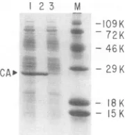
FIG. 1. Isolation of recombinant HIV-1 CA proteins fromE. coli. Recombinant CAwasisolated by aFrench pressof induced cellscarrying FSII.The lysatewasclarified by centrifugation and chromatographed on a DEAE-cellulose column as described in Materials and Methods. Proteins in the unbound column fraction wereconcentrated to-30 mg/ml by precipitation with ammonium sulfate and keptat5°C overnight. Under these conditions,
recom-binant CA proteins separate as a white precipitate that readily redissolves in low-ionic-strength buffer. The figure shows a Coomassie-stained SDS-polyacrylamide gel containing proteins in the unbound fraction of the DEAE-cellulose column (lane 1), precipitated recombinant CA redissolved in buffer (lane 2), and proteins remaining in thesupernatantafter centrifugation of precip-itatedCA (lane 3). Molecular weightsare ontheright.
greater detail in an earlier report (11). The isolation of CA protein in the final stageof thepurification is illustratedby
theCoomassie-stained gelshown inFig. 1. Lane 1 inFig. 1
shows the unbound proteins that coeluted with CA in the flowthroughfraction afterchromatographyon DEAE-cellu-lose. These proteins were concentrated byammonium sul-fate precipitation, resuspended at about 30 mg/ml (total protein concentration), and left unperturbed at 5°C. CA protein separated as a white precipitate (Fig. 1, lane 2), leavingmostof the otherproteinsinthesupernatant(Fig. 1, lane3).
The CA protein expressed in bacteria shares several
properties with CA protein isolated from the virus. The
recombinant CA preparation consists of two isoelectric forms that exhibitpIvalues of 6.55 and 6.75. ThepI = 6.75
form predominated in most preparations (44; also data not
shown).Both of thesepIvaluesaresimilartothosereported
forcapsid proteinfound inmaturevirus(pI = 6.6 andpl =
6.7;forareview,seereference30).RecombinantCAprotein was recognized in the native form by monoclonal and
polyclonal antibodies directed against viral CA protein in
immunoprecipitation experiments (data not shown).
Analy-sisbyEdmandegradation anddigestionwith
carboxypepti-dase P (data not shown) indicated that both the N- and C-terminal amino acid sequences of the recombinant CA
protein were identical to that of CA protein isolated from virus(22).
Concentration-dependent dimer formation. Observations that AKR murine leukemia virus (41, 43), Moloney murine leukemia virus (4), avian myeloblastosis virus (42), or recombinant HIV CA(11, 37) proteins exhibit self-associa-tive properties have been previously reported. To identify
stable multimers thatmight give insight into the mechanism
ofcapsidshellassembly,wemeasured the molecular size of CA protein under various conditions in vitro. Samples of purified recombinant CA protein at 1 or 10 mg/ml were chromatographed on a Sephadex G-200 column that had been equilibrated with 50 mM Tris-HCl buffer, pH 8, con-taining30 mMNaCland 1 mM EDTA.The elutionpositions
of the CA polypeptides and the protein markers used to
calibrate the column were determined by SDS-PAGE
anal-ysis of aliquots of collected fractions. Panel A in Fig. 2 is a
Coomassie-stained gel of fractions collected during column calibration and shows the elution positions of BSA (Mr =
68,000), chymotrypsinogen
(M,
=25,000), and cytochromec(Mr = 13,000) standards. The position of Blue Dextranwas
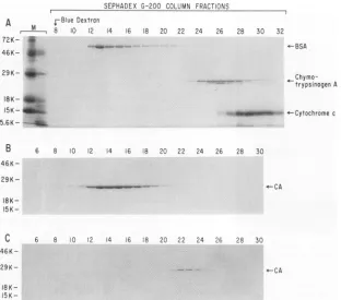
located spectrophotometrically and is indicated. Figure 2B shows the elution position of the 10 mg/ml CA protein sample. Under these conditions, CA protein eluted slightly behind BSA (Mr = 68,000) and ahead of chymotrypsinogen (Mr = 25,000). Assuming that the protein is globular, this
patternisconsistent with the molecular size ofaCAprotein dimer (50 kDa). This conclusion was supported by the results obtained upon dilution of the protein. Figure 2C showstheelution profile of CA when chromatographed at 1
mg/ml. The CA protein eluted as a monomer with an estimated molecular mass of about 25 kDa. The elution of CA protein as a monomer when chromatographed as a 1
mg/ml solution and as a dimer when chromatographed as a 10 mg/ml solution reflects the dependence of CA dimer formation on protein concentration. Similar results were obtained when the recombinantCA protein was sedimented through a 4 to 40% sucrose density gradient (data not shown).
CA proteindimers cross-link innonrandom arrays. Chem-ical cross-linking agents were used to determine whether the recombinant CA protein dimers were organized in larger structures. Cross-linkers capable of bridging distances
be-tween arrays should permit determination of higher-order
structure. The results ofcross-linking studies using DTSSP
(spanlength = 1.2nm)areshown inFig.3.CAprotein (1.7
mg/ml) was incubated with increasing concentrations of
cross-linking agent for60 min priorto analysis of reaction products by Western analysis. In the absence of DTSSP,
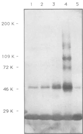
most of the CAprotein migrated with anMrof 25,000 (Fig. 3, lane5).A faintband (Mr= 50,000) was also visible. This
subpopulation of dimeric CA protein was not observed consistently and may have been due to occasional oxidiza-tion of sulfhydryl groups leadingto theformation of inter-molecular disulfide bridges. Increasing amounts of
cross-linked CA molecules thatmigrated with Mrs of -50,000were
detected withincreasingamountsof DTSSP(Fig. 3, lanes 1
to4). Itislikely thatsome orall of theproteins detectedas monomersunder these conditionswereinvolved in nonpro-ductiveinteractions because ofhydrolysisofoneof thetwo
reactive groups in the reagent prior to bindingto a second CA molecule. Atthehighest concentration of DTSSP(Fig.
3, lane4), dimers, tetramers, andmonomers weredetected in excess overtrimers andhigheroligomers. Similar results wereobtained in studiesusingtwoother bifunctional
cross-linking agents, sulfoethylene glycolylbis(succinimidylsucci-nate) and dithiobis(succinimidylpropionate) (data not shown).Since random collision would generatemoretrimers than tetramers (20), the results suggest that CA protein
dimers form the basic subunit of higher-order structures.
Cross-linked dimers, tetramers, and
higher-molecular-weight forms alsowere observed in previous studiesusing
native particles ofavian myeloblastosisvirus (42) and
mu-rine leukemia virus(43).
Recombinant HIVcapsid proteincan formhigher-ordered oligomers. The fact thattetramers andhigher oligomers of CAproteinweredetectedby chemical cross-linkingbutnot
begelfiltrationorsedimentationanalyses suggestedthatCA
protein association states might be unstable or reversible under some experimental conditions. The migration of recombinant CA innondenaturingpolyacrylamide gels sup-J. VIROL.
on November 9, 2019 by guest
http://jvi.asm.org/
HIV-1 CAPSID PROTEIN 4877
SEPHADEX G-200 COLUMN FRACTIONS 3-Blue Dextran
8 10 12 14 16 18 20 22
I8K.
15
K--46
5.6 ---6,24 26 28 30 32
-BSA
Chymo-trypsinogenA
;I~WIM_-Cytochrome:-' c
6 8 10 12 14 16 18 20 22 24 26 28 30
-CA
6 8 10 12 14 16 18 20 22 24 26 26 30
18K-15K
-FIG. 2. Gel filtration of purified recombinant HIV-1capsid protein. Two-hundred-microliter samples ofa10 mg/ml anda1 mg/mlsolution ofpurified recombinant CA proteinwerechromatographedon aSephadex G-200 column thatwaspreequilibrated with 50 mM Tris (pH8)-30 mM NaCl buffer. Column eluates were collected, and 20-plA aliquots were analyzed for CA protein by electrophoresis on SDS-12.5% polyacrylamide gels. (A) Gel showing the elution positions of the following column calibrationprotein standards: monomeric BSA (molecular weight [MW] = 68,000),chymotrypsinogen (MW=25,000), and cytochromec(MW= 13,000). (B) Gel showing the elution position of CA
appliedtothecolumnas a10mg/mlsolution.(C) Gel showing the elutionposition of CA protein appliedas a1mg/ml solution. Molecular weights for each panelareontheleft.
ported this view (Fig. 4). Denatured CA protein migrated
with the expected molecular mass of 24 kDa, as shown in Fig. 4A. When analyzed under nondenaturing conditions (Fig.4B andC),the CAprotein migratedaslarge oligomers.
At a protein concentration of 2 mg/ml, the CA protein migratedas abroad band withanapparentmolecularmassof approximately300 kDa(Fig.4B). Ataproteinconcentration of 5 mg/ml, CA protein exhibited an apparent molecular massthatwasgreaterthan that ofthe545-kDaureasemarker (Fig. 4C).Oligomers thatcomigratedwith the ferritinmarker (-800 kDa) have been observed with CA protein prepara-tions at concentrations of 10 mg/ml or higher (data not shown). Smaller amounts of oligomers thatwere approxi-matelythe sizeof trimersordimersalso have beendetected (Fig. 4; alsoseeFig. 5 below).Weinterpretthepresenceof aspecieswithanMrof about300,000tosuggestthatthe CA
proteindimersformed dodecamerswhichwerestableunder these conditions (predicted Mr = 288,000). The -300-kDa
oligomer was stable to dilution and was still detected at proteinconcentrationsaslowas100,ug/ml (datanotshown).
Thedetection of multimers of this -300-kDaoligomerwith
increasing protein concentration suggests that these form repeatingunits (predicted Mr = 576,000, 864,000, etc. [38]).
CAprotein oligomers arestable tohigh-ionic-strength and nonionicdetergents.We examinedthe effects ofhigh-saltand nonionic detergents on the stability ofCA protein interac-tions. Figure 5A shows themigration of CAproteins in6to
15% nondenaturing gels afterincubation in buffers
contain-ing 1 M NaCl or a nonionic detergent (0.1% octyl-beta
glucopyranoside) (OBG). The CA protein in the untreated
sample migrated as two major oligomeric species with
ap-parentmolecularmassesof >545 and -300kDa andastwo minor species of -200 and -70 kDa (Fig. SB). The two major speciesweredetected inaratio of 2:1(>545:300 kDa). Incubation of the protein with NaCl or OBG altered this molecular size distribution. The ratio of the >545- and
300-kDa forms decreased to 1:1, and smaller oligomers migrating at about 250 and 132 kDa appeared. Neither monomers nor dimers were detected. The new molecular size distribution indicated that both saltand detergent pro-moted small degrees of dissociation. However, this limited dissociation occurredonlyafter several weeks ofincubation,
indicatingthat theoligomerswereactuallyquitestable under theseconditions.
Oligomerizationisaffectedbylimitedproteolysis. Sincethe cross-linking experiments described above indicated the presenceof residues with accessible primaryaminogroups in or near regions ofprotein-protein interaction,we exam-ined the possibility that limited proteolysis with trypsin
affects CAprotein oligomerization. Trypsin cleaveson the carboxyside of aminoacids withprimaryaminogroups. In contrast tothe minimaleffects ofhighsaltconcentrationand
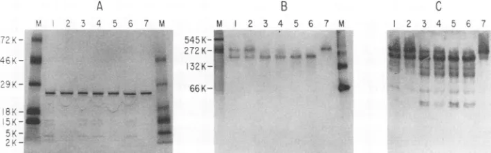
nonionic detergent, the incubation of CA proteins with trypsin even at a low enzyme-to-substrate (E/S) ratio re-sulted in dramatic changes in the apparent size of the oligomers (Fig. 6). Panel Ain Fig. 6 shows SDS-PAGE of
untreated CA (Fig. 6A, lane 7) and CA protein whichwas
incubated withincreasingamounts oftrypsin (Fig. 6A,lanes
A M
72K-
46K-29K- *
B
46K-
29K- 18K-15K
-C
46K-
29K-VOL. 66,1992
*--CA
on November 9, 2019 by guest
http://jvi.asm.org/
[image:4.612.156.468.73.348.2]2 3 4 5
200 K
109 K
72 K
46 K
-29K
--FIG. 3. Chemical cross-linking of purified recombinant HIV-1
capsid protein.Reaction mixturescontaining24 ,ugofpurified CA protein and 0.01 to 10 mM DTSSP in 10 mM sodium phosphate
buffer (pH 7) were incubated at room temperature for 60 min. Proteins were then denaturedbyincubationat100°Cin the presence of1%SDS, electrophoresedon anSDS-6to12.5%polyacrylamide gel,andtransferred onto nitrocellulose. CA-specific protein bands
weredetectedbyprobingwithanti-CAmonoclonalantibodies.The
figure shows an immunoblot of reaction mixtures incubated with 0.01(lane 1),0.1(lane2),1.0(lane 3), and 10(lane 4)mMDTSSP. CAproteinincubated for 60 minwithoutDTSSP is shown inlane5.
1 to 6). Protein was visualized with Coomassie blue stain. Limited proteolysis occurred under all conditions tested,
resultinginaminorpopulationoffragments thatmigrated at
2.5, 4, 14, and 16kDa. Themajorityof the CAprotein in all
samples comigrated with the CA protein in the untreated
sample.
To identify the sites cleaved by trypsin, the N-terminal amino acids of the 2.5-, 4-, 14-, and 16-kDa fragments
A
MARKERSI
CA109K- -_-72K- _W 46K-
29K-produced from the digestion of CA proteinat anE/Sratioof 1:100 (datanotshown)weredetermined by usingautomated Edman degradation. An E/S ratio of 1:100 was used to
increase the relative yields of the digestion products. The 16-kDa fragmentis apartialdigestion fragmentthat starts at
the first amino acid of the CA protein (P) and thusrepresents
the N-terminal half of thepolypeptide. The 2.5-kDafragment was derived by cleavage after K-30 within this N-terminal portion of CA protein. The 14-kDa fragment is a partial digestion productthatstarts afterR-100and thus represents
the C-terminal half of thepolypeptide. The 4-kDa fragment, which starts at M-144, is a product of partial digestion derivedbycleavage after R-143 within this C-terminal halfof CAprotein. The 16-, 14-,and 4-kDafragments allcontained multiple Arg and Lys residues, suggesting the presence of higher-order structures in these regions which protected these fragmentsfrom further digestion.
In contrast to the results obtained with denaturing gels,
distinct differences in the apparent molecular sizes of
un-treated andtrypsin-treatedoligomers were detected by using nondenaturingPAGE(Fig. 6B).Asexpected, CA proteinin
the untreated sample migrated as a single population of
oligomers with an apparent molecular mass of -300 kDa
(Fig. 6B, lane 7).Following incubation withtrypsinat anE/S ratio of1:5,000 for 30 and 60 min (Fig. 6B, lanes 1 and 2), oligomers thatmigrated withamolecularmassof-200kDa were detected in addition to the -300-kDa species. In samples treatedathigherE/S ratios (i.e., 1:500 and 1:1,000), only the smalleroligomeric formwasdetected. To determine whetherthissmalleroligomeric formrepresentedan associ-ation of truncatedCAmolecules,thisbandwas electroblot-ted onto a membrane ofpolyvinylidene difluoride (Immo-bilon) foranalysisof the N terminus by automated Edman
degradation or waseluted for analysis of the C terminus by hydrolysis with carboxypeptidase Pfollowedby aminoacid
analysis. Evidence for neither amino-norcarboxy-terminal hydrolysiswasobtainedby theseanalyses (datanotshown).
Thesedataareconsistent withtheinterpretation thatlimited
trypsinhydrolysis resulted in the removal of four CA protein molecules (-100 kDa) from the 300-kDa oligomer. It is possible that the interactions between subunits within the
B
MARKERS
m CA
545K-
a
K- '
66K-<
45K-C
MARKERSI
CA545 K- 272K-
132K-66K- a 45K- S
*
18K-15K-'4
FIG. 4. Migrationofpurifiedrecombinant HIV-1 on native polyacrylamide gels. Twenty-microgram samples of purified recombinant CA wereelectrophoresedonLaemmli gels underdenaturing and nondenaturing conditions. Proteins were visualized by staining with Coomassie blue. (A)Adenaturing10 to20%polyacrylamide gradient gel with 20 ,ug of CA protein from a 2 mg/ml solution. (B) A nondenaturing 10 to 20%polyacrylamidegel with 20 ,ug of CA protein from a 2 mg/ml solution. (C) A nondenaturing 4 to 20% polyacrylamide gel with 20 ,ug of CAproteinfroma5mg/mlsolution. Molecular size markers: ovalbumin (MW= 45,000), BSA monomer (MW= 66,000), BSA dimer (MW
= 132,000),ureasetrimer(MW = 272,000), and urease hexamer (MW =545,000).
amaw-,.,w2i ;- 'Ab-, "''
on November 9, 2019 by guest
http://jvi.asm.org/
[image:5.612.112.246.71.287.2] [image:5.612.160.460.517.661.2]HIV-1 CAPSID PROTEIN 4879
MARKERS
890
K-545Ko *
445K'r
I
-.
132 K-K
66K-
45K-FIG. 5. Migration of purifiedrecombinant CAproteinonnondenaturing gelsaftertreatmentwith1 MNaCland 0.1%OBG.NaCland OBG
were added to aliquots ofa 2 mg/ml solution of purified recombinant CA. These sampleswere kept at5°C for several weeks priorto
electrophoresis on a6to 15% acrylamide gel undernondenaturingconditions. Protein bandswerevisualized bystainingwith silver.(A) Markersandsamples containing20p,gofCAproteinwith 1 MNaClor0.1%OBG; (B)markers and 20,ugofuntreated CAprotein.Molecular
weight markers: ovalbumin (MW=45,000), BSAmonomer(MW= 66,000),BSA dimer(MW= 132,000), jackbeanureasehexamer(MW
= 545,000),ferritinmonomer(MW= 445,000),and ferritindimer(MW= 890,000).
300-kDa oligomer are not identical and that susceptible regions exposedin onesubunitwere shielded in another.
Mostlikely, the observed change in molecularmass was attributabletodigestionof accessibleCA molecules into the
2.5-,4-, 14-, and 16-kDaproductsdetected inFig.6A. These minorproductswereidentifiedonthe nativegel(Fig. 6C) by N-terminal and immunological analyses (data not shown).
Thesefragmentswerenotdetectedatthemigration positions
of the 300- or 200-kDa oligomers, as shown by N- and C-terminalanalysesof theproteinsintheseoligomersandby excisingthebands andsubjectingthemtoreelectrophoresis
under denaturingconditions in SDSgels (data not shown).
The fact thatneither the 16-nor14-kDafragmentthatspans the N-terminal and C-terminal halves of the CApolypeptide, respectively,wasassociated with the 200-or300-kDa oligo-merssuggeststhat theseputativestructureswereinsufficient
A
for, or not involved in, maintaining oligomer association. Perhaps theconcerted action of structural domains located in both halves of the CApolypeptide is required. Alterna-tively, cleavage may have effected conformational changes
inregionsof theprotein involved inoligomerassociation. In
eithercase,these results indicate that the structuralintegrity
of a region in the middle of CA protein is required for oligomer association.
Morphology of structures formed by recombinant HIV capsid protein in vitro. Purified coat proteins of tobacco mosaic virus, polyomavirus, and several simple spherical plantviruses have been observedtoform functionally
rele-vantstructures invitro (20, 45, 48). To determinewhether the recombinant CA protein oligomers participate in the formation of higher-ordered structures invitro, samples of thepurified proteinwere examinedbyusing negative
stain-B
.
_
5 c7M M 1 2 3 4 5 6 7M-< 0" 545K-I
272K- * w
132K-..
_
66K-8V
C 2 3 4 5 6u 7
gW
m.nEs
FIG. 6. MigrationofCAprotein onnative gelsafter limitedtrypsin digestion. Trypticdigestion wascarried out in reaction mixtures
containing100 mM Tris-HCl buffer(pH8.5),3 p.gofpurifiedrecombinant CAprotein, and0.6, 3.0,or6.0 ng oftrypsin.Eachmixturewas
made in triplicate and incubated at 37°C for 30 or 60 min. (A) Electrophoresis of trypsin-treated samples in 10 to 20% denaturing polyacrylamide gradient gels. Protein was visualized by Coomassie blue staining. (B) Electrophoresis in 10 to 20% nondenaturing polyacrylamide gradient gel. ProteinswerevisualizedasdescribedforpanelA. (C)An immunoblot of thetrypsin-treated samples analyzed
on anondenaturing polyacrylamide gel probedwithpolyclonalanti-CAantibody.Lanes:1,incubation with 0.6 ng oftrypsin (E/S=1:5,000), 60min;2,incubationasin lane 1,30min;3, incubation with 3ngoftrypsin (E/S = 1:1,000),60min;4, incubationasin lane3,30min; 5,
incubation with 6ngoftrypsin (E/S = 1:500), 60min;6,incubationasin lane5,30min;7,CAproteinincubated for 60min in the absence
oftrypsin; M,molecularweightmarkers.
A
MARKERS
7I I
CA 0.1% IM 0A-G NoCI
B
CA I I
890 K-545 K 445
132
K-6 K-6K
-45 K -VOL.66, 1992
*a
on November 9, 2019 by guest
http://jvi.asm.org/
[image:6.612.159.472.81.268.2] [image:6.612.136.491.532.643.2]ing and electron microscopy. The most abundant structure detected resembled flexible elongated fibers of variable length(-200nmandlonger) with frequent points of branch-ing and bendbranch-ing (Fig. 7A). The preparation also contained
minoramounts ofotherstructureswhich wereeither spher-ical or rodlike in appearance. These observations suggested that the CA protein was capable of different assembly
pathwaysin vitro.
Toexamine thispossibility, samplesof CA protein (orig-inallyin pH 8 buffercontaining30 mMNaCl)weredialyzed
againstbuffersof various pHs and ionic strengths for 2 days
at 5°C and then examined by using negative staining and electron microscopy. Dialysis of the sample against pH 8 buffercontaining 0.1 to 1.0 M NaCl oragainst pH 6 buffer
containing0.1 to 0.3 MNaCl induced the formation of the structures having exclusivelythe spherical shape (Fig. 7B). These imperfectspheres had diameters of about 10 to 20nm
andwerecomparable in sizetonegatively stained spheres of horse ferritin (440 kDa [33]) and simian virus 40 T-antigen hexamers(556kDa [34]).Byanalogytohorseferritin, these
structuresmayrepresent theoligomers detected under non-denaturing conditions (Fig. 4).This suggests that the flexible
fibers present in the original CA protein preparation were dissociated under these conditions and that the smaller
spheresaretheunassembled form ofthese structures.
Dial-ysisof theoriginal sample againstpH 6 buffer that contained 1.0 M NaCl induced the formation ofstructures with
pre-dominantly rigidrodshapes(Fig. 7C).Theseweredistinctly
more organized and structured than the flexible fibers and
spheres. Althoughtherodlikestructuresalso had the tubular
shape of the flexible fibers, they lacked the bends and branches that characterized the fibers andwere ofuniform width (-10 nm). The addition of divalent cations such as
Zn2+, Mg2+,
andCa2+
todialysisbuffersdidnotchangethemorphologyof thestructuresdetected
(data
notshown).
The results suggest that the conformation of the CAprotein
and its capacity for interactions at its surfacecan be altered by specificin vitroconditions.DISCUSSION
Theresults described inthis report indicate that
recombi-nantHIV CAproteincan form stableoligomeric structures underdifferentconditions in vitro. These observations
indi-cate that associative properties are intrinsic to the CA
protein.Itmaybe that thehighlymultimerizedstatein which
Gag is found in immature
particles
is contributed to byinteractions through this domain. Alternatively, these bio-chemical properties of CA protein may relate
solely
to interactions involvedin mature capsidformation.Themoststable forms ofCAprotein oligomers detected
were adimer(-48 kDa)andamultimer of -300 kDa. On the basis of analytical ultracentrifugation (data not shown),
migration in nondenaturing polyacrylamide gels, and the
molecular dimensions of negatively stained samples, the -300-kDa multimer could represent adodecameror a hex-amer of dimeric arrays. The dimerwas detected under all
experimentalconditions, although in variable relative yields.
Incontrast,the-300-kDa multimerwasdetectedonly under conditions of minimal sample perturbation. The liability of this multimer under conditions ofsucrose density gradient
sedimentationorgel filtration chromatographyonSephadex columnscontrastswith itsstabilityto high-ionic-strengthor
nonionic detergent and suggests that the structure may be destabilized by interactions with sucrose or components in thegelmatrix. The influence ofligandsontheequilibriumof
the variousoligomeric speciesofanumber of self-associat-ingproteinsis well documented(39).Itisnotknownwhether the association and dissociation of CAoligomers are
simi-larly ligand influenced. It is possible that the various tech-niques used for determining molecular size distribution introduce orremove pertinent ligandsfrom the CAsample
under analysis and, in so doing, affect the molecular size distribution.
Mapping of the trypsin-sensitive regions that appear to participate in stabilization of the dimeric arrays in the -300-kDa multimer provided insight into the sequences involved. Incubation of the -300-kDa oligomer with the enzyme resulted in cleavage of an exposed region in the
centerof the CApolyproteinand released N- and C-terminal
fragments of 16 and 14 kDa, respectively. This observation suggests that a region oftrypsin susceptibility localized in the middle of the CA polypeptide plays a role in the stabilization ofprotein-proteininteractions in the multimer.
Interestingly,thisregionof themolecule containsasegment which ishighlyconserved in all retroviruses
(51).
Thismajor homology region couldplay a role inprotein-protein
inter-actions of the CA protein. One of thetrypsin-susceptible
sites thatwehavemapped islocated inaregion
predicted
to beexposedinamodel of CAproteinproposed by Argos(2).
Paradoxically,noneofseveral anti-CA monoclonal antibod-ies recognized this region
(29).
Perhapsits involvement inprotein-proteininteractions accountsforthisobservation. The fiberlike structures identified by using electron mi-croscopy presumably resulted from the assembly of the -300-kDa multimer subunits. Flexible fibers and
rigid
rodswere readily formed and dissociated by adjustment of the
protein concentration, the pH, or the ionicstrength ofthe solution. The sensitivityofthese
higher-order
structures to suchfactors indicates thatpolar
interactionsonthemultimer surface regulate their formation. The results also suggest that capsid shell assembly occursthrough
association ofpreassembled
intermediates rather thanby
directpolymer-ization of monomericordimeric forms of the
capsid protein.
An assembly pathway that utilizes
preassembled
subunits wouldpermitstrictregulation
ofmorphogenesis by
environ-mental factorsor other gag geneproducts.In mature HIV particles, the CA protein forms a
cone-shaped
capsid
shell 120 to 130 nminlength,
55 to62nm in diameter on thebroad end, and 25 nm in diameteron thetaperedend
(14).
Incontrast, thestructures weassembled in vitroexhibited tubularshapes.Althoughthesestructuresdonotresemble the
cone-shaped
shell found in virusparticles,
theymaynonetheless representintermediatesin the
assem-blypathway. Longtubularcoreshavebeendescribed in thin sections and
negative-stained samples
of viralparticles
ob-tained fromHIV-infected cells(6, 14).
Theseaberrant cores, whicharevariable inlength, display
thesameantigenicity
as the more abundant conical cores(6, 14).
Interestingly,
mutantsof the
bacillus-shaped
alfalfamosaic virus that formaberrant tubular structures of variable
lengths
also have been described. This phenotype is thought to be due to defects in theswitching
mechanism thatregulates
theassem-blyof dimeric subunits into hexameric arrays in the
cylin-drical body and into
pentameric
arrays in the icosahedral ends (7, 12). RNAdirectlyregulates thisswitching
mecha-nismbyalteringtheconformation of the N-terminal segment of the coatprotein.
In the case ofHIV,
formation of thecone-shaped shell also may
require
alternateconformations ofCAproteininducedbyinteraction withnucleicacid,
otherGag
proteins,
orposttranslational
modifications.Experi-mentswith
purified
CAprotein
in vitroprovide
anapproach
on November 9, 2019 by guest
http://jvi.asm.org/
HIV-1 CAPSID PROTEIN 4881 VOL. 66, 1992
on November 9, 2019 by guest
http://jvi.asm.org/
for the manipulationofassembly conditions to test the role of suchfactors incapsidshell assembly.
In summary, we have demonstrated thatpurified
recom-binant HIV-1 CA protein has self-associative properties
which provide a basis for a capsid shell assembly process
drivenby CA-CA protein association.Wehaveshown that CA
proteins
in vitro can form dimers that associate intooligomers.
Ourevidence suggeststhatacentralregionof theprotein whichisaccessible to trypsin is involved in stabili-zation of the
protein-protein
interactionsrequired for oligo-mer formation in vitro. The association of oligomers into largerstructuresand thedissociationof thelargerstructurescan be induced readily upon dilution or changes in ionic
strength and pH. The results suggest that the HIV capsid assemblyprocessinvolves the association ofpreassembled subunits.The aberrantmorphologyof structuresobtained in
vitro suggests theparticipationofcontrolmechanismsinthe
assembly
of the characteristiccone-shaped lentiviral core.ACKNOWLEDGMENTS
We thank D.Bynumfor hisenthusiasmand encouragement, J.J.
Dunn and F. W. Studier for the bacterial strain BL21(DE3), I.
Jayatilaka,Y.Ding,andG.Phillipsbergfor technicalassistance,T.
Fischerofthe CenterforAnalysis and Synthesis of Macromolecules
(CASM) at Stony Brook for amino acid sequence analyses, D. Colfieshand G. Rudomen ofthe University Microscopy Imaging
Centerforultrastructuralanalyses,J.Schirmer forartwork,and C. Helmke forphotography.
This workwas supported byNIH grantAI25993. The CASM is
supported byNIH grantRR02427 and the Center forBiotechnology. REFERENCES
1. Abdella, P. M., P. K. Smith, and G. P. Royer. 1979. Anew
cleavagereagentforcross-linkingandreversible immobilization ofproteins. Biochem. Biophys. Res.Commun. 87:734-742.
2. Argos, P. 1989. A possible homology between
immunodefi-ciencyvirusp24coreproteinandpicornaviralVP2coatprotein: predictionofHIVp24antigenicsites. EMBO J. 8:779-785.
3. Bryant, M., and L. Ratner. 1990. Myristoylation-dependent replication and assemblyof human immunodeficiencyvirus 1. Proc. Natl.Acad. Sci. USA87:523-527.
4. Burnette, W. N., L. A. Holladay, and W. M. Mitchell. 1976.
Physicaland chemicalproperties ofMoloneymurineleukemia virusp30 protein:amajorcorestructuralcomponentexhibiting high helicityand self-association. J.Mol. Biol. 107:131-143. 5. Cann,A.J.,andJ.Karn.1989.Molecularbiologyof HIV:new
insightsintotheviruslife-cycle. AIDS3(Suppl. 1):S19-S34. 6. Chrystie, I. L., and J. D. Almeida. 1988. Further studies on
human immunodeficiency virus (HIV) by negative staining.
AIDS 2:459-464.
7. Cremers,A.F.M.,G. T.Oostergetel,M.J.Schilstra,andJ.E. Mellema. 1981. An electron microscopic investigation of the structureofalfalfa mosaicvirus. J.Mol. Biol. 145:545-561.
8. Debouck, C.,J.G.Gorniak,J.E.Strickler,T. D.Meek,B. W.
Metcalf, and M. Rosenberg. 1987. Human immunodeficiency virus protease expressed in
Escherichia
coli exhibitsauto-processingandspecificmaturation ofthe gag precursor. Proc.
Natl.Acad.Sci. USA84:8903-8906.
9. Dickson, C., R. Eisenman, H. Fan, E. Hunter,and N. Teich. 1984. Protein biosynthesis and assembly, p. 513-648. In R. Weiss,N.Teich, H.Varmus,and J. Coffin(ed.), RNAtumor viruses. Cold Spring HarborLaboratory, ColdSpring Harbor,
N.Y.
10. Earl,P.L.,B.Moss,and R.W. Doms. 1991.Folding,interaction
withGRP78-BiP,assembly,and transportof human immunode-ficiencyvirus type 1 envelope protein.J. Virol.65:2047-2055.
11. Ehrlich,L.S.,H.-G.Krausslich,E.Wimmer,andC. A. Carter. 1990.ExpressioninEscherichiacoliandpurificationof human immunodeficiencyvirus type 1capsid protein (p24).AIDS Res. Hum. Retroviruses6:1169-1175.
12. Fukuyama, K., S. S. Abdel-Meguid, J. E. Johnson, and M. G.
Rossmann.1983.Structureof a T = 1 aggregate of alfalfa mosaic
virus coat protein seen at 4.5 A resolution. J. Mol. Biol.
167:873-894.
13. Garcea, R. L., D. M. Salunke, and D. L. D. Caspar. 1987.
Site-directed mutation affectingpolyomavirus capsid assembly
in vitro. Nature (London)329:86-87.
14. Gelderblom,H., P. A. Marx, M. Ozel, D. Gheysen, R. J. Munn, K. I.Joy, andG. Pauli.1990. Morphogenesis, maturation and
finestructureof lentiviruses,p. 159-180. In L. H. Pearl (ed.),
Retroviralproteases:control of maturation and morphogenesis.
StocktonPress,NewYork.
15. Gelderblom,H. R., E. H. S. Hausmann, M. Ozel, G. Pauli, and M. A. Koch. 1987. Fine structure of human immunodeficiency
virus (HIV) and immuno-localization of structural proteins.
Virology 156:171-176.
16. Gheysen, D.,E.Jacobs,F.de Foresta, C. Thiriart, M. Francotte,
D. Thines, and M. De Wilde. 1989. Assembly and release of HIV-1 precursorPr55Ga9 virus-like particlesfrom recombinant
baculovirus-infected insect cells.Cell 59:103-112.
17. Gorelick,R.J., S. M. Nigida, J. W. Bess, L.0.Arthur, L. E.
Henderson,and A.Rein. 1990. Noninfectioushuman
immuno-deficiencyvirus type 1 mutantsdeficient in genomicRNA. J.
Virol.64:3207-3211.
18. Gottlinger, H. G., T. Dorfman, J. G. Sodroski, and W. A. Haseltine.1991. Effect of mutationsaffectingthep6gagprotein
onhumanimmunodeficiency virus particle release. Proc. Natl. Acad.Sci. USA 88:3195-3199.
19. Gottlinger, H.G.,J. G. Sodroski, and W. A. Haseltine. 1989.
Role of capsid precursor processing and myristoylation in
morphogenesis andinfectivity of human immunodeficiency vi-rustype 1. Proc.Natl. Acad.Sci.USA 86:5781-5785. 20. Harrison, S. C. 1983. Virusstructure: high resolution
perspec-tives.Adv. Virus Res.28:175-240.
21. Harrison, S.C.1991. Viruses. Curr.Opin.Struct. Biol.
1:288-295.
22. Henderson,L. E., T.Copeland, R.C.Sowder,A. M. Schultz,
andS.Oroszlan.1988.Analysisofproteinsandpeptidespurified from sucrose gradient banded HTLV-III, p. 135-147. In D.
Bolognesi (ed.), Human retroviruses, cancer and AIDS:
ap-proaches toprevention andtherapy. Alan R. Liss, Inc.,New York.
23. Hockley, D.J., R. D. Wood,J. P. Jacobs, and A. J. Garrett.
1988. Electronmicroscopyof humanimmunodeficiencyvirus. J. Gen. Virol.69:2455-2469.
24. Hoglund, S.,L.-G.Ofverstedt,A.Nilsson, M.Ozel,T.Winkel,
U.Skoglund,andH.Gelderblom. 1990.Analysis of the assembly
of the HIV corebyelectron microscope tomography, p. 149-157. In L. H. Pearl (ed.), Retroviral proteases: control of maturationandmorphogenesis.StocktonPress,New York. 25. Jacks, T.,M. D.Power,F. R.Masiarz,P. A.Luciw, P. J. Barr,
and H. E. Varmus. 1988.Characterizationof ribosomal
frame-shiftinginHIV-1gag-polexpression.Nature(London) 331:280-283.
26. Kohl, N. E., E. A. Emini, W. A. Schleif, L. J. Davis, J. C.
Heimbach,R. A. F.Dixon, E. M.Scolnick,andI.S.Sigal.1988. Active humanimmunodeficiencyvirus protease isrequired for
viralinfectivity.Proc. Natl.Acad. Sci.USA85:4686-4690. 27. Krausslich, H.-G.,H.Schneider,G.Zybarth,C. A.Carter,and
E.Wimmer. 1988.Processingofinvitro-synthesizedgag pre-cursorproteinsof humanimmunodeficiencyvirus(HIV)type 1
by HIV proteinase generated in Escherichia coli. J. Virol. 62:4393-4397.
28. Laemmli,U. K.1970.Cleavageofstructuralproteins duringthe assembly of the head ofbacteriophage T4. Nature (London)
227:680-685.
29.
Langedik,
J. P. M., J. J. Schalken, M. Tersmette, J. G.Huisman,and R. H. Meloen.1990. Location ofepitopesonthe
major protein p24of human immunodeficiencyvirus. J. Gen.
Virol.71:2609-2614.
30. Laurent,A.G.,B.Krust,M.-A.Rey,L.Montagnier,and A.G. Hovanessian.1989.Cellsurfaceexpression of several speciesof human immunodeficiency virus type 1 major core protein. J.
on November 9, 2019 by guest
http://jvi.asm.org/
HIV-1 CAPSID PROTEIN 4883
Virol. 63:4074-4078.
31. Lightfoote, M. M., J. E.Coligan,T. M. Folks, A.S. Fauci, M. A. Martin, andS. Venkatesan. 1986. Structural characterization of reverse transcriptase and endonuclease polypeptides of the acquired immunodeficiency syndrome retrovirus. J. Virol. 60: 771-775.
32. Lomant, A. J., and G. Fairbanks. 1976. Chemical probes of extendedbiological structures: synthesis and properties of the
cleavableproteincross-linkingreagent
[35S]dithiobis(succinim-idyl propionate). J. Mol. Biol. 104:243-261.
33. Malech, H. L., and J. P. Albert. 1979. Negative staining of
protein macromolecules: asimple rapidmethod.J.Ultrastruct. Res. 69:191-195.
34. Mastrangelo, I.A., P. V. C.Hough, J. S. Wall, M. Dodson, F. B. Dean, and J. Hurwitz.1989.ATP-dependent assembly of double
hexamers ofSV40Tantigenattheviralorigin ofDNA
replica-tion. Nature(London)338:658-662.
35. Meric, C., and S. Goff. 1989. Characterization of Moloney
murineleukemia virusmutantswithsingle-amino-acid
substitu-tions intheCys-Hisbox of the nucleocapsidprotein. J.Virol. 63:1558-1568.
36. Mervis, R. J., N.Ahmad, E. P.Lillehoj, M. G. Raum, F. H. Rick Salazar, H. W. Chan, and S. Venkatesan. 1988. The gag gene products of human immunodeficiency virus type 1: alignment
within the gag openreadingframe, identificationof
posttrans-lationalmodifications, andevidence for alternativegag precur-sors.J.Virol. 62:3993-4002.
37. Mills, H. R., and I. M. Jones.1990.Expressionandpurification
ofp24, thecoreprotein of HIV, usingabaculovirus-insectcell
expression system.AIDS 4:1125-1131.
38. Nichol, L. W. 1981. Protein interactionpatterns, p.1-29. In C.
Friedanand L. W. Nichol (ed.), Protein-protein interactions. JohnWiley and Sons,New York.
39. Partin, K., G. Zybarth, L. Ehrlich, M. DeCrombrugghe, E. Wimmer, and C. Carter. 1991.Deletion ofsequences upstream of theproteinaseimprovestheproteolytic processing of human immunodeficiency virus type 1. Proc. Natl. Acad. Sci. USA 88:4776-4780.
40. Peng, C.,B. K.Ho,T. W.Chang,andN.T.Chang.1989. Role of human immunodeficiency virus type 1-specific protease in core proteinmaturation and viralinfectivity. J. Virol. 63:2550-2556.
41. Pepinsky,R. B.1983. Localization oflipid-protein and protein-protein interactions within themurine retrovirusgag precursor
by a novel peptide-mapping technique. J. Biol. Chem. 258: 11229-11235.
42. Pepinsky, R. B., D. Cappiello, C. Wilkowski, and V. M. Vogt. 1980. Chemical crosslinkingof proteins in avian sarcoma and leukemia viruses. Virology 102:205-210.
43. Pinter, A., and E. Fleissner. 1979. Structural studies of retrovi-ruses:characterization ofoligomericcomplexesofmurineand feline leukemiavirus envelope and core components formed uponcross-linking.J. Virol.30:157-165.
44. Prongay, A. J., T. J. Smith, M. G. Rossmann, L. S. Ehrlich, C.A. Carter,andJ. McClure. 1990. Preparation and
crystalli-zation of a human immunodeficiency virus p24-Fab complex.
Proc. Natl. Acad.Sci.USA 87:9980-9984.
45. Raghavendra, K., D. M. Salunke,D. L. D.Caspar, and T. M. Schuster.1986. Diskaggregatesoftobacco mosaic virusprotein
in solution: electron microscopy observations. Biochemistry
25:6276-6279.
46. Roberts, M. M., and S. Oroszlan.1990. The actionof retroviral proteasesinvariousphasesof virusreplication,p.131-139.In L. H. Pearl (ed.), Retroviral proteases: control ofmaturation andmorphogenesis.Stockton Press,NewYork.
47. Rossmann, M. G. 1988. Antiviral agentstargetedtointeract with
viral capsid proteins and a possible application to human immunodeficiency virus.Proc. Natl. Acad. Sci.USA
85:4626-4627.
48. Salunke, D. M., D. L. D. Caspar, and R. L. Garcea. 1989. Polymorphismintheassembly ofpolyomavirus capsid protein
VP1.Biophys. J. 56:887-900.
49. Staros, J. V. 1982. N-hydroxysulfosuccinimide active esters:
bis(N-hydroxysulfosuccinimide) estersoftwodicarboxylic ac-idsarehydrophilic,membrane-impermeant, protein cross-link-ers.Biochemistry 21:3950-3954.
50. Towbin, H., T.Staehelin,andJ.Gordon. 1979.Electrophoretic transferofproteins from polyacrylamide gelsintonitrocellulose sheets:procedureandsomeapplications.Proc. Natl. Acad.Sci.
USA76:4350-4354.
51. Wills, J. W., and R. C. Craven. 1991.Form,function anduseof retroviralGagproteins. AIDS 5:639-654.
52. Yoshinaka, Y., and R. B.Luftig. 1977. Murine leukemia virus
morphogenesis:cleavageofp7Oin vitrocanbeaccompanied by
ashiftfromaconcentricallycoiled internal strand("immature")
toa collapsed ("mature") form of the virus core. Proc. Natl. Acad. Sci. USA74:3446-3450.
VOL. 66,1992