0022-538X/93/010497-10$02.00/0
Copyright ©1993, AmericanSocietyforMicrobiology
Expression and Analysis of the
Human
Cytomegalovirus
UL80-Encoded
Protease:
Identification of
Autoproteolytic Sites
ELLENZ. BAUM,* GERALDINEA. BEBERNITZ,JEFFREY D. HULMES,
VIERA P.MUZITHRAS, THOMAS R.JONES, ANDYAKOV GLUZMAN MolecularBiology Section, Medical Research Division, Lederle Laboratories,
American Cyanamid Company, Pearl River, New York 10965 Received 22 July 1992/Accepted 23October 1992
The45-kDaassemblyprotein ofhuman cytomegalovirus is encoded by theC-terminal portionof theUL80 open reading frame (ORF). Forherpes simplex virus, packaging of DNA isaccompanied by cleavage ofits
assemblyproteinprecursoratasitenearitsCterminus, byaproteaseencoded by theN-terminalregion ofthe
sameORF(F. Liu and B. Roizman, J. Virol. 65:5149-5156, 1991). By analogy with herpessimplexvirus,we
investigated whetheraproteaseis contained within the N-terminalportionof the humancytomegalovirusUL8O ORF. Theentire UL8OORFwasexpressedinEscherichiacoli, under thecontrol of the phage T7promoter.
UL80 should encode a protein of85 kDa. Instead, the wild-type construct produces a set of proteins with
molecularmassesof 50, 30, 16, 13, and 5 kDa. Incontrast,whenmutantUL8O is deleted of the first 14 amino
acids, it produces only an 85-kDa protein. These results suggest that the UL8O polyprotein undergoes
autoproteolysis. We demonstrate bydeletional analysis and by N-terminalsequencingthat the 30-kDa protein is theproteaseand that it originates from the N terminus ofUL80. The UL80 polyprotein is cleaved atthe following three sites: (i) atthe C terminus of theassembly protein domain, (ii) between the 30- and 50-kDa
proteins, and (iii)within the30-kDaprotease itself, which yields the 16-and 13-kDa proteins and maybe a
mechanismtoinactivate the protease.
Human cytomegalovirus (HCMV) is a clinically serious pathogeninimmunocompromised adults, including patients with AIDS and recipients of organ orbone marrow
trans-plants. This betaherpesvirus has a 230-kb double-stranded
DNAgenomewhichwas recently sequenced in its entirety
and contains over 200 significant open reading frames (ORFs) (4). Although HCMV has been studied widely for severaldecades, thefunctions ofmost oftheprotein prod-uctsof thesegenes areunknown.
In 1983, Preston et al. (24) reported on a
temperature-sensitive mutant, tsl201, of herpes simplex virus type 1
(HSV-1), an alphaherpesvirus. At the nonpermissive
tem-perature,progenyviral DNAwasnotpackagedintovirions, resulting inemptynucleocapsids. Protein analysesrevealed that an -45-kDaprecursor ofaviral protein, called ICP35 (orVP22aorp40) (3, 33),wasnotprocessed toits -40-kDa mature form, suggesting a linkage between ICP35 and the maturation of nucleocapsids. Therefore, ICP35 has been referred to as the assembly protein and is proposed to be analogoustothebacteriophageT4scaffoldingprotein (7, 10, 11, 14). The assembly protein is associated with capsids
which lack DNA (29). Marker rescue studies of tsl201
indicated that the defectwasin theN-terminalregion ofthe
UL26ORF(24, 25),which couldencodean -80-kDaprotein (15, 20). Recently, Liu and Roizman (16) reported that the UL26 ORF encodes a protease and its assembly protein precursor substrate from its N- and C-terminal regions, respectively. They demonstrated thatthe UL26protease is
responsiblefor thecleavageof theassemblyproteinnearits C terminus. The assembly protein and protease are trans-lated from separate UL26-derived mRNAs which are 3'
*Correspondingauthor.
coterminal (9, 15), and the amino acid sequence of the
assembly proteinisidenticaltothatof theC-terminalregion
of the -80-kDaprotease.
The HCMVUL80 ORFwaspreviously reported tohave homologywith theHSV-1 UL26 ORF(4).TheHCMVUL80
ORF also has homology with the assembly protein nested
genes (APNGs) from simian cytomegalovirus (SCMV) (26, 30). Like HSV-1 UL26, several 3'-coterminal mRNAs are
transcribed from both HCMVUL80 and SCMVAPNGs at late timespostinfection; multipleproteinswereshowntobe encoded by these transcripts (30). Gibson and coworkers have demonstrated that a protein corresponding to the C-terminal region of the SCMV ORF is posttranslationally
cleavednearits C terminus(8, 28).Thecorresponding region
of HCMVUL80 encodes aproteinwhich isproteolytically processed in a similar fashion (8, 28), and both the HCMV and theSCMVproteinsmaybefunctionally analogoustothe HSV-1 assembly protein.
On the basis of the identification of the HSV-1 UL26
assembly protein protease (16), itwas of interest to deter-mine whether HCMVUL80 also encodes a protease func-tion. Recently, by transientexpression in eukaryotic cells,
the SCMV APNG1 ORFwas shown to encode aprotease (31).OurstrategywastoexpresstheUL80ORF and several truncated derivatives in Escherichiacoli. Wereport that the
N-terminal domain ofUL80encodesa30-kDa protease.The protease cleaves the UL80 polyprotein at three sites,
pro-ducing fivedifferentproducts. TheUL80-encoded protease
waspartially purified andwas shown to cleave the HCMV assembly protein precursor in trans. This cleavage was
sensitive to inhibitors of cysteine proteases. A model for
proteolytic processing and its possible regulatory role in virus replication arediscussed.
497
on November 9, 2019 by guest
http://jvi.asm.org/
498 BAUM ET AL.
MATERIALSANDMETHODS
Cells and virus. Growth conditions for human foreskin fibroblasts (HFF) were described previously (13). HCMV strain AD169 wasobtained from the American Type Culture Collection and propagated according to standard protocols (13).
Plasmid constructions. The numerical base designation of the HCMV strain AD169 is according to the system de-scribed by Chee et al. (4). The HCMV ORF UL80 (4) was placed under the control of the T7 promoter in the bacterial expressionvectorpT7K(23, 27)toyieldpCPRT3asfollows. pHindIII-L, which contains the HCMV strain AD169 genomic HindIII-L DNA fragment in pAT153 (22), was digested with SspI and XbaI, and the UL80 DNA fragment corresponding to bases 115078 to 117485 was made blunt with Klenow enzyme, which was followed by partial diges-tion with AccI at base 115238. Asynthetic oligonucleotide duplex whichbegins with an NdeI restriction site was used
to rebuild thecoding sequence of UL80 from the initiating methionine codontotheAccI site. The UL80 fragment plus
oligonucleotideswasligatedinto the NdeI and BamHIsites
(the latterwasmade blunt with Klenow enzyme)ofpT7K. Theligation mixturewastransformedinto E. coli DH5a. To
create pAcc33,which has the first 14 amino acids of UL80 deleted, the AccI site at base 115238 was made bluntwith Klenow enzyme andligated into the EcoRI site ofa deriva-tive ofpT7Kpreviously made blunt with Klenow enzyme, placing Tyr-15 of UL80 immediately downstream of an
in-frame Met-Glu-Phe of thevector.
The3'-translational truncations of UL80 were constructed bylinearizing pCPRT3orpAcc33atthe indicatedrestriction sites (Fig. 1), making blunt ends as necessary, and ligating theXbaInonsense linkerCTAGTCTAGACTAG (New En-glandBiolabs).
To construct p30k, which encodes the 30-kDa protein encodedby the N-terminal region of UL80, pCPRT3 was
digested withSpel (at Leu-244) andBamHI (in thevector)
and a syntheticoligonucleotide duplexwas inserted which regenerates the sequence from the SpeI site to Ala-256, followedby a translation termination codon and aBamHI site.Severalinsertions and deletions of the Cterminus of the 30-kDa protein were also generated. p3Ok(-8) has nucle-otide G115943 deleted, resulting in aframeshift and subse-quenttranslation termination. Thus, the C terminus of this
protein is Thr-248 followed by the frameshift sequence
Asp-Ala-Ser-His-Thr. p30k(-3) is deleted of nucleotide
G115947, resultinginatranslation termination codon
imme-diatelyafterTyr-253. The C terminus of the protein encoded
byp3Ok(+12) is Ala-256 followedby Gly-Gly-Glu-Gln-Lys-Leu-Ile-Ser-Glu-Glu-Asp-Leu; the last 10 amino acidsarea c-mycepitope (6).
PlasmidpBX49,encoding the C-terminal portion of UL80 and containing the putative assembly protein cleavage site
(16),wasconstructed byligating theBamHI-XbaI fragment of UL80(bases 116508 to117485) into an EcoRI site (previ-ously made blunt with Klenow enzyme) downstream of an in-framemethionine in a derivative of pT7K.
E. coliexpression. UL80 plasmids were transformed into the expression strain, E. coli BL21pLysS (27). This strain containsachromosomal copy of T7RNApolymerase under thecontrol ofaninduciblelacUV5 promoter and a
chloram-phenicol resistance plasmid encoding T7 lysozyme, which inhibitstranscription by low levels of T7 RNA polymerase in the absence of induction (21). For expression of UL80
A
nt: 1 250 500 750 1000
Ia
aa: 100 sos 300
Proirs:
B
CPRT3
Acc 33 Acc 16
BgiI
sty11
1250 1500 1750 2000 2250 400 500 600 700
1
30kDi
501M77
5D
Ala256/Ser 257 Ala643I Ser644
16kDE 13kD
Ala 143/Ala144
Mal1 Gkb708
Tyr15 Glu708
Tyr15 A.p694
Met1 Asp694
Met
Bamt71
mall1 Bap Mat1
Mdll Pro260
E
0401 1
Me
4 VW 207 BX49
Lys 631
A-p438
&pill
Thr 354
[image:2.612.317.553.76.334.2]Asp438 ¢uh708
FIG. 1. (A)Map of HCMV UL80proteins.The 16-kDa-13-kDa and30-kDa-50-kDa cleavage sites were determined byN-terminal sequencing of the 13- and 50-kDa proteins, respectively. The 50-kDa-5-kDa site wasdeterminedby Welchetal. (31) by C-terminal sequencing of theSCMVmatureassembly protein.aa, aminoacid;
nt, nucleotide. (B)UL80 constructs used in thisstudy. Each region of UL80picturedis under thecontrol of the phageT7promoter in thebacterialexpressionplasmidpT7K.
proteins, cellswereinduced with
isopropyl-p-D-thiogalacto-pyranoside(IPTG) asdescribed previously (23).
Radiolabelling of E. coli. E. coli cells harboring UL80 expression plasmids were metabolically labelled with
[35S]cysteine
and/or[35S]methionine
asdescribedpreviously(23). For the pulse-chase experiment (see Fig. 6), E. coli cells(2ml)weregrownwithshaking at 37°C to anA595of 0.5 in Luria broth (18) containing kanamycin (50 ,ug/ml) and
chloramphenicol (30
,ug/ml).
IPTG(1 mM)was thenadded, and the incubation was continued for 30 min. Cells werepelletedandresuspendedin 2 mlof M9 minimalmedium(18) containing 1 mM IPTG. After continued incubation for 30
min,
rifampin (20,g/ml)wasaddedtosuppresstranscription andsubsequenttranslation from E. coli(but not T7)promot-ers. After 30 min, the cells were pulsed with 200 ,uCi of
[35S]methionine
(1,000 Ci/mmol), followed bya chase 25 slater with 0.2 ml of unlabelled1% methionine. Aliquots (0.25 ml each) were withdrawn at the indicated times and precip-itated with 0.5 ml of 10% trichloroacetic acid on ice for 30 min, and theprecipitates were pelleted in a microcentrifuge.
Sampleswerewashed with cold acetone, dried, and
resus-pended in Laemmli sample buffer (18), and the suspensions
wereboiled prior to sodium dodecyl sulfate-polyacrylamide
gelelectrophoresis(SDS-PAGE) and autoradiography. HCMV protease purification. Crude E. coli lysates were usedas asourceof protease for the experiment depicted in Fig. 4. E. coli BL21pLysS cells (1 ml) harboring the indi-catedplasmidsweregrownto an
A595
of0.5 inLuriabroth, induced with1 mMIPTG for 2h, pelleted, and resuspended J. VIROL.on November 9, 2019 by guest
http://jvi.asm.org/
in 0.1 volume of 10 mM Tris-Cl (pH
7.5)-i
mM dithiothrei-tol-50 mM NaCl (TDN). The cells were lysed either by sonicationorbyfourcycles of freezingondry ice followed by thawing in ice water. After centrifugation, the clearsupernatant was used as a source of protease. To prepare
substrate,asimilar procedurewasfollowed,exceptthat the cells were labelled with
[355]methionine.
Some proteins (e.g., the protein from bacteria harboring pAcc16) did notpartition into thesupernatantbut rather into inclusion bodies
in thepellet fraction. In thesecases,thepelletwasextracted with 8 Murea-10 mM Tris-Cl (pH 7.5)-i mM dithiothreitol-1
M NaCl. To renature the extracted proteins, this material was dialyzed against 10 mMTris-Cl (pH 7.5)-i mM dithio-threitol-1 M NaCl-10% glycerol and then against TDN-10% glycerol.
Proteaseassays. Protease andradiolabelled substratewere incubated in TDN bufferat37°C for 2 h, priortoSDS-PAGE
andautoradiography.
Protein sequence analysis. Proteins were electroblotted ontopolyvinylidene difluoride membranes (Trans-Blot; Bio-Rad) (19) and visualized byCoomassie blue staining. The N-terminal sequence of the membrane-bound protein was
determined byautomatedEdmandegradation, usingaBlott cartridge in an Applied Biosystems model 477A protein
sequencer.
Antibodies. Preparative reverse-phase high-performance liquid chromatography on crude protease was performed withaC4 column (Vydac; 250 by 10 mm) andagradient of
acetonitrile in0.1%trifluoroaceticacid(0to28% acetonitrile
in 10 min; 28 to 56% acetonitrile in 60 min; 56 to 80%
acetonitrile in10minat1.5
ml/min).
Thisprocedure resultedin the separation of the 13-kDa protein from the 30- and
16-kDa proteins. After lyophilization, the 30- plus 16-kDa proteinfractionwas coupledtokeyhold limpet hemocyanin
and injected into New Zealand Whiterabbits for antibody production by standard techniques (Pocono Rabbit Farm andLaboratory, Canadensis, Pa.).
Immunoprecipitation of HCMV proteins. Uninfected or infected-cellproteinswereradiolabelled and
immunoprecip-itated asdescribed elsewhere (13).
RESULTS
UL8Opolyprotein expressed inE. coliundergoes
autopro-teolysis. To determine whether the HCMV UL80 ORF encodesaproteasefunction analogoustothat of itsreported homolog inHSV-1, UL26 (16), UL80was expressed in E.
coliBL21pLysS underthe control of the17 promoter. We havepreviously reportedtheexpression of functional human immunodeficiency virus and poliovirus proteases in this
system(1, 2). pCPRT3 containsthe entireUL80 ORF (Fig. 1B)andwasexpectedto encodeaproteinwith amolecular mass of approximately 80 kDa. Instead, upon metabolic
labellingof E. coliBL21pLysScellsharboringpCPRT3with
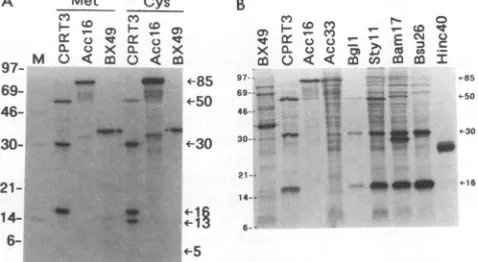
[35S]methionine, proteinswithmolecularmassesof approx-imately 50, 30, 16,and 5 kDawereobserved(Fig. 2A).When
[35S]cysteine was used instead of[35S]methionine, the 50-, 30-, 16-,and5-kDaproteinswereagain detected, aswell as a 13-kDa protein which lacks a methionine residue. In
contrast, bacteria harboring pAcc16 (Fig. 1B), which is deleted of boththe first 14amino acids of the N terminus and the last 14 amino acidsof the Cterminus ofUL80, produced a single proteinwithamolecularmassofapproximately85 kDa(Fig. 2A).These resultssuggestedthattheUL80 protein
encoded by pCPRT3 undergoes autoproteolytic cleavage,
whereasthedeletionproteinencodedbypAcc16isunableto
A Met
_to0
0L 0 X
a'7 M C) < co
69- 46-
30-Cys
Cf)
D
21- 14-
6-B
c,)
-x m X cA
97 5
69--_ -50
46- .
*~~~~'3
30-;
21~~~~~~~~~~~1
_ ._z16
14_
G-4-.5
FIG. 2. (A) Metabolic labelling of UL80 constructs in E. coli
BL21pLysS. Cells harboringthe indicated plasmidswere labelled with either [35S]methionine (Met) or[35S]cysteine (Cys) and then
subjectedtoSDS-PAGE and autoradiography.The5-kDa protein is
notobvious at this exposure level butis clearlyvisible with longer exposureof thegel (datanotshown) and comigrateswith the 5-kDa
protein generated byinvitro cleavageof theassemblyprotein-like
precursor (protein encoded by pBX49; see Fig. 4, lane 3). (B) Labelling of UL80 truncation constructs. Cells harboring the indi-catedplasmidswerelabelled with[35S]methionine asdescribed for panelA. The positions ofmolecular massmarkers(inkilodaltons) areshowntotheleft of each panel.
autodigest. A series of additional experiments described below support this view.
Mapping of the UL80 proteolytic products. Two ap-proacheswereused tolocalize the products of the apparent
autoproteolytic cleavage of the UL80 protein. The protein
mapdeduced from theseexperimentsappearsinFig. 1A and is consistent with the independently obtained map for the simian homolog of this gene, APNG1 (31). In the first strategy, the 50-, 30-, 16-, and 13-kDa proteins were
sub-jectedtoN-terminal sequenceanalysis (Table1).The
result-ing amino acid sequences were then compared with the translated UL80 gene sequence (4) and aligned (Fig. 1A).
The N terminus of the 30-kDa product is Met-1, the N terminus of UL80. The N terminus of the 50-kDaproductis Ser-257, which is about 30 kDa from the N terminus of UL80. Thissuggestedthatthe 50-kDaproteinresulted from cleavage of the UL80polyproteinprecursoratthe Ala-256-Ser-257junction. Since C-terminalsequencingwas not per-formed, the C terminus of the 30-kDa protein isinferred to
beAla-256, although thiswas notdemonstrated directly.
[image:3.612.318.557.82.213.2]Like that of the 30-kDa protein, the N terminus of the
TABLE 1. N-terminalsequencesof HCMVproteinsexpressed
frompCPRT3
Amt Amino acid
(pmol)
position
50 kDa 10
X1_V_X2_P-E-A-A-X3-D_Ib_
Ser-257 30 kDa 200 MC-T-M-D-E-Q-Q-S -Q-A - Met-1 16kDa 400 MC-T-M -D-E-Q-Q-S -Q-A- Met-113 kDa 350 A -T-S-L-S-G-S-E -T-T - Ala-144
a Thefirst 10 amino acidsareindicated.
bFrom alignment ofthis peptide with the UL80ORF, X1 and X2 are
inferred to be Ser-257 andSer-259;theamounts werebelowdetectable levels. Cysteineresiduesare notdetectedbythismethodology; X3isinferredtobe Cys-264.
cApproximately20to25%of theproteincontainsnoN-terminal methi-onine.
on November 9, 2019 by guest
http://jvi.asm.org/
500 BAUM ET AL.
16-kDa protein is Met-1. The N terminus of the 13-kDa protein is Ala-143, about 16 kDa from the N terminus of UL80. Thus, the 16- and 13-kDa proteins appear to result fromcleavage within the 30-kDaprotein,atthe
Ala-143-Ala-144junction. Examination of the putative coding region of the13-kDa protein indicates that this sequence is devoid of methionine residues(4),consistent with the results of label-ling experimentsdescribed above(Fig. 2A; compare CPRT3 cysteine and methioninelanes).
The second strategywas toanalyzetheproteins produced by a series of 3' truncations of the UL80 gene. These truncationswereconstructedbytheinsertionofanonsense
linker at the indicated restriction sites (Fig. 1B). E. coli BL21pLysS cells harboring the indicated truncation
con-structs were metabolically labelled with
[35S]methionine,
and induced proteins were displayed by SDS-PAGE and then by autoradiography (Fig. 2B). pCPRT3 produced the 50-, 30-, and 16-kDa proteins. The 13-kDa band was not
detected because it lacks methionine, but the 5-kDa band wasdetected upon prolongedexposure of thegel(datanot shown). When the experiment depicted in Fig. 2B was
repeated with
[35S]cysteine
instead of [35S]methionine, es-sentially identical results were obtained for all constructs, exceptthat whenever the 16-kDaproteinwasproduced,the 13-kDaproteinwas also detected(datanotshown).Construct pAcc16 again produced the -85-kDa
polypro-tein, as did pAcc33, from which 14 amino acids at the N
terminusaredeleted but which hasanintact Cterminus(Fig.
1B). These data suggest that sequencesattheNterminusare
required for theputative autodigestionof the UL80
polypro-tein. Furthermore, the lack of significant levels of discrete 50-, 30-, and 16-kDa cleavage products from the UL80
polyprotein encodedbyboth pAcc16and pAcc33indicates that thisproteolysis isnotcarriedoutbyanE. coliprotease,
since all of theproposed cleavage sitesarepresentin these
two mutantUL80proteins.Somenonspecific degradationof the pAcc16 and pAcc33 polyproteins does occur (Fig. 2B)
and isprobablydueto cell proteases.
TheUL80 ORFsencodedbyconstructspBgllandpStyll
should endatAsp-694 andLys-631,respectively, compared
with Glu-708 ofpCPRT3 (Fig. 1B). Both thepBgll and the
pStyll constructs produced the 50-, 30-, and 16-kDa pro-teins(Fig. 2B).The5-kDaproteinwas notdetected forpBgll
and pStyll, even upon prolonged exposure of a
higher-percentagegelwhen the 5-kDaprotein produced bypCPRT3
wasvisible(datanot shown).TheAla-643-Ser-644junction
ispredicted to be the site ofassembly protein cleavage (8,
28,31) and is also likelytobethejunctionbetween the 50-and5-kDa proteins which we observe (Fig. 2A). Since the pBgllconstructterminatesatAsp-694, which is downstream of thiscleavagesite,the50-kDaproteinwould beunaffected. ThepStyll constructterminatesatLys-631, 12amino acids upstreamof theputative 50-kDa-5-kDa junction. Therefore,
the5-kDaprotein shouldnotbemade and the 50-kDa protein should be reduced in size by -1 kDa, which is not a detectable difference in this gel system.
The pBaml7 construct terminates at Asp-438 (which is within the 50-kDaprotein) and produced the 30- and 16-kDa proteinsbut notthe50-kDa protein, as expected (Fig. 2B). In
addition, an -28-kDa protein, which may be the truncated
version of the 50-kDa protein, is observed. The pBsu26
construct terminates at Pro-260, just downstream of the Ala-256-Ser-257 junction which apparently separates the 30-and 50-kDa proteins. pBsu26 also produced the 30- and 16-kDa proteins (as well as the 13-kDa protein if
[35S]cys-teine isused; datanot shown), suggestingthat the protease
1 2 3 4 5 6 7 8 9 10 11 1213
859*97.
69;-Z to
504 46
3043 s -Kan
16-4
FIG. 3. Coupled transcriptionand translation ofUL80plasmids. The indicated plasmids(1to2pg)wereincubatedwithE. coli S30
extract containing [35S]methionine asdescribed elsewhere (5) and then subjectedtoSDS-PAGE andautoradiography.Lanes:3,pBX49;
4,pCPRT3; 5, pAcc16; 6, pAcc33; 7, pBgll, 8, pStyll; 9, pBaml7; 10,pBsp6; 11,pBsu26; 12,pHinc40; 13,noDNA. Forcomparison,
[35S]methionine-labelled proteins produced in BL21pLysS by con-structs pCPRT3 (lane 1) and pAcc16 (lane 2) are shown. The plasmid-encoded kanamycinresistance protein (Kan) is indicated.
Molecularmassmarkers(inkilodaltons)areshownontheleft.
domain is foundattheNterminus ofUL80, analogouslyto
the UL26 protease of HSV-1 (16). Consistent with this hypothesis is the observation that the pHinc40 construct, which contains asynthetic termination codon after Val-207, produceda25-kDaprotein,which is the sizeexpectedfrom this ORF. The 25-kDaprotein apparently lacks autoprote-olytic activity, since the 16-kDa proteinwas notproduced,
even though the Ala-143-Ala-144 cleavage site is present. Theotherputative cleavage product (-7 kDa, corresponding
to Ala-144-Val-207 and containing two cysteine residues) was also not detected, consistent with lack ofproteolytic activity. These experiments strongly suggest that the 30-kDa protein is the protease responsible for cleavage of the UL80 polyprotein; however, the possibility that the 13-kDa protein is actually the protease couldnotbe excluded. From datato
be discussed elsewhere, the 13- and 16-kDa proteinswere shown to lack protease activity; pCPRT3, which has been mutated at the 16-kDa-13-kDajunction, still generates the
30-, 50-, and5-kDaproteins, despite the absence of the 16-and 13-kDaproducts, suggestingthat the 30-kDaspecies is theactive protease (1).
Toverifythat all ofthese truncationconstructsdoin fact encode the expected ORFs, theseplasmidsweresubjected tocoupledinvitrotranscriptionandtranslationinanE. coli S30 extract (Fig. 3). We had previously observed that a
human immunodeficiency virus protease precursor which
autodigests to completionwhen expressed in intact E. coli
cells (la) produces detectable amounts of the protease precursorwhen expressed inthe S30 system(1). Similarly,
pCPRT3 and the UL80 truncation constructs displayed less autoproteolysis in the S30 system than in intactE. coli cells
(Fig. 3).Foreach construct,protein of the size expected for its ORFwas detected. Welch et al. (31) also noted that the
proteinfrom thecorrespondingSCMV ORF, APNG1, failed
to autodigest upon translation of its mRNA in the rabbit
reticulocyte lysate.
Proteolysisintrans of theassemblyprotein cleavagesite. To
determine the abilities of various derivatives of UL80 pro-tein to carry out proteolysis in trans,
[35S]methionine-la-belled substratewas prepared from E. coli cells harboring pBX49(Fig. 1B). pBX49 encodesa35-kDaprotein (Fig. 4, lane 1) corresponding to amino acids Asp-438-Glu-708 of
UL80, containing the predicted assembly protein cleavage J. VIROL.
21
on November 9, 2019 by guest
http://jvi.asm.org/
[image:4.612.340.535.73.175.2]EXPRESSION OF HCMV UL80-ENCODED PROTEASE 501
1 2 3 4 5 6 7 8 9 10 690
[image:5.612.83.273.71.174.2]
14--Cleavage
FIG. 4. Transdigestion of the substrate derived frompBX49 by UL80proteins. Lysates ofE. coli BL21pLysS cells harboring the indicated plasmids were incubated with
[35SJmethionine-labelled
pBX49-derivedproteinas described in Materials and Methods and thensubjectedtoSDS-PAGE andautoradiography.Molecularmass markers(in kilodaltons)areshowntothe left. Thepositionsofintact pBX49-derived proteinandof the 30- and 5-kDacleavage products
are shown on theright. Lane 1, unincubatedpBX49-derived sub-strate. Labelled pBX49-derived substrate Was incubated with
ly-satesofbacteriaharboring pBX49 (lane2), pCPRT3 (lane 3), pAccn6 (lane4), pAcc33 (lane 5), pBgll (lane 6), pStyll (lane 7),
pBaml7
(lane 8), pBsu26 (lane 9),or
pHinc40
(lane 10).siteatAla-643-Ser-644
(which
alsoappears
tobe thecleav-age site between the So- and 5-kDa
proteins
encodedby
HCMwUL8o; Fig.
LA).
Note thatthe bona fideassembly
protein corresponds
to Met-336-Glu-708 and ispresumably
translated from the 1.1-kb UL80.s mRNA,
compared
with the 2.1-kb UL8Oa mRNAencoding
the entire UL80 ORF(30).
Thus, thepBX49-derived
substrate is an -10-kDa N-terminal deletion ofthe HCMVassembly
proteinprecur-sor.
Lysates
wereprepared
from induced, unlabelledE. colicells harboring pCPRT3 or the truncation constructs (Fig. 1B) and incubated with the radiolabelled pBX49-derived substrate. Proteolytic cleavage of this 35-kDa substrate encodedby pBX49 was monitored by SDS-PAGE followed
by autoradiography (Fig. 4). The lysate from E. coli cells
A
B
harboring pCPRT3 cleaved the 35-kDa pBX49-derived
sub-strate into the expected 30- and 5-kDa digestion products (Fig. 4, lane 3). These products were not observed upon
incubation of the pBX49-derived substrate alone (Fig. 4, lane 2)orwithpAcc16-orpAcc33-derived lysate (lanes4and 5).
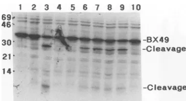
In contrast, lysates from bacteria harboring truncation con-structspBgll, pStyll, pBaml7, and pBsu26 (Fig. 4, lanes 6 to9)were abletocleave thepBX49-derived substrate into the 30- and 5-kDa products. However, the lysate from bacteria harboring pHinc4o failed to cleave the pBX49-derived substrate (Fig. 4, lane 10). These data suggest that the ability to cleavethe HCMVassembly protein substrate intransis retained by the N-terminal 30-kDa domain of the UL80 protein.
Inhibition ofprotease activity. HCMV proteasewas
pre-pared frominclusion bodies from BL21pLysS cells harbor-ing pBaml7. This material is amixture of the 30-, 16-, and 13-kDa proteins (Fig. 5A, lane 5). After the protease was titrated to determine the minimal amount needed tocleave the pBX49 substrate (Fig. SB), a battery of protease inhibi-tors were tested fortheir abilities to inhibit the protease (Fig. SC). Aprotinin (Fig. 5, lane 3 [1 ,ug/ml]), E-64 (lane4 [0.5 ,ug/ml]), calpain inhibitors I (17 ,ug/ml) and II (7 ,ug/ml), TLCK (N-tosyl-L-lysine chloromethyl ketone; 40 ag/ml),
TPCK (N-tosyl-L-phenylalanine chloromethyl ketone; 80
p,g/ml),
pepstatinA(0.7 ,ug/ml), leupeptin (0.5 ,ug/ml),cys-tatin (100 pg/ml), and EDTA (1 mM) did not inhibit the digestion of the pBX49-derived substrate (data not shown). However,ZnCl2 (2.5 mM) completely inhibited the proteol-ysisof that substrate(Fig. 5,lane6),suggesting thatHCMV
protease is a cysteine protease. Phenylmethylsulfonyl
fluo-ride (100 ,g/ml), which inhibits cysteine and serine
pro-teases,was alsoslightly inhibitory towardHCMVprotease
(Fig. 5, lane5). In contrast to our resultswith HCMV UL80 protease,Liu andRoizman (17) recently demonstratedthat
HSV-1 UL26 protease exhibitsserine protease-like features. The 30-kDa protein is not the precursor of the 16- and 13-kDa proteins. Since the N-terminal sequence analysis
C
M 1 2 3 4 5
46 "
30 w au. -3OkD
14
-6IkD
6 s * i -13kD
0 2 5 10 25 50
jw ...
46
30"--
--_ -BX49_ w-Cleavage
-%.
14
-6.
-Cleavage
FIG. 5. (A)Purification of HCMV protease. E. coli BL21pLysScellsharboringpBaml7areshownpriorto(lane 1)and 2 hafter(lane2) addition of IPTG. The induced cells werelysedin a French press, and thelysates werecentrifugedtoseparatesupernatant(lane 3)frompellet (lane4).Thepelletwassolubilized with urea anddialyzed as described in Materials and Methods(lane 5).Lanes 1to4contain
100-tlA
cellequivalents; lane 5contains 500-,ulcellequivalents. Thisis aCoomassie blue stain ofa 15% polyacrylamidegel. (B)Titration of HCMV protease.Theindicatedquantities(inmicrograms)of 30-kDa proteasepreparedfrom thepelletshownwereincubated withpBX49-derived substrate and thensubjectedtoSDS-PAGEandautoradiography.(C)Inhibitortest.A20-,ug sampleofHCMVproteasewasincubated with
pBX49-derivedsubstratein the presence of the indicated protease inhibitors and thensubjectedtoSDS-PAGE andautoradiography.Lanes: 1,noprotease;2,noinhibitor; 3,aprotinin (1pg/ml); 4,E-64(0.5,Lg/ml); 5,phenylmethylsulfonyl fluoride(100
1±g/ml);
6,ZnCl2(2.5mM).2 3 4 5 6 .li:",
*.
Z-4n
wn-P4j6I
_-BX49*sCleavaoe
M I
46i
21
1 4
6 VOL. 67,1993
on November 9, 2019 by guest
http://jvi.asm.org/
[image:5.612.137.481.475.642.2]502 BAUM ET AL.
CPRT3
Acc16
1' 5' 15'30'60' 1' 5'15'30'60'
504 _UI*
304 -
[image:6.612.122.240.73.236.2]-134 7
FIG. 6. Pulse-chase of E. coli BL21pLysS cells harboring pCPRT3 or pAcc16. Cells were pulsed with [35S]methionine as described in Materials and Methods and chased with unlabelled
methionine for the indicated times.The13-kDaprotein lacks
methi-onineresidues; thelabelling of this protein probablyresults from
reincorporationoflabel from[35S]methionineintocysteine.
discussed above demonstrated that the 16- and 13-kDa proteins are organized inalinear fashion within the 30-kDa protein, itwasof interest to determine whether the 30-kDa protein is the precursor of thetwosmallerproteins. Accord-ingly, a pulse-chase radiolabelling experiment was per-formed onE. colicells harboring pCPRT3 orpAcc16 (Fig. 6).Asexpected, pCPRT3produced[35S]methionine-labelled
proteins with molecular masses of 50, 30, and 16 kDa. A smallamountoflabellingof the 13-kDaproteinwasdetected and probably results from degradation of methionine and reincorporation of the
35S
into cysteine. None of theseproteins is present in the lysates of bacteriaharboring the pAcc16 control. Between 5 and 60 min, theamounts of the 16-and 13-kDaproteinsincreasedapproximately 10-fold,yet the amount of 30-kDaprotein stayed constant. These data suggestthat the 30-kDaproteinisnotthe precursor ofthe 16-and 13-kDa proteins. Moreover, the fact that intact 85-kDa UL80 polyprotein or presumptive cleavage intermediates (e.g., 13- plus 50-kDa proteins are equal to a63-kDa
inter-mediate)were notdetected suggests thatcleavageatall sites
(16-kDa-13-kDa, 30-kDa-50-kDa, and 50-kDa-5-kDa junc-tions)occurs cotranslationally.
Ifthe 16- and 13-kDa proteins are not direct products of the 30-kDa breakdown, what then is the origin of these proteins? Experiments with the UL80 truncationconstructs pBaml7and pBsp6 suggest thatcleavageat the 16-kDa-13-kDa junction occurs on the UL80 precursor polyprotein rather than directlyfrom preexisting 30-kDa protein. Each
constructcontains a stopcodon within the 50-kDaregion of UL80(Fig. 1B).pBaml7 terminates atAsp-438, and pBsp6 terminatesatThr-354, compared with Ala-643 for the 50-kDa protein.Asexpected, the30-, 16-, and 13-kDa species were detected in radiolabelled E. coli cells harboringpBaml7or pBsp6(Fig. 7A). As noted for the experiment shown in Fig. 2B,an -28-kDaproteinwhich may be the truncated version
ofthe 50-kDaproteinwas observed forpBaml7; the corre-sponding truncation product for pBsp6 was not observed and maybe unstable. A new protein which is not present from
parentalpCPRT3wasobserved for each construct (Fig. 7A
[arrows]). Thesebands are consistent with full-length
poly-proteinswith molecular masses of -52 kDa forpBaml7and
A
XL CO ea
B
Baml7
1 5' 15'30'45'
Bsp6 1' 5115'30' 45'
69-_ _ _.. ., e~f52
46-30-* _3
21-14- -1
3-FIG. 7. (A) Metabolic labelling ofUL80 constructs inE. coli
BL21pLysS. Cells
harboring
the indicatedplasmidswere labelledwith[3S]methionine plus [3S]cysteineand thensubjectedto
SDS-PAGE and autoradiography. The putative full-length polyproteins from bacteria harboring the pBaml7 and pBsp6 constructs are shown (arrows). (B) Pulse-chase radiolabelling of E. coli
BL21pLysScellsharboringpBam17orpBsp6.Theexperimentwas performed as described in the legendto Fig. 6, except that both [35S]methionineand[35S]cysteinewereusedfor thepulse andboth
methionine andcysteinewereused for the chase.The times ofchase areshown in minutes.
of -42 kDa for pBsp6. Full-length 85-kDapolyproteinwas
notobserved for pCPRT3,suggesting eitherthatcleavageof pBaml7 and pBsp6 polyprotein is less efficient than that of
pCPRT3orthat therateofsynthesisofpBaml7andpBsp6 polyproteins is higherthan that ofpCPRT3polyprotein.
A pulse-chase experiment with E. coli cells harboring pBaml7 and pBsp6wasperformed(Fig. 7B). The full-length
polyprotein (Fig. 7B, arrows) decreased in abundance
be-tween 1 and 5 min,as theamountsof30-, 16-, and 13-kDa proteins increased. Aswas seen in the pulse-chase experi-mentwithE. coli cellsharboring pCPRT3 (Fig. 6), the 16-and 13-kDa species did not appear at the expense of the 30-kDaspecies. Thedisappearanceof thepolyproteins dur-ing the chase coupled with the apparent stability of the 30-kDa species strongly suggests that cleavage at the 16-kDa-13-kDajunctionoccursonly in the polyprotein and not within preexisting30-kDa protein. Thus, the N terminus of UL80polyproteinappearstohavetwopossiblefates: cleav-agetogeneratethe 16- and13-kDaproteinsandcleavageto
generate the30-kDaprotein.
Detection of cleavage at the 16-kDa-13-kDa junction in
HCMV-infectedcells.Ifcleavageatthe16-kDa-13-kDa junc-tion isapotential mechanismtodownregulate the expression of the 30-kDa protease, it should bepossible to detect the
16-and/or13-kDa product duringinfectionbyHCMV.
Accord-ingly, HFF were infected with HCMV strain AD169 and labelled with [35S]methionine plus
[35S]cysteine
from 68 to 72 h postinfection. Lysates from infected and uninfected cells were immunoprecipitated with polyclonal antiserum raisedagainstthe 16-kDaprotein. This serum recognizes the 16- and 30-kDa, but not the 13-kDa, UL80 proteins ex-pressed in E. coli(Fig.8, lower panel, lane E). Four proteinswerespecificallyimmunoprecipitated from HCMV-infected cells: -85- and -80-kDa proteins (Fig. 8, upper panel) and 30- and 16-kDaproteins (lowerpanel). These proteins were
not immunoprecipitated from uninfected cells or with pre-immuneserum.
The 30- and 16-kDa proteins from virus-infected cells
comigratedwith their counterparts expressed in E. coli cells
(Fig. 8, lower panel). Thus, these proteins are produced J. VIROL.
on November 9, 2019 by guest
http://jvi.asm.org/
[image:6.612.321.554.73.199.2]200-~
97- &IR
69- _
46-
30-A
ISME
-50
o-30
*-19
*-30
B
MI 2 34 5 46,
30 --- +-BX49
i...
_CI-CIeavage 21..14f. 6
*-Cleavage
UNINF AD169I
.PI P I E
-Ig
4-30
21-.
*wF
[image:7.612.125.224.73.295.2]14- A16
FIG. 8. UL80 proteins produced during HCMV infection. Unin-fected HFF(UNINF) orHFF infected withHCMV strain AD169 wereradiolabelled with [35S]methionine and [35S]cysteine from 68to 72 h postinfection. Lysates were immunoprecipitated with either preimmuneserum(P)orimmuneserum(I) raised againstthe 16-and 30-kDaUL80 proteinsandsubjectedtoSDS-PAGEand autoradiog-raphy. Theupperpanel isa1-dayexposureof theupperportion, and
the lowerpanel isa14-dayexposureof the lowerportionof thesame
gel. Lane E,immunoprecipitate of radiolabelled E. coli cells har-boring pBaml7. Molecularmassmarkers(in kilodaltons)areshown
onthe left. An unidentifiedprotein (Ig) with amolecular massof approximately 35 kDa is nonspecifically immunoprecipitated from AD169-infected cellsby almostevery serumsampletested andmay
be animmunoglobulin receptor-like protein (12a).
duringthe HCMVreplicative cycle. The 85-kDa species is
full-length UL80 polyprotein, and the 80-kDa species is
probablyUL80 polyprotein which has been cleaved at the
assembly protein cleavage site (Ala-643-Ser-644; Fig. 1A),
with 5 kDa removed from the C terminus as aresult. The
relative abundance of these four proteins at 68 to 72 h
postinfection (85 80 > 30 > 16kDa)isquitedifferent from that ofUL80 expression in E. coli (16 30 > 80, 85kDa) (from pCPRT3; Fig. 2A). This result maybe due to differ-entialcleavagesatthe various sites in theUL80polyprotein
in HCMV-infected cells or to differential stabilities of the various UL80-derived proteins. The results from Western blots (data not shown) areconsistent with the immunopre-cipitation data.
Separation ofcisandtransactivities byalteration of theC terminus of the 30-kDa protein. Several constructs which result inproduction of the 30-kDaproteinwith either small deletions or additions at its C terminus were generated.
Constructs p3Ok(-8)andp3Ok(-3) aredeleted ofeight and three aminoacids, respectively, fromthe C terminusof the
30-kDa protein. Similarly, p3OK(+12) contains an extra 12
amino acids (a c-myc epitope) at the C terminus. The
proteins produced byE.coli cellsharboringtheseconstructs
were labelled with [35S]methionine and [35S]cysteine and examinedbySDS-PAGE andautoradiography.p30k,
[image:7.612.351.517.74.185.2]encod-ingthewild-type30-kDaprotein, producedthe30-, 16-,and 13-kDaproteins in ratios comparabletothose producedby pCPRT3 (Fig. 9A). In contrast, deletion ofasfew asthree
FIG. 9. (A) Proteins generated by the 30-kDa protease construct andby C-terminalmutants. E. coli BL21pLysS cells harboringthe
indicated plasmids(seetext)werelabelled with[35S]methionineplus
[35S]cysteine andthen subjected toSDS-PAGE and
autoradiogra-phy. 30k, the wild-typeproteinwhichterminates at Ala-256;
dele-tion or addidele-tion of amino acids at the Cterminus is indicated in
parentheses. Wild-type UL80 proteins encoded by pCPRT3 are shown as a control. (B) Cleavage ofpBX49-derived substrate by
30-kDa protease and by C-terminalmutants. Protein (2 to 10 pLg) from urea-solubilized inclusion bodies from E.coliBL21pLysScells harboring the indicatedplasmids was incubated with
[5S]methio-nine-labelledpBX49-derived substrate and thensubjectedto SDS-PAGE andautoradiography. Lanes: 1,substrateonly; 2,pCPRT3;
3,p3Ok(-8); 4,p30k; 5,p3Ok(+12).
amino acids from the C terminusdrasticallyreduced produc-tionof the 16- and 13-kDa proteins, byatleast90% [Fig.9A, lane 30k(-3)]. Similar results are obtained upon deletion of 8 amino acids[Fig.9A,lane30k(-8)]oraddition of 12 amino acids[lane 30k(+ 12)]. These data indicate that the authentic C terminus of the 30-kDa protein is required for efficient
cleavage at the 16-kDa-13-kDajunction. This suggests that inE. coli cellsharboringpCPRT3, the 30-kDa proteasemust
be released from the precursorpolyprotein forcleavage at
the 16-kDa-13-kDajunction to occur. The 16- and 13-kDa bands arealso observed forpBsu26 (Fig. 2B),which
termi-nates at Pro-260, four amino acids downstream of the C terminus of the 30-kDaprotein.Thisimplieseither that these
extrafour amino acids are cleaved from the C terminusor
thattheydonotimpede16-kDa-13-kDacleavageif present. Theabilities of the 30-kDa protease and of the C-terminal
mutants to cleave pBX49-derived substrate in trans were
examined. Thesemutants were able tocleave the
50-kDa-5-kDa junctionof that substrate(Fig.9B, lanes 3to5).Thus,
mutations at the C terminus severely diminish protease
activityatthe 16-kDa-13-kDa bond butnot at the 50-kDa-5-kDa bond in trans. Note that when unlabelledwild-type
proteasewasaddedtolabelledlysates of bacteriaharboring p3Ok(-8), p3Ok(-3), or p3Ok(+12), the labelled 30-kDa
mutant proteins were not further cleaved into the 16- and 13-kDaproducts (datanotshown). Itispossiblethat cleav-age at the 16-kDa-13-kDa bond can occuronly
intramolec-ularlyorthatcleavage occurscotranslationallyand that the 16-kDa-13-kDa bond is inaccessibleoncethe 30-kDaprotein
is released from the UL8O polyprotein. In either case, it appearsthat thewild-typeC terminus of the 30-kDa protease isrequiredfor thiscleavage.
DISCUSSION
WehaveusedexpressioninE.colitodemonstratethat the HCMV UL80 ORF encodes a protease atits N terminus. The presenceofaproteasein HCMVUL80was
postulated
byLiu and Roizman(17)from studies of HSV-1 ORF
UL26,
on November 9, 2019 by guest
http://jvi.asm.org/
504 BAUM ET AL.
50k
Make "active" 5k protease
4-
NWmmm-.
30k
4 Make"inactive"protease
16k 13k 50k 5k
"inactive"
30k isnotdirectly cleaved into 16k + 13k proteins
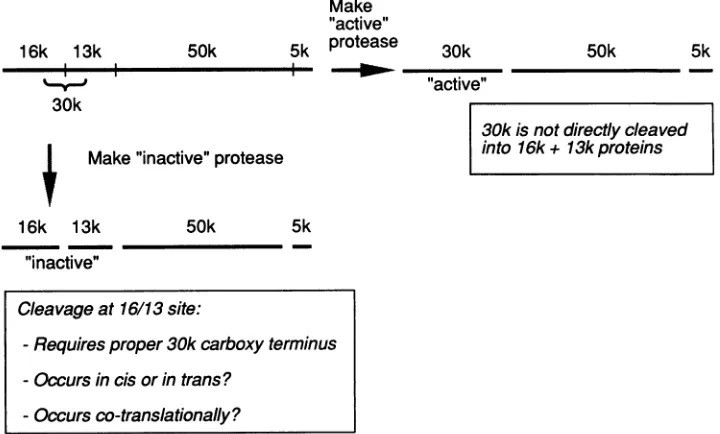
FIG. 10. Model of UL80polyprotein-processing pathway. A two-branch pathwayispostulated (see Discussion), whichresults inthe productionof active30-kDaproteaseorinactive16- and13-kDaproteins.Onceformed, the 30-kDaproteaseappearstobestableand isnot furthercleaved into the16- and13-kDaproteins.
which, likeUL80, encodes the assembly protein. The SCMV homologofUL80, APNG1,wasrecentlyshowntoencodea protease (31). Previous studies with HSV, SCMV, and
HCMV demonstrated that the assembly protein undergoes proteolytic cleavage near its C terminus (8, 24, 28); in
HSV-1, this cleavage ispresumably required for packaging
ofprogeny DNA into the capsid (11, 14, 24, 25). We now
show that a 30-kDaprotein isgenerated fromautocatalytic digestionof85-kDaUL80 polyproteinand is abletocleavea
substrate containing the assembly protein cleavage site.
Thus, the30-kDaprotein encodedbytheN-terminal region
ofUL80appearstobe theprotease.
The85-kDaUL80 polyproteinis cleavedatthree different sites (the 16-kDa-13-kDa, 30-kDa-50-kDa, and
50-kDa-5-kDajunctions; Fig. 1), generatingfivemajorproducts (with
molecularmassesof16, 13,30,50, and 5 kDa). Cleavageat
the30-kDa-50-kDajunctionpresumablyreleases themature
30-kDaproteaseanda50-kDaproteinwhichisanN-terminal
extension of the 40-kDa assembly protein. The 50-kDa protein isprobably analogousto theSCMV 45-kDa protein
observed bySchenk et al. (28), and its function, ifany, is
unknown.
For HCMV-infectedcells at late times postinfection, we
show that the 85-kDa species (intact UL80 polyprotein) is
slightly more abundant (approximately threefold) than the
80-kDaspecies(UL80 polyproteinwhich hasbeencleavedat
the assembly protein cleavage site) (Fig. 8). Together, the
85- and 80-kDa proteins are approximately 10- to 20-fold more abundant than the 30-kDa protein. Thus, the 30-kDa species is a relativelyminorcomponentinHCMV-infected
cells. Incontrast, forUL80expressed inE. coli,cleavage at both the50-kDa-5-kDa and the 30-kDa-50-kDa junctionsis
essentially complete, since no polyprotein precursors are
detected. This suggests that the extent of cleavage at the
50-kDa-5-kDa andthe30-kDa-50-kDa junctions is regulated
invirus-infectedcells. Alternatively, the cleavage products mayexhibit differentialstabilities.
It is notknownwhether the 80- and 85-kDa UL8O
poly-proteins possess protease activity. Mutagenesis of the
30-kDa-50-kDa junction of theUL80geneexpressedin HCMV
would addressthispoint morecompletely. Cleavage at the 30-kDa-50-kDa site does appear to be required for the 30-kDaprotein toacquire its fullrangeofproteolytic
activ-ity, sincefusion to the C terminus of the 30-kDa protease [e.g., Fig. 9,lane30k(+ 12)]abolished theabilitytocleaveat
the 16-kDa-13-kDa junction but not at the 50-kDa-5-kDa
junction. This is reminiscent of the poliovirus 3CD (poly-meraseprotease) polyprotein,whichexhibits protease activ-itybut has substratespecificitydifferent from that ofmature
3C protease(32).
A recent publication by Welch et al. (31) also demon-strated the existence ofa30-kDa proteaseatthe Nterminus oftheUL80 homolog (APNG)fromSCMV.By transfecting portions of the APNG ORF into mammalian cells, they
deduced thatcleavageoccursatthe30-kDa-50-kDa junction (Ala-256-Ser-257) and at the 50-kDa-5-kDa junction (Ala-643-Ser-644),in agreementwithourresults withprokaryotic
cells. We have detected an additional cleavage event at Ala-143-Ala-144, which generates 16- and 13-kDa proteins whose sequences areentirely contained within the 30-kDa protein.The16-kDa-13-kDacleavage site(Ala-Ala)is-50%
cleaved in E. coli, compared with quantitative cleavage at
the Ala-Ser sites of the 30-kDa-50-kDa and 50-kDa-5-kDa
junctions. Computer alignment indicates that the SCMV APNG studied by Welch et al. (30, 31) lacks the exact sequenceofthe HCMVUL80 16-kDa-13-kDa cleavage site
(datanot shown); cleavagemayoccur elsewhere, ornot at all,within the SCMV30-kDaprotease. Furthermore, since Welch etal. (31) monitored cleavage by epitope taggingat
either the C terminus of APNG or within a region that apparently inactivated the protease, the 16- and 13-kDa proteins,ifpresent, would notbedetected.
We initially detected the 16- and 13-kDa proteins by expression ofUL80 in E. coli, but, more importantly, this cleavage event also occurs during virus replication, as
demonstratedby immunoprecipitationof the16-kDaprotein
16k 13k 30k
"active"
50k 5k
Cleavageat16/13site:
-Requires proper30k carboxy terminus -Occurs in cisorintrans?
-Occurs
co-translationally?
J. VIROL.
on November 9, 2019 by guest
http://jvi.asm.org/
[image:8.612.134.494.76.293.2]by antibodies tothe N terminus ofUL80. However, atlate
times postinfection, the 16-kDa protein is relatively rare
(-1/10 the abundance ofthe 30-kDa protein, which is itself
-1/10 the abundance ofthe 80- plus 85-kDa proteins). Thus,
it remains to be determined whether the 16-kDa-13-kDa
cleavage event plays a role in the virus life cycle. Cleavage at the 16-kDa-13-kDa site separates the two conserved
domains, CD2 and CD1, which are present in all putative
herpesvirus proteases and which may be important for
function(31). Thus, thiscleavage possibly is a mechanism to
inactivate the protease. The individual 16- and 13-kDa proteins lack detectable protease activity (1). Additionally, the 16- and/or 13-kDa protein may serve some unknown function inHCMV replication; this is currently under
inves-tigation.
Theproduction of the 16- and 13-kDa proteins has several
interesting features. Although the 16- plus13-kDa sequences
constitute the 30-kDa protein, the resistance of the 30-kDa species to further cleavage and its stability in pulse-chase experimentssuggest that the 30-kDa protein is not the direct
precursorof the 16- and 13-kDa proteins. Instead, cleavage atthe16-kDa-13-kDa junction appearsto occur on theUL80
polyprotein. Since the 30-kDa protein is stable, itis possible
thatcleavageatthe16-kDa-13-kDajunction occurs cotrans-lationally and that this site is inaccessible once the 30-kDa proteinisreleased from the UL80 precursor. These features are summarized in the model diagrammed in Fig. 10. A
two-branch pathway is postulated: the first cleavage occurs
either at the 30-kDa-50-kDa junction, producing active
30-kDa protease, or at the 16-kDa-13-kDa junction, producing proteolytically inactive 16- and 13-kDa proteins. The two
branches can be separated by mutation of the C terminus of
the 30-kDa protease; whereas wild-type protease carries out
bothbranches, C-terminal mutants are defective for
cleav-age at the 16-kDa-13-kDa junction.
In E. coli,the amount of 30-kDa protein produced is about the sameas the amount of 16- or 13-kDa protein produced;
this suggests that the two putative branches are utilized approximately equally. In contrast, in virus-infected cells at
late timespostinfection, the 30-kDa species is -10-foldmore
abundant than the 16-kDa species, suggesting that the
branch to generate active protease is utilized almost
exclu-sively. Ifcleavage at the 16-kDa-13-kDa junction is a regu-latory mechanism in HCMV, then a shift in the ratio of
active toinactive branch utilization may occur during virus replication. To determine the relevance of cleavage at the 16-kDa-13-kDa junction and at the other UL80 junctions, mutations of these sites can be analyzed within the context
of the virus via gene replacement (12, 13).
ACKNOWLEDGMENTS
We thank J. Morin, M. Vitek, S. Plotch, and L. Li for useful
criticismof themanuscript.
REFERENCES
1. Baum,E. Z., and G. A. Bebernitz.Unpublished data.
la.Baum,E.Z., G. A.Bebernitz, and Y.Gluzman. 1990. Isolation ofmutantsof human immunodeficiency virus protease based on thetoxicityof the enzyme inEscherichiacoli. Proc. Natl. Acad. Sci. USA 87:5573-5577.
2. Baum,E.Z., G. A. Bebernitz,0. Palant, T. Mueller, and S. J. Plotch.1991.Purification, properties, and mutagenesis of polio-virus 3C protease. Virology185:140-150.
3. Braun,D.K., B. Roizman, and L.Pereira. 1984. Characteriza-tion ofposttranslationalproducts of herpes simplex virus gene 35products bindingtothesurface of full but not empty capsids. J.Virol. 46:103-112.
4. Chee, M. S., A. T. Bankier, S. Beck, R. Bohni, C. M. Brown, R. Cerny, T. Horsnell, C. A. Hutchinson, T. Kouzarides, J. A. Martignetti, E. Preddie, S. C. Satchwell, P. Tomlinson, K.
Weston, and B. G.Barrell. 1990. Analysis of the protein-coding content of the sequence of human cytomegalovirus strain AD169. Curr. Top. Microbiol. Immunol. 154:125-169. 5. Collins, J. 1979. Cell-free synthesis of proteins coding for
mobilization functions ofColEl and transposition functions of Tn3. Gene 6:29-42.
6. Evan, G. I., G. K.Lewis, G. Ramsay, and J. M. Bishop. 1985. Isolation of monoclonal antibodies specific for human c-myc proto-oncogene product. Mol. Cell. Biol. 5:3610-3616. 7. Gibson, W. 1983. Protein counterparts of human and simian
cytomegaloviruses. Virology 128:391-406.
8. Gibson, W., A. I. Marcy, J. C. Comolli, and J. Lee. 1990.
Identification of precursor to cytomegalovirus capsid assembly protein and evidence that processing results in loss of its carboxy-terminal end. J. Virol.64:1241-1249.
9. Holland, L. E., R. M. Sandri-Goldin, A. L. Goldin, J. C. Glorioso, and M. Levine. 1984. Transcriptional and genetic analyses of the herpes simplex virus type 1 genome: coordinates 0.29 to 0.45. J. Virol. 49:947-959.
10. Irmiere, A., and W. Gibson. 1983. Isolation and characterization of a noninfectious virion-like particle released from cells in-fected with human strains of cytomegalovirus. Virology 130: 118-133.
11. Irmiere, A., and W. Gibson. 1985. Isolation of human cytomeg-alovirus intranuclear capsids, characterization of their protein constituents, and demonstration that the B-capsid assembly protein is also abundant in noninfectious enveloped particles. J. Virol. 56:277-283.
12. Jones, T. R., and V. P. Muzithras. 1992. A cluster of dispensable genes within the human cytomegalovirus genome short compo-nent: IRS1, US1 through US5, and the US6 family. J. Virol. 66:2541-2546.
12a.Jones,T. R., and V. P. Muzithras. Unpublished data. 13. Jones, T. R., V. P. Muzithras, and Y. Gluzman. 1991.
Replace-mentmutagenesis of the human cytomegalovirus genome: US10 and US11 gene products are nonessential. J. Virol.
65:5860-5872.
14. Lee, J. Y., A. Irmiere, and W. Gibson. 1988. Primate cytomeg-alovirus assembly: evidence that DNA packaging occurs subse-quent to B capsid assembly. Virology 167:87-96.
15. Liu, F., and B. Roizman. 1991. The promoter, transcriptional unit, and coding sequence of herpes simplex virus 1 family 35 proteins are contained within and in frame with the UL26 open reading frame. J. Virol. 65:206-212.
16. Liu, F., and B. Roizman. 1991. The herpes simplex virus 1 gene encoding a protease also contains within its coding domain the gene encoding the more abundant substrate. J. Virol. 65:5149-5156.
17. Liu, F., and B. Roizman. 1992. Differentiation of multiple domains in the herpes simplex virus 1 protease encoded by the UL26gene. Proc. Natl. Acad. Sci. USA89:2076-2080. 18. Maniatis, T., E. F. Fritsch, and J. Sambrook. 1982. Molecular
cloning: a laboratory manual. Cold Spring Harbor Laboratory, Cold Spring Harbor, N.Y.
19. Matsudaira, P. 1987. Sequence from picomole quantities of proteins electroblotted onto polyvinylidene difluoride mem-branes. J. Biol. Chem. 262:10035-10038.
20. McGeoch, D. J., M. A. Dalrymple, A. J. Davison, A. Dolan,
M. C. Frame, D. McNab, L. J.Perry,J. E. Scott, and P. Taylor. 1988. The complete DNA sequence of the long unique region in the genome of herpes simplex virus type 1. J. Gen. Virol. 69:1531-1574.
21. Moffatt, B. A., and F. W. Studier. 1987. T7lysozyme inhibits transcription by T7 RNA polymerase. Cell 49:221-227. 22. Oram, J. D., R. G. Downing, A. Akrigg, A. A. Dollery, C. J.
Duggleby, G. W. G. Wilkinson, and P. J. Greenaway. 1982. Use of recombinant plasmids to investigate the structure of the human cytomegalovirus genome. J. Gen. Virol. 59:111-129. 23. Plotch, S. J.,0.Palant, and Y. Gluzman. 1989. Purification and
properties of poliovirus RNA polymerase expressed in
on November 9, 2019 by guest
http://jvi.asm.org/
506 BAUM ET AL.
cherichiacoli. J.Virol.63:216-225.
24. Preston,V. G., J. A. V. Coates, and F. J. Rixon. 1983. Identifi-cation and characterization of a herpes simplex virus gene
product required for encapsidation of virus DNA. J. Virol. 45:1056-1064.
25. Preston, V. G., F. J. Rixon,I.M.McDougall, M.McGregor, and
M. F. Al Kobaisi. 1992. Processing of the herpes simplex virus assemblyproteinICP35nearits carboxy terminal end requires theproduct of the whole of the UL26 reading frame. Virology 186:87-98.
26. Robson, L., and W. Gibson. 1989. Primate cytomegalovirus assemblyprotein: genomelocation and nucleotidesequence. J.
Virol.63:669-676.
27. Rosenberg, A. H., B. N. Lade, D.-S. Chui, S.-W. Lin, J. J. Dunn, and F. W. Studier. 1987. Vectors for selective expression of clonedcDNAs by T7 RNA polymerase. Gene 56:125-135. 28. Schenk, P., A. S. Woods, and W. Gibson. 1991. The
45-kilodalton protein ofcytomegalovirus (Colburn) B-capsids isan
amino-terminal extension form of the assembly protein. J. Virol. 65:1525-1529.
29. Sherman, G., and S. L. Bachenheimer. 1988. Characterization of intranuclear capsids made by ts morphogenic mutants of HSV-1. Virology163:471-480.
30. Welch, A. R., L. M.McNally,andW. Gibson. 1991. Cytomeg-alovirus assembly protein nestedgenefamily: four 3'-coterminal transcriptsencode four in-frame, overlapping proteins. J. Virol. 65:4091-4100.
31. Welch, A. R., A. S. Woods, L. M. McNally,R.J. Cotter, andW. Gibson. 1991. Aherpesvirus maturational proteinase, assemb-lin: identification of its gene, putative active site domain, and cleavage site. Proc. Natl. Acad. Sci. USA 88:10792-10796. 32. Ypma-Wong, M. F., P. Gillis Dewalt, V. H. Johnson, J. G.
Lamb, and B. L. Semler. 1988. Protein 3CD is the major poliovirus proteinase responsible for cleavage of theP1capsid
precursor.Virology 166:265-270.
33. Zweig, M., C. J. Heilman, H. Rabin, and B. Hampar. 1980. Shared antigenic determinantsbetweentwo distinct classesof proteins in cells infected with herpes simplex virus. J. Virol. 35:644-652.
J.VIROL.

![FIG. 3.The4,plasmid-encodedstructsthenextractMolecular[35S]methionine-labelled10, pCPRT3; Coupled transcription and translation of UL80 plasmids](https://thumb-us.123doks.com/thumbv2/123dok_us/1304018.83582/4.612.340.535.73.175/plasmid-encodedstructsthenextractmolecular-methionine-labelled-coupled-transcription-translation-plasmids.webp)