JOURNAL OFVIROLOGY, Aug. 1989, p. 3220-3226 0022-538X/89/083220-07$02.00/0
Copyright© 1989, American Society for Microbiology
Activation
of
Endogenous
c-fos
Proto-Oncogene
Expression
by
Human
T-Cell
Leukemia Virus
Type
I-Encoded
p4Otax
Protein in the
Human
T-Cell
Line, Jurkat
KINYA NAGATA, KIYOSHI OHTANI, MASATAKA NAKAMURA, AND KAZUO SUGAMURA* DepartmentofMicrobiology, Tohoku University School of Medicine, 2-1 Seiryomachi, Sendai 980, Japan
Received 23 December1988/Accepted 10 April 1989
Weexamined theability ofthetrans-acting factor p4O"'0 ofhuman T-cellleukemiavirus type I (HTLV-I), which isthoughttobeacrucialmolecule in T-celltransformationby HTLV-I,toactivateexpressionofaset ofendogenouscellulargenesrelatedtoT-cellproliferation.For thispurpose, weestablishedasubclone(JPX-9)
ofJurkat cells that was stably transfected with an expression plasmid containing the p40' gene, whose expression isdefinitively dependentonheavy-metal ions. Expressionof the interleukin-2 receptorachain in
JPX-9 cellswasinducedinresponsetotheinductionofp40"' expression,ashasbeen demonstratedbyothers
intransient transfection experimentswithJurkatcells. Inaddition, wefoundthatsignificantenhancement of
expression ofthe nuclear proto-oncogene c-fos wasclosely associated withexpression ofp40"''. Continuous
enhancement inthe level ofc-fosmRNAwasobserved in thepresenceofp40'. Incontrast, mRNA levels of other nuclear proto-oncogenes (c-myc, c-myb, andc-jun) were notappreciably affected by the expressionof
p40''. These results suggest that(i) inaddition to the interleukin-2-interleukin-2 receptor system, cellular genessuchasc-fos,whichregulatenormalT-cellgrowth,arealso activateddirectlyorindirectly by p40"" and
(ii) p40w-induced modulation ofgeneexpression playsacrucialrole in T-cell transformationby HTLV-I.
HumanT-cell leukemia virustypeI(HTLV-I) is ahuman
retrovirus known to be an etiological agent of adult T-cell leukemia (ATL) (16, 33, 59) andtotransform normal human T cells in vitro (26, 34, 58). The mechanism by which HTLV-I induces leukemogenesis or cell transformation is stillunclear.
Unlike many other acutely transforming animal
retrovi-ruses, HTLV-I has no typicaloncogene (48). Furthermore,
since the HTLV-I proviral genome is integrated randomly into the cellular genome in leukemic cells of ATL, the promoter insertion model for retrovirus-induced leukemo-genesis may not be applicable to ATL (46). On the other hand, it has been shown that HTLV-I has a unique gene,
designated pX (48). The pX gene encodes at least three distinct products, one of which, p40"", functions as a
trans-acting transcriptional activatorfor itsownenhancer in along terminalrepeat (10, 47,51). Ithasalso beenreported thatp4ofaxcanactivatenoncognate viral enhancers(41, 50). These findings led us to postulate that the trans-acting functionofp40f"'xmayplayacentral role inHTLV-I-induced
celltransformation orleukemogenesis bymeans of activat-ing certain cellulargenesthat regulate T-cell growth.
In this regard, activation of interleukin-2 (IL-2) and IL-2 receptor a chain (IL-2Roa) genes by p401ax has been
docu-mented (8, 17, 23, 40, 49). These genes are considered
candidates for the keytarget genes that p40.,x acts on(54)
because the IL-2-IL-2R system is known to be the central systemfor normal T-cellgrowth (6)and abnormal, constitu-tive expression of IL-2Rcx is observed inalmost all HTLV-I-transformed cell lines andATL leukemiccells (15, 54, 55, 57). IL-2productionis, however, rarely observed in HTLV-I-transformed cell lines and ATL leukemic cells (2, 3). In addition, we have no direct evidence proving that
unregu-latedconstitutive expression of IL-2Ra alone is sufficientto
induce transformationinnormalTcells. On theother hand,
* Correspondingauthor.
intransgenic micecarryingthep40"" gene, development of tumors of multiple tissues was observed (29). Hence, one may assumethat p4O"'-. causesanotheralternation in expres-sion of cellular genes which is decisive to the immortal growth of Tcells. But theeffect ofp40"'" on expression of
cellulargenesotherthan the IL-2 andIL-2Ra geneshas not been investigated in depth.
Innormal T cells, sequential expressionof several
proto-oncogenes, such as c-fos, c-myc, and c-myb, is observed during lectin-stimulated mitogenesis (36), indicating that these proto-oncogene products may be involved in activa-tion andproliferationof T cells. It isthereforepossiblethat some of these proto-oncogenes or other cellulargenes are also activatedby p40(IX, resultinginabnormal cellgrowth in HTLV-I-infectedT cells.
In thispresentstudy, weexamined the effectofp4Ofax on
expression of nuclear proto-oncogenes by using a Jurkat
clone stably transfected withaheavy-metal-inducible p40far expression plasmid. The levels of mRNA of the cellular
genes tested were compared before and after induction of
p4Ofax. Our results show thatexpression of theendogenous c-fosgeneis facilitatedby p4Qf(,xaswellasthe IL-2Ragene. In contrast, the expression of other nuclear
proto-onco-genes, i.e., c-myc, c-myb, and c-jun, was not appreciably affectedbyp40'ax.
MATERIALS AND METHODS
Cells. The humanacute lymphocytic leukemia T-cellline Jurkat (a gift from N. Kondo, Ajinomoto Co., Inc.) was
maintained in RPMI 1640 medium supplemented with 10% fetal calfserum, 2 mML-glutamine, and antibiotics at37°C under 7%CO2 inair. TheHTLV-I-producingcell line MT-2
was maintained under thesame conditions.
Plasmids and transfection. The plasmids and transfection procedures used have been described elsewhere (30). The p40"" expression plasmid pMAXRHneo-1 isaderivativeof pMAXneo (31, 32), whichcontainsacoding regionfor
p40"'-3220
Vol.63,No. 8
on November 10, 2019 by guest
http://jvi.asm.org/
precededbyamurinemetallothioneinpromoterunit and the
bacterial aminoglycoside phosphotransferase (neo) gene
driven by the simian virus 40 early promoter. Although p4Otax from pMAXRHneo-1 is a fused protein carrying seven amino acids from the murine metallothionein gene at the
amino terminal, its activity in transactivating the HTLV-I enhancer is indistinguishable from that of the authentic product (our unpublished result). pMAXneo/M is a mutant plasmid in which the frameshift mutation is introduced in the coding region ofp40.cx in pMAXneo (31). To obtain stable transfectant cell lines, plasmids were introduced into Jurkat cells by the electroporation method. Stable transfectants were then isolated by selection with G418 (600 ,ug/ml as the
activemoiety;GIBCOLaboratories), followedbycloningby the limiting-dilution method. For the transient transfection assay, theplasmid was transfected into cells by the DEAE-dextranmethod as described previously (31).
Immunofluorescence. Cells were washed with
phosphate-buffered saline,smeared on a glassslide,andfixedwith cold acetone-methanol (1:1) for 20 min. p4Of(Jx was detected by the anticomplement immunofluorescence method (37). Cells were first incubated with a mixture of
anti-p40fi-positive
human serum (final dilution, 1:10) and fresh anti-p4otx-negative human serum (complement source; final dilution, 1:10) at 37°C for 1 h, washed with phosphate-buffered saline, andthenincubatedwith rhodamine-labeled goat antibody to
humancomplement (Organon Teknika) at 37°C for 30 min. Tovisualize IL-2Rc molecules, cells were serially incubated with monoclonal antibody H-31 (10 j,g/ml), specific to hu-manIL-2Ro (56), at 37°C for 30 min, with a biotinylated goat
antibody specific to mouse immunoglobulin (Organon
Teknika) at 37°C for 30 min, and with fluorescein-labeled
avidin D (Vector Laboratories) at roomtemperature for 20 min, withaphosphate-buffered saline wash after each step. Immunoblotting. Cells werelysed with asodium dodecyl
sulfate (SDS)-polyacrylamide gel electrophoresis sample
bufferconsisting of 2% SDS, 5% 2-mercaptoethanol,50 mM
Tris hydrochloride (pH 7.5), and
10%
glycerin. After boilingfor 5 min, the cell lysate was subjected to electrophoresis, using 10% SDS-polyacrylamide gel, bythe methodof
Laem-mli(19). Proteins separated by electrophoresiswere electri-cally transferred onto a Durapore filter (Millipore Corp.).
p4Orax
was detected by incubating the blot serially withanti-p40fax-positive
human serum(1:200dilution,room tem-perature, 1 h) and1251I-labeled
sheep antibody specific to human immunoglobulin G (Amersham Corp.) (0.5 ,uCi/ml,room temperature, 1 h) as recommended by the supplier. After extensive washing,the blot wasexposedto X-rayfilm (X-AR; Eastman KodakCo.)with an intensifyingscreen at
-700C.
Northern (RNA) blotting and probes. Total cellular RNA was isolated from cells by extraction with guanidinium thiocyanate,and Northern blothybridizationwasperformed
as described previously (42). The IL-2Ra probe was a
0.4-kilobase (kb) PstI fragment from pIL-2R-6 (14). The c-mycprobewas a1-kb
Clal-EcoRI fragment
encompassing thethird exon of human c-myc from pMyc6514 (4). Themyb probe was a 0.9-kbEcoRI-BamHI fragment ofv-myb frompmyb(5). Thec-fos probewas a2.1-kb EcoRIfragmentfrom
pSPT-fos (53). The pX probe was a0.5-kb MluI-StuI
frag-ment of the pX region from pXS6 containing a 1.5-kb pX fragment atthe NcoI site converted fromthe SmaI site of
pUC9. Theprobefor theglyceraldehyde-3-phosphate dehy-drogenase gene was a 1.2-kb PstI fragment frompGAD-28
(9). The
c-jun
probe was a 52-base syntheticoligodeoxynu-cleotide derived from the putative DNA-binding domain
encoded by the c-jun gene (nucleotides 1039 to 1190; 1). Plasmids
pMyc6514,
pmyb,
andpSPT-fos
wereprovided by
the Japanese Cancer Research Resources Bank. Probes were labeled withrandomprimers and [a-32P]dCTP (Amer-sham) except for the c-jun
probe,
which was end labeled with T4polynucleotide kinase and[_y-32P]ATP.RESULTS
Establishmentandcharacterization ofaJurkatclone induc-iblyexpressing
p40tYl.
Weused the T-cellline Jurkat because this cellline retainsitsoriginal
nature as ahelper
Tcellwithregard to the inducibility of both IL-2 and IL-2Ra in re-sponsetoextracellular stimuli
(12,
21)andbecauseleukemiccells ofATL and HTLV-I-transformed cell lines have the cell surfacephenotype of
helper
Tcells (15, 55).To make stable transfectants
containing
the induciblep40fa-
gene, we introduced a metallothionein promoter-drivenp40f"'- expression plasmid,
pMAXRHneo-1, into Jur-katcellsbytheelectroporation
method.After selectionwith G418 andrepeated
cloning by limiting dilution,
we obtainedatransfected clone that
expressed
littlep40a'r spontaneously
at either the mRNAor
protein
level butbegan
toexpress itmarkedly
upontreatmentwithCdCl2.
Thisclonewasdesig-nated as JPX-9. The level of
CdCl2-induced
p40,.xexpres-sionwasdose
dependent,
and the maximallevel,
whichwascomparable
tothatinMT-2,anHTLV-I-producing
cellline,
was observedat
CdCl2
concentrations of10to 30 ,uM(Fig.
1A).
Toxicity
ofCdCl2
to JPX-9 cells was seen at concen-trationshigher
than 30 ,uM. Immunofluorescence studieswith
anti-p40fc-l-positive
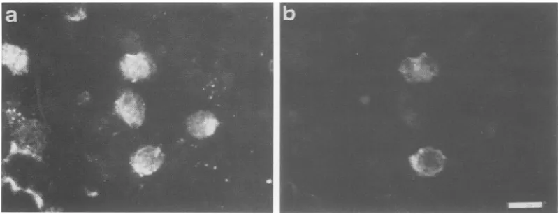
serumdemonstrated that thenuclei of up to half of the JPX-9population
were stained after treatment withCdCl2 (Fig.
2a), whereas nostaining
was observed in the absence ofCdCl2
treatment(data
notshown).
As a control transfectant we used
JPX/M,
which wassimilarly generated by
introduction ofa mutantplasmid,
pMAXneo/M,
into Jurkatcells,
followedby
selection withG418. JPX/M was
apparently polyclonal,
because it was derived from a mass culture in which many G418-resistantcellcolonieswereobserved.No
p40fax antigen
wasdetectedby
immunofluorescenceanalysis
inJPX/M cells even aftertreatmentwith
CdCl2 (data
notshown)
despite
induction oftranscript
from the introducedplasmid by CdCl2
(seeFig.
5A).
We tested the
activity
ofp4Oax
inducedby
CdCl2
totransactivate the HTLV-I enhancer. A reporter
plasmid,
pCHL4 (31),
which contains thechloramphenicol
acetyl-transferase
(CAT)
genelinked tothe HTLV-Ilong
terminal repeat, wastransiently
transfected intocells,
and CATactivity
was determined as describedpreviously (31).
Anapproximately
13-foldincrease inCATactivity
wasinducedby
treatment of JPX-9 cells withCdCl2.
In contrast, no increase in CATactivity
was found inparental
Jurkat cells andJPX/McellsevenafterCdCI2
treatment(Fig.
1B).
Theseresults indicated that a functional
p40,.x
was induced inJPX-9 cellsby
CdCl2
treatment.Thefunctional
product
ofp40fax
in JPX-9 cells was alsoconfirmed
by
examining
the induction ofexpression
of theIL-2Ra
gene, which has beenreported
tobe transactivatedby
transient introduction of thep40tax
gene to some T-celllines,
including
Jurkat(8, 17, 23,
49).
JPX-9 cells weretreated with
CdCl2,
andexpression
ofp401(Ix
andIL-2Ro wasexamined
by
double immunofluorescencestaining.
Expres-sionofIL-2Ra was seen
only
in cellsthatexpressed
p40fa
(Fig.
2),suggesting
that induction of the IL-2Ra geneby
on November 10, 2019 by guest
http://jvi.asm.org/
3222 NAGATA ET AL.
A
JPX-9
CdCl2(LiM)
00.3
1 3 10 30tax
p4O-
-. lo O0 0 a 0 0
%/6Conv"erson
FIG. 1. Induction of p4Ofax by CdCl2 in JPX-9 cells. (A)Dose-dependent induction ofp40("' proteininJPX-9 cellsby CdCl2. JPX-9 cells
wereincubated with variousconcentrations of CdCl2at37°C for 24 hand harvested for examination of the expression of p4O"'. protein.Cells
(2 x 10⁵perlane)werelyseddirectly with SDS sample buffer. After boiling for5min,thecelllysatewassubjectedtoSDS-polyacrylamide gelelectrophoresis (10% gel) and immunoblot analysis, using anti-p40"'-t-positive humanserumand1251I-labeled anti-humanimmunoglobulin G. The blotwasthen exposedtoX-ray film withanintensifyingscreenat-70°C. (B) trans-Activating activity of p4O"'. inducedinJPX-9 cells. JPX-9, JPX/M, and Jurkat cellsweretransfected with pCHL4 by the DEAE-dextran method. After12 h ofincubation, the culturewasdivided
inhalf, andeach halfwascultured forafurther 24 h in thepresence (+)orabsence (-) of 10 ,uM CdCl2. The cellswerethenharvested for the CATassay. CAT activity isexpressedasthepercentageof[14C]chloramphenicol converted into acetylated forms.
p40(axoccursother thanthroughaparacrinemanner involv-ing somehumoral factor(s), which is induced by p401"X. In
parental Jurkat cells and JPX/M cells, expression ofIL-2ROt
wasnotdetected either beforeoraftertreatmentwith CdCl2 (data not shown). These results show that JPX-9 cells retained the characteristics of the parental Jurkat cells in that they could express T-cell-specific genes in response to p4Oax. Therefore, JPX-9 cells can be considered useful in
studies of thefunction of p4Ofx as a modulator of cellular
geneexpression in T cells.
Effectof p4Ot onexpression ofproto-oncogenes. We next examined the effects of p4O'fx on expression of a set of nuclearproto-oncogenesthat are knownto be activated by T-cell activators such as phytohemagglutinin in normal T cells(36). JPX-9 cellswerecultured in thepresenceof 20 pLM CdCl2 and harvested atthe indicated times. Levels ofgene
expressionweredetermined byNorthern blot hybridization.
Expression of the p4Ofax gene wasinduced within 2 h after
addition ofCdCl2 and reachedanearly maximal levelwithin
4to6h(Fig. 3A). Similarly,theamountofp40ax proteinwas
nearly maximal after 4 to 6 h (Fig. 3B). Expression of the IL-2Rco genewas undetectable until 6 h but became
detect-able within 9 h after addition ofCdCl2. A similartime lag between appearance of p40(tO and onset of IL-2Ra gene
expression wasobserved in otherexperiments.
We also foundasignificant increasein thelevel of
expres-sion ofc-fosmRNA. The levelofc-fos mRNA begantorise 4h after additionofCdC12; themaximal levelwas seenat6 to9h, after which the level declined. Incontrast,thelevels ofmRNAsofc-myc andc-myboncogenes were not signifi-cantly affected by induction ofp4"'X. We also examinedthe effect ofp401(x on expression of the c-jun proto-oncogene, which has been shown to be activated in fibroblasts in
responsetovarious stimuli thatgive risetoactivation of the
c-fos gene. No appreciable change in the level of c-jun
FIG. 2. Immunofluorescence analysis of p40'a'and IL-2Ra. JPX-9 cellswereincubated in thepresenceof10F.MCdCl2 for20 h,harvested,
fixed withcold acetone-methanol, and doubly stainedforp40"t (a)andIL-2Rac (b) withanti-p40""-positive humanserumandanti-IL-2Ra monoclonal antibody H-31, respectively,asdescribedin Materials and Methods. Thesamecellswereobserved by fluorescent microscopy
withappropriate filters forrhodamine (a) and fluorescein (b). Bar, 10 ,um.
B
(~ R'
MW
(10-)
94 67
BN*.
.'I.
CdC!-,
43
4_.- 30
J. VIROL.
on November 10, 2019 by guest
http://jvi.asm.org/
[image:3.612.136.491.79.247.2] [image:3.612.110.510.542.695.2]Atime(h)
0 2 4 6 9 15 kbpX
c-myc
c-myb
c-fos
IL-2RC
T 9
a A
*PI
3.4 2.2 2.3
3.8
2.4
3.5
HaW
1.5
GAPDH
*b*bSN
1.8_
_I... 1.8
B
p4ax
wt 4UkdFIG. 3. Northern blot analysis of expression of nuclear proto-oncogenes after induction of p4OfeLx in JPX-9 cells. (A) Growing JPX-9cellswere recovered andsuspended in fresh medium contain-ing 20 ,uM
CdCl2
at 106 cellsper ml. At the indicated times, total RNA wasisolated from the cells and analyzed (15p.gper lane) for relative levels of mRNAs with radiolabeled probes. The relative amount of glyceraldehyde-3-phosphate dehydrogenase (GAPDH) mRNA was monitored to check the amount of total RNA applied to each lane. Sizes of themRNAs areshown at theright.(B) Aportion (2 x 10⁵cells per lane) ofthe same cell sample was analyzed forp40"xexpression by immunoblotting. kd, Kilodaltons.
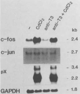
mRNAafterinduction ofp40t'xin JPX-9 cells was seen(Fig. 4).
Sincethec-fos gene generally responds rapidly to various
stimuli,thefact that enhancement ofc-fosexpression always followed the appearance of p40"'x suggested that this
en-C!-)
A.
AN Ahf
r) * * k
/b 1 ~ k
Lh
c-fos ,
c-jun
2.4
2.7
hancement may have been due to the trans-acting function of p4Qfx but not to the direct effect of CdCl2. To confirm this notion, we examined the effect ofCdC12 treatment on the expression of c-fos mRNA in control cells. The c-fos mRNA level in JPX-9 cells reproducibly increased aftertreatment with CdCl2 for 6 to 9 h in several experiments (Fig. 4 and SA). In contrast, parental Jurkat cells and control transfec-tantJPX/M cells, in which expression of the mutant p4Ofax gene wasprofoundly induced atthe mRNAlevel, reproduc-ibly showed nosignificant change in the level of c-fos mRNA upon treatment withCdCl2 for at least 6 to 9 h after addition ofCdCl2 (Fig. 5A). However, thepossibility that activation of thec-fos gene by CdCl2 depends on the clones used could not be completely ruled out. To testthis, wefurther exam-ined the correlation between expression of p4Otax and in-creaseinthec-fos mRNA level in newly isolated subclones of JPX-9 cells. Enhancement ofc-fos expression by CdCl2 wasobservedin subclones thatexpressed p4Ofax in response toCdCl2, whereas no increase in c-fos expression was seen in subclones that did not express the p4Ofax gene (Fig. 5B). These results led us toconclude that the increased expres-sionof the c-fos gene observed inCdCl2-treatedJPX-9cells was mediatedby the functional p4Otaxprotein.
Anti-T3 antibody, which significantly augmented induc-tion of IL-2Rcx expression by p4Otax (data not shown), exhibitednoeffectoneither the basal or the enhanced level ofc-fos mRNA in JPX-9cells (Fig. 4).
Effects of long-term treatment with CdCl2 on c-fos and IL-2Ra expression. Since induction of IL-2Ro expression
and enhancement of c-fos expression are transient in a normal environment, we were interested in investigating whether p4Of'x can activate the IL-2Rcx or c-fos gene con-tinuously. To dothis, JPX-9 cells were cultured for several
daysin the presenceofCdCl2at aconcentration of10 ,uM,
which was nontoxic and still effective in the induction of
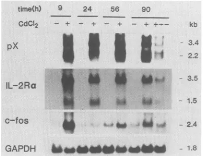
p4ota';attheindicatedtimes, the levels ofc-fos and IL-2Ra mRNAs were determined and compared with thoseof un-treated JPX-9 cells. Expression of the p4Oax gene was
maintained at a substantially constantlevel in the presence ofCdCl2 throughoutthecultivation; after removal ofCdCl2,
the level declined (Fig. 6). Similarly, a constant level of
expression ofthe IL-2Ra gene was seen in the presence of CdCl,; after removal ofCdCl2, the level declined. On the otherhand, the levelofc-fosmRNAbecamehighlyelevated at9hafter the addition ofCdCl2, but then, unlike the IL-2Ra mRNA level, it decreased even in the presence of CdCl2 (Fig.6).This reduction may beexplained,atleastin part,by
the negative-feedback regulatory function of the c-fos pro-tein itself(45). Thereafter, however, a slight but definitely
significant increase (two- to fourfold) in the c-fos mRNA level wasobserved in cells treated withCdCl2 as compared with untreated control cells (Fig. 6). These results indicate thatp4Ofax,ifit isconstitutively expressed,cancontinuously
activate expression ofboth the IL-2Ra andc-fos genes.
- 3.4
pX
2.2
GAPDH ____ 1.8
FIG. 4. Effects of anti-T3antibodyonc-fosmRNA levels. JPX-9 cells were incubated in medium alone or in medium containing
CdCl2 (20 ,uM), anti-T3 antibody (0.5 ±g/ml), orboth. Cells were
harvested 9hlater,and total RNAwasanalyzed (25 ,ugperlane)for
relative amounts of c-fos, c-jun, and pX mRNAs by Northern
blotting.
DISCUSSION
The trans-actingfunction of HTLV-I p4O'ax in viral and cellular genes has beeninvestigated mainly throughtransient cotransfection experiments in which a
p4Qtax
expression plasmid and plasmids containing regulatory regions oftest genes linked to reporter genes such as the CAT gene are transfected together into various types of cell lines. Theseexperiments have revealed that
p4Ofx
can transactivate enhancersof several viral andcellular genes suchasthose of simian virus 40(41),humanimmunodeficiency
virus type 1A. Jm.M~--- N
-k.4
on November 10, 2019 by guest
http://jvi.asm.org/
[image:4.612.92.261.72.331.2] [image:4.612.111.240.521.673.2]3224 NAGATA ET AL.
l-.:..A
pX
GAPDH _
B
JPX-9 subclones5 6 8 12 14 17 19
kb CdCt2
c-fos
2.4
3.4
2.2
1.8
GAPDH
kb
- 2.4
[image:5.612.105.521.80.274.2]3.4 2.2
FIG. 5. Effects ofCdCl2treatmentonlevels of c-fos mRNA in JPX-9 and control cells. (A) JPX-9, parental Jurkat, and JPX/Mcellswere
incubated inthepresence(+)orabsence (-) of 20 F.M CdCl2 for 6 h. Cellswereharvested, and total RNAwasisolatedandanalyzed (25 p.g
perlane) for relative levels of c-fos mRNA by Northern blotting. (B) JPX-9subcloneswereincubated with(+)orwithout(-)20 puM CdCl2 for9 h and harvestedtodetermine relative levels of c-fos mRNA (25 p.gof totalRNAperlane) by Northern blotting.
(50), IL-2 (8, 23, 49), granulocyte-macrophage colony-stim-ulating factor (25), IL-3 (25), IL-4 (25), and IL-2Rc (8, 23,
49). On the other hand, it has also been reported that the resultsobtainedfromtransient cotransfection assays are not
necessarily applicabletoendogenous genes (25).Therefore, to evaluate the role of
p4Ot(Xx
in T-cell transformation, it isimportant to examine the effect of
p40(x
on endogenous geneexpressionin Tcells. However, with the exception ofIL-2and
IL-2Rc
genes,theeffects ofp4Ofax
onexpression of endogenous genes has not been investigated in detail (17,40).
Ingeneral, transfection efficiency is unacceptably lowin
such human lymphoid cells as T-cell lines; therefore, it is
difficult to quantitatively examine the cellular changes
in-time(h) 9 24 56 90
CdC12 - + -+ -+ -++- kb
pX
*3*f
2.2
3.5
IL-2Ra
c-fos 2.4
GAPDHg-1
FIG. 6. Effects of long-term treatment with CdCl2 on mRNA levels ofIL-2Raand c-fos genes. JPX-9 cells were incubated with (+) or without (-) 10
FiM
CdCl2
for the indicated periods. Total RNA was analyzed (25F.g
per lane) for relativelevels ofIL-2Raand c-fos mRNAs by Northern blotting. The total medium was ex-changedforfreshmediumwith or without 10 ,uMCdCI256 h after initiationof theculture. At that time, theCdCl2-containingculture wasdividedin half, and the two halves were further cultured in the presence(+)orabsence(+---*-)
ofCdCl2 for 34 h.duced by p4Ofa'in transient transfection experiments. To
overcomethisdifficulty,weestablishedastabletransfectant
clone in whichexpression ofp4Ofx iscompletely inducible by heavy-metaltreatment. Thisclone thereby allowedusto carry out a quantitative analysis and kinetics study ofthe p40at"-induced expression of cellulargenes.
In this study, we examined the effects of p4Ofax on
expression of some oncogenes involved in cell growth:
c-myc,c-fos, c-myb,andc-jun.Theseoncogenesarenuclear proto-oncogenes and areknown to respond well to various extracellular activating stimuli such as lectins, growth fac-tors,andphorbolestersinvarious types ofcells,includingT cells(13, 18, 20, 36, 42, 52). We chose these oncogenes as
targets ofp4Ofax because oftheirhigh levels of
responsive-ness to extracellular stimuli and because ofan assumption
that p40fx can mimic such extracellular stimuli in part by interacting with a cellular component(s) that is involved in transduction of theactivating signaisfrom the cell membrane to nuclear target genes. Our results clearly show that the c-fos gene responds to p4Q("X, whereas, unexpectedly, the other nuclear oncogenes tested apparently do not. These results indicate that p4Otax can act on a limited number of
cellulargenes,which suggeststhat it interacts witha
partic-ular cellpartic-ular factor(s) involved in the activation ofspecific
genes.
HTLV-I-transformed cellshavenotbeen reportedtohave
ahigh levelofc-fos expression. Suchastudyis underway,
but it maybedifficultto drawageneralconclusion because
basal levels of expression of the c-fos gene seem to vary
from cell line to cell line(Fig. 5). In this regard, an
abnor-malityindevelopment of the thymus has been independently observedboth intransgenic mice carrying the p4Otax
expres-sionplasmidand inthosecarryingac-fos expression plasmid
(29, 39). These results suggest that p4Oax may activate
continuously the c-fos gene even in in vivo tissue such as
thatof thethymus, resultinginabnormaldevelopment of the thymusin transgenic micecarrying thep4Otaxgene.
Several lines ofevidencedemonstrate that thec-fos prod-uctisimplicatedincellgrowth (38), differentiation (28),and development(27). Ithasalsobeenreported that
overexpres-.. 8
--&LAndL 00
J. VIROL.
on November 10, 2019 by guest
http://jvi.asm.org/
[image:5.612.81.287.488.646.2]sion of the c-fos gene results in cell transformation in
fibroblasts (24). Nevertheless, no lymphoid malignancy has
beenobserved in transgenic mice carrying the c-fos expres-sion plasmid, although a high level of c-fos mRNA has been found in lymphoid tissues such as splenocytes (39). Thus, the question of whether continuous activation of the c-fos gene can directly cause transformation of normal human mature Tcells, which are the natural targets ofHTLV-I, is stillcontroversial. Recently, c-fos protein has been shown to
associate with several nuclear proteins (43), and the major
fos-associated protein has been identified as the
sequence-specific transcription factor AP-1 (the product of the c-jun
gene), which is known to mediate the action of the tumor promoter 12-0-tetradecanoylphorbol-13-acetate (7, 35, 44). Furthermore, it has been demonstrated that the c-fos prod-uct stimulates AP-1-responsive genes (7, 44), and it is also expected to participate in formation of the transcription
machinery responsible for other genes (44). Therefore, con-tinuous activation of the c-fos gene by
p40"'X,
if this is the case in normal Tcells, may causeunregulated activation of some genes, such as AP-1-responsive genes, resulting in direct transformation of cells or maintenance of cells in anactivated state such as occurs with 12-0-tetradecanoyl-phorbol-13-acetate-treated cells, which may be a crucial step required for complete transformation.
The molecular mechanism by which p4O0"x transactivates itstarget genesis still unclear. Recent studies have indicated that
p40(ax
functions in at least two distinct ways (41): for HTLV-I itself, and for IL-2Ro,simian virus 40, and human immunodeficiency virus type 1. In the latter case, exertion ofthistrans-acting activity has been shown to occur through a
tissue-specific transcription factor, NF-KB (22). In fact, significant enhancement of NF-KB activity in JPX-9 cells was induced by CdCl2 treatment (our unpublished data). Another nuclear transcription factor, cyclic
AMP-respon-sive-element binding factor, is shown to be involved in
p40fax-dependent
activation of the HTLV-I enhancer (M. Nakamura, M. Niki, K. Ohtani, and K. Sugamura,submit-ted for publication). However, it is still unclear how
p4Ofax
activates these factors and whether thesefactorsareused in
activation ofthe c-fos gene by
p4Ofax.
Identification of theprimary target molecule of p4Otax should lead to a much better understanding of the molecular basis of the
p4Ofdx
function. Inducible transfectants of the
p40(ax
expression plasmid, suchasJPX-9, willbe usefulforthis purpose.During the preparation of this article, we learned of a recentreportdemonstrating theability of
p401ax
totransac-tivate thec-fos gene in atransienttransfection assay (11).
ACKNOWLEDGMENTS
WethankI.Hondaforprovidinghumanseraand Y. Kirihata for technical assistance.WealsothankE.Ohkubo forhelp in prepara-tion of themanuscript.
This work was supported in part by grants-in-aid from the Ministry of Education, Scienceand CultureofJapan.
LITERATURECITED
1. Angel, P.,E. A.Allegretto,S. T.Okino,K.Hattori,W.J.Boyle, T. Hunter, and M. Karin. 1988. Oncogene jun encodes a sequence-specific trans-activator similar to AP-1. Nature (London) 332:166-171.
2. Arima, N.,Y.Daitoku, S.Ohgaki, J. Fukumori,H.Tanaka, Y.
Yamamoto,K.Fujimoto,andK.Onoue.1986. Autocrinegrowth ofinterleukin2-producingleukemic cells inapatientwith adult Tcellleukemia. Blood 68:779-782.
3. Arya,S.K.,F.Wong-Staal,andR.C.Gallo.1984.T-cellgrowth factor gene: lack of expression in human T-cell
leukemia-lymphomavirus-infected cells.Science 223:1086-1087. 4. Battey, J.,C. Moulding,R. Taub,W.Murphy, T.Stewart, H.
Potter, G. Lenoir, and P. Leder. 1983. The human c-myc oncogene: structural consequences of translocation into theIgH locus inBurkittlymphoma. Cell34:779-787.
5. Bergmann, D. G., L. M. Souza, and M. A. Baluda. 1981. Vertebrate DNAs contain nucleotide sequences relatedto the transforminggene of avian myeloblastosis virus. J. Virol. 40:
450-455.
6. Cantrell,D.A.,and K. A. Smith.1984. The interleukin-2 T-cell system: anewcellgrowthmodel. Science224:1312-1316. 7. Chiu, R., W.J. Boyle, J. Meek, T.Smeal, T. Hunter, and M.
Karin. 1988. The c-fos protein interacts with c-jun/AP-1 to
stimulate transcriptionofAP-1responsivegenes. Cell 54:541-552.
8. Cross, S. L., M. B. Feinberg, J. B. Wolf, N. J. Holbrook, F. Wong-Staal,andW.J.Leonard. 1987.Regulationof the human interleukin-2 receptor ax chain promoter: activation of a
non-functionalpromoterbythetransactivatorgeneofHTLV-1.Cell 49:47-56.
9. Dugaiczyk, A., J.A.Haron,E. M.Stone,0. E.Dennison,K. N. Rothblum,and R.J.Schwartz.1983.Cloningandsequencingof
a deoxyribonucleic acid copy of glyceraldehyde-3-phosphate dehydrogenase messenger ribonucleic acid isolated from chicken muscle. Biochemistry22:1605-1613.
10. Felber,B.K.,H. Paskalis,C.Kleinman-Ewing,F.Wong-Staal, and G. N. Pavlakis. 1985. The pX protein of HTLV-I is a
transcriptional activator of its long terminal repeat. Science 229:675-679.
11. Fujii, M., P. Sassone-Corsi, and I. M. Verma. 1988. c-fos promoter trans-activation by the tax1 protein ofhuman T-cell leukemia virustype I. Proc. Natl.Acad.Sci.USA85:8526-8530. 12. Fujita, T., H. Shibuya, T. Ohashi, K. Yamanishi, and T. Taniguchi. 1986. Regulationofhumaninterleukin-2gene: func-tional DNA sequences in the 5' flanking region for the gene expressioninactivatedTlymphocytes. Cell46:401-407. 13. Greenberg,M.E.,and E. B.Ziff.1984.Stimulation of3T3cells
induces transcription of the c-fos proto-oncogene. Nature (London)311:433-438.
14. Hatakeyama,M., S.Minamoto,T.Uchiyama, R. R. Hardy,G. Yamada, andT. Taniguchi. 1985. Reconstitution of functional receptor for human interleukin-2 in mouse cells. Nature (London)318:467-470.
15. Hattori, T., T. Uchiyama, T. Toibana, K. Takatsuki, and H. Uchino. 1981. Surfacephenotype ofJapanese adult T-cell leu-kemia cells characterized by monoclonal antibodies. Blood 58:645-647.
16. Hinuma, Y.,K.Nagata,M.Hanaoka,M.Nakai,T.Matsumoto,
K. Kinoshita,S. Shirakawa,andI. Miyoshi. 1981. Adult T-cell leukemia:antigeninanATLcellline and detectionofantibodies to the antigen in human sera. Proc. Natl. Acad. Sci. USA 78:6476-6480.
17. Inoue, J., M. Seiki, T. Taniguchi, S. Tsuru,and M. Yoshida. 1986. Induction ofinterleukin 2 receptor gene expression by
p40xencodedbyhuman T-cellleukemia virus typeI.EMBO J. 5:2883-2888.
18. Kelly, K., B. H. Cochran, C. D. Stiles, and P. Leder. 1983. Cell-specific regulationofthe c-myc genebylymphocyte mito-gens andplatelet-derived growthfactor.Cell 35:603-610. 19. Laemmli,U. K.1970.Cleavageof structuralproteinsduringthe
assembly ofthe head of bacteriophage T4. Nature (London)
227:680-685.
20. Lamph,W.W.,P.Wamsley,P.Sassone-Corsi,andI. M. Verma. 1988.Inductionofproto-oncogenejun/AP-1 byserumandTPA.
Nature(London)334:629-631.
21. Leonard,W.J., J.M.Depper, M.Kanehisa, M.Kronke,N.J. Peffer, P. B. Svetlik, M. Sullivan, and W. C. Greene. 1985. Structure of the human interleukin-2 receptor gene. Science 230:633-639.
22. Leung, K., and G. J. Nabel. 1988. HTLV-1 transactivator induces interleukin-2 receptor expression through an NF-KB-like factor. Nature(London)333:776-778.
23. Maruyama, M., H. Shibuya, H. Harada, M. Hatakeyama, M.
on November 10, 2019 by guest
http://jvi.asm.org/
3226 NAGATA ET AL.
Seiki, T. Fujita, J. Inoue, M. Yoshida, and T. Taniguchi. 1987.
Evidence for aberrant activation ofthe interleukin-2 autocrine
loop by HTLV-I encoded p4Ox and T3/Ti complex triggering. Cell 48:343-350.
24. Miller, A. D., T. Curran, andI.M. Verma. 1984. c-fos protein
caninduce cellulartransformation: anovel mechanism of
acti-vation ofacellularoncogene. Cell 36:51-60.
25. Miyatake, S., M. Seiki, R. D. Malefijt, T. Heike, J. Fujisawa, Y. Takebe, J. Nishida, J. Shlomai, T.Yokota, M. Yoshida, K. Arai,
and N. Arai. 1988. Activation of T cell-derived lymphokine
genesin T cells and fibroblasts: effects of humanTcellleukemia
virustypeI p40X protein and bovine papilloma virus encodedE2
protein. Nucleic Acids Res. 16:6547-6566.
26. Miyoshi, I.,I.Kubonishi,S.Yoshimoto,T.Akagi,Y. Ohtsuki, Y.
Shiraishi, K. Nagata, and Y. Hinuma. 1981. Type C virus
particles in acord T-cell line derivedbyco-cultivating normal
human cord leukocytes and human leukaemic T cells. Nature
(London) 294:770-771.
27. Muller, R., D. J. Slamon, J. M. Tremblay,M. J.Cline,andI.M.
Verma.1982. Differential expression of cellularoncogenes
dur-ing pre- and postnatal development of the mouse. Nature
(London) 299:640-644.
28. Muller, R., and E. F. Wagner. 1984. Differentiation of F9
teratocarcinoma stem cells after transfer of c-fos
proto-onco-genes. Nature(London) 311:438-442.
29. Nerenberg, M., S. H. Hinrichs, R. K. Raynolds, G. Khoury, and
G. Jay.1987. Thetatgeneof humanT-lymphotropic virus type 1 induces mesenchymal tumors in transgenic mice. Science
237:1324-1329.
30. Ohtani, K., M. Nakamura, S. Saito, K. Nagata, K. Sugamura, andY. Hinuma. 1989. Electroporation: application to human
lymphoid cell lines for stable introduction ofa transactivator
geneof human T-cell leukemia virustype Nucleic Acids Res.
17:1589-1604.
31. Ohtani, K., M. Nakamura, S. Saito, T. Noda, Y. Ito, K.
Sugamura, andY. Hinuma. 1987. Identification oftwodistinct elementsin the longterminalrepeatof HTLV-I responsiblefor
maximumgeneexpression. EMBOJ. 6:389-395.
32. Paskalis,H., B. K. Felber, and G. N. Pavlakis. 1986.Cis-acting
sequencesresponsible for the transcriptional activation of
hu-man T-cell leukemia virus type I constitute a conditional
en-hancer. Proc. Natl. Acad. Sci. USA 83:6558-6562.
33. Poiesz, B.J., F. W. Ruscetti, A. F. Gazdar, P. A. Bunn, J. D. Minna, and R. C. Gallo. 1980.Detectionandisolation oftypeC
retrovirus particles from fresh and cultured lymphocytes ofa
patient withcutaneousT-celllymphoma. Proc. Natl. Acad. Sci.
USA77:7415-7419.
34. Popovic, M.,G.Lange-Wantzin,P.S.Sarin, D. Mann, and R.C.
Gallo. 1983. Transformation of human umbilical cord blood T
cells by human T-cell leukemia/lymphoma virus. Proc. Natl.
Acad. Sci. USA80:5402-5406.
35. Rauscher F. J.,III, D. R. Cohen, T. Curran, T. J. Bos, P. K.
Vogt, D. Bohmann, R. Tjian, and B. R. Franza, Jr. 1988.
Fos-associated protein p39 is the product of the jun
proto-oncogene.Science 240:1010-1016.
36. Reed,J. C., J. D. Alpers, P. C. Nowell, and R. G. Hoover. 1986.
Sequential expression ofprotooncogenes during
lectin-stimu-lated mitogenesis of normal human lymphocytes. Proc. Natl.
Acad. Sci. USA83:3982-3986.
37. Reedman,B. M., and G. Klein. 1973. Cellular localization ofan Epstein-Barr virus (EBV)-associatedcomplement-fixing antigen
inproducer and nonproducer lymphoblastoid cell lines. Int. J.
Cancer 11:499-520.
38. Riabowol, K. T., R. J. Vosatka,E. B. Ziff, N. J. Lamb, and J.R. Feramisco. 1988. Microinjection of fos-specific antibodies
blocks DNA synthesis in fibroblast cells. Mol. Cell. Biol.
8:1670-1676.
39. Ruther, U., W. Muller, T. Sumida, T. Tokuhisa, K. Rajewsky,
and E. F.Wagner.1988. c-fos expressioninterferes withthymus
development in transgenic mice. Cell53:847-856.
40. Sabe, H., A. Tanaka, H. Siomi, S. Koyasu, S. Yonehara, I.
Yahara, Y. Tagaya, K. Sugie,K.Teshigawara, J. Yodoi, and M.
Hatanaka.1988. Differential effectsonexpression ofIL-2
recep-tors (p55 and p70) by the HTLV-I pX DNA. Int. J. Cancer 41:880-885.
41. Saito, S., M. Nakamura, K. Ohtani, M. Ichijo, K. Sugamura, and Y. Hinuma. 1988. trans-Activation of the simian virus 40 enhancer by a pX product of humanT-cellleukemiavirus type I. J. Virol. 62:644-648.
42. Sakitani, M., M. Nakamura, M. Fujii, K. Sugamura, and Y. Hinuma. 1987. High affinity interleukin 2 receptors in HTLV-I-infected T cells can mediate signals for gene expression. Virus Genes 1:35-47.
43. Sambucetti, L. C., and T. Curran. 1986. The fos protein complex is associated with DNA in isolated nuclei andbindsto DNAcellulose. Science 234:1417-1419.
44. Sassone-Corsi, P., W. W. Lamph, M. Kamps, and I. M. Verma. 1988. fos-associated cellular p39 is related to nuclear transcrip-tion factor AP-1. Cell54:553-560.
45. Schonthal,A., P.Herrlich, H. J.Rahmsdorf, and H. Ponta. 1988. Requirement for fos gene expression in the transcriptional activation of collagenase by other oncogenes and phorbol Ws-ters. Cell 54:325-334.
46. Seiki, M., R. Eddy, T. B. Shows, and M. Yoshida. 1984. Nonspecific integration of the HTLV provirus genome into adult T-cellleukemia cells. Nature (London) 309:640-642. 47. Seiki, M., J. Inoue, T. Takeda, and M. Yoshida. 1986. Direct
evidence that p4oX of human T-cell leukemia virus type I is a trans-acting transcriptional activator. EMBO J. 5:561-565. 48. Seiki, M., S. Hattori, Y. Hirayama, and M. Yoshida. 1983.
Human adult T-cell leukemia virus: complete nucleotide se-quenceof the provirus genome integrated in leukemia cell DNA. Proc. Natl. Acad. Sci. USA80:3618-3622.
49. Siekevitz, M., M. B.Feinberg, N. Holbrook, F. Wong-Staal, and W. C. Greene. 1987. Activation ofinterleukin 2 and interleukin 2receptor (Tac)promoter expression by thetrans-activator (tat) geneproduct of human T-cellleukemia virus, type I. Proc. Natl. Acad. Sci. USA84:5389-5393.
50. Siekevitz, M., S. F.Josephs, M. Dukovich, N. Peffer, F. Wong-Staal, and W. C.Greene. 1987.Activation of the HIV-1 LTR by T cell mitogens and the trans-activator protein of HTLV-I. Science 238:1575-1578.
51. Sodroski, J. G., C. A. Rosen, W. C. Goh, and W. A.Haseltine. 1985. A transcriptional activator protein encoded by the x-lor region of the human T-cell leukemia virus. Science 228:1430-1434.
52. Stern, J. B., and K. A.Smith. 1986. Interleukin-2 induction of T-cell G1 progression and c-myb expression. Science 233: 203-206.
53. Straaten, F. V., R.Muller,T.Curran, C. V. Beveren, and I. M. Verma. 1983. Complete nucleotide sequence of a human c-onc gene:deduced amino acid sequence of the human c-fos protein. Proc. Natl. Acad. Sci. USA80:3183-3187.
54. Sugamura, K., M. Fujii, T. Ishii, and Y. Hinuma. 1986. Possible role of interleukin 2 receptor in oncogenesis ofHTLV-I/ATLV. Cancer Rev. 1:96-114.
55. Sugamura, K., M. Fujii, M. Kannagi, M. Sakitani, M. Takeuchi, and Y. Hinuma. 1984. Cell surfacephenotypes and expression of viral antigens of various human cell lines carring human T-cell leukemia virus.Int. J. Cancer34:221-228.
56. Tanaka, Y., H. Tozawa, M. Hayami, K. Sugamura, and Y. Hinuma. 1985. Distinctreactivities of four monoclonal antibod-ies with human interleukin 2 receptor. Microbiol. Immunol. 29:959-972.
57. Uchiyama, T., T. Hori, M. Tsudo, Y. Wano, H. Umadome, S. Tamori, J. Yodoi, M. Maeda, H.Sawami, and H. Uchino. 1985. Interleukin-2 receptor (Tac antigen) expressed on adult T cell leukemia cells. J. Clin. Invest. 76:446-453.
58. Yamamoto, N., M. Okada, Y. Koyanagi, M. Kannagi, and Y. Hinuma. 1982. Transformation of human leukocytes by cocul-tivation with an adult T cellleukemia virus producer cell line. Science 217:737-739.
59. Yoshida, M., I. Miyoshi, and Y. Hinuma. 1982. Isolation and characterization of retrovirus from cell lines of human adult T-cell leukemia and its implication in the disease. Proc. Natl. Acad. Sci. USA79:2031-2035.
J. VIROL.