Vol. 63, No. 10
Processing of Yellow
Fever
Virus
Polyprotein:
Role of Cellular
Proteases in Maturation of the Structural
Proteins
ANDRES RUIZ-LINARES,t ANNIECAHOUR,t PHILIPPE
DESPRkS,
MARCGIRARD,AND MICHELEBOULOY*
Unit ofMolecular Virology, CentreNational de laRecherche
Scientifique
UA545, InstitutPasteur, 75724ParisCedex 15, France Received10 April 1989/Accepted 27 June 1989The yellow fevervirus (YFV)cDNAsegmentcoding for thepartoftheprecursorpolyprotein generating the structural proteins C (capsid), prM (precursortothe membrane protein M), and E (envelope)was expressed
invitro by using theT7 promoter-polymerase transcriptionsystemcoupledtotranslation in rabbitreticulocyte
lysates. A polypeptide of the expected molecular weight was observed to accumulate in the assay and was processed into proteins C, prM, and Eonly when dog pancreas microsomal membranes wereadded tothe
translation system. Proteins prM and E were translocated inside the endoplasmic reticulum, where prM
underwent glycosylation. Regions essential for translocation ofthese proteins were localizedto the 18- and
15-amino-acid C-terminal hydrophobic regions of proteinsC and prM, respectively. Translocation of protein
prM appearedtobelessefficientthan thatofprotein E. Maturation of these proteins followeddifferentkinetics,
indicatingthattheprM signal is probably cleavedoff moreslowly. A polypeptide composed of proteins Cand
prM,similartothe NVxpolypeptide describedin yellow fever virus-infected cells,wasalso produced in the in
vitro system inthepresenceofmembranes. Nomatureprotein Mwasdetected,suggesting thatthe cleavageof prMtoM isalateprocessing eventmediated byaproteasedifferent from endoplasmicreticulum signalases.
Yellow fevervirus(YFV) is theprototype of the Flavivir-idaefamilywhich includes about 65 viruses, someof which
are ofmajor human health concern, such as yellow fever virus, dengue virus, and Japaneseencephalitis virus (27, 48).
Thevirus is small andenveloped, consisting ofan
icosahe-dral nucleocapsid containing the single-stranded RNA
ge-nome complexed with multiple copies ofthe basic capsid
protein (C; molecular weight [MW], 13,000 to 16,000) sur-rounded by a host-derived membrane in which two viral proteins, the membraneproteinM(MW, 8,000) derived from a glycosylated precursor prM (MW, 27,000) and the enve-lope protein E(MW, 51,000to59,000) (48),areinserted. The genomic RNA ofpositive polarity is the only mRNA pro-duced during infection (14).
Sequence analysis of the genomesof several flaviviruses
(6, 15, 16, 33, 34)has shownasimilarorganization.The viral
RNA is approximately 11 kilobases long and has a single
open readingframe expanding over more than 90% of the genome,makingthe flavivirus RNAamongthelongestof the
eucaryotic messengers. By sequencing the N terminus of
purified viral proteins from several flaviviruses, the gene order in the openreadingframehas been determined tobe
C-prM-E-NS1-NS2A-NS2B-NS3-NS4a-NS4B-NS5 (1, 2, 4,
7, 32, 34, 37).Itwasconcluded that all the viralproteinsare
derived fromahigh-MWprecursorwhichmustbecleavedto
producetheindividual structural andnonstructuralproteins.
The proteases responsible for these cleavages are un-known,but it has beensuggestedthatboth cellular and viral proteases could be involved. According to the favored
hypothesis, cellular signalases would be responsible for
cleavage of the polyprotein precursor at hydrophobic re-gionswhicharelocatedattheN and Ctermini of each of the
*Correspondingauthor.
t Present address: DepartmentofGenetics, University of
Cam-bridge, CambridgeCB23EH, UnitedKingdom.
tPresent address: Laboratory of Infectious Diseases, National
InstitutesofHealth, Bethesda, MD 20892.
structural proteins and of the nonstructural protein NS1. These regions presumably act as signal peptides and stop
transfer sequences and determine the translocation of pro-teinsprM, E,andNS1 into theendoplasmic reticulum (ER). The cleavage of protein prM to generate matureprotein M
wouldbealateevent performed byaGolgiprotease
recog-nizing the consensus sequenceArg-X-Arg/Lys-Arg present in severalviral glycoproteins and insome hormone
precur-sors (41). Alternatively, thepresence of thetripeptide Cys-Trp-Cys in the prMamino acid sequence suggests that this
protein might be an autoprotease, as this sequence is con-served atthe active site of thiol proteases(33).
Theprocessing of thepolyprotein moiety comprising the
nonstructuralproteinsNS2Ato NS5 would require another
enzyme(s)of cellularorviraloriginwhichrecognizes pairsof basic amino acids, eitherArg-Arg or Lys-Arg, surrounded
by short-side-chain amino acids. These cleavages would occur in the cytosolic phase, generating the nonstructural
proteins, someofwhich couldplayaroleasviral proteases, cappingenzymes, orreplicases (33, 40, 41).
The strategy offlavivirus protein synthesis is stillpoorly
understood because of a lack of experimental data. In
infected cells thecomplete polyprotein precursorhas never
been detected, although when infection was carried out
underspecial conditions, some high-MWpolypeptideswere
observed(9, 11, 30). Furthermore,in vitrotranslation of the
genomic RNA in rabbit reticulocyte lysate (RRL) gave a complex pattern ofpolypeptides inwhich no specific viral
products could be identified (28, 47). However, discrete
bandscorrespondingtoproteinsEand Ccould be observed
when tick-borne encephalitis virus RNA was translated in
Krebs IIcellextracts(22, 42, 43). Interestingly, the
produc-tion of these proteins was dependent on the membrane
fraction of the extract, indicating that cellular proteases
included in this fractionprobably were responsible for the
maturation ofthepolyprotein.
In thepresentwork, we haveusedanin vitrotranslation
systemtostudytheroleof the cellularproteasesinvolved in
4199 JOURNALOFVIROLOGY, OCt.1989, p. 4199-4209
0022-538X/89/104199-11$02.00/0
Copyright © 1989,American
Society
for Microbiologyon November 10, 2019 by guest
http://jvi.asm.org/
the
cleavage
of the YFVpolyprotein
precursorgenerating
proteins C,
prM,
and E. In vitrotranscripts
weresynthe-sized
by using
the T7promoter-polymerase
system andsubsequently
translated in RRL in the presence orabsenceof
dog pancreatic
microsomal membranes. The transcriptcoding
for the structuralproteins C-prM-E
was translatedinto a
polyprotein
precursor, which wasefficiently
cleavedinto
polypeptides corresponding
to authenticproteins
C,
prM,
andEin the presence of membranes. Wealso detectedan
incompletely
cleaved precursorcomposed
ofproteins
Cand
prM.
Progressive
deletions of the N-terminalregion
ofthe
polyprotein permitted
usto localizetheregions
respon-sible for the translocation of
proteins
prM
and E into membrane vesicles. We also showed that theputative
N-type
glycosylation
sites ofprotein
prM
wereefficiently
recognized,
whereas those ofprotein
E were not. Thisconfirms
previous
observationsindicating
thatprotein
Efromthis
specific
strain ofYFV is notglycosylated
(17;
P.Despres
etal., unpublished
results).
MATERIALSANDMETHODS
Materials. All restriction endonucleases and
DNA-modi-fying
enzymes,endoglycosidase
H,
phenylmethylsulfonyl
fluoride,
and ribo- anddeoxyribonucleotides
were obtainedfrom
Boehringer
Mannheim Biochemicals. VectorpGEM4,
T7 RNA
polymerase,
RNaseinhibitors,
RQ1 DNase, RRL,
dog
pancreatic
microsomalmembranes,
and unlabeledamino acids were
purchased
fromPromega
Biotec. ProteinA-Sepharose
waspurchased
fromPharmacia,
Inc.Protein-ase Kwas obtained from
Sigma
Chemical Co.[35S]methio-nine
(800
Ci/mmol)
waspurchased
from AmershamCorp.
All
oligodeoxyribonucleotides
used forsequencing
weresynthesized
at the Institut Pasteur.Construction ofYFV in vitro
expression plasmids.
PlasmidpGX.1S,
containing
thecomplete region coding
for the structuralproteins
and the first one-fourth ofprotein NS1,
was obtainedby subcloning
the AvaI-BamHIfragment
de-rived fromthe YFV cDNAinsert of
plasmid pAP51
(12) into theAvaI-BamHI sites ofplasmid pGYF5' (Fig. 1A),
which containsa1,000-base-pair
insertstarting
with the firstnucle-otide ofthe YFV
genomic
sequence located 5 nucleotidesdownstream of the T7 promoter
transcription
start point(35).
PlasmidpGX.lS/prM,
deleted from part of the prMregion,
was obtainedby
successive treatments ofpGX.1S
with
NdeI,
AvaI,
and Klenowfragment
asindicated in Fig.1A.
Figure
1B shows the construction of plasmid pGX.4,which is derived from
pGYF5'
and has conserved the YFV5'noncoding
region, including
theinitiating ATG,followed bythe
polylinker region
ofpGem4.
Several restriction
fragments
isolated frompGX.1S
andrepresenting
various parts of the YFV structural proteinswere subcloned into
pGX.4
digested withAccI,
Sall, orHinclI
(depending
on the phase of the insert), Klenowtreated,
anddigested
with BamHI in order to orient theinsert
(Fig.
1C).General DNA methods. Restriction endonucleases,
Kle-nowenzyme, and
ligase
were used as recommended by themanufacturer;
specific
deoxynucleotide triphosphates wereomitted when
partial
Klenowfilling
in was desired.Treat-mentwith S1exonucleasewas performedin atotal volume of 50 ,ul
containing
up to 10ig
ofplasmid DNA, 250 mMNaCl,
1 mM zinc acetate, and 50 mg of bovine serumalbumin perml in the presence of5 U of S1nuclease. The
reaction mixture was incubated for 30 min at 25°C, phenol
extracted, and ethanol
precipitated.
Samples
were treated with Klenow before ligation in order torepair
overhangs.
Restoration of the reading frame was verified in each
con-struct by direct sequencing of the plasmid (8) by using an oligodeoxynucleotide
complementary
tothe sequence of T7 promoter, except for construct pGX.lS/prM, for which an oligodeoxynucleotide hybridizing at the 3' end of theprM
coding region (position 810 to
794)
was used. HB 101 bacteriawererendered competent andtransformed with the plasmids. Other molecular biological manipulations were performed by usingstandardprotocols
(25).
In vitro transcription and
analysis
oftranscripts. Linear-ized plasmids(1Rg)
were transcribed in 50 ,ul ofamixture containing 20 mM KHPO4(pH7.5), 10 mMdithiothreitol,
2 mMMgCl2, 4 mMspermidine, 50,uMof eachribonucleotidetriphosphate, RNasin(1
U/Ipl),
and T7 RNApolymerase
(0.5
U/pl). The reaction mixture was incubated for 1 hat
37°C,
and the DNAtemplatewaseliminatedbytreatmentwith 2 U of RQ1 DNase for 15 min at 37°C. The samples were
phenol-chloroform extracted and ethanol precipitated twice with 2 M ammonium acetate. mRNAs thus obtained were
finally taken up in sterile water, and their concentrations weredeterminedbymeasuring theopticaldensityat260nm.
In vitro translation and analysis oftranslation products.
Translation was performed in a rabbit reticulocyte
lysate
system. The standardreaction mixture(12,ul) contained 60%
of the commercialpreparationofRRL, 20puMof eachamino acid exceptmethionine, 1 mCi of[35S]methionine per ml(800
Ci/mmol), 1 mM magnesium acetate, 170 mM
potassium
acetate, and RNA. Incubationwas carriedoutfor 60 minat
30°C, and the radioactivity incorporated into hot
trichloro-aceticacid-precipitable material wasestimated for 3-pA
sam-ples by using the protocol recommended by the RRL
man-ufacturer(PromegaBiotec).
Translation in the presenceofmicrosomalmembraneswas
carried out by the addition of 1 pA ofmembranes into the reaction mixture described above.
Translation products were analyzed on 14%
polyacryl-amide gels after heat denaturation in the presence of 2%
sodium dodecyl sulfate and 1%
P-mercaptoethanol
(21).When immunoprecipitated, the translation products were diluted to 100plIinabuffercontaining1%Nonidet P-40,0.15 M NaCl, 50 mM Tris hydrochloride, pH 7.5, and 1 mM EDTA and were incubatedovernight at4°C in the presence of antibodies (up to 5 pA) and protein A-Sepharose. After extensivewashing, the immunoprecipitates were eluted from protein A-Sepharose by boiling them in Laemmli denatur-ation buffer.
Enzymatic treatmentof translation products.Proteinase K treatment wasperformed in a volume of 10 pA containing 6plI of translation mix and proteinase at a concentration of0.2
mg/ml. After incubation on ice, phenylmethylsulfonyl fluo-ridefreshly dissolved in isopropanol was added at a concen-tration of 1to 2mg/ml. After a further 5-min incubation on
ice, 30 plI of sodium dodecyl sulfate-Laemmli buffer was added and the samples were boiled immediately for 5 min and loaded onto gels (26).
Endoglycosidase H treatment was carried out with 8-pul
samples of the translation mix, which were diluted into 100 pL of0.1 M sodium citrate (pH 5.5) containing 0.1% sodium
dodecyl sulfate. After denaturation at 100°C for 2 minand
cooling, phenylmethylsulfonyl fluoride at a final concentra-tion of1 to 2 mg/ml and 5 mU of endoglycosidase H was added and the samples were incubated overnight at37°C.
Reactions were stopped by adding 1 ml of 20% trichloro-acetic acid. After 30 min on ice, the precipitates were
on November 10, 2019 by guest
http://jvi.asm.org/
EL
Bamill
AvaI
IAvaI
Aval
SamEl BemilH
I
'""I N"I "del
Aval
Paircia Ceke" sI
p
S0SphI NlalIll
NiaSII
Spb
HI
Xmnl
Hindlt
X-a1 Xmnl
YFV5'N.C..AT C@3GCATGC
OCTGCAVTCGA
q..M.CS..
Hindill PsLI SailAccl
M-4,,,, HincIl
PstI
SI
p
YFV5'N.C...ATGCA
LGTCG]...
M.C.S.. SallAcci Hinc I C.
Acci Kienow Bauilm
SIl
Klanuw
(G .4 ) lioci
Acc FL mHt Acci
Hincil Kicauw
armill
BamHI
Avell
p .4 /
Bamlnl
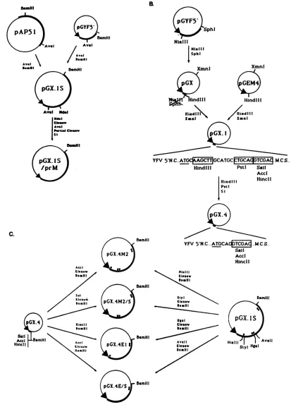
FIG. 1. Schematic diagram of the strategy used to construct YFV in vitro expression plasmids. (A) Reconstruction of the complete
structuralregion of YFV cDNA from overlapping plasmidspAP51 and pGYF5'. Plasmid pGX.1S includesthefirst2,725 bases ofthe YFV
genomecoding for proteins C, prM, E, and the first 92 amino acids of NS1.Aninternal deletionof theN-terminal half ofprotein prMwas
introduced inpGX.1Sto obtainpGX.lS/prM by usingappropriate restriction sites. YFV sequences are shownasbold lines,andvector
sequencesareshownasthinlines(PBR327 in plasmidpAP51 and pGEMinpGYF5'). TheT7 promoterisindicatedbyanarrowhead. (B)
Constructionofplasmids containingthe YFV 5' noncoding (YFV5'NC)regiondownstreamof the T7 promoter. Themultiple cloningsite
(MCS) ofvectorpGEM4 wasplaced downstream of the YFV 5'NC region andwassubsequently modified with S1nuclease toeliminate
excessnucleotides betweeninitiating ATG(underlined) and the recognitionsites for theenzymesSall,AccI,andHincII. This sitewaschosen
becauseitpermits cloninginallthreereadingframes.Nucleotidesequencesatthe YFVjunctionaregivenforpGX.1andpGX.4.Relevant
restrictionsitesareboxed.Othersymbolsare asinpanelA.(C) Construction of plasmids designedtolocalize translocationsignalsofproteins prM and E. All plasmidswerederived frompGX.1S by subcloning of appropriate fragmentsofthe YFV cDNA insert intopGX.4downstream of the YFVuntranslated leader. Hydrophobic regionsin theYFV structuralproteinsareindicated(O).Othersymbolsare asinpanelA.
4201
A.
Be-HI
on November 10, 2019 by guest
http://jvi.asm.org/
[image:3.612.105.518.46.623.2]Hga
A
5NCR
C-SfaN Pstl EcoRI BamHI
,=-NS I
T
3'1f
B
EXPRESSIONPLASMII)S
C
I
,
122pGX. I S
TRANSLATION PRODUCTS
prM
285
372
1Arg 270
1E-778
39aaI
NSI
869
I1
Arg754
prM and
E prM and
E
prM and
E pGX./ M'2/S
pGX.4EI
PGX.4E/S
104
Az1
7/'Z_
.z
i7
AFTrLN VAL ASPLuVal... 124
I'
~~~~zzz7/zzzzzz7/ZJ~~~~~~-MEr GL'VAtLVot Vat lie AlaLeu LeuValLeuAla ValGlyPro AlaTyrSer..
I...
r
7/zY
_7Z"
NONEMA7CLM ValGly.. 280
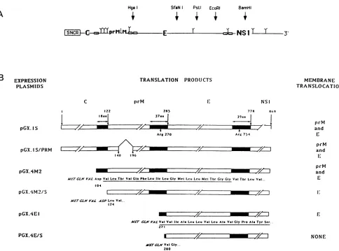
FIG. 2. Schematic diagrams oftheproteins synthesized bythe YFV in vitroexpression plasmids. (A) Schematicrepresentationofthe
YFVcDNAcomprising the 5' noncoding region (5'NCR)and theregion codingforproteins C, prM, E,andpartof NS1. Thepositionsofthe
restriction sitesusedtolinearize theplasmidsareindicated,as arethepositionsof thehydrophobic regions (E ),delimitingthe individual
proteins. (B) Representation of thepolyproteinsencodedbydifferenttranscripts synthesizedfromBamHI-linearizedplasmids.Limits of the
structuralproteinsareindicated,asisthepositionof the N-terminal residue of eachprotein.Protein NS1 isrepresented only byits first 90
aminoacids, whicharepresentin theprecursorsynthesized bythesetranscripts. Hydrophobic regions (_)of theprecursorareindicated.
The Nterminus of eachpolypeptideencodedbythe deletedconstructsis indicated. Amino acids in italictypewerederived fromthe construct
anddonot belongtoYFV proteins. The complete sequencesofthehydrophobic regionsofproteins prMand E areindicated under the
polypeptides encoded by pGX.4M2 and pGX.4E1, respectively. All numbers refertoamino acidpositionsonthecompleteYFVpolyprotein. Theproteins translocated in thepresenceof membranesareindicatedontheright.
centrifugedandthepelletswerewashed twice with ethanol-ether(1:1), suspendedin40
RI
of Laemmlibuffer,and loadedontopolyacrylamide gels.
RESULTS
YFV structural protein precursor has no autoproteolytic
activity.Since itwassuggestedthatprotein prM mightbean
autoprotease,wefirst tested the stability of the polypeptide comprising the amino acid sequence of proteins C-prM-E.
This polypeptide was synthesized in vitro in RRL pro-grammedby synthetic mRNAtranscripts. Forthispurpose we constructed a plasmid, pGX.1S, which contains the region coding for proteins C, prM, and E and for the first 90
aminoacids ofprotein NS1 (Fig. 1and2). PlasmidpGX.1S wasderived by recombinationfrom plasmids pAP51(12)and pGYF5' (35). Runoff transcripts obtained from plasmid pGX.lS linearized at the BamHI site were translated in
RRL,andsampleswerewithdrawn after15, 30,60, 120,and
180minof incubation andanalyzedon apolyacrylamide gel (Fig. 3A). A polypeptide of90,000 MW, the MWexpected
for a C-prM-E-NS1 precursor, was synthesized with no evidence forprocessing to lower-MW products. This indi-cated that underourexperimentalconditions, no
autoprote-olytic activitywasdetectedin the structuralprotein
precur-sor. However, it cannot be excluded that the prM protein
possessesaproteaseactivity whenglycosylated.
Induction of processing of the YFV structural protein
precursorbymicrosomal membranes. Totestthehypothesis
that the processing leading to the formation ofproteins C, prM, and E is dueto cellular signalases, we supplemented
the RRL withdog pancreaticmicrosomal membranes. Such
asystem hasprovedtobe extremelyuseful forstudyingthe
topogenic signals for the transport ofa number of cellular
and viral proteinsinto the ER(3, 18, 19).
Transcripts of increasing length were synthesized from
plasmid pGX.1S linearized at the SfaNI, PstI, and EcoRI MEMBRANE TRANSLOCATION
pGX.IS/PRM
E4
4=ZZ7/
I/ /140 196
pGXAM2
---ZZ
z72zzzzzz
,.
MTrCLNVt4 AspVal Leu Tbr ValGlnPheLeulic LeuGlyMCt Leu Leu MetTt.rGlyGlyValTbr LeuVal...
lx
-/,Z-id
-W M--/I-1
on November 10, 2019 by guest
http://jvi.asm.org/
[image:4.612.71.555.72.431.2]YFV PROTEIN PROCESSING 4203
A
I-) 4 5 6
s....- 4__ w
B.
pGX.lS
C YCp ECORI Barn" v
2 2 3 4 5 6 I89t
-92 -92
-69
-46
-46
-30o -.
-30 -14
C
pGXAlS CV1-17D
1 2 3 4 5 6 7 8
to a
4
U'
M M M M M
-14
MMM
[image:5.612.93.521.79.313.2]p
FIG. 3. Analysis ofthetranslation products synthesized in RRL programmed withpGX.1Stranscripts. (A) BamHI runoff transcripts
coding forapolypeptide endingatposition 869 of the YFV polyprotein (Fig. 1C and 2)weretranslated in the absence of microsomes. Samples
werecollected after 0, 15,30, 60, 120,and 180min(lanes 1to6, respectively).MWs(in thousands)areindicatedattheright. (B)pGX.1S
transcripts of increasing size were synthesized from pGX.1S templates linearized at the SfaNI, PstI, EcoRI, and BamHI sites. These
transcriptsencodepolypeptides ending, respectively,atpositions340, 674, 717,and769of theYFVpolyprotein(Fig. 2).Translationproducts
synthesized from these transcripts inthe absenceorpresence of membranes werecompared with those obtained with the genomic RNA extracted from YFV virions. Products loaded on each lane are indicated. M, translations carried out in the presence of microsomal membranes. MWs(inthousands)areindicatedatthe right.(C)Immunoprecipitation of YFV proteins synthesized in vitroorinYFV-infected cells.Productsobtainedbyinvitro translation of pGX.1S BamHI transcriptswereimmunoprecipitatedwithamousepolyclonalascitic fluid
directedagainst infectiousviralparticles(lanes 1, 2, and 3),a mousemonoclonal antibody (5H3)specificfor protein E (lane 4), oranormal
mouse serum(lane8). Extractsof monkeykidney CV-1 cells infected with YFVwereimmunoprecipitated with YFV polyclonal antibodies
(lane5),anti-E monoclonal antibody(lane 6),or anormalmouse serum(lane 7). Where indicated, in vitro translationswereperformedin the
presenceof membrane (M[lanes2, 3,and 4])and submittedtoproteinaseKtreatment(P)priortoimmunoprecipitation.Lane M,Molecular
sizemarkers.
restriction siteswithinthe region coding for protein E orat
the BamHI site within the region coding for protein NS1
(Fig. 2A). In the absence of membranes, the SfaNI, PstI,
EcoRI, or BamHI transcripts induced the synthesis of a polypeptideof the MWpredictedfrom thecodingcapacityof the corresponding mRNAs (Fig. 3B, lanes 1, 3, 5, and 7).
With regardtothe YFVgenomicRNAtranslationproducts (lane 9), thepattern ofbands was extremelycomplex, thus making difficult the identification ofany viral protein and
confirming previous reports on the in vitro translation of otherflavivirusgenomic RNAs(28, 47).
(i) Description of the products. When membranes were
included in the translation reaction (Fig. 3B,lanes2, 4, 6, 8,
and 10), the polypeptide precursor was clearly processed. Every transcript led to the synthesis ofatleast four poly-peptidesof15, 27, 34,and37 kilodaltons(kDa), respectively.
On the basis of theirmolecularmasses, the 15-and27-kDa
polypeptides are likely to be proteins C and prM,
respec-tively.InmanyexperimentstheproteinCbandwasdifficult todetect unless thegelwasexposedforaverylongtime.As
will beshownbelow,the34-to37-kDa doubletcorresponds
toapolypeptide comprising proteinsC andprM.Inaddition, afifthbandwasobserved when thePstI, EcoRI,andBamHI
transcripts were translated. This polypeptidevaried in size
from approximately 40 kDa in the PstI transcript-derived products (lane 4)to 54 kDain thetranslationproducts from
theBamHI transcript (lane 8) or from genomic YFV RNA
(lane 10). As the N terminus ofprotein E corresponds to
amino acid 285 in the polyprotein (32, 33), it could be deduced that the regionof the mature protein Eexpressed
from the SfaNI, PstI, and EcoRI runofftranscripts
repre-sents55, 389, and 482 amino acids, respectively, while the BamHI transcript contains the entire sequence of the E
protein (Fig. 2A). Therefore, on the basis of the apparent MWs of theproducts, theseresults strongly suggest that in
thepresenceofmembranes,theprecursorwasprocessed to
generateproteinE. However, definite identification ofthe E
proteinwasperformed by immunoprecipitations.
(ii) Identification of the envelope protein. Two envelope-specific antibodies were used: a polyclonal ascitic fluid
which reacted with most of the structural proteins and several nonstructural proteins and monoclonal antibody 5H3,which reacted withproteinE(36). All thepolypeptides synthesizedfromtheBamHIrunofftranscriptswere
precip-itatedbythepolyclonalantibodies(Fig. 3C;comparelanes 1
and 2 with lanes7and8),butonlythe 54-kDaproteinandthe
unprocessedprecursor were recognized by monoclonal an-tibody 5H3 (Fig. 3C, lane 4). Other monoclonal antibodies
specific for the envelope protein (kindly provided by A.
Barrett and J. Schlesinger)werealsoreactive. Asacontrol, [35S]methionine-labeled proteins from YFV-infected CV1
cells were immunoprecipitated with either the polyclonal
serum(lane 5)orthe monoclonalantibody5H3(lane 6). As
expected,theEproteins synthesizedin vitroand in infected
M
a
VOL.63, 1989
on November 10, 2019 by guest
http://jvi.asm.org/
pG
X -1S aYFV pGX-lS"iprMaYFV
Gx.tS
aC2 5 6 7 8 9 to0
12)
Y....':
Al:l
M M
p M'Mp
M M
p
FIG. 4. Immunoprecipitation of YFV proteins from RRL
pro-grammedwithtranscriptsfrom pGX.1SandpGX.lS/prM linearized
with BamHI. Products obtained were immunoprecipitated with a
normalmouse serum(lanes1and5);ananti-YFVmousepolyclonal
serum(lanes2to4and 6to8);anormal rabbit serum(lane9);or a
rabbitantipeptideserumdirectedagainst protein C(lanes10to12).
M,Translationinpresenceofmicrosomalmembranes; P,proteinase
Ktreatment.ProteinCwasclearlyvisibleonlyafteralongexposure time. The 54-kDa band obtained in lanes 10 and 11 does not correspond to protein E, as it was observed in translation mixes
performedintheabsence of membranes (lane 10)and was
protein-ase K sensitive (lane 12); it represents an artifact ofprecipitation withtheantipeptide antibody. MWs(in thousands)are indicatedat the right.
cells comigrated. Toconfirm that theE proteinwas translo-cated into the ER, we showed that in the presence of
membranes,thepolypeptidewasprotected from degradation by proteinase K (lane 3)and became sensitive tothe prote-aseinthepresenceof Triton X-100(not shown). Thus, these
experiments demonstrate that generation of protein E oc-curred in the presence of membranes. The fact that the apparent MW of the protein was not affected bytreatment
with proteinase K indicated that its C terminus was not
exposedonthecytoplasmicsideof the membraneorthatits
exposure was limited to a few amino acids, in agreement
with the model presented by Mandlet al. (23, 24).
(iii) A C-prM precursor is synthesized and partially proc-essed in vitro. We next attempted to identify the 34- to 37-kDapolypeptideastheC-prMprecursor.Thepresenceof protein C sequences in this doublet was demonstrated by immunoprecipitation using a rabbit protein C-specific
im-mune serumraisedagainstasynthetic peptidecorresponding
toproteinCaminoacids20to40.Theserumrecognized the unprocessed polyprotein,the34-to37-kDa polypeptide,and
the proteinC band, which appeared as afaint band onthis autoradiogram (Fig. 4,lane 11). Inmany experiments, pro-tein C was difficult to detect; it might be unstable, and
cleavage of the C-prM precursor was a slow process (see
Fig. 7andbelow). The 27-kDa protein wasnot
immunopre-cipitated byeitherananti-C immuneserumorby the anti-E
antibodies. It must therefore represent the prM protein
moiety. One can observe in this gel, in the absence or
presence of membranes, a non-specific band comigrating
with protein E which was considered an artifact. The fact thattheC-prMprecursormigratedas adoubletsuggeststhat
theprM region underwent various degrees ofglycosylation,
a phenomenon frequently observed in in vitro translation systems (31, 38).
Toverify that prM sequences were also presentin the 34-to 37-kDa doublet, plasmid pGX.1S wasdeleted in frame of theN-terminal half of theprMprotein.Thenewplasmidwas
denoted pGX.1S/prM (Fig. 1 and 2). Translation products
obtained from the corresponding BamHI runofftranscripts
wereimmunoprecipitated by using YFVpolyclonal
antibod-ies and compared onpolyacrylamide gels with
immunopre-cipitated translation products from pGX.1S (Fig. 4, lanes 1
to 8). This deletion induced a reduction in the MW ofthe precursorsynthesized in the absence of membranes(lanes2 and 6) as well asintheprotein prMand in thedoublet
band,
whichwere produced in the presenceof membranes (lanes 3 and 7). The MWs of these products were reduced
by
approximately 8 kDa,which is close to thecalculated 6-kDa deletion. The loss of twopotential glycosylation sites in the deleted prM protein could explain the 2-kDa difference between the theoretical and the experimental values. This deletion affected neither the processingofproteins prMand E nor the sizeofprotein E. Protein prM wasfully protected
against proteinase K treatment (Fig. 4, lanes 4 and 8),
indicating that it was translocated efficiently into the ER
vesicles and that it possesses an extremely shortcytoplasmic
tail, if any.
Localization of the signals for translocation of proteinsprM
and E. Experiments were next designed to localize the regions in the polypeptide precursor responsible for the translocation of proteins prM and E inside the membrane vesicles.
Computer analysis of the YFV polyprotein (20) identified three hydrophobic regions localized between each of the structural proteins (Fig. 2). The first oneis located between
proteins C and prM and spans amino acids 105 to 121 of the precursor, the second one is present between proteins prM and E ataminoacids 250to287,and the thirdoneislocated
atthe Cterminus of protein Eatamino acids 740to778. The last two hydrophobic regions are in fact divided into two
peaks spaced by a single positively charged residue (Arg),
suggesting that they would act as a stop transfer signal and signal peptide, respectively. More precisely, the hydropho-bic region at the end of protein C would be the signal for translocation of protein prM, and its stop transfer signal would correspond to the firsthalf of the second hydrophobic
region (amino acids 250 to 270). The second half of this hydrophobic domain (amino acids 271 to 285) would direct
the translocation of protein E. In the third hydrophobic
region, also divided into two peaks, the first domain would
serve as a stoptransfer signal for protein E and the second
would serve as a signal peptide for protein NS1. According
tothis model, proteins prMand E would be type I membrane
proteins, with their N termini exposed on the exoplasmic
side of the membrane and their hydrophobic C termini
embedded inthelipidbilayer. The nonstructural proteinNS1
would also be translocated, in accordance with the factthat
it is found in a glycosylated formwhich is either soluble or
associated with the cell surface of the infected cell (5, 39).
As no C-terminal sequence data are available for any of the YFVproteins, it is not known whether small intergenic peptides are produced by cleavages at the C termini of proteins C and prM during the maturation of the structural proteins. Such cleavages could be necessary to expose the N terminus of thehydrophobic regions preceding proteins prM and E. Alternatively, these hydrophobic regions could func-tion as internal signal sequences, as has been found for polytopic membrane proteins (18, 29). In the latter case, the C termini of the structural proteins would be determined by signalase cleavages at the interior of the ER and thus could
on November 10, 2019 by guest
http://jvi.asm.org/
[image:6.612.64.302.79.252.2]YFV PROTEIN PROCESSING 4205
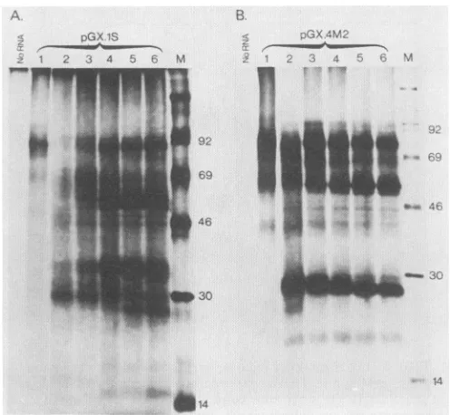
c;X-M2 G.X4M2_S XGX.X4E4 'S
:123 4 5 t 8 3 1C li. 1s' 1 14115 1M
12 4.35 !
A
nGX 4M2
1 2 3 4 5 -67
.-. l 2 3 4 5 6 7
46
* -92
_ -69
-46
.W. _-30
-w...'
..v.
* _ .O~~~~~~~1
30
B
< pGX.4El
z 1 2 3 4 5
-69
46
1 m _
-30
M M M M
EH P P
T
MM9T
P P P P
T
FIG. 5. Localization of regions essential forthe translocation of
proteinsprM and E. Transcripts obtained fromtemplateslinearized
atthe BamHI site were translated in the presence or absence of
membranes(M) and submittedtoaproteinaseKtreatment(P)in the
presence orabsence of Triton X-100 (T). Details of the polypeptides
encodedbyeach transcriptaregiven in Fig. 2. MWs(in thousands) areindicatedattheright.
coincide with the N termini of proteins prM and E. How-ever,thisalso implies that the C protein would beassociated with membranes.
To define the role played by the hydrophobic regions of
theprecursor assignals for translocation of proteinsprMand
E, several plasmids were constructed.
Plasmid pGX4 was derived from plasmid pGYF5' by
deletion of the YFVcoding region and insertiondownstream
of theinitiatingATGcodon of the multiple cloning sitefrom plasmid pGem4 (Fig. 1B). This plasmid produces in vitro T7 transcripts containing the 5' noncoding region of YFV. The initiation codonwasconserved and followed by themultiple cloning site, which allowed the cloning in phase and efficient
in vitro expression of different open reading frames (35).
Differentfragments of cDNA coding for various YFV
struc-tural proteins were then subcloned into plasmid pGX4 in orderto produce plasmidswith N-terminaldeletions in the
polyproteinprecursor (Fig. 1C and 2). Four plasmids were
thus constructed. pGX.4M2 coded fora polypeptide begin-ning with the hydrophobic region from the C terminus of protein C, andplasmid pGX.4M2/S coded for apolypeptide lacking this hydrophobic region but beginning at the third amino acid ofprotein prM. PlasmidpGX4.E1 expressedthe
region coding for protein E preceded by the second hydro-phobic peak located afterarginine 270 andpresent at theC terminus of protein prM. The last construct, pGX.4E/S,
coded for a polypeptide beginning 5 amino acids upstream
from the N terminus of the matureEprotein.
Thepolyprotein expressedfrompGX4M2 transcripts was processed, andproteins prM andEwere efficiently
translo-cated(Fig. 5,lanes 1to4), indicatingthat the first103 amino acidsofthecapsid proteinareunnecessaryfor translocation.
Furthermore, the 37- to 34-kDa doublet disappeared, as
would beexpected forthe C-prMprecursor.
The sequence codingfor thehydrophobic regionatthe C terminus ofprotein Cwas deleted in plasmid pGX.4M2/S.
Thus theresulting transcript expressedapolyprotein,the N
terminus ofwhich corresponded to the third amino acid of
M M M M M M
EH P P P
T
FIG. 6. Invitro glycosylation of proteins prM andE.Translation products were submitted to proteinase K (P) or endoglycosidaseH (EH) treatment as indicated. M, Microsomal membranes; T, Triton
X-100. (A) Transcripts obtained from pGX.4M2 linearized at the HgaI orBamHI site (position 270 or 869 of the YFV polyprotein [Fig. 2]). (B) Transcripts obtained frompGX.4E1 linearized at the PstI site (position674 of the YFV polyprotein [Fig. 2]). MWs (in thousands) are indicated at the right.
protein prM. In this case, protein E was translocated but protein prM was not. Indeed, protein E was resistant to
protease treatment,butthe 16-kDa polypeptide which must
represent the unglycosylated form of protein prM was not
(Fig. 5, lanes 5 to 8). This result indicates that the region
essential forprotein prM translocation is located within the
last 18aminoacidsof protein C and that proteinsprMand E
bearindependent translocation signals.
Deletionofmostof theprotein prM,withthe exception of
itsC-terminalhydrophobic region (plasmid pGX.4E), didnot
alter thetranslocation ofproteinE(Fig. 5, lanes 9to12),but the absence ofthis hydrophobiczoneinplasmidpGX.4E/S
preventedthe translocationoftheprotein (Fig. 5, lanes 13to
16).Thissuggeststhat the last 15amino acidsinprotein prM are necessary for the translocation of protein E, which confirms thepredictionbasedonthe rule ofvonHeijne (44, 45)that thisregionexhibits all thecharacteristics ofasignal peptide. This also confirms the resultsofourrecentin vivo studies using a simian virus 40 expression vector which indicate that the regionwithinamino acids 271to285of the YFVpolyprotein acts as asignal peptide for the
transloca-tion of the envelope protein aswell asfor the heterologous poliovirus VPOprotein (13; P.Despresetal., manuscriptin
preparation).
From the experiments carried out with transcripts from
pGX.4E/S, it is notclear whether NS1 wastranslocated or not (Fig. 6, lanes 13 to 16). However, the polypeptide
contains the putative signal sequence for its translocation,
and ifprocessingofprotein NS1 had occurred in the
pres-ence of membranes, the apparent MW of the precursor
wouldhave been reduced by approximately 10 kDa,
gener-ating twopolypeptides of 10and 60kDa, respectively. The 10-kDapolypeptidewasnotdetected, possiblybecause ofits
small size. In addition, analysis ofthe processed
polypep-tides(lane 14)did revealthepresenceofaband about 10kDa smaller than the precursor. However, since a similar poly-peptide was synthesized in the absence ofmembranes, an
unambiguous conclusion could notbe drawn. These
exper-iments indicatemerelythat thetranslocation ofproteinEdid
M M NI M M M
P P P P
T T
VOL.63, 1989
on November 10, 2019 by guest
http://jvi.asm.org/
[image:7.612.56.293.76.267.2] [image:7.612.310.554.77.250.2]not occur, since nopolypeptidewasprotected from
protein-ase K digestion (lane 15).
Protein prM is efficiently glycosylated in vitro, whereas
protein E is not. The precursor toprotein M, prM, possesses four potential N glycosylation sites located at amino acids
134, 150, 172, and 266 of the YFV polyprotein. The site at
position 266 is probably not used, since it is located in the transmembrane hydrophobic regionof theprotein.The three
remaining sites are located in the N-terminalhalf ofprotein
prM, a region not conservedinprotein M. As thepredicted
MW for the prM backbone is 21kDa, theobserved MWof27 kDa in our in vitro experiments suggests thatthis protein is efficiently glycosylated.
In order to demonstrate the in vitro glycosylation of
protein prM, we synthesized transcripts from pGX.4M2
linearized at the HgaI site (Fig. 1Cand 2). Thesetranscripts
should code for a polypeptide which contains at its N terminus the signal for translocation of protein prM but has deleted at its C terminus the 15 amino acids which precede
the sequence of the envelope protein. The encoded polypep-tide should have a MW close to that of the native prM protein. The
pGX.4M2/HgaI
transcripts code fora polypep-tide of 18 kDa that is partially transformed into a 27-kDa polypeptide in the presence of membranes (Fig. 6A).This polypeptide is glycosylated, as evidenced by its sensitivity to endoglycosidase H treatment (lane 3). As expected, the glycosylated form of prM is protected from proteinase K digestion (lane 4) but is digested in the presence of detergent (lane 5). A similar 27-kDa polypeptide is also synthesized and processed in translation reactions carried out with longer
BamHI
transcripts (lanes 6 and 7), indicating that the C terminus of prM is in close proximity to theHgaIsite.
Protein E possesses two potential N-linked glycosylation sites at asparagine residues 594 and 755 of the YFV poly-protein. As in the case of protein prM, the second site is thought to be nonfunctional because it is located in the C-terminal hydrophobic region at the C terminus of the protein. To investigate whether asparagine 594 could be effectively recognized, in vitro transcripts were synthesized from plasmid pGX4.E1 linearized with PstI. These tran-scripts should code for the N-terminal region of protein E containing asparagine 594 followed by 20 amino acids. An expected polypeptide of 40 kDa was synthesized upon translation (Fig. 6B, lane 1). In this size range it should be easy to detect small variations in MW due to the addition of polysaccharide residues. Translocation products obtained in the presence of membranes (Fig. 6B, lane 2) were treated with proteinase K (lanes 4 and 5) and endoglycosidase H (lane 3). The observed product did not change in MW after the addition of membranes or after endoglycosidase H treatment, indicating that no glycosylation had occurred. However, the polypeptide was efficiently translocated, as demonstrated by its resistance to proteinase K in the
ab-sence of Triton X-100. The lack of glycosylation of proteinE is in agreement with previous observations (17) and with the fact that the only accessible glycosylation site in the protein is found in a weak context for glycosylation.
Kinetics of YFV structural protein synthesis. To study the
events leading to the production of the individual C,
prM,
and E proteins, pGX.lS-BamHI transcripts were translated in the presence of membranes, and the products synthesized afterdifferent times of incubation were analyzed (Fig. 7A). A product with an apparent MW close to 30,000 was first observed, followed by the simultaneous appearance of the 55-kDa protein E and the 37- to 34-kDa C-prM precursor.
A
pGX IS
_ 2 3 4 5 6 M
.i
pI.-B.
-:- i , 45D
-_ .X..) .}
c92
69
..
wo-M
L
46
,303
o~~~~~~~~~~~~~~~~~~~~~~~~~~~~~~~~~~~~~~~~~~~~~~~
.3.14~~~~~~~~~~~~~1
FIG. 7. Kinetics of processing of the YFV structure protein
precursor. Transcripts from BamHI-linearized pGX. 1S (A) or
pGX.4M2 (B) were translated in the absence (lane 1) or presence (lanes 2 to 6) of microsomal membranes. Samples of translation products obtained in the presence of membranes were collected after 15, 30, 45, 60, and 120 min (lanes 2, 3, 4, 5, and 6,respectively). Addition of membranes posttranslationally did not induce any protein processing (not shown). MWs (in thousands) are indicatedat
the right.
The latest event was the production ofproteins prM (27kDa)
and C (15 kDa), presumably after cleavage of the C-prM precursor. The 30-kDa product possibly corresponds to an
untranslocated C-prM precursor which after translocation increases in MW to 37 kDa due to glycosylation. It would thus seem that the prM signal peptide workswith a moderate efficiency for translocation and is not cleaved cotranslation-ally but as a late event.
To confirm the kinetics of the
processing
of theC-prM
precursor,transcripts derived frompGX.4M2 were analyzedin asimilar kinetics experiment. Thepolypeptideencoded by
this transcript begins with the 18-hydrophobic-amino-acid stretch found at the C terminus of the capsid protein (Fig. 2).
When the products synthesized after various incubation periods were analyzed, protein E was already detectable after 15 min of incubation (Fig. 7B) and did not change in MW with time, while the protein prM was first synthesized as a 30-kDa product which was progressively processed into a 27-kDa product. This 3-kDa shift in MW most probably
corresponds to the cleavage ofthe N-terminal region
repre-senting the signal peptide. Inaddition, aproduct of 18 kDa,
probably corresponding to the unglycosylated
(untranslo-cated) prM, was observed in many experiments.
Capsid protein is closely associated with the membrane of
the ER in the C-prM precursor. Proteinase K treatment of
pGX.iS translation productssynthesized in the presence of
membranes induced a reduction ofapproximately 3 kDa in the apparent MW of theC-prM precursor (Fig. 4, lanes 3, 4, 11, and 12). This implied that only the first 20 to 30 amino
acids were exposed outside the membrane vesicles.
Immu-noprecipitation ofthe translation products of pGX.1S
tran-scripts with the anti-C antibody provided confirmation of
this observation (Fig. 4, lanes 9 to 12). The 30- to 31-kDa
:.ml T -
---A. :.-,la, "
'.. T
f
on November 10, 2019 by guest
http://jvi.asm.org/
[image:8.612.318.566.77.306.2]YFV PROTEIN PROCESSING 4207
doublet observed after proteinase K digestion was still recognized by the C-specific antibodies (Fig. 4, lanes 11 and 12). Since this antibody is directed against capsid protein amino acids 20 to 40, the region digested by proteinase K must not extend very much further than the first 20 amino
acids. As the capsid protein is fairly hydrophobic from residue 42 onward, it is possible that the rest of the protein is closely associated with the membrane and thus is pro-tected from proteinase Kdigestion.
Unfortunately, it was not possible to verify whetherthis
partial resistance of protein C to proteinase K digestion also occurred when it was cleaved from the C-prM precursor, because protein C could only be detected clearly on gels
after long exposure times. This problem was worse when
immunoprecipitations were performed. For this reason and because of the poor resolving power of these gels for
low-MW polypeptides, we could not determine whetherthe
capsid protein was partially or totally degraded after protein-ase K treatment.
DISCUSSION
We have established an in vitro transcription-translation system to study the processing of the YFV polyprotein and
to define the role played by cellular proteases in the
matu-ration of the viral structural proteins.
By using this approach, it was found that (i) productionof
proteins C, prM, and E was dependent on the addition of
microsomal membranes to the translation system. Proteins
prM and E were translocated inside the ER membrane,
where protein prM, but not protein E, wasglycosylated.The
translocated proteins are totally resistant to proteinase K
digestion, indicating that they do nothave a domainlocated in the cytoplasmic side of the membrane but most likely are anchored in the membrane by the hydrophobic regions present at their C termini. Proteins prM and E are thus
membrane proteins of the type I, with their N termini exposed on the exoplasmicside of themembrane andtheir C
termini anchored in it.
(ii) Translocation of proteins prM and E inside the ER
vesicles is dependent on thelast 18 or 15 amino acidspresent at the carboxylic ends of proteins C and prM,respectively. These two signals functionindependently ofeach other; they
are active when located in anN-terminal positionfrom the
protein they translocate, and the prM signal, at least, can
also function as aninternal translocation sequence. (iii) The prM signal seems to be less efficient for translo-cation than the E signal, since a significant amountof prM is
not translocated inside the ERmembrane, whereasnearly all
of the E protein is translocated. Furthermore, cleavage of the prM signal is not complete and takes place after
trans-location of the protein, as evidenced by the constant
pres-ence of a translocated C-prMprecursor.
(iv) Protein C is not translocated insidethe ERvesiclesbut
remains very closely associated with the ER membrane by
its C terminus, at least in the formof thetranslocatedC-prM
precursor. The existenceof the translocated C-prM
precur-sor suggeststhat previousexposureofthe Nterminusof the
hydrophobic region preceding protein prM is not essential
for its translocation. Thus, it would function as an internal signal sequence.
These results confirmedthe schematicrepresentation
pro-posed by Coiaet al. (10),which demonstratethedependence of the processing of the YFV polyproteinon cellular
prote-ases. Thecellular protease involvedmust be of the signalase
type, recognizing cleavage sites located after signals for
translocationattheN-terminalpartsofprMand E
proteins.
Cleavage of prM toproduce
M was not effected in oursystem, suggesting that it is mediated
by
aprotease present in theexport vesicles of the cell.Cleavage
of the C terminusofthecapsid protein seems tobe
initially
performed
by
thesignalasethat liberates the N terminus of
prM.
Thiswouldleave a capsid
protein
associated withmembranes,
the C terminus of which hasto be cleaved asecondtime in ordertomake thecapsidprotein available forRNA
encapsidation.
Itispossiblethat thesecond
cleavage
iseffectedby
the viralprotease responsible for the
cleavages
ofthepolyprotein
precursor yielding the nonstructural
proteins.
This enzyme recognizespairs
of basic residues followedby
a small-side-chain amino acid. Such a sequence is foundjust
Nterminal to the
membrane-spanning
domain of thecapsid
protein and is conserved in mostflaviviruses
(23).
Matura-tion ofthevirus could thus be
regulated
by
complex
kinetics of cleavage,leading
to isolatedcapsid,
membrane,
andenvelope proteins.
Processing ofthe YFV
polyprotein
seemstobe an inter-esting model ofpolytopic
protein
processing.
Indeed,
a series of translocation and stop transfersignals
are foundseparating
each structuralprotein
andprotein
NS1. The structuralproteins
of the Flaviviridae seem to beunique
in that thestoptransfer andsignal
for translocationarelocated in a singlehydrophobic
region,
separated
only
by
a basic residue. Inthepresent reportwehavedelimited theregions
essential for translocation ofthe
prM
and Eproteins.
Fur-ther studies are needed to show whetherthese
regions
are sufficient for translocation and whetherthey
aresignal
recognition
particle
dependent,
which would confirm their role as signalpeptides (46).
Furthercharacterization of the regionsacting
as stop transfersignals
is neededin ordertorelate theinternal translocation
signal
tothose found intype
II membrane
proteins.
This seemsparticularly
relevant inthe caseofthe
hydrophobic
region
separating proteins
Cand prM, as theC-prM
precursor has anorientation
relative tothe ER membranewhich is
typical
of typeIIproteins
with acytoplasmic
Nterminus andaluminal C terminus.Interest-ingly, the two
potential
signal
peptides
differ in theircom-petence for translocation as well as in the kinetics with
which they are cleaved off. Such a
difference
insignal
efficiency
could beimportant
fortheregulation
ofthe viral lifecycle, asit wouldregulate
therateof maturation of virus particles.ACKNOWLEDGMENTS
We thank R. Landenheim for stimulating discussions and J.
Schlesinger, A. Barrett, andF. Rodhain for
providing
monoclonal antibodiesandhyperimmune sera.Thisworkwassupportedinpartbycontract 85/120 fromDRET. A.R.L.was afellowof theInternational Network of
Biotechnology.
LITERATURE CITED
1. Bell, J. R., R. M. Kinney, D. W. Trent, E. M. Lenches, L.
Dalgarno, and J. M. Strauss. 1985. N-terminal amino acid
sequences of structuralproteins ofthreeflaviviruses. Virology
143:224-229.
2. Biedrizycka, A.,M.R.Cauchi,A.Darthomoeusz,J. J.Gorman,
andP.Wright.1987.Characterization ofproteasecleavagesites
involved in theformation ofthe
envelope
glycoprotein
andthreenonstructural proteins ofdengue virus type 2, New Guinea C
strain.J. Gen. Virol. 68:1317-1326.
3. Blobel,G.,and B.Dobbertstein.1975.Transfer of
protein
across membranes. I. Presence ofproteolytic
processed
andunproc-essednascentimmunoglobulin
light
chainsonmembrane-bound ribosomes ofmurine myeloma.J. Cell. Biol.67:835-851. VOL. 63,1989on November 10, 2019 by guest
http://jvi.asm.org/
4. Boege, U., P. X. Heinz, G.Wengler, and C. Kunz. 1983. Amino
acidcomposition andamino terminal sequences of the structural
proteins ofaflavivirus, Europeantick-borneencephalitisvirus.
Virology 126:651-657.
5. Cardiff,R. D., andJ. K.Lund. 1976. Distribution ofdengue-2 antigens by electron immunocytochemistry. Infect. Immun. 13:1699-1709.
6. Castle, E., U. Leidner, T.Nowak,G.Wengler,andG.Wengler. 1986. Primary structure of the West Nile flavivirus genome
region coding for all nonstructuralproteins.Virology149:10-26. 7. Castle,E., T.Nowak,U.Leidner,G.Wengler,andG.Wengler. 1985. Sequence analysis of the viral core protein and the
membrane-associated proteins Vl and NV2 of the flavivirus
WestNile virus and of thegenome sequence for theseproteins. Virology 145:227-236.
8. Chen, E. G., and P. H. Seeburg. 1985. DNA Lab. Methods 4:165-170.
9. Cleaves, G. R. 1985. Identification of dengue type 2 virus-specific high molecularweight proteins in virus-infected BHK
cells. J. Gen. Virol. 66:2767-2771.
10. Coia, G., M. D. Parker, G. Speight, M. E. Byrne, and E. G. Westaway.1988. Nucleotide andcomplete amino acidsequence
ofKunjin virus. Definitivegeneorder and characteristicsof the
virus-specified proteins. J.Gen. Virol. 69:1-21.
11. Crawford, G. R., and P. J. Wright. 1987. Characterization of
novelviralpolyproteins detected incells infected bythe flavi-virus Kunjin and radiolabelled in the presence ofthe leucine analogue hydroxyleucine.J. Gen. Virol. 68:365-376.
12. Despres,P., A. Cahour, A.Dupuy, V.Deubel,M.Bouloy, J.P. Digoutte, and M. Girard. 1987. High genetic stability ofthe
region coding for the structural proteins of yellow fevervirus
strain17D.J. Gen. Virol. 68:2245-2247.
13. Despres, P., A. Cahour, C. Wychowski, M. Girard, and M. Bouloy. 1988. Expression of the yellow fever virus envelope
protein using hybrid SV40/yellow fever viruses. Ann. Inst. PasteurVirol.139:59-67.
14. Despres, P., V. Deubel, M. Bouloy, and M. Girard. 1986.
Identification and characterization of intracellular yellow fever virus-specific RNA: absence ofsubgenomic RNA. Ann. Inst. PasteurVirol. 137:193-204.
15. Deubel,V., R. M. Kinney, and D. W. Trent. 1986. Nucleotide
sequence and deduced amino acid sequence ofthe structural proteins of denguetype 2virus, Jamaican genotype. Virology 155:365-377.
16. Deubel, V., R. M. Kinney, and D. W. Trent. 1988. Nucleotide
sequenceand deduced amino acidsequenceof the nonstructural proteins of Denguetype 2virus, Jamaicagenotype:comparative
analysis of the full lengthgenome.Virology 165:234-244.
17. Deubel, V., J. J. Schlesinger, J. P. Digoutte, and M. Girard. 1987. Comparative immunochemical and biologicalanalysis of AfricanandSouthAmerican yellow feverviruses. Arch. Virol.
94:331-339.
18. Friedlander, M., and B. Blobel. 1985. Bovine opsin has more
thanonesignal sequence. Nature(London) 318:338-443. 19. Garoff, H. 1985. Usingrecombinant DNAtechniques to study
protein targeting in the eukaryotic cell. Annu. Rev. Cell Biol.
1:403-445.
20. Kyte, J., and R. F. Doolittle. 1982. A simple method for displayingthehydropathiccharacterofaprotein. J. Mol. Biol. 157:105-132.
21. Laemmli, U. K. 1970. Cleavage of the structural proteins during
the assembly ofthe head of bacteriophage T4. Nature (London) 227:680-685.
22. Lyapustin, V. N., Y. V. Svitkin, T. Y. Ugarova, V. A.
Lash-kevich,and V. I.Agol. 1986. A tentative model of formation of structural proteinsoftick-borne encephalitis virus (flavivirus).
FEBSLett. 200:314-316.
23. Mandl, C. W., F. Guirakhoo, H. Holzmann, F. Heinz, and C. Kunz. 1989. Antigenic structure of the flavivirus envelope
protein Eatthe molecular level, using tick-borne encephalitis
virusas amodel. J. Virol. 63:564-571.
24. Mandl, C. W., F. X. Heinz, and C. Kunz. 1988. Sequence of the
structuralproteins ofthetick-borneencephalitisvirus(Western
subtype) andcomparative analysiswith other flavivirus. Virol-ogy166:197-205.
25. Maniatis, T.,E. F.Fritsch,andJ. Sambrook. 1982. Molecular
cloning: alaboratorymanual. ColdSpringHarborLaboratory,
Cold Spring Harbor, N.Y.
26. Melancon,P.,and H.Garoff. 1986. Reinitiation of translocation in theSemlikiforest virus structuralpolyprotein:identification of the signalfor theElglycoprotein. EMBO J. 5:1551-1560. 27. Monath,T.P.1986.Pathologyof theflaviviruses,p. 375-440. In
S.SchlesingerandM. J.Schlesinger (ed.),TheTogaviridaeand Flaviviridae. AcademicPress, Inc.,NewYork.
28. Monckton,R.P.,and E.S.Westaway.1982.Restricted
transla-tion ofthe genome ofthe flavivirus Kunjin in vitro. J. Gen.
Virol. 63:227-232.
29. Mueckler, M., and H. F. Lodish. 1986. The human glucose
transportercaninsertposttranslationallyinto microsomes. Cell
44:629-637.
30. Ozden, S.,and B. Poirier.1985.Denguevirus induced
polypep-tidesynthesis. Arch. Virol. 85:129-137.31. Perara,E.,and V. R.Lingappa. 1985.Aformer amino terminal
signalsequenceengineeredto aninternal location directs
trans-location ofboth flanking protein domains. J. Cell. Biol. 101:
2292-2301.
32. Rice, C. M., R.Aebersold, D. B. Teplow, J. Pata, J. R. Bell,
A. V.Varndam,D.W.Trent,M.W.Brandiss,J. J.Schlesinger,
andJ.H.Strauss. 1986. PartialNterminiamino acid sequences
of three nonstructural proteins of two flaviviruses.
Virology
151:1-9.33. Rice,C.M.,E. M.Lenches,S. R.Eddy,S.J.Shin,R. L.Sheets,
andJ. H. Strauss. 1985. Nucleotide sequence ofyellowfever virus:implicationsfor flavivirusgeneexpressionandevolution. Science229:726-733.
34. Rice,C.M.,andJ.H.Strauss. 1986.Structure of the flavivirus
genome, p. 279-326. In S. Schlesinger and M. J. Schlesinger
(ed.),TheTogaviridaeandFlaviviridae. AcademicPress,Inc.,
NewYork.
35. Ruiz-Linares, A., M.Bouloy,M.Girard,and A. Cahour.1989. Modulations of the in vitro translationalefficiencies ofyellow
fever virus inRNAs: interactions betweencodingand
noncod-ing regions. NucleicAcidsRes. 17:2463-2476.
36. Schlesinger, J. J., M. W. Brandiss, and T. P. Monath. 1983. Monoclonal antibodies distinguish between wild and vaccine strains of yellowfever virus by neutralisation,
hemagglutina-tion,inhibition and immuneprecipitationof the virusenvelope.
Virology125:8-17.
37. Speight,S.,G.Coia,M. D.Parker,and E.G. Westaway. 1988. Gene mapping and positive identification of the nonstructural
proteins NS2A, NS2B, NS3 and NS5 of the flavivirus Kunjin
andtheircleavage sites.J. Gen. Virol. 69:23-34.
38. Spiess, M.,and H. F. Lodish.1986.Aninternalsignalsequence: the asialoglycoprotein receptor membrane anchor. Cell 44: 177-185.
39. Stohlman, S. A., C. L. Wisseman, 0. R. Eylar, and D. J.
Silverman. 1975. Dengue virus-induced modifications of host cellmembranes. J. Virol. 16:1017-1026.
40. Strauss,J.H.,E.G.Strauss,C. S.Hahn,andC. M. Rice.1987.
The genomesofalphavirusesandflaviviruses:organizationand
translation, p. 75-104. In D. J. Rowlands, M. A. Mahy, and
B. W. J. Mahy (ed.), The molecular biology of the positive
strand RNA viruses. Academic Press, Inc. (London), Ltd.,
London.
41. Strauss,J.H.,E.G.Strauss,C.S.Hahn,Y.S.Hahn,R.Galler,
W.R.Hardy,andC. M. Rice. 1987. Replicationofalphaviruses
andflaviviruses: proteolytic processingofpolyproteins. UCLA
Symp. Mol. Cell. Biol. 49:209-225.
42. Svitkin, Y. V., T. Y. Ugarova, T. V. Chernovskaya, V. N.
Lyapustin, V. A.Lashkevich, and V.I. Agol. 1981.Translation of tick-borne encephalitis virus (Flavivirus) genome in vitro:
synthesisoftwostructuralpolypeptides. Virology110:26-34.
43. Svitkin, Y. V., V. N. Lyapustin, V. A. Lashkevich, and V. I.
Agol. 1984. Differences between translation products of tick-borneencephalitisvirusRNAincell-freesystems fromKrebs-2
cellsand rabbitreticulocytes:involvement of membranes inthe
on November 10, 2019 by guest
http://jvi.asm.org/
YFV PROTEIN PROCESSING
processing ofnascent precursors of flavivirus structural
pro-teins. Virology 135:536-541.
44. vonHeijne, G. 1985.Signalsequences.Thelimits of variation. J. Mol. Biol. 184:99-105.
45. von Heijne, G. 1986. A new method for predicting signal
sequencecleavage site. Nucleic Acids Res. 14:4683-4690.
46. Walter, P., and V. R. Lingappa. 1986. Mechanism of protein translocation across the endoplasmic reticulum membrane.
Annu. Rev. Cell Biol. 2:499-516.
47. Wengler, G., M. Beato, and G. Wengler. 1979. In vitro transla-tion of 42S virus specific RNA from cells infected with the flavivirus West Nile virus. Virology 96:516-529.
48. Westaway, E. G., S. Y. Gaidamovitch, M. S. Horzinek, A.
Igarashi,L.Kaariainen, D. K. Lvov, J. S. Porterfield, P.K.P. Russel, and D. W. Trent. 1985. Flaviviridae. Intervirology 24:183-192.
VOL.63, 1989 4209