Vol. 62, No. 3 JOURNALOF VIROLOGY, Mar. 1988, p. 887-893
0022-538X/88/030887-07$02.00/0
Copyright © 1988, AmericanSociety for Microbiology
Tissue Tropism and Target Cells of Bluetongue
Virus
in
the
Chicken Embryo
LIWANG,' MAURICE C. KEMP,' POLLY
ROY,2
AND ELLEN W.COLLISSON1*
DepartmentofVeterinary Microbiology and Parasitology, College ofVeterinaryMedicine, and TexasAgricultural ExperimentStation, Texas A&M University, College Station, Texas
778434467,'
and SchoolofPublicHealth,University of AlabamainBirmingham, Birmingham, Alabama 352942 Received6August 1987/Accepted19November 1987
In situ cytohybridization was used to determine the tissue tropism and target cells for replication of bluetongue virus (BTV) in the developing chickenembryo. Hybridization withabiotinylated probe specific for segment 3of BTV serotype 17 detected viral replication in embryos inoculated with U.S. serotypes2, 10, 11, 13, and 17 or sheep blood containing a BTV field strain. At the final stages ofinfection, when the embryos were hemorrhagic, viral infection could consistently be detected in the brain,kidney,spinalcord,heart,lung, and liver, with the brain and kidney most severely affected. Other tissues, such as the retina, skin, tongue, and intestinalvilli,also supported viral replication in some embryos. Greater concentrations of virus tended to be localized within epithelial cells, such as those lining the kidney tubules and tertiary bronchi of the lungs. Kinetics studieswith BTV serotypes 11 and 17 and a field strain indicated that within 24 h after inoculation, viralreplicationoccurred initially in the brain and kidney. By 48 h, viral replication was also detected in the lungs, heart, and spinal cord, with the liver being severely infected by 72 h. Low levels of hybridization could be detected inembryos infected with epizootic hemorrhagic disease virus, which isantigenically related toBTV.
Bluetongue virus (BTV), which has a 10-segment double-stranded RNA genome, is the prototype ofthe orbivirus genusin the Reoviridaefamily. Bluetongue, whichis trans-mitted by Culicoides spp., is an infectious, noncontagious, and, under certain circumstances, hemorrhagic disease in ruminants. Infection during gestation may result in congen-italmalformationsor deathofthedeveloping fetus(16, 19) or lead to persistently infected carriers (13, 15). Studies de-finingthe tissuetropism ofBTVhavedependedprimarilyon virus recovery techniques. Virus may be isolated from lymphocytes, neutrophils, or erythrocytes, as well as from semen,after insect-mediated infectionwith BTV(2, 12).The involvement of these cellsmayindicate that theyaresites of viralreplication,but could alsoreflectthe strong
affinity
that BTV apparently has for cellular membranes (24). In deer, BTV has beenisolated from anumber of tissues, including thelung, liver, kidney, spleen,andtongue, afterinoculation ofegg-adapted virus (23). However, thefrequency of isola-tion fromeach
organranged from55 to70%,
and nospecific tissue could be recommended for optimal BTV recovery. Likewise,virushasbeenreisolated fromavarietyof organs ofinutero-infected fetuses (A.J. Luedke, personal commu-nication).Thefetal brainhasbeenfoundtobe apredominant targetfor viral replicationandpathogenesis, asindicated by thedevelopment of hydranencephalyandporencephaly(16, 19).Although Osburn etal. (18) reported that BTV could be detected in fetal tissue by immunofluorescent techniques, the preferred method for confirmation of BTV infection generally involves intravascular (i.v.) inoculation of embr-yonating chicken eggs (ECE) followed by several blind passages ofembryo homogenates on cultured cells (3, 5). Because of difficulties encountered in isolating virus and, thus,in identifying carriers, regulatory agencies throughout
*Correspondingauthor.
the world impose restrictions on the movementofcattle and importation of otherwise desirablegermplasmandembryos. A sensitive and economical assay for detection of carriers would be valuable in the certification of BTV-free animals. Nucleic acidhybridization techniques haveproven valuable in demonstratingtarget organsfor viral replication, as well as providing sensitive, rapid alternatives for diagnosis of viral andgenetic diseases (1, 4, 7). Thesetechniquesareless dependent on genomic expression, making it possible to detect defective or nonexpressed genomes. Target tissues and viral infection of individual cells, evenwhenrelatively few cells are infected, can bereadilydetected (7).
Inthisreport, in situ hybridization techniques were used to identify the specific target cells and organs for viral replication in longitudinal sections of ECE infected with several BTV serotypes indigenous to the United States. Since all themajororgans could be viewedsimultaneouslyin each section, thismodelprovedtobeconvenient for study-ing tissue tropism ofBTV, as well as theeffects ofdose and the kinetics of infection in adeveloping animal.
MATERIALS AND METHODS
Viruses. Theserotype 10, strain 8, collected inCalifornia; serotype 11, Station strain, Texas; serotype 13, strain 67-41B, Idaho; serotype 17, strain 62-45S, Wyoming; and epizootic hemorrhagicdisease viruses(EHDVs), U.S. sero-type 1, New Jersey, and serotype 2, Alberta, laboratory prototype strains used were obtained from the Arthropod-Borne Animal Diseases Research Laboratory, Laramie, Wyo. All prototype strains wereadapted toBHK-21 cells. Blood from afield-infected sheepfrom westernTexas was used as the sourceofBTVinonestudy.BTVhad
previously
beenisolated from this bloodbyi.v.inoculation of ECE(5). The avian infectious bronchitis virus (IBV) Arkansas and Newcastledisease virus(NDV)GB usedfor thisstudywere
egg-adaptedviruses. 887
on November 10, 2019 by guest
http://jvi.asm.org/
Tissuepreparation. Eleven-day-old ECEwereinfectedi.v. witheach viral strain, andat various intervals of time after
inoculation,
theembryos were harvested. Afterharvesting,the
wings
andlegswereremovedfromtheembryos,and thetissue was embedded in OCT (Miles Scientific) at -70°C.
Longitudinal
sections(8lim)
ofthespecimenswere cutwitha
Lipshaw
cryotome at -20°C, transferred to glass slidestreated as described by Haase et al. (7), and coated with Histostik(Accurate ChemicalandScientific
Co.,
Westbury, N.Y.). The sections were then airdried,
dipped in phos-phate-buffered saline, and fixed in freshly prepared 4%paraformaldehyde
for 15 min (22). Afterfixation,
sectionswere rinsed in phosphate-buffered saline, dehydrated in
increasing
concentrations ofethanol, and stored at4°C (7).Probe preparation. The biotinylated probe was prepared
by
nick translation ofaBTV cDNAforserotype 17 segment 3, whichwaspreviously
showntobespecific forall theU.S. serotypes(20, 21). The BTVcDNA insertwasexcisedfromthe
plasmid
withHindlIl,
separated from the plasmid on anagarose
gel,
andelectroelutedasdescribedinManiatisetal. (17). The insert was labeled by nick translation in the presence ofbiotinylated
dUTP(instruction
manual ofEnzoBio-Chem,
NewYork,
N.Y.). Theefficiency
of nicktrans-lation was determined by monitoring the incorporation of
[3H]dATP.
Optimal
labeling of the probe was achieved bylowering
the concentration of DNA (<0.5p.g/50
ILI)
andincreasing
the concentration ofDNase(>1ng/50
,ul).Hybridizationassays. Thein situ hybridization procedure used in this study was a modification of the methods de-scribed by
Singer
etal. (22) and Haaseet al. (7). Prehybri-dizationwas foundto be unnecessary. Sections wereover-laid with 30 to 40 ,ul of hybridization solution
(50%
formamide,
2x SSC [0.15 M sodiumchloride,
0.015 Msodium
citrate],
5x Denhardtsolution, 10%dextransulfate,100ngof salmon spermDNA per
ml,
1 mgofwholechickenembryo
RNA perml,
and 20to50 ngofbiotinylated
cDNA,per
slide).
Silicon-treated coverslips
wereplaced
over thesections covered with
hybridization
solutionand sealedwith rubber cement. The slides were then heated for 10 min at80°C,
andhybridization
wasallowedtoproceed overnight
at37°C. Posthybridization, washing
and visualization wereperformed
asdescribedfortheEnzo kit. Positivehybridiza-tionwasdetected
by
thereactionofstreptavidin-horseradish
peroxidase,
diaminobenzidinetetrahydrochloride,
andhy-drogen peroxide.
The nuclei were counterstained with a0.25% solution of
methyl
greenin 0.03 Msodiumacetate,pH
4.8. Positive
hybridization
wasindicatedby areaswith dark red-browngrains surrounding blue-green
nucleicounter-stained with
methyl
green.Dotblot
hybridization
wasdone with RNA extracted withphenol-sodium
acetate, pH 5, buffer fromuninfectedBHK-21 cells orfrom cells infected with BTV 11 or EHDV 1. A modification of the procedures described by Kafatos et al.
(11)
andSchleicher &Schuell, Inc., Keene, N.H.,wasused.Briefly,
the RNA was pretreated with formaldehyde,form-amide,
andphosphate-SSC
buffer. Afterheating
to 65°C for10 min, the solution with 1
pLg
of RNA or fourfold serial dilutionswasblottedontonitrocellulosepretreatedwith20x SSC (17) with aBio-Dot Apparatus (Bio-Rad Laboratories) manifold.Afterdrying,thesampleswereprehybridizedfor4 h at37°C
in 50% formamide-5x Denhardt solution-0.1% sodium dodecyl sulfate-150 ,ug of calf thymus DNA perml-5x SSC(17)and
hybridized
for 24 hat37°Cinasolutionof 50% formamide-2x Denhardt solution-150
jg
of calfthymus
DNA perml-5x SSC and 106 cpm ofprobelabeledby
nick translation with[ax-32P]dATP
(17)
perml.RESULTS
Tissuetropism ofBTVand probespecificity. Insitu cytohy-bridization with a recombinant cDNA probe specific for segment3 of BTV 17wasusedtodetect viralreplicationin
ECE individuallyinfected with laboratory strains of theU.S. serotypes of BTV: 2, 10, 11, 13, and 17. At least three embryos were infected per virus. Hemorrhagic embryos were harvested after they had died, which was between 3
and 5 daysafterinoculation of BTV. Thin (8 jim) longitudi-nal sections of the OCT-embedded embryos were usedfor
these in situ studies. Figure 1 shows such awhole embryo
section stained withhematoxylin and eosin. Various organs
including brain, eye, kidneys, liver, spinal cord, heart,
intestines, and lungscanbe readily identified in sections. In
addition, the spleen could also be seen inoccasional sections.
Althoughsome tissueorganization can bediscerned only at the lower magnifications, higher-power (about 1,OOOx) objectiveswerenecessarytoaccuratelyscoreBTV
infectiv-ity in the cytoplasm. Figures 2a and b illustrate a control
section of brain from an uninfected 15-day-old embryo at 250x and 1,000x magnification. This control is
repre-sentative of the assay with uninfected cells from other
tissues, since only nuclei were stained, and thus all the
tissueswere very similar at the higher magnifications. The viral RNA-probe hybrids in the following black and white figuresareseen asdarkgranules in the cytoplasm
surround-ing lightergray nuclei. In embryos infected withanyof the
five U.S. serotypes, positive hybridization could consis-tently be detected throughout the brain, kidneys, lungs, heart, liver, and spinal cord. However, greater concentra-tions of virus were demonstrated in the external granular
layer of the cerebellum of the brain (Fig. 2c and d), in renal tubuleepithelial cells (Fig. 3a, b, and c), and cells lining the tertiary bronchi of the lungs (Fig. 3d and e). In additionto the hybridization found in these organs, viral replication
could be demonstrated occasionally in other tissues of embryos, suchastheretina(Fig. 3f and g), skin,tongue,and breastmuscle.
Of the six tissues in which virus wasconsistently
demon-strated, more viral RNA was found in the brains and
kidneys, with less in the spinal cord and liver, whereas the amount of BTV RNA found in the hearts and lungs was
intermediaterelativetotheother tissues (Table 1). Although theembryoswereseverelyhemorrhagic, the positive signals
in BTV-infected sections were specific and not a result of
FIG. 1. Hematoxylin-and-eosin-stainedlongitudinalsection ofa
15-day-old chicken embryo. Individualorgans:B,brain; E,eye;H, heart; Li, liver; Lu, lung;I,intestine; S, spleen; SC, spinal cord; K, kidney.
on November 10, 2019 by guest
http://jvi.asm.org/
[image:2.612.320.560.547.683.2]BTV TARGET CELLS IN CHICKEN EMBRYOS 889
b
.I! 6
0-I , e
..
41..
.F
,;.-.S f
r..*
.*
[image:3.612.316.557.618.698.2]*;~* P WE;
FIG. 2. Insituhybridizationofsections of thebrain fromanuninfected15-day-old embryo (a, x250; b, x1,000)andaBTV11-infected 15-day-old embryo (c, x250; d, x1,000) harvested4days after infection.
extensive lesions in blood vessels, because hybridization
wasnotseenin the embryosinfected with NDV, which, like
BTV, causes hemorrhages in embryos. Embryo sections infectedwith IBV, another unrelated virus which served as an additional control,werealso negative.
Hybridization, however,wasfoundinboth the brains and kidneys of embryos harvested 5 days after infection with either serotype ofEHDV (Table 1), although therewas no evidence ofgrosspathology in these ECEand these strains did not kill embryos. Therelative amount ofhybridization
was less in the brain than the kidneys of EHDV-infected embryos, and the amount in both thesetissueswas
consid-erably less than that seen in the brain and kidneys of
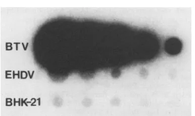
BTV-infected embryos. Since it had been reportedthat the segment3 BTV 17probewasnotspecificfor EHDV 1 RNA intransfer blot assays (20), adot blot assay withextracted RNA was used to compare EHDV, BTV serotype 11, and BHK-21RNA(Fig. 4).Under theconditionsused,theprobe did have specificity for EHDV as well as BTV RNA. However, theamountofhybridization to BTV was consid-erablygreaterthan thattoEHDV RNA. Attempts to elimi-nate the in situ hybridization to EHDV by increasing the hybridizationtemperature to42°Cresulted inless hybridiza-tion inboth the EHDV- and BTV-infected tissues. At45°C, hybridization was notfound in tissues infected with either virus; however, thismaybe theresult of celldisintegration foundatthis temperature.
Kinetics and dose-response studies. BTV infection was
examined by in situ hybridization of ECE at various inter-vals of time after infection with various concentrations of virus. Tenfold dilutions rangingin concentrationfrom
102.5
to 105 50% egg-lethal doses (ELD50s) per ml of BTV serotype11 wereused,and theembryoswereharvestedat4, 8, 16, 24, 48, 72, 96, and 120 h after infectionor when the embryosdied(from72h on).The earliestthatreplication
of either virus could be detectedwas24 hafter viral inoculation with the104-5
and105-5ELD50doses(Table 2), and the effect ofdecreasing concentrations of virus tended to extend the time at which viral RNA could bedetected, so thathybrid-TABLE 1. Viral specificity ofBTV 17 segment3 cDNA in insituhybridization assays
Virusdetected"
Virus
Brain Kidney Heart Lung Spordl Liver
BTVb ++++ ++++ +++ ++ ++ +++
EHDV' + ++ - _ _ _
IBV - - - _ _ _
NDV - - - _
"Concentrations
fromhigh(++ ++)toundetectable(-). bBTVserotypes 2, 10, 11, 13,and 17.EHDV serotypes 1 and 2.
VOL.62, 1988
4
.4 :: .1,
ii `4
OF
.IL4L''
on November 10, 2019 by guest
http://jvi.asm.org/
J. VIROL. 890 WANG ET AL.
.
~~~~~4.
*.; ...4 ..g. 4
2 1
I I.'
I ~ ~v.. . *:^
[image:4.612.67.557.66.575.2].. x
FIG. 3. Insituhybridizationof sections of BTV 11-infectedkidney (a, b,and c,asinglecell
supporting
viralreplication), lung (d
ande), and retina of theeye(fandg)of a15-day-old embryo.Magnification:a,d,andf,40x;b,e,andg,1,000x;c,2,000x.Arrowsindicateportionsof the40x magnification whichareenlargedinpanelsdand f. ization could notbe detected until 72 hafter infection with
the
102.5
and 103.5 ELD50SAt 24 h, when there were no visible signs of virus-induced lesions, infection ofkidney and brain tissues was evident with the higher concentrations of inoculum (Table 3). By 48 h, BTV could also be detected in the cells surroundingthespinalcord and in thelungand heart. By72 h,viruswasreadilydetectablethroughouttheembryo,when thehybridization wassimilartothatfoundin thepreviously described hemorrhagic embryos. Parallel experiments with
BTV 17 gavesimilar results. Becauseof the low
background
occasionally seen in the
liver,
itwas sometimes more diffi-cultto identify positivehybridization
at the lower concen-trations ofvirus, andconsequently,
there may have beenmore BTV
replication
present in this organ than we havereportedat24and 48 h.
DetectionofBTVfromafieldspecimen. The
sensitivity
of this assay indetecting
viralRNAearly
ininfection,
thatis,
before
signs
ofdisease, suggested
thatit hadpotential
asadiagnostic procedure
toconfirm BTV in fieldsamples.
on November 10, 2019 by guest
http://jvi.asm.org/
BTV TARGET CELLS IN CHICKEN EMBRYOS 891 andaffected tissues earlier than the BTV 11 and 17 labora-torystrains.
DISCUSSION
In situcytohybridizationwasusedtodetect BTVinfection
in developing chicken embryos.Since all themajor organs of
BTV
EHDV
BHK-21
FIG. 4. Dot blot hybridizations of[ox-32P]dATP-labeled BTV 17
segment3 cDNAwith RNA fromuninfected andBTV- and
EHDV-infected chicken embryos. Fourfold dilutions of the RNA were addedtoeach well, startingwith 1,ug ofRNA in thefirstwellonthe
left.
lation ofa strain of BTV had been made inour laboratory
fromablood specimen ofasheep during a1985 bluetongue
outbreak in western Texas. Embryos infected with sheep blood(about 3.7 ELD50s) from which the isolation had been madewereexaminedat24, 48, and 72 h after inoculationor
atthe time of death (Table 3). Five embryoswereexamined
eachday. After 24 h, viral replicationwasdetected through-out the brain and kidneys and in a few cells of the spinal
cord. As with the laboratory strains used above, after the second day, replication in this unmodified strain could be detected inanumberof cells in the heart andlungs, but also
in the liver. Also at48 h, positive replication could beseen
in other tissues throughout the embryos, including the epi-thelial cells of the intestinal mucosa(Fig. Saand b), which
hadnotbeen found tosupport viral replication in embryos infected with the laboratory strains; the retina; breast
mus-cle;tongue;and skin. FigureScisa1,OOOx magnificationof
an uninfected intestinal mucosa. This Texas field strain appearedtobemore virulent, since it affectedmoretissues
TABLE 2. Effects of time and viral concentrationon
the detection of BTVinfectivity
Dose Hybridizationaat time postinoculation:
(loglo
ELD50) 16 h 24h 48 h 72 h 96h 120h2.5 - - - + + +
3.5 - - _ + + +
4.5 - + + + + +
5.5 - + ND ND ND ND
aAfter i.v. inoculation of 11-day-old chicken embryos with BTV 11.
Symbols:-,hybridizationwasnegativeinthelongitudinal embryo sections;
[image:5.612.83.276.65.180.2]+,positivehybridizationwasfound in theembryosections; ND,notdone.
TABLE 3. Time that RNAwasdetectable after infection with BTV serotype 11 or 17 orthe westernTexasfield strain
Time of detection (hpostinoculation)
Tissue
11or17a Fieldstrain'
Brain 24 24
Kidney 24 24
Spinalcord 48 24
Heart 48 48
Lung 48 48
Liver 72 48
a Virustiter,4.5ELD50.
"Virus
titer, 3.7ELD50.
b
FIG. 5. In situ hybridization of sections of BTV field strain-infected intestines at (a) 40x and (b) 1,000x and of uninfected intestine at1,000x (c).
VOL.62, 1988
0
As 4. -.eo
-p
0
A
..
on November 10, 2019 by guest
http://jvi.asm.org/
[image:5.612.317.554.159.700.2] [image:5.612.58.296.620.708.2]a longitudinal section of the embryo can be viewed simulta-neously on a single slide, the chicken embryo was a conve-nient model for examining the pathogenesis of BTV infec-tion, which is known to cause congenital malformations in the developing fetus (16, 19). The initialtarget organs where replication could consistently be detected as early as 24 h after inoculation of BTV were the brain and kidneys. Simi-larly, congenital malformations in in utero-infected fetal calves and sheep were also reported to be targeted to the brain (16, 19). At 48 h after BTV inoculation of ECE,viral infection was not only present in the brain and kidneys, but had alsospread to the spinal cord, heart, and lungs. The liver wasunquestionably involved by the third day. At the time of death, when the lesions were extensive, viral RNA was detected throughout the embryo. Additional tissues such as the retina, tongue, breast muscle, intestinal villi, and skin could also support viral replication.
Although our procedure could identify specific organs associated with viral replication, it was difficult to define the specific cells involved since only nuclei werecounterstained. However, the location ofthe positive hybridization within tissue sections and comparisons with hematoxylin-and-eo-sin-stained sections suggested that many cell types, espe-cially epithelial cells, were infected. For example, the cells lining tubules in the kidney, the retina, the epithelial mucosa ofthe intestines, and external layer of the cerebellum of the brains appeared to be highly susceptible to viral replication. Erythrocytes or their precursors may also harbor virus but didnot appear to be major targets of viral replication, since viral replication was detected throughout tissues, and espe-cially in epithelial cells, rather thanin luminal areas, where high concentrations oferythrocytes arefound. In addition, target organs for viral infection did notcorrelate with those organs having high concentrations oferythrocytes; that is, the brain supported replication earlier than theliver.
Whereas the segment 3 BTV probe could not detect IBV-or NDV-infected cells, it could detect viral RNA of the closely related EHDV. This segment in EHDV and BTV has been shown to have sequence homology (6), and the corre-sponding proteins of EHDV and BTV have also been shown to share antigenic epitopes (8). Viral replication in EHDV-infected embryos, however, was only detected in the brain and kidneys. Hybridization may not have been found in other tissues either because of the low level of EHDV replication or because of the low level of specificity of the probe for EHDV RNA. Gross lesions or other signs of viral infection were not detected in the EHDV-infectedembryos. The relative specificity of the probe used for EHDV RNA was considerably less than for BTV.
Along with in situ hybridization techniques, the chicken embryo, which is the preferred medium for unmodified BTV isolation, was also shown in these studies to offer apotential alternative source for rapid and sensitive diagnosis of BTV infection. BTV from field samples can be confirmed in infected embryos as early as 24 h postinfection and in the absence of lesions. Since we have recently been able to demonstrate BTV replication after only 4 h of hybridization (unpublished observations), the procedure can be done within an 8-h day. Therefore, the amount of time required for confirmation of BTV can conceivably be reduced from several weeks to 2 days.
ACKNOWLEDGMENTS
We gratefully acknowledge the help of Paul F. Frelier and David L. Graham in the interpretation of data and Albert J. Luedke for valuable suggestions.
Thiswork wassupportedby theTexas Agricultural Experiment Station,TexasA&MUniversity, CollegeStation, Tex.
LITERATURE CITED
1. Caskey, C. T. 1987. Disease diagnosis by recombinant DNA methods. Science 236:1223-1229.
2. Collisson, E. W., and T. L. Barber. 1983.Bloodcells associated with bluetongue virus infection in cattle. Annu. Proc. Am. Assoc. Vet. Lab. Diagn. 26:287-300.
3. Collisson,E.W., and T. L. Barber. 1985. Isolation and identi-fication ofbluetongue virus: a serotype new to the U.S., p. 319-328. In Proceedings of the International Symposium of Bluetongue and RelatedOrbiviruses,Asilomar,Calif., 16to20 January 1984.
4. Dunn, D. C., C. D. Blair, D. C.Ward, and B.J. Beaty. 1986. Detection of bovineherpesvirus-specific nucleic acidsbyin situ hybridization with biotinylatedDNAprobes. Am J. Vet. Res. 47:740-746.
5. Foster, N. M., and A. J. Luedke. 1968. Direct assay for bluetonguevirus by intravascular inoculation ofembryonating chickeneggs. Am J. Vet. Res. 29:749-753.
6. Gould, A. R. 1987. Thecomplete nucleotidesequenceof blue-tongue virus serotype 1 RNA3 and acomparison with other geographic serotypes from Australia, South Africa and the United States of America, and with other orbivirus isolates. Virus Res. 7:169-183.
7. Haase,A., M.Brahic,L.Stowring, and H. Blum.1984. Detec-tion of viral nucleic acidby in situhybridization.Methods Virol. 7:189-226.
8. Huismans, H., C. W. Bremer, and T. L. Barber. 1979. The nucleic acid and proteins of epizootic haemorrhagic disease virus.OnderstepoortJ. Vet. Res.46:95-104.
9. Jochim, M. M., T. L. Barber, and B. M. Bando. 1974. Identifi-cationofbluetongue andepizootichemorrhagicdisease viruses by the indirect fluorescent antibody procedure. Proc. Am. Assoc. Vet.Lab. Diagn. 17:91-103.
10. Jochim, M.M., and S. C.Jones. 1976. Plaque neutralization of bluetongue virus and epizootic hemorrhagic disease virus in BHK-21 cells. Am. J.Vet. Res. 37:1345-1347.
11. Kafatos, F.C., C. W. Jones, and A. Efstratiadis. 1979. Deter-mination of nucleic acid sequence homologies and relative concentrationsbyadothybridizationprocedure.NucleicAcids Res. 7:1541-1552.
12. Luedke, A. J. 1970. Distribution of virus in bloodcomponents during viremia of bluetongue. Proc. Annu. Meet. U.S. Anim. Health Assoc. 74:9-21.
13. Luedke, A.J.,M. M.Jochim,and T. L. Barber.1982.Serologic andvirologic responsesofaHereford bullpersistentlyinfected withbluetongue virus for elevenyears. Proc. Annu. Meet. Am. Assoc.Vet. Lab. Diagn. 25:115-134.
14. Luedke, A. J., M. M. Jochim, J. G.Bowne, and R. H.Jones. 1970. Observations on latent bluetongue virus infection in cattle. J.Am. Vet. Med. Assoc. 156:1870-1889.
15. Luedke, A. J., M. M.Jochim, and R. H.Jones.1977. Bluetongue in cattle: effects of Culicoides variipennis-transmitted blue-tongue virus onpregnantheifers and their calves. Am. J. Vet. Res. 38:1687-1694.
16. MacLachlan, N. J., and B. I. Osburn. 1983. Bluetongue virus-inducedhydranencephaly in cattle. Vet. Pathol. 20:563-573. 17. Maniatis, T., E. F. Fritsch, and J.Sambrook. 1982. Molecular
cloning: alaboratory manual.Cold SpringHarbor Laboratory, Cold Spring Harbor, N.Y.
18. Osburn, B.I., R. T.Johnson, A. M.Silverstein, R. A. Prender-gast, M. M. Jochim, and S. E. Levy. 1971. Experimental viral-inducedcongenitalencephalopathies. II.Thepathogenesis of bluetongue vaccine virus infection in fetal lambs. Lab. Invest. 25:206-210.
19. Osburn, B. I., A. M. Silverstein, R. A. Prendergast, R. T. Johnson, and C. J. Parshall, Jr. 1971. Experimental viral-induced congenital encephalopathies. Lab. Invest. 25:197-205.
on November 10, 2019 by guest
http://jvi.asm.org/
BTV TARGET CELLS IN CHICKEN EMBRYOS 20. Purdy, M., J. Petre, and P. Roy.1984.Cloning of the bluetongue
virusL3gene. J. Virol. 51:754-759.
21. Roy, P., G. D. Ritter, H. Akashi, E. Collisson, and Y. Inaba. 1985. Agenetic probe for identifying bluetongue virus infections in vivoandin vitro.J. Gen.Virol. 66:1613-1619.
22. Singer, R. H., J. B. Lawrence, and C. Villnave. 1986.
Optimiza-tion of in situ hybridization using isotopic and non-isotopic detection methods. Biotechniques 4:230-249.
23. Thomas, F. C., and D.0. Trainer. 1970. Bluetongue virusin
white-tailed deer. Am.J. Vet. Res.31:271-278.
24. Verwoerd, D. W. 1969. Purification and characterization of bluetongue virus.Virology 38:203-212.
VOL. 62, 1988 893