Differential Phosphatidylinositol-3-Kinase-Akt-mTOR Activation by
Semliki Forest and Chikungunya Viruses Is Dependent on nsP3 and
Connected to Replication Complex Internalization
Bastian Thaa,aRoberta Biasiotto,aKai Eng,aMaarit Neuvonen,bBenjamin Götte,aLara Rheinemann,aMargit Mutso,cAge Utt,c Finny Varghese,dGiuseppe Balistreri,bAndres Merits,cTero Ahola,dGerald M. McInerneya
Karolinska Institutet, Department of Microbiology, Tumor and Cell Biology, Stockholm, Swedena
; University of Helsinki, Institute of Biotechnology, Helsinki, Finlandb ; University of Tartu, Institute of Technology, Tartu, Estoniac
; University of Helsinki, Department of Food and Environmental Sciences, Helsinki, Finlandd
ABSTRACT
Many viruses affect or exploit the phosphatidylinositol-3-kinase (PI3K)-Akt-mammalian target of rapamycin (mTOR) pathway, a crucial prosurvival signaling cascade. We report that this pathway was strongly activated in cells upon infection with the Old World alphavirus Semliki Forest virus (SFV), even under conditions of complete nutrient starvation. We mapped this activation to the hyperphosphorylated/acidic domain in the C-terminal tail of SFV nonstructural protein nsP3. Viruses with a deletion of this domain (SFV-⌬50) but not of other regions in nsP3 displayed a clearly delayed and reduced capacity of Akt stimulation. Ec-topic expression of the nsP3 of SFV wild type (nsP3-wt), but not nsP3-⌬50, equipped with a membrane anchor was sufficient to activate Akt. We linked PI3K-Akt-mTOR stimulation to the intracellular dynamics of viral replication complexes, which are formed at the plasma membrane and subsequently internalized in a process blocked by the PI3K inhibitor wortmannin. Replica-tion complex internalizaReplica-tion was observed upon infecReplica-tion of cells with SFV-wt and SFV mutants with deleReplica-tions in nsP3 but not with SFV-⌬50, where replication complexes were typically accumulated at the cell periphery. In cells infected with the closely related chikungunya virus (CHIKV), the PI3K-Akt-mTOR pathway was only moderately activated. Replication complexes of CHIKV were predominantly located at the cell periphery. Exchanging the hypervariable C-terminal tail of nsP3 between SFV and CHIKV induced the phenotype of strong PI3K-Akt-mTOR activation and replication complex internalization in CHIKV. In con-clusion, infection with SFV but not CHIKV boosts PI3K-Akt-mTOR through the hyperphosphorylated/acidic domain of nsP3 to drive replication complex internalization.
IMPORTANCE
SFV and CHIKV are very similar in terms of molecular and cell biology, e.g., regarding replication and molecular interactions, but are strikingly different regarding pathology: CHIKV is a relevant human pathogen, causing high fever and joint pain, while SFV is a low-pathogenic model virus, albeit neuropathogenic in mice. We show that both SFV and CHIKV activate the prosur-vival PI3K-Akt-mTOR pathway in cells but greatly differ in their capacities to do so: Akt is strongly and persistently activated by SFV infection but only moderately activated by CHIKV. We mapped this activation capacity to a region in nonstructural protein 3 (nsP3) of SFV and could functionally transfer this region to CHIKV. Akt activation is linked to the subcellular dynamics of rep-lication complexes, which are efficiently internalized from the cell periphery for SFV but not CHIKV. This difference in signal pathway stimulation and replication complex localization may have implications for pathology.
A
lphaviruses are positive-sense RNA viruses grouped into the family Togaviridaeand differentiated into Old World and New World alphaviruses. Prominent examples of Old World al-phaviruses comprise well-studied model viruses such as Semliki Forest virus (SFV) and Sindbis virus (SINV) as well as human pathogens, such as chikungunya virus (CHIKV). CHIKV is spread by tropical mosquitoes of theAedesfamily and causes chikungu-nya fever, an illness characterized by high fever and debilitating joint pain. In recent years, several big chikungunya outbreaks have occurred in the Indian Ocean area, in Asia, and, recently, in the Caribbean, according to the CDC (www.cdc.gov/chikungunya/geo).
SFV is not associated with major disease in humans but has been employed as a model for viral pathogenesis in mice (1). SFV also serves as a basis for viral vectors for gene therapy and vacci-nation (2–4).
SFV and CHIKV, though different in terms of disease and pa-thology, are very closely related, as evidenced by their
classifica-tion as members of the same serological group, the Semliki Forest antigenic cluster (5). All Old World alphaviruses are very similar in terms of their cell biology and replication processes (for a re-view, see references6and7). After cell entry and uncoating of the virus, the viral genome serves directly as mRNA for translation of the viral nonstructural proteins (nsPs) as a polyprotein, cleaved
Received19 June 2015 Accepted26 August 2015
Accepted manuscript posted online2 September 2015
CitationThaa B, Biasiotto R, Eng K, Neuvonen M, Götte B, Rheinemann L, Mutso M, Utt A, Varghese F, Balistreri G, Merits A, Ahola T, McInerney GM. 2015. Differential phosphatidylinositol-3-kinase-Akt-mTOR activation by Semliki Forest and chikungunya viruses is dependent on nsP3 and connected to replication complex internalization. J Virol 89:11420 –11437.doi:10.1128/JVI.01579-15.
Editor:R. M. Sandri-Goldin
Address correspondence to Bastian Thaa, bastian.thaa@ki.se.
Copyright © 2015, American Society for Microbiology. All Rights Reserved.
on November 7, 2019 by guest
http://jvi.asm.org/
Downloaded from
on November 7, 2019 by guest
http://jvi.asm.org/
Downloaded from
on November 7, 2019 by guest
http://jvi.asm.org/
successively by nsP2 into nsP1 (mRNA capping enzyme), nsP2 (RNA helicase, protease), nsP3, and nsP4 (RNA-dependent RNA polymerase). The functions of nsP3 have long been enigmatic, but there is growing evidence that the protein is a relevant player for virus-host interaction. Old World alphavirus nsP3 comprises an N-terminal macro domain that binds ADP-ribose moieties (8,9), an essential zinc-binding region in the middle of the protein (10), as well as a C-terminal hypervariable domain (HVD). This intrin-sically unstructured region serves as a hub for protein-protein interactions (11); it contains a hyperphosphorylated/acidic do-main, a proline-rich dodo-main, and a C-terminal region with two FGDF motifs. These motifs mediate binding to the cellular protein G3BP (Ras-GAP SH3 domain binding protein), an interaction which counteracts the formation of stress granules (12–14). These are dynamic RNA/protein aggregates, known as a cellular re-sponse to stress such as virus infection and possibly linked to cel-lular signaling (15).
After processing from the polyprotein, the nsPs stay connected by protein-protein interactions and form the viral replication complex, which is bound to cellular membranes by nsP1. (The nsPs also have other, replication complex-independent, subcellu-lar localizations and functions.) The replication complex is ini-tially formed at the plasma membrane and comprises bulb-shaped membrane invaginations termed spherules, which contain dou-ble-stranded RNA (dsRNA) replication intermediates, shielded from recognition by cytosolic pattern recognition factors. Later, the spherules are internalized from the plasma membrane to form large intracellular cytopathic vacuoles (CPV-I), modified endo-somes/lysosomes containing multiple replication complexes. Replication complex internalization depends on the phosphati-dylinositol-3-kinase (PI3K)-Akt signaling pathway as well as on the actin cytoskeleton and microtubules (16). In the replication complex, the various viral RNA species are generated in a regu-lated manner; they comprise the complementary negative strand, the full-length positive strand, and a subgenomic mRNA encod-ing the structural proteins of the virus, which ultimately give rise to progeny virus particles.
Virus infection generally interferes with and exploits numer-ous cellular functions and pathways. One such pathway is the PI3K-Akt-mammalian target of rapamycin (mTOR) pathway, which transduces growth factor signals to convey a “prosurvival” state (reviewed in references17and18) (Fig. 1Agives a simplified schematic representation). The pathway involves PI3K catalyzing the generation of the membrane phospholipid phosphatidylino-sitol-3,4,5-triphosphate (PtdInsP3), a cue for recruitment of the protein kinase Akt to the plasma membrane. This recruitment is a prerequisite for activation of Akt by phosphorylation at first thre-onine-308 and then serine-473. Activated Akt phosphorylates a multitude of targets, many of which mediate proliferation, act in an antiapoptotic fashion, or modulate cytoskeleton dynamics. One (indirect) downstream target of Akt is mTOR, the central metabolic regulator in cells. mTOR, forming a large protein com-plex termed mTORC1, senses nutrient availability (e.g., amino acids, ATP, and growth factors, with the last of these signaled through Akt). Active mTOR ensures efficient cellular translation by inducing phosphorylation of downstream targets such as eu-karyotic initiation factor 4E-binding protein 1 (4EBP1) and the ribosomal protein S6. These phosphorylation events commonly serve as readouts for mTOR activity and are impeded by nutrient starvation.
The PI3K-Akt-mTOR pathway is affected by many viruses and is often required for efficient replication (19–21). While in partic-ular mTOR activation is a common feature especially of DNA virus infection (22), it has also been found that many— but not all—RNA viruses activate (23) and others inhibit the PI3K-Akt-mTOR pathway (24). Thus, viruses differ in their modes of influ-encing this signaling pathway.
In spite of its high functional relevance for virus infection, the PI3K-Akt-mTOR pathway has been investigated only slightly in Old World alphavirus infection, yielding partially conflicting re-sults: SINV infection was found to suppress PI3K-Akt-mTOR late in infection (25) but to activate the pathway in arthropod cells (26), and Akt was recently suggested to be stabilized and activated in CHIKV-infected cells (27). Pharmacological inhibition of the PI3K-Akt-mTOR pathway generally has no or only little effect on alphavirus titers (16,25), but it was shown that at least one feature of virus replication is affected by such treatment: the internaliza-tion of replicainternaliza-tion complexes, which is blocked by the PI3K inhib-itor wortmannin (16). It is at present unclear how the virus regu-lates the dynamics of the replication complexes, but nsP3 has been suggested to be relevant for the internalization process (28).
In order to clarify these issues, we endeavored to assess system-atically how infection with the Old World alphaviruses SFV and CHIKV interferes with the PI3K-Akt-mTOR pathway and to eval-uate the implications for the subcellular localization of replication complexes. We report here that the PI3K-Akt-mTOR pathway was gradually activated in cells upon infection with SFV or CHIKV. We observed that SFV but not CHIKV infection boosted PI3K-Akt activity to promote replication complex internalization, and we mapped this to the hyperphosphorylated/acidic domain of SFV-nsP3.
MATERIALS AND METHODS
Cell culture.Human osteosarcoma cells (HOS, ATCC CRL-1543) and immortalized mouse embryonal fibroblasts (MEF) (13) were cultured in Dulbecco’s modified Eagle’s medium (DMEM) supplemented with 10% fetal bovine serum (FBS), 2 mML-glutamine, and penicillin-streptomycin at 37°C in 5% CO2at 95% humidity. Baby hamster kidney (BHK) cells
(ATCC CCL-10) were kept in Glasgow’s modified Eagle’s medium (GMEM) supplemented with 10% FBS, 10% tryptose phosphate broth, 20 mM HEPES, 1 mML-glutamine, and penicillin-streptomycin. For starva-tion experiments, cells were supplied with Earle’s balanced salt solustarva-tion (EBSS; Sigma). Where applicable, the following drugs were used: rapamy-cin (final concentration, 400 nM [Sigma]), torin-1 (final concentration, 10 nM [Tocris Bioscience]), wortmannin (final concentration, 400 nM [Sigma]). Stock solutions were prepared in dimethyl sulfoxide (DMSO). Viruses and virus infections.Wild-type SFV (SFV-wt) stocks were generated from an SFV4 infectious clone (pSP6-SFV4) as described pre-viously (29).-Galactosidase (-Gal)-expressing SFV (SFV--Gal) was derived from the plasmid pSFV-b7lacZ (30) and packaged (31). The SFV mutants with the deletions in nsP3 (seeFig. 4Aand7A) were constructed and produced from the infectious plasmid pCMV-SFV4 (where CMV indicates the cytomegalovirus immediate early promoter) (32):⌬50, lack-ing residues 319 to 368 (33);⌬P(1⫹2) (34), lacking the proline-rich region at residues 408 to 440 (here referred to as⌬P);⌬789, lacking the G3BP interaction domain at residues 449 to 472 (35);⌬26 and⌬26-4S-4A (33,36), lacking C-terminal residues 343 to 368 and this mutant with four serines (S320, S327, S332, and S335) replaced by alanines, respectively; and⌬24, lacking N-terminal residues 319 to 342 (generated by inverse PCR, sequenced, and cloned into the Bsu36I and XhoI sites of the pSFV1 replicon, and further cloned into pCMV-SFV4 using Bsu36I and NotI sites).
on November 7, 2019 by guest
http://jvi.asm.org/
CHIKV-wt was derived from the wild-type infectious clone CHIKV LR2006-OPY1 (37), which is also the basis for CHIKV-⌬5 (⌬5nsP3, a mutant with a deletion in the hypervariable region of nsP3 [38]). For generation of SFV/CHIKV chimeras, the hyperphosphorylated/acidic do-mains or the hypervariable dodo-mains of nsP3 were swapped between SFV and CHIKV. The nsP3 of SFV/CHIKV5 comprises amino acid residues 1 to 318 of the SFV protein, followed by residues 323 to 384 of CHIKV-nsP3 and 369 to 482 of SFV-nsP3; the nsP3 of CHIKV/SFV50 consists of resi-dues 1 to 322 of CHIKV-nsP3 plus 319 to 368 of SFV-nsP3 and 385 to 530 of CHIKV-nsP3. The nsP3 sequence of SFV/CHIKV-HVD comprises res-idues 1 to 322 of SFV-nsP3 followed by resres-idues 323 to 530 of CHIKV-nsP3; nsP3 of CHIKV/SFV-HVD encompasses residues 1 to 322 of CHIKV-nsP3 plus residues 323 to 482 of SFV-nsP3. The corresponding DNA sequences were obtained by gene synthesis (GenScript USA, Inc.) and subcloned into pCMV-SFV4 (for SFV/CHIKV5 and
SFV/CHIKV-HVD) or pCMV-CHIKV-ICRES (CHIKV/SFV50 and CHIKV/SFV-HVD) using Bsu36I and NotI or SanDI and AgeI sites, respectively. All viruses were rescued and propagated on BHK cells. Titers were deter-mined by plaque assay on BHK cells; infectivity of SFV--Gal was assessed by immunofluorescence.trans-Replication assays were performed as de-scribed previously (39), except that the constructs had a CMV promoter and the replication-competent template contained aGaussialuciferase (Gluc) reporter under the control of subgenomic promoter (details of the system will be published elsewhere). Replication efficiency was estimated by comparing the activity levels of Gluc produced in the presence of con-structs expressing the replicase under study. The efficiency of infectious virus rescue was determined on BHK cells (40).
For experimental infection, virus suspensions were diluted in infec-tion medium (DMEM with 0.2% bovine serum albumin [BSA] and 20 mM HEPES) and added to the cells for 1 h at the multiplicity of infection
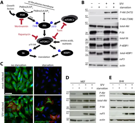
FIG 1Activation of the PI3K-Akt-mTOR pathway in SFV-infected cells, even upon starvation. (A) The PI3K-Akt-mTOR pathway. Only proteins and inhibitors used in this study are shown. Arrows indicate (indirect or direct) activation. Starvation denotes complete absence of growth factors, amino acids, and nutrients to shut off PI3K-Akt and mTOR activation. PDK, phospholipid-dependent kinase; S6K, S6 kinase. PtdIns(3,4,5)P3, phosphatidylinositol-3,4,5-triphosphate; PtdIns(4,5)P2, phosphatidylinositol-4,5-bisphosphate. For other abbreviations, see the text. (B) HOS cells were infected with SFV-wt (MOI of 10 for 1 h) or mock infected and then supplemented with growth medium (no starvation) or EBSS (starvation) prior to lysis and Western blot analysis for the indicated proteins (P, phosphorylated). Positions of molecular mass markers (in kDa) are indicated on the left. (C) Immunofluorescence analysis of HOS cells, infected for 1 h with SFV-wt (MOI of 0.2) or mock infected, incubated in growth medium (no starvation) or EBSS (starvation) for another 7 h, fixed, and stained for phosphorylated S6 (pseudocolored green) and SFV-nsP1 (red). Nuclei were stained with Hoechst 33258 (blue). Scale bar, 25m. (D and E) MEF and BHK cells were treated as described for panel B and assessed for activation of PI3K-Akt-mTOR by Western blotting with the indicated antibodies.
Thaa et al.
on November 7, 2019 by guest
http://jvi.asm.org/
[image:3.585.75.508.66.462.2](MOI) indicated in the figure legend. Phosphate-buffered saline (PBS) was used as a diluent in the experiments shown inFig. 2AandB. After infection, cells were washed with PBS and supplemented with growth medium or EBSS (for starvation experiments) and typically kept under cell culture conditions for another 7 h (if not indicated otherwise) and processed for Western blotting or immunofluorescence analysis as de-scribed below.
Cloning and transfection.Expression plasmids were cloned by stan-dard molecular biology techniques. The nsP3 coding sequences of SFV and CHIKV were amplified by PCR using pCMV-SFV4 (wt and muta-tions thereof) and pCMV-CHIKV-ICRES (CHIKV LR2006-OPY1 and mutations thereof), respectively, as templates and oligonucleotide prim-ers (Eurofins) containing restriction sites for cloning. For generation of Myr-Pal-nsP3-FLAG constructs, the sequences encoding the myristoyl-ation and palmitoylmyristoyl-ation signals of murine Lyn kinase (amino acids MG CIKSKRKDNLNDDE) and the C-terminal FLAG tag (amino acids DYK DDDDK) were added by PCR. The PCR products were subcloned into pcDNA3.1(⫺) using NheI and BsrGI (New England BioLabs). Plasmids were checked by sequencing (Eurofins) prior to transient transfection using Lipofectamine 2000 (Life Technologies) according to the manufac-turer’s instructions.
Cell lysis, SDS-PAGE, Western blotting.After treatment and washing with PBS, cells in six-well plates were lysed with 250l of lysis buffer (20 mM HEPES, pH 7.4, 110 mM potassium acetate, 2 mM magnesium chlo-ride, 0.1% [vol/vol] Tween 20, 1% [vol/vol] Triton X-100, 0.5% [wt/vol] sodium deoxycholate, and 500 mM sodium chloride [41], supplemented with complete protease inhibitor and PhosSTOP phosphatase inhibitor cocktails [Roche]) on ice. Lysates were cleared by centrifugation at 20,000
⫻gfor 10 min at 4°C, mixed with 4⫻reducing NuPAGE lithium dodecyl sulfate (LDS) sample buffer (Life Technologies), and heated (95°C for 5 min). Samples were electrophoresed using precast Novex NuPAGE Bis-Tris (4 to 12% polyacrylamide) gels (Life Technologies) and subsequently transferred onto Hybond P polyvinylidene difluoride (PVDF) mem-branes (GE Healthcare). Memmem-branes were blocked with either 3% BSA in PBS with 0.1% (vol/vol) Tween 20 (PBST) or 5% skim milk powder in Tris-buffered saline with 0.1% (vol/vol) Tween 20 (TBST), followed by incubation with primary antibodies as listed below (16 h at 4°C), washing with either PBST or TBST, incubation with a horseradish peroxidase-coupled secondary antibody (Sigma) (1 h at room temperature), another washing step, and detection by enhanced chemiluminescence (ECL, Pierce) using Hyperfilm chemiluminescence films (GE Healthcare) and a Curix 60 film developer (AGFA). The following antibodies were used according to the manufacturers’ instructions: rabbit anti-phospho-Akt (S473) (catalog number 4060; Cell Signaling), rabbit anti-phospho-Akt (T308) (2965; Cell Signaling), rabbit anti-total Akt (4691; Cell Signaling), rabbit phospho-4EBP1 (T37/46) (2855; Cell Signaling), rabbit anti-total 4EBP1 (9644; Cell Signaling), rabbit anti-phospho-S6 (5364; Cell Signaling), mouse anti-total S6 (2317; Cell Signaling), goat anti-actin (1616; Santa Cruz), and mouse anti-SFV-nsP2 (42) (a cocktail of 2H3, 2C7, and 3B5 at 1:1,000 each). Rabbit antisera directed against SFV-nsP3 (43), CHIKV-nsP3 (44), and CHIKV-nsP2 (45) were generated in the Merits lab and used at 1:5,000. All Western blot data are representative of at least three independent experiments. Densitometry was performed on scanned films using ImageJ; phospho-Akt (S473) signal intensities were normalized to the corresponding total-Akt signals.
Immunofluorescence and microscopy.For assessment of Akt phos-phorylation, cells were seeded on coverslips in 12-well plates and kept in serum-free medium for 32 h before and during treatment to reduce back-ground activation of Akt signaling. After treatment, cells were washed with PBS, fixed with 3.7% formaldehyde in PBS for 15 min at room tem-perature, and subsequently treated with 5% horse serum (Sigma) in PBS– 0.3% Triton X-100 for 90 min, followed by incubation with a primary antibody cocktail for 16 h at 4°C consisting of rabbit anti-phospho-Akt (S473; 1:100) (4060; Cell Signaling) and either mouse anti-SFV-nsP1 (1: 300) (28) or mouse anti-FLAG tag M2 (1:500) (F1804; Sigma) in PBS–
0.3% Triton X-100 –1% BSA. Then, coverslips were washed with PBS and treated with Alexa 488-conjugated donkey anti-rabbit (1:200) and Alexa 555-conjugated donkey anti-mouse-IgG (1:1,000) antibodies (Molecular Probes) plus Hoechst 33258 (1g/ml; Life Technologies) in PBS– 0.3% Triton X-100 –1% BSA (30 min, room temperature). Coverslips were then washed, mounted on glass slides using Vinol, and imaged by epifluores-cence microscopy with a Leica DMRB microscope using a 63⫻objective, a Hamamatsu charge-coupled-device (CCD) camera, and HiPic soft-ware. Images were processed using Adobe Photoshop.
For visualization of replication complexes after infection, cells (grown on coverslips in 12-well plates) were washed with PBS and fixed with 3.7% (vol/vol) formaldehyde in PBS for 10 min at room temperature, followed by permeabilization with methanol at⫺20°C for 10 min, blocking with 5% horse serum (Sigma) in PBS for 16 h at 4°C, and treatment with primary antibody cocktail in blocking solution for 5 h at room tempera-ture. Mouse anti-dsRNA (1:200; English and Scientific Consulting) and either rabbit anti-SFV-nsP3 (1:300) (43) or rabbit anti-CHIKV-nsP3 (1: 300) (44) were used. Subsequently, coverslips were washed with PBS and incubated for 30 min with Alexa 488-conjugated donkey anti-rabbit (1: 200), Alexa 555-conjugated donkey anti-mouse-IgG (1:1,000) (Molecular Probes), and the DNA dye DRAQ5 (1:1,000; Biostatus). Coverslips were then washed, mounted, and imaged by confocal laser scanning micros-copy using a Leica TCS SP5 X microscope equipped with a supercon-tinuum pulsed white laser. Microscopy samples were typically processed in a double-blinded manner to avoid bias.
RESULTS
Characterization of SFV-induced PI3K-Akt-mTOR activation. First, we determined whether and how the PI3K-Akt-mTOR pathway is affected by infection of cells with SFV and to what extent this is influenced by nutrient availability, generally a pre-requisite for this prosurvival pathway. We mock infected or in-fected human osteosarcoma (HOS) cells with SFV wild type (wt; strain SFV4) at a multiplicity of infection (MOI) of 10 for 1 h. Cells were then either kept in standard growth medium or starved for all nutrients and growth factors. Cell lysates were prepared at 8 h postinfection (p.i.) and subjected to Western blot analysis to de-tect phosphorylation of Akt at either activation residue (threo-nine-308 or serine-473) as well as phosphorylation of 4EBP1 and the ribosomal protein S6, downstream readouts for mTORC1 ac-tivity. Infection was assessed by probing for nsP3. These analyses
(Fig. 1B) revealed that the phosphorylation status of Akt was
strongly increased in infected cells compared to the Akt phosphor-ylation levels in noninfected cells (very low under standard con-ditions and undetectable under starvation concon-ditions). Akt phos-phorylation upon infection was seen both in nonstarved and starved cells, pointing toward an active, infection-induced stimu-lation. Concomitantly, the mTOR targets S6 and 4EBP1 were phosphorylated in infected cells under not only normal growth conditions (where mTOR is active) but also starvation conditions (where mTOR is normally inactive).
In a complementary approach, we assessed S6 phosphorylation by immunofluorescence at 8 h p.i. (Fig. 1C, costaining for phos-pho-S6 [green] and viral nsP1 [red]) and obtained comparable results: S6 was phosphorylated in nonstarved cells irrespective of their infection status; S6 was not phosphorylated in starved cells except those cells that had been infected (Fig. 1C, rightmost panel). Thus, mTOR was kept active by infection, not only under normal growth conditions but even in starved cells. The stimula-tion of the PI3K-Akt-mTOR pathway by infecstimula-tion was not limited to HOS cells as we obtained similar results also in other mamma-lian cell types, such as immortalized mouse embryonal fibroblasts
on November 7, 2019 by guest
http://jvi.asm.org/
(Fig. 1D, MEF) and baby hamster kidney cells (Fig. 1E, BHK). We therefore conclude that PI3K-Akt-mTOR activation is a common feature of SFV infection.
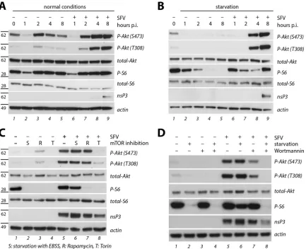
Next, we performed a time course experiment to assess the kinetics of PI3K-Akt-mTOR activation upon infection. HOS cells were infected with SFV at an MOI of 10 or mock infected for 1 h, employing phosphate-buffered saline (PBS) for dilution to pre-vent any activation of mTOR by nutrients in the infection me-dium. Cells were subsequently kept under standard or starvation conditions; cell lysates were prepared at different times after in-fection and assessed by Western blotting (Fig. 2AandB). Akt and S6 phosphorylation was reduced after 1 h of starvation (mock infection or infection in PBS (Fig. 2A), compare lanes 2 and 6 to lane 1) and reappeared after 1 h of incubation in nutrient-rich medium (Fig. 2A, lane 3). Levels of S6 phosphorylation subse-quently stayed high throughout the time frame of analysis (up to 8 h) in both noninfected and infected cells (lanes 3 to 5 and 7 to 9, respectively). Akt activation levels in noninfected cells reached starting levels (Fig. 2A, compare lanes 4 and 5 to lane 1), with a
transient boost shortly after resupply of nutrient-rich medium (lane 3). In contrast, Akt activation levels increased gradually in infected cells, exceeding the starting levels (Fig. 2A, compare lanes 8 and 9 to lane 1). When the same experiment was performed under conditions of starvation (Fig. 2B), significant Akt activation was seen 4 h after infection (lane 8) and increased further up to 8 h p.i. (lane 9), while staying undetectably low in noninfected, starved cells (lanes 2 to 5). S6 phosphorylation faded out in the course of starvation in noninfected cells (Fig. 2B, lanes 2 to 5) but was restored to full levels in infected cells in spite of starvation (compare lane 9 to lane 1). These data indicate that the PI3K-Akt-mTOR pathway was actively stimulated by infection, even in starved cells, starting as early as 4 h p.i.
In order to better understand the mode of virus-induced PI3K-Akt-mTOR activation, we employed specific inhibitors of the pathway. Cells were infected for 1 h with SFV or mock infected and were then kept in standard medium or under conditions that differentially block mTOR: starvation (general deactivation of the pathway by nutrient withdrawal) or treatment with rapamycin FIG 2Characterization of SFV-induced PI3K-Akt-mTOR activation. (A and B) Time course experiment in HOS cells infected for 1 h with SFV-wt (MOI of 10) in PBS or mock infected and then supplemented with growth medium (normal conditions) or EBSS (starvation). Cell lysates were prepared at the indicated times p.i. and probed by Western blotting. Positions of molecular mass markers (in kDa) are indicated on the left. (C) Effect of mTOR inhibition. HOS cells were infected with SFV-wt (MOI of 10 for 1 h) or mock infected, followed by incubation in growth medium (no mTOR inhibition), EBSS (starvation, S), growth medium with rapamycin (R; 400 nM), or growth medium with torin-1 (T; 10 nM) for another 7 h prior to lysis and Western blotting. (D) Effect of PI3K inhibition and starvation. HOS cells were infected as described for panel C, followed by incubation in growth medium or EBSS (starvation), without or with 400 nM wortmannin as indicated, for another 7 h, and then lysed and analyzed by Western blotting.
Thaa et al.
on November 7, 2019 by guest
http://jvi.asm.org/
[image:5.585.78.511.66.418.2](an inhibitor of mTORC1) or torin-1 (an inhibitor of mTORC1 as well as of another mTOR complex, mTORC2, the kinase for Akt activation residue S473). Cell lysates were prepared at 8 h p.i. and analyzed by Western blotting (Fig. 2C). In noninfected cells, S6 phosphorylation, the downstream readout for mTOR activity, was not detected upon each of these treatments (Fig. 2C, lanes 2, 3, and 4), which confirms their efficacy. In infected cells, S6 phos-phorylation was sustained in untreated and in starved cells (Fig. 2C, lanes 5 and 6) but not upon application of either rapamycin (lane 7) or torin (lane 8), indicating that the infection could not overcome mTOR inhibition by these drugs and thus acted up-stream of mTOR itself. When we assessed Akt phosphorylation, we observed that this modification was prevented by starvation (absence of growth factors) (Fig. 2C, lane 2) and torin-1 (inhibi-tion of mTORC2) (lane 4) but not by rapamycin (lane 3), while in infected cells (lanes 5 to 8), very strong Akt activation was seen under all conditions except for torin-1 treatment (lane 8). This indicates that the virus-induced stimulation of mTOR acts up-stream of or at the level of Akt activation.
We then applied the PI3K inhibitor wortmannin for further characterization of the virus-induced Akt activation. After mock infection or infection at an MOI of 10 for 1 h, cells were kept in growth or starvation medium, with or without wortmannin at a high concentration (400 nM). Cell lysates were prepared at 8 h p.i. and analyzed by Western blotting (Fig. 2D). The high level of Akt phosphorylation seen in infected cells (Fig. 2D, lane 5) was reduced— but not abolished— upon treatment with wortmannin (lane 7). S6 phosphorylation was not affected by wortmannin (owing to intact mTOR stimulation through other pathways). Phosphorylation of Akt and S6 was not detected when wortman-nin treatment was combined with starvation (Fig. 2D, lane 8).
In summary, the data inFig. 1and2show that infection of cells with SFV led to strong and permanent, partially wortmannin-insensitive, activation of Akt. This activation occurred even under starvation conditions and resulted in sustained activity of mTOR. Viral requirements for SFV-induced PI3K-Akt-mTOR acti-vation.Next, we investigated the viral requirements for PI3K-Akt-mTOR activation. To evaluate whether the structural
pro-teins play a role for this activation, we employed SFV--Gal, an SFV replicon in which the open reading frame for the structural proteins had been replaced by the gene encoding-galactosidase. Western blot analysis of cell lysates prepared at 8 h p.i. showed that Akt was phosphorylated upon infection with SFV--Gal virus replicon particles to a similar extent as observed with SFV-wt (Fig. 3A), indicating that expression of the structural proteins is dis-pensable for SFV-induced Akt activation. In addition, we em-ployed immunofluorescence analysis for phosphorylated Akt to assess the activation of Akt at a single-cell level and corroborated that Akt was phosphorylated only in cells that had been infected with SFV-wt or SFV--Gal but not in the noninfected neighbor-ing cells (Fig. 3B). In addition, we transfected cells with a plasmid encoding nsP1 to nsP3 (P123) to determine whether Akt activa-tion depends on the presence and acactiva-tion of the RNA-dependent RNA polymerase nsP4, which is lacking in P123. We employed the immunofluorescence assay and detected that Akt was consider-ably phosphorylated in the cells expressing P123 (Fig. 3B, right-most panel). Thus, virus-induced Akt activation in cells could be narrowed down to the presence of the replication complex pro-teins nsP1, -2, and -3 but did not require the presence and action of nsP4 or the structural proteins.
It has been proposed that nsP3 mediates the internalization of SFV replication complexes from the plasma membrane (28), a process that is linked to the PI3K-Akt pathway (16). We therefore asked whether mutations of nsP3 in the viral context would affect the virus-induced Akt activation. We hypothesized that the C-ter-minal hypervariable region of nsP3, a scaffold for interaction with cellular factors, could be relevant for PI3K-Akt activation and fo-cused on mutations in this region. We employed virus mutants characterized earlier, encoding deletion variants of nsP3 as de-picted inFig. 4A. In SFV-⌬50, 50 residues (319 to 368) comprising the hyperphosphorylated/acidic region of nsP3 are deleted (33); SFV-⌬P does not contain the proline-rich regions (residues 408 to 440) (34); SFV-⌬789 lacks the G3BP interaction domain (residues 449 to 472) (35). We infected BHK cells with SFV-wt or either of these viruses and assessed Akt activation at 8 h p.i. by Western blotting (Fig. 4B) and phospho-Akt immunofluorescence (Fig. 4C). FIG 3Viral requirements for SFV-induced PI3K-Akt-mTOR activation. (A) BHK cells were infected with SFV-wt or SFV--Gal (MOI of 10, 1 h) or mock infected, followed by incubation in growth medium for another 7 h, lysis, and Western blotting for the indicated proteins. (B) HOS cells were infected at an MOI of 0.2 with the indicated viruses for 1 h or mock infected and fixed at 8 h p.i. or were transfected with the nsP123-encoding plasmid and fixed 24 h later, followed by immunofluorescence for phospho-Akt (S473; green) and SFV-nsP1 (red). Nuclei were stained with Hoechst 33258 (blue). Scale bar, 25m.
on November 7, 2019 by guest
http://jvi.asm.org/
[image:6.585.41.545.67.244.2]While the levels of Akt phosphorylation were similar for SFV-wt, -⌬P, and -⌬789, infection with SFV-⌬50 led to considerably weaker phospho-Akt signals. All the mutant viruses were similarly attenuated with respect to the wild-type level (as judged from the
intensities of the nsP3 signals). The result indicates that the stretch of nsP3 deleted in SFV-⌬50, but none of the other nsP3 regions under study, substantially contributed to SFV-induced Akt acti-vation. The weak Akt activation by SFV-⌬50 was completely FIG 4Requirements in nsP3 for SFV-induced PI3K-Akt-mTOR activation. (A) Schematic depiction of SFV-nsP3 (482 amino acids) and sketch of the nsP3 deletions in the virus mutants employed below. (B) BHK cells were infected with the indicated variants of SFV (MOI of 10, 1 h) or mock infected, incubated in growth medium, lysed at 8 h p.i., and assessed by Western blotting for the indicated proteins. Positions of molecular mass markers (in kDa) are indicated on the left. Relative phospho-Akt signal intensities (as a percentage of the wt level) were as follows: SFV-⌬50, 59%⫾14%; SFV-⌬P, 107%⫾10%; SFV-⌬789, 99%⫾10% (n⫽3). (C) HOS cells were infected with the indicated SFV variants (MOI of 0.2, 1 h) or mock infected, followed by incubation in DMEM for 7 h, fixation, and staining for phospho-Akt (S473; green) and SFV-nsP1 (red). Nuclei were stained with Hoechst 33258 (blue). Scale bar, 25m. (D) BHK cells were infected with SFV-⌬50 at an MOI of 10 for 1 h; incubation was continued in growth medium (no starvation) or EBSS (starvation), without or with 400 nM wortmannin, for another 7 h prior to lysis and Western blotting for the indicated proteins. Compare the results with those shown inFig. 2D. (E) BHK cells were infected with SFV-wt or SFV-⌬50 at an MOI of 10 for 1 h and incubated for another 3 h (left panel) or another 7 h (right panel), followed by lysis and Western blotting for the indicated proteins.
Thaa et al.
on November 7, 2019 by guest
http://jvi.asm.org/
[image:7.585.137.450.64.576.2]blocked by application of wortmannin (Fig. 4D) and exhibited slower kinetics than in the case of SFV-wt. The Akt phosphoryla-tion in SFV-⌬50-infected cells was at background levels at 4 h p.i., a time where Akt was already strongly activated by infection with SFV-wt (Fig. 4E).
After observing that the hyperphosphorylated/acidic region of SFV-nsP3 is relevant for Akt activation, we next asked whether expression of nsP3 alone is enough to induce Akt phosphoryla-tion. To test this, we cloned expression vectors for nsP3 (wt and
⌬50) with a C-terminal FLAG tag (nsP3-FLAG), transfected HOS cells, and performed immunofluorescence analysis with anti-phospho-Akt (S473) and anti-FLAG tag antibodies. We could not detect Akt phosphorylation above background levels in cells ex-pressing SFV-nsP3-FLAG, either the wt or the⌬50 variant, indi-cating that nsP3 expression did not detectably activate Akt (Fig.
5AtoC).
There is a significant difference in the subcellular localizations between nsP3 expressed on its own (cytosolic) and nsP3 in asso-ciation with the replication complex (stably membrane-associ-ated through interaction with nsP1, which binds to membranes [28,46]). Since P123 activated Akt (Fig. 3B), we postulated that nsP3 needs to be membrane associated to mediate Akt phosphor-ylation, a process that requires plasma membrane recruitment of Akt (47). We thus engineered a membrane-attached version of nsP3 by fusing the N-terminal myristoylation and palmitoylation signal of Lyn kinase (Myr-Pal) to the N terminus of nsP3-FLAG in the context of the expression vector. This signal leads to covalent modification of the protein with fatty acids (myristic acid at gly-cine-2 and palmitic acid at cysteine-3) with the potential to target the protein to the plasma membrane (48). When we expressed Myr-Pal-nsP3-FLAG in HOS cells, the protein was typically pres-ent at intracellular vesicles; some cells exhibited a concpres-entration of the signal at the cell periphery (Fig. 5D). When we assessed Akt phosphorylation by immunofluorescence, cells expressing mem-brane-attached Myr-Pal-nsP3-FLAG displayed strong signals for phosphorylated Akt (Fig. 5D); expression of Myr-Pal-nsP3-FLAG with the⌬50 mutation, however, failed to induce phosphorylation of Akt (Fig. 5E). Taken together, the data presented inFig. 5show that nsP3, when attached to membranes (thus imitating the situ-ation in the replicsitu-ation complex), activated Akt, with a depen-dence on the hyperphosphorylated/acidic region.
Subcellular localization of SFV replication complexes.In the course of SFV infection, the viral replication complexes (spher-ules) are formed at the plasma membrane and subsequently inter-nalized in a process that depends on PI3K-Akt signaling (16). We hence hypothesized that the activation of the PI3K-Akt-mTOR pathway upon SFV infection is linked to replication complex dy-namics, possibly resulting in spherule internalization. To assess this, we infected BHK cells with either SFV-wt, SFV-⌬50, SFV-⌬P, or SFV-⌬789 at an MOI of 10, fixed cells at 8 h p.i., and performed immunofluorescence staining for double-stranded RNA (dsRNA) to detect replication complexes; costaining for nsP3 was per-formed to confirm nonstructural protein expression. Representa-tive confocal micrographs are shown inFig. 6. In cells infected with SFV-wt, dsRNA-positive foci were present throughout the cytosol without prominent staining of the cell surface (Fig. 6A). Application of wortmannin (for 7 h; added at 1 h p.i.) resulted in accumulation of replication complexes at the cell periphery (Fig. 6B), which is indicative of a block in spherule internalization and corroborates earlier results (16). Administration of either torin or
FIG 5Activation of Akt by ectopically expressed SFV-nsP3 variants. HOS cells were mock transfected or transfected with plasmids encoding FLAG-tagged nsP3-wt, nsP3-⌬50, Myr-Pal-nsP3-wt, or Myr-Pal-nsP3-⌬50, as indicated, fixed at 24 h posttransfection, and stained for phospho-Akt (S473; green) and the FLAG tag (red). Nuclei were stained with Hoechst 33258 (blue). Scale bar, 25m.
on November 7, 2019 by guest
http://jvi.asm.org/
[image:8.585.317.521.63.666.2]rapamycin did not affect replication complex internalization (Fig. 6CandD). When cells were infected with SFV-⌬50, which does not induce strong PI3K-Akt-mTOR activation (Fig. 4), the sub-cellular localization of replication complexes (Fig. 6E) was notice-ably different from the appearance in SFV-wt-infected cells (Fig. 6A) but strongly resembled the situation with SFV-wt-infected cells treated with wortmannin (Fig. 6B). Thus, replication com-plexes accumulated at the cell periphery in most cells infected with SFV-⌬50. Conversely, the localizations of replication complexes upon infection with SFV-⌬P (Fig. 6F) or SFV-⌬789 (Fig. 6G) were indistinguishable from SFV-wt localization, thus indicating un-perturbed spherule internalization. The nsP3 signals were either punctate, sometimes colocalizing with dsRNA and thus represent-ing replication complex-associated nsP3, or diffusely spread in the
cytosol. This is in accordance with the notion that nsP3 is engaged in various complexes (49).
In summary, the results presented inFig. 6provide a link be-tween strong PI3K-Akt-mTOR activation and replication com-plex internalization and show that both functions are disrupted by the⌬50 mutation in nsP3, where the hyperphosphorylated/acidic region of nsP3 is lacking.
Next, we endeavored to decipher whether a specific part of the hyperphosphorylated/acidic region is responsible for the effect on PI3K-Akt-mTOR signaling and replication complex dynamics. To this end, we employed recombinant viruses with partial dele-tions in this domain of nsP3 (residues 319 to 368). These deledele-tions comprise either the 24 N-terminal residues (⌬24; i.e., residues 319 to 342) or the 26 C-terminal residues (⌬26; residues 343 to 368) of FIG 6Subcellular localizations of SFV replication complexes. BHK cells were infected with the indicated SFV variants (a schematic depiction is given inFig. 4A) at an MOI of 10 for 1 h or mock infected, incubated in growth medium for 7 h, and fixed. Drug treatments (added at 1 h p.i. and present until fixation at 8 h p.i.) were as follows: 400 nM wortmannin (B), 10 nM torin (C), and 400 nM rapamycin (D). Immunofluorescence staining for dsRNA (red) and SFV-nsP3 (green) was performed; nuclei were stained with DRAQ5 (blue). Representative confocal micrographs are shown, with the inset areas shown magnified in the respective lower frames. Scale bar, 25m.
Thaa et al.
on November 7, 2019 by guest
http://jvi.asm.org/
[image:9.585.77.510.67.491.2]the region (Fig. 7A) (33). Also, to assess the influence of phos-phorylation, we made use of the mutant⌬26-4S-4A, which carries the⌬26 deletion and an additional replacement of four serines— the major remaining phosphorylation sites— by alanines, a set of mutations that eliminates detectable phosphorylation of nsP3 (36). We infected BHK cells with either of these recombinant vi-ruses and assessed the subcellular localizations of replication com-plexes by immunofluorescence staining for dsRNA and SFV-nsP3 at 8 h after infection (Fig. 7BtoD). For each of these viruses, we observed that dsRNA-positive foci were localized in the cytosol, very similar to the situation for SFV-wt (Fig. 6A); no prominent accumulation of replication complexes at the cell periphery (as in the case of SFV-⌬50) (Fig. 6E) was seen. In complementary exper-iments, BHK cells were infected with SFV-wt, SFV-⌬50, or one of the partial deletion mutants at an MOI of 10 to assess Akt phos-phorylation at 8 h p.i. by Western blotting (Fig. 7E). As before, we observed high levels of Akt phosphorylation in SFV-wt-infected cells but clearly reduced levels in cells infected with SFV-⌬50. Conversely, infection with SFV-⌬24, -⌬26, and also -⌬26-4S-4A induced Akt phosphorylation to levels comparable to infection with SFV-wt and higher than the level of infection with SFV-⌬50. Thus, the capacity to strongly activate Akt was not lost with any of the partial deletions within the hyperphosphorylated/acidic re-gion of nsP3.
Taking these results together, the partial deletions within the hyperphosphorylated/acidic region prevented neither strong PI3K-Akt-mTOR activation nor replication complex internaliza-tion. This implies that individual portions of this region are suffi-cient to support these functions and that phosphorylation is not essential.
PI3K-Akt-mTOR activation and replication complex
dy-namics in CHIKV infection. Next, we assessed whether the
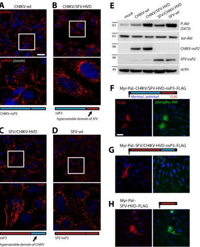
closely related CHIKV also activates the PI3K-Akt-mTOR path-way in a fashion similar to that of SFV. To this end, we infected BHK cells with CHIKV-wt at an MOI of 10 and prepared cell lysates at different times after infection (up to 16 h p.i., before the onset of extensive cell death) and determined Akt phosphoryla-tion by Western blotting (Fig. 8A). The extent of Akt phosphory-lation increased within 8 h after infection with CHIKV and was not further augmented by 16 h p.i. Hence, Akt was activated by CHIKV infection as well; strikingly, however, the extent of Akt activation was much weaker than the intensity seen 8 h after in-fection with SFV-wt, performed in parallel under the same condi-tions (Fig. 8A).
To assess whether CHIKV-induced Akt activation requires the same domain in nsP3 as that for activation by SFV, we made use of a CHIKV mutant, termed CHIKV-⌬5, which carries a deletion in nsP3 comparable to the SFV-nsP3-⌬50 deletion (38). The respec-tive stretches of nsP3 are equivalent in position but not sequence. When BHK cells were infected with the CHIKV-⌬5 mutant and assessed by Western blotting, we observed time-dependent phosphorylation of Akt, which reached levels comparable to the Akt activation level seen with CHIKV-wt infection (Fig. 8A). Fur-ther, we noted that the CHIKV-induced Akt stimulation in BHK cells was completely blocked in the presence of wortmannin in both CHIKV-wt and CHIKV-⌬5 infections (Fig. 8B). This is rem-iniscent of the situation with SFV-⌬50 (Fig. 4D) but unlike that with SFV-wt, where Akt stimulation was only partially wortman-nin sensitive (Fig. 2D). When we assessed whether CHIKV-wt is capable of activating the PI3K-Akt-mTOR pathway also under
FIG 7Akt activation and replication complex internalization of recombinant SFV with mutations in the hyperphosphorylated/acidic domain of nsP3. (A) Mu-tations of nsP3 in the indicated SFV variants. nsP3-⌬26-4S-4A is not phosphory-lated. (B to D) BHK cells were infected with SFV-⌬24, SFV-⌬26, or SFV-⌬ 26-4S-4A, as indicated, at an MOI of 10 for 1 h, incubated in growth medium for another 7 h, fixed at 8 h p.i., and stained for dsRNA (red) and SFV-nsP3 (green). Nuclei were stained with DRAQ5 (blue). Representative confocal micrographs are shown, with the inset areas shown magnified in the respective adjacent frames. Scale bar, 25m. (E) BHK cells were infected with the indicated viruses at an MOI of 10 for 1 h or mock infected, followed by incubation in growth medium for 7 h, lysis, and Western blotting as indicated. Positions of molecular mass markers (in kDa) are indicated on the left. Relative phospho-Akt signal intensities (as a per-centage of wt levels) were as follows: SFV-⌬50, 65%⫾3%; SFV-⌬24, 94%⫾16%; SFV-⌬26, 102%⫾17%; SFV-⌬26-4S-4A, 93%⫾18% (n⫽3).
on November 7, 2019 by guest
http://jvi.asm.org/
[image:10.585.322.520.64.591.2]starvation conditions, as seen above for SFV (Fig. 1), we found that neither Akt nor S6 was strongly phosphorylated in CHIKV-infected, starved cells, indicating that the CHIKV-induced PI3K-Akt-mTOR activation is sensitive to starvation (Fig. 8C). Taken together, these results imply that CHIKV infection led to only moderate activation of the PI3K-Akt-mTOR pathway, which was prevented by wortmannin or starvation and not dependent on the region of nsP3 that corresponds to the hyperphosphorylated/ acidic domain of SFV-nsP3.
Next, we assessed the subcellular localization of replication complexes in BHK cells 8 h after infection with CHIKV-wt or -⌬5 at an MOI of 10 by immunofluorescence staining for dsRNA and CHIKV-nsP3 and by confocal microscopy (Fig. 8D to F). We noted a difference in nsP3 localizations between CHIKV-wt and -⌬5, probably reflecting engagement of nsP3 in various complexes in cells (49). The dsRNA-positive replication complexes were markedly accumulated at the cell periphery in most cells for both CHIKV-wt and -⌬5 at 8 h p.i. (Fig. 8EandF) and also at later times (data not shown). This is a clear difference from the typical repli-cation complex localization upon infection with SFV-wt and re-sembles the situation for infection with SFV-⌬50 (compare with
Fig. 6). These results suggest that the hyperphosphorylated/acidic
region of SFV but not that of CHIKV mediates strong PI3K-Akt-mTOR activation and replication complex internalization.
PI3K-Akt-mTOR activation and replication complex dy-namics of chimeric SFV/CHIKV viruses.We next asked whether the hyperphosphorylated/acidic domains can be functionally ex-changed between SFV and CHIKV, i.e., whether the SFV domain induces the SFV phenotype (strong PI3K-Akt-mTOR activation and replication complex internalization) also in the context of CHIKV. To this end, we generated recombinant SFV and CHIKV genomes in which the residues deleted in SFV-⌬50 were replaced by the sequence deleted in CHIKV-⌬5 and vice versa, giving rise to CHIKV/SFV50 (CHIKV with the SFV domain) and SFV/CHIKV5 (SFV with the CHIKV domain), respectively. The efficiency of infectious virus rescue was drastically reduced compared to the levels of the respective wild-type viruses. In particular, rescue ef-ficiency of CHIKV/SFV50 was reduced by at least 10,000-fold; correspondingly, virus stocks had very low initial titers and ac-quired secondary (adaptive) mutations upon propagation in BHK cells. Interestingly, these mutations did not reside in the swapped region or in the rest of the C-terminal region of nsP3; instead, a compensatory methionine-to-isoleucine mutation was detected in the zinc-binding domain of nsP3 (position 1552 in P1234, where the corresponding SFV-wt residue is leucine). Further-more, another compensatory mutation occurred even outside nsP3 (N1318I in P1234; C-terminal region of nsP2). The ineffec-tive rescue and low infectivity of the CHIKV/SFV50 virus most likely resulted from severely compromised replicase activity in the recombinant virus: we could not detect any replicase activity above the background for the CHIKV/SFV50 replicase in atrans -replication system. The activity of the SFV/CHIKV5 replicase was also severely diminished (reduced to approximately 1% compared to the SFV-wt level); consequently, the rescue of infectious SFV/ CHIKV5 virus was reduced by at least 1,000-fold. Hence, while deletion of the hyperphosphorylated/acidic regions in SFV- and CHIKV-nsP3 was well tolerated (SFV-⌬50 and CHIKV-⌬5), swapping of the domains was not. This precluded biochemical experiments.
We employed the recombinant CHIKV/SFV50 and SFV/
CHIKV5 viruses to determine the subcellular localizations of rep-lication complexes in infected BHK cells at 8 h p.i. Staining for dsRNA revealed that replication complexes were predominantly localized at the cell periphery for SFV/CHIKV5 (Fig. 9A) as well as for CHIKV/SFV50 (Fig. 9B). Thus, the hyperphosphorylated/ acidic domain of CHIKV could not functionally replace the do-main in SFV, and the SFV dodo-main did not mediate replication complex internalization when it was introduced into CHIKV.
We also addressed the activation of the PI3K-Akt-mTOR path-way. To this end, we generated expression constructs for mem-brane-associated nsP3 (Myr-Pal-nsP3-FLAG) for use in phospho-Akt immunofluorescence experiments (Fig. 5), with the nsP3 sequences of CHIKV-wt as well as the chimeric variants. Note that adaptive mutations do not occur in this virus replication-inde-pendent approach. These constructs were expressed in HOS cells and assessed by immunofluorescence microscopy for phospho-Akt. In contrast to the result with Myr-Pal-SFV-nsP3-FLAG (Fig. 5Dand9C), we did not detect significant phosphorylation of Akt above background levels in cells expressing Myr-Pal-nsP3-FLAG in the CHIKV, CHIKV/SFV50, or SFV/CHIKV5 variant (Fig. 9D
toF). Taken together, the results shown inFig. 9indicate that the 50 residues of the hyperphosphorylated/acidic domain of SFV-nsP3 (deleted in SFV-⌬50) are necessary but not sufficient for replication complex internalization and Akt activation.
Our approach using domain swapping likely disturbed correct folding of nsP3 and accurate assembly of replication complexes, probably due to sequence incompatibility upon insertion. We supposed that the risk of such adverse effects would be reduced if the entire C-terminal hypervariable domain of nsP3 were swapped between CHIKV and SFV. Hence, we generated a recom-binant CHIKV genome where the sequence encoding the hyper-variable domain of nsP3 (starting at residue 323, where the high sequence conservation ends) was replaced by the corresponding sequence of SFV, yielding CHIKV/SFV-HVD, and a recombinant SFV genome where the C-terminal tail of nsP3 was analogously replaced by that of CHIKV (SFV/CHIKV-HVD). Using this ap-proach, infectious chimeric viruses could be rescued efficiently. Subsequently, BHK cells were infected with these viruses to assess the subcellular localizations of replication complexes by immuno-fluorescence staining for dsRNA at 8 h p.i. Confocal microscopy showed that the replication complexes of CHIKV/SFV-HVD (Fig. 10B) were efficiently internalized from the cell periphery, in con-trast to the situation with CHIKV-wt (Fig. 10A) and resembling the phenotype of SFV-wt (Fig. 10D). Conversely, replication com-plexes of SFV/CHIKV-HVD (Fig. 10C) were predominantly local-ized at the cell periphery, similarly to those of CHIKV-wt and unlike the situation with SFV-wt. Hence, the C-terminal domain of nsP3 determined the subcellular localization of replication complexes: the domain of SFV-nsP3 induced efficient replica-tion complex internalizareplica-tion in the context of both SFV and CHIKV, while the corresponding domain of CHIKV largely failed to promote replication complex internalization in CHIKV as well as SFV.
We then analyzed activation of the PI3K-Akt-mTOR pathway and asked whether the presence of the hypervariable domain of SFV-nsP3 confers the SFV phenotype (strongly activated Akt sig-naling) to the recombinant CHIKV/SFV-HVD and whether this phenotype is lost in SFV/CHIKV-HVD. BHK cells were mock infected or infected with either of the wild-type or chimeric vi-ruses at an MOI of 10 and lysed at 8 h p.i., followed by SDS-PAGE
Thaa et al.
on November 7, 2019 by guest
http://jvi.asm.org/
and Western blotting (Fig. 10E). Efficient infection was verified with antisera against the nsP2 proteins of CHIKV and SFV. Akt phosphorylation was significantly stronger in cells infected with CHIKV/SFV-HVD than in those infected with CHIKV-wt,
reach-ing levels similar to those infected with SFV-wt. Conversely, the Akt phosphorylation levels upon infection with SFV/CHIKV-HVD were clearly lower than those for infection with SFV-wt. This is evidence that the hypervariable domain of SFV, but not FIG 8PI3K-Akt-mTOR activation and replication complex dynamics upon infection with CHIKV. (A) BHK cells were mock infected or infected with the indicated viruses at an MOI of 10 for 1 h, followed by incubation in growth medium, lysis at 8 h (SFV) or at 4, 8, or 16 h (CHIKV), and analysis by Western blotting for the indicated proteins. CHIKV-⌬5 has a deletion of the hyperphosphorylated/acidic domain in nsP3. Positions of molecular mass markers (in kDa) are indicated on the left. (B) Sensitivity to PI3K inhibition. BHK cells were infected as described for panel A, followed by incubation in growth medium, without or with 400 nM wortmannin, for another 15 h, and then lysed and analyzed by Western blotting. (C) Effect of starvation. BHK cells were infected as described for panel A, followed by incubation in growth medium or EBSS (starvation) for another 7 h prior to lysis and Western blotting. (D to F) Immunofluorescence analysis of replication complexes. BHK cells were mock infected or infected with CHIKV-wt or CHIKV-⌬5, as indicated, at an MOI of 10 for 1 h, followed by incubation in growth medium for 7 h, fixation, and immunofluorescence staining for dsRNA (red) and CHIKV-nsP3 (green). Nuclei were stained with DRAQ5 (blue). Representative confocal micrographs are displayed, with the inset areas shown magnified in the respective adjacent frames. Scale bar, 25m.
on November 7, 2019 by guest
http://jvi.asm.org/
[image:12.585.95.494.68.576.2]that of CHIKV, boosted PI3K-Akt-mTOR signaling in the context of both SFV and CHIKV infection.
In a complementary approach, we generated expression con-structs for membrane-associated nsP3 (Myr-Pal-nsP3-FLAG) and performed phospho-Akt immunofluorescence analysis in transfected HOS cells as described above (Fig. 5and9). In contrast to results with the CHIKV version of this construct (Fig. 9F), expression of membrane-attached CHIKV-nsP3 with the hy-pervariable domain of SFV induced clearly detectable levels of Akt phosphorylation (Fig. 10F), similar to the analogous SFV-nsP3 construct (compare withFig. 5Dand9C). Conversely, no Akt activation above background levels was visible for membrane-attached chimeric SFV/CHIKV-HVD-nsP3 (Fig. 10G). Thus, the hypervariable C-terminal tail of SFV-nsP3 had the capacity to confer strong Akt activation in the context of the membrane-at-tached nsP3 of both SFV and CHIKV. Finally, we assessed whether the hypervariable domain of SFV-nsP3 (residues 319 to 482) is sufficient to induce Akt phosphorylation when equipped with an N-terminal myristoylation and palmitoylation signal and a C-ter-minal FLAG tag (Myr-Pal-SFV-HVD-FLAG). When we expressed
this construct in HOS cells and performed phospho-Akt immu-nofluorescence analysis, we observed prominent Akt activation in most transfected cells (Fig. 10H). This indicates that the essential elements of the PI3K-Akt-mTOR activation domain reside in the C-terminal tail of SFV-nsP3.
DISCUSSION
In this work, we report that infection of a variety of cells with SFV-wt led to a very strong and persistent phosphorylation and thus activation of the pivotal prosurvival kinase Akt, leading to sustained activation of the downstream metabolic regulator mTOR (Fig. 1and2). This was very likely due to activation of the Akt pathway rather than to inhibition of a negative regulator or mere stabilization of activated components since SFV infection could overcome the inactivation of Akt and mTOR by starvation (as shown in the time course inFig. 2B).
Expression of membrane-bound nsP3 in the absence of other viral proteins was sufficient to activate Akt, as demonstrated by using nsP3 with an N-terminal membrane anchor (Myr-Pal-nsP3-FLAG) (Fig. 5), indicating that (plasma) membrane local-FIG 9The hyperphosphorylated/acidic domain of SFV-nsP3 is necessary but not sufficient for replication complex internalization and Akt activation. (A and B) BHK cells were infected with recombinant SFV in which the hyperphosphorylated/acidic domain of nsP3 was replaced with its counterpart from CHIKV-nsP3 (SFV/CHIKV5) or with CHIKV containing the reciprocal swap (CHIKV/SFV50) at an MOI of 0.1 for 8 h, fixed, and stained for dsRNA (red). Nuclei were stained with DRAQ5 (blue). Representative confocal micrographs are shown, with the inset areas shown magnified in the respective adjacent frames. Scale bar, 25m. (C to F) HOS cells were transfected with the indicated membrane-anchored, FLAG-tagged nsP3 constructs for 24 h, fixed, and stained for phospho-Akt (S473; green) and FLAG (red). Nuclei were stained with Hoechst 33258 (blue). Representative micrographs are shown. Scale bar, 25m.
Thaa et al.
on November 7, 2019 by guest
http://jvi.asm.org/
[image:13.585.94.491.65.422.2]ization of the viral protein is necessary for Akt activation. During infection, stable membrane association of nsP3 is achieved by in-teraction with the peripheral membrane protein nsP1 within the replication complex (46,50,51). There are other, nonmembra-nous nsP3-containing structures in infected cells (49), which are, however, unlikely to be relevant for Akt activation since mem-brane anchorage of nsP3 was required.
Even though there have been several comprehensive investiga-tions into molecular interacinvestiga-tions of Old World alphavirus nsP3 (41,
49,52), none of these reported an interaction partner of nsP3 known to have a direct function in PI3K-Akt-mTOR signaling. Presumably, the interaction of nsP3 with the Akt signaling module is transient or dynamic. Since proximity to membranes appears to be required (Fig. 5), an intriguing possibility is that the protein mimics a cellular acti-FIG 10The hypervariable domain of SFV-nsP3 induces replication complex internalization and Akt activation in CHIKV. (A to D) BHK cells were infected with CHIKV-wt, CHIKV containing the hypervariable domain of SFV-nsP3 (CHIKV/SFV-HVD), SFV with the hypervariable domain of CHIKV-nsP3 (SFV/CHIKV-HVD), or SFV-wt, as indicated, at an MOI of 10 for 8 h, fixed, and stained for dsRNA (red). Nuclei were stained with DRAQ5 (blue). Representative confocal micrographs are shown, with the inset areas shown magnified in the respective lower frames. Scale bar, 25m. (E) BHK cells were mock infected or infected with the indicated virus at an MOI of 10, lysed at 8 h p.i., and subjected to Western blotting with the indicated antibodies. (F to H) HOS cells were transfected with the indicated membrane-anchored, FLAG-tagged nsP3 constructs for 24 h, fixed, and stained for phospho-Akt (S473; green) and FLAG (red). Nuclei were stained with Hoechst 33258 (blue). Representative micrographs are shown. Scale bar, 25m.
on November 7, 2019 by guest
http://jvi.asm.org/
[image:14.585.96.491.64.552.2]vation feature for the Akt module. Such molecular mimicry might also explain why the SFV-induced Akt activation was not completely blocked by the PI3K inhibitor wortmannin.
We identified that the hyperphosphorylated/acidic region of nsP3, deleted in the virus mutant SFV-⌬50, is necessary for strong and persistent PI3K-Akt-mTOR activation (Fig. 4). Interestingly, PI3K-Akt-mTOR activation did not require the proline-rich re-gion of nsP3 (deleted in SFV-⌬P), a motif shown to mediate bind-ing to the SH3 domain of amphiphysin (34). This is markedly different from the situation with other viruses such as influenza A virus (IAV), where the protein NS1 activates Akt through the in-teraction of a proline-rich sequence with the SH3 domain of p85, the regulatory subunit of PI3K (53). Thus, the proline-rich regions of different viral proteins differ in their specificities for cellular proteins containing SH3 domains; specificity is likely conferred by additional adjacent binding motifs. In the case of IAV-NS1, a (pre-sumably phosphorylated) tyrosine interacts with the SH2 domain of p85 (54). The nonstructural proteins of SFV, however, have never been shown to contain phosphorylated tyrosines (55).
While our results show that the hyperphosphorylated/acidic region of SFV-nsP3 is the key part for the strong stimulation of the PI3K-Akt pathway (Fig. 4and5) and for efficient replication com-plex internalization (Fig. 6), it remains unclear by what molecular mechanism this is achieved. A distinctive feature of the domain is that it comprises a high density of negative charges: 8 aspartic acid residues as well as 6 threonine and 12 serine residues (in SFV4), most of which are phosphorylated (at least in the case of SFV and also SINV) by largely undefined cellular kinases, presumably in a nonregulated manner (33,55,56). Tyrosine phosphorylation of nsP3 has never been detected; thus, the region is unlikely to mimic activated growth factor receptors which employ phospho-ty-rosine for binding to PI3K. Phosphorylation of nsP3 was not crit-ical for Akt activation and replication complex internalization since these phenotypes were also observed for the virus mutant SFV-⌬26-4S-4A, where no phosphorylation is detectable (36). Strong and persistent Akt activation upon SFV infection might require the presence of a certain number or density of negative charges in the structural context of nsP3 (as is the case in nsP3 of SFV-wt, -⌬24, -⌬26, and also -⌬26-4S-4A but not -⌬50).
In cells infected with SFV-⌬50, the PI3K-Akt-mTOR pathway was activated to a moderate level (clearly above the background in Western blotting but not above the detection threshold in immu-nofluorescence assays). This stimulation was completely blocked by wortmannin and was thus entirely dependent on PI3K activity. Taken together with the finding that SFV-wt induced much stron-ger and partially wortmannin-insensitive Akt activation, we pro-pose that there are two types of alphavirus-induced PI3K-Akt-mTOR activation (Fig. 11): basal, completely wortmannin-sensitive (PI3K-dependent) activation upon infection (Fig. 11A), which is also seen with SFV-⌬50 as well as CHIKV (Fig. 8), and additional strong Akt activation (Fig. 11B), which is observed for SFV-wt but not SFV-⌬50 or CHIKV. This activation is at least partially resistant to starvation or wortmannin treatment (but not both) (Fig. 2D) and leads to potent and sustained activation of Akt-mTOR signaling. This strong PI3K-Akt-mTOR activation phenotype can be induced in CHIKV by swapping the hypervari-able C-terminal domain of nsP3 with that of SFV (Fig. 10).
Taken together with the data on the subcellular localization of replication complexes, it becomes evident that boosted (Fig. 11B) but not basal (Fig. 11A) Akt activation is linked to internalization
of replication complexes from the cell periphery. All viruses that were capable of very strong Akt activation exhibited replication complex internalization; SFV-⌬50 and CHIKV, which induced only moderate Akt activation, did not. Thus, the hyperphospho-rylated/acidic region in SFV-nsP3 is required both for the boost in PI3K-Akt-mTOR activation and the internalization of replica-tion complexes. The corresponding domain of CHIKV-nsP3 did not mediate these phenotypes, either in CHIKV (Fig. 8) or when it was introduced into SFV (Fig. 9and10). Conversely, the C-terminal domain of SFV-nsP3 led to strong PI3K-Akt-mTOR activation and efficient replication complex internalization in the context of both SFV and CHIKV infections (Fig. 10).
The PI3K-Akt-mTOR activation domain is not congruent with the 50 residues deleted in SFV-⌬50. This is evidenced by the find-ing that partial deletions within this region did not prevent strong PI3K-Akt activation and replication complex internalization (Fig. 7). Also, the 50 residues did not induce the SFV phenotype when transferred into CHIKV-nsP3 (Fig. 9), while the complete C-ter-minal hypervariable region did (Fig. 10). This implies that PI3K-Akt-mTOR activation and replication complex internalization do not critically depend on any specific amino acids within the 50 residues; several regions inside and outside this region may act cooperatively and form an (potentially discontinuous) activation feature. Elements downstream of the 50 residues are likely part of the PI3K-Akt activation domain, which is disrupted by the⌬50 deletion. The conserved N-terminal domains of nsP3 (macro domain and zinc-binding region), however, do not signifi-cantly contribute to the PI3K-Akt activation structure since the hypervariable C-terminal region of SFV-nsP3 alone induced detectable Akt phosphorylation when equipped with a mem-brane anchor (Fig. 10H).
The nonstructural proteins of alphaviruses are initially tar-geted to the plasma membrane, where the replication complexes (spherules) are formed (16,57). It is hence likely that SFV-nsP3 mediates activation of the PI3K-Akt signaling module at this stage, at the plasma membrane, the general cellular site of PI3K action and Akt activation. Replication complex internalization would then follow, likely involving additional cellular features such as the cytoskeleton (16). The interdependence of infection-induced PI3K-Akt activation and replication complex dynamics is evident from the finding that SFV replication complexes are stalled at the cell periphery by PI3K inhibition (Fig. 6B) (16). The signal for replication complex internalization is, however, not downstream of Akt since torin blocked SFV-induced Akt phosphorylation but not replication complex internalization (Fig. 6C). It remains un-clear for what purpose the replication complexes of SFV are effi-ciently internalized, but it is conceivable that this subcellular localization ensures spatial proximity of the viral replication products to the cellular translation machinery.
The robust stimulation of Akt and mTOR may have other functional consequences for the cell biology of SFV infection. Thus, the strong perturbation of cellular signaling may pose a potential caveat for the use of SFV-based vectors for studies of molecular pathogenesis, vaccination, or gene therapy. The poten-tial to activate this survival pathway might have evolved to allow for establishment of virus persistence in the mosquito host, as proposed for SINV (26); such a function is not manifested in mammalian cells, where infection is lytic. There, activation of the PI3K-Akt-mTOR pathway may help the virus to ensure efficient virus replication under suboptimal growth conditions such as
Thaa et al.
on November 7, 2019 by guest
http://jvi.asm.org/
starvation. Intriguingly, the presence of the hyperphosphorylated/ acidic domain in nsP3 is one of the features that have been linked to pathogenicity (especially neuropathology) of SFV infection in mice (33, 58), possibly connected to the strong and persistent PI3K-Akt-mTOR activation. Yet the domain and its functional roles are unlikely to be the decisive reasons for neurovirulence; virulence determinants have been mapped to several features of the nonstructural and also the structural proteins of SFV (59–61). The question remains why SFV but not CHIKV robustly activates PI3K-Akt-mTOR signaling and efficiently internal-izes replication complexes. It is surprising that SFV and CHIKV show these clear differences because these two Old World alphaviruses are otherwise very closely related and are thus expected to be very similar in many aspects of the infected cell’s biology. Indeed, they do not differ regarding their
repli-cation processes and other cell-biological features such as the nsP2-dependent degradation of RNA-polymerase II (62) or the nsP3-mediated sequestration of the stress granule component G3BP (12). The differential activation of the PI3K-Akt-mTOR pathway by SFV and CHIKV may have a cellular connection to disease and pathology, which are distinctly different for these two related viruses.
ACKNOWLEDGMENTS
We thank Karl Ljungberg, Peter Liljeström, and Dan Grandér (Karolinska Institutet) for provision of reagents and Mohammedyaseen Syedbasha (University of Helsinki) for technical assistance.
Funding was provided by the Swedish Cancer Foundation (CAN 2012/789 to G.M.M.), the Swedish Research Council (621-2014-4718 to G.M.M.), the German Research Foundation (TH1896/1-1 to B.T.), the
FIG 11Model of SFV- and CHIKV-induced PI3K-Akt-mTOR activation and replication complex dynamics. (A) CHIKV and SFV-⌬50 induce basal, wort-mannin-sensitive Akt activation in which replication complexes remain at the cell periphery, as shown in the schematic at the top right of the figure. (B) SFV carrying the hyperphosphorylated/acidic domain in nsP3 boosts PI3K-Akt-mTOR activation, and replication complex internalization is promoted, as shown in the schematic in the bottom right section of the figure. Interactions of the hypervariable domain of SFV-nsP3 are depicted by the schematic in the middle, boxed panel on the right. Representative confocal micrographs are shown of replication complexes (staining for dsRNA) in cells infected with CHIKV-wt (A) and SFV-wt (B) as described in the legends ofFig. 8Eand6A, respectively.
on November 7, 2019 by guest
http://jvi.asm.org/
[image:16.585.83.505.65.483.2]Estonian Ministry of Education and Research (IUT20-27 to A.M.), and the Academy of Finland (grant 265997 to T.A.).
We declare that we have no competing interests.
REFERENCES
1.Atkins GJ, Sheahan BJ, Liljeström P.1999. The molecular pathogenesis of Semliki Forest virus: a model virus made useful? J Gen Virol80:2287– 2297.http://dx.doi.org/10.1099/0022-1317-80-9-2287.
2.Karlsson GB, Liljeström P.2004. Delivery and expression of heterologous genes in mammalian cells using self-replicating alphavirus vectors. Meth-ods Mol Biol246:543–557.
3.Ljungberg K, Liljeström P.2015. Self-replicating alphavirus RNA vaccines. Expert Rev Vaccines14:177–194.http://dx.doi.org/10.1586/14760584.2015 .965690.
4.Quetglas JI, Ruiz-Guillen M, Aranda A, Casales E, Bezunartea J, Smer-dou C.2010. Alphavirus vectors for cancer therapy. Virus Res153:179 – 196.http://dx.doi.org/10.1016/j.virusres.2010.07.027.
5.Karabatsos N.1975. Antigenic relationships of group A arboviruses by plaque reduction neutralization testing. Am J Trop Med Hyg24:527–532. 6.Jose J, Snyder JE, Kuhn RJ.2009. A structural and functional perspective of alphavirus replication and assembly. Future Microbiol4:837– 856.http: //dx.doi.org/10.2217/fmb.09.59.
7.Rupp JC, Sokoloski KJ, Gebhart NN, Hardy RW.24 July 2015. Alpha-virus RNA synthesis and nonstructural protein functions. J Gen Virol http://dx.doi.org/10.1099/jgv.0.000249.
8.Malet H, Coutard B, Jamal S, Dutartre H, Papageorgiou N, Neuvonen M, Ahola T, Forrester N, Gould EA, Lafitte D, Ferron F, Lescar J, Gorbalenya AE, de Lamballerie X, Canard B.2009. The crystal structures of chikungunya and Venezuelan equine encephalitis virus nsP3 macro domains define a conserved adenosine binding pocket. J Virol83:6534 – 6545.http://dx.doi.org/10.1128/JVI.00189-09.
9.Neuvonen M, Ahola T.2009. Differential activities of cellular and viral macro domain proteins in binding of ADP-ribose metabolites. J Mol Biol
385:212–225.http://dx.doi.org/10.1016/j.jmb.2008.10.045.
10. Shin G, Yost SA, Miller MT, Elrod EJ, Grakoui A, Marcotrigiano J.
2012. Structural and functional insights into alphavirus polyprotein pro-cessing and pathogenesis. Proc Natl Acad Sci U S A109:16534 –16539. http://dx.doi.org/10.1073/pnas.1210418109.
11. McInerney GM.23 June 2015. FGDF motif regulation of stress granule formation. DNA Cell Biol 34:557–560. http://dx.doi.org/10.1089/dna .2015.2957.
12. Panas MD, Ahola T, McInerney GM.2014. The C-terminal repeat do-mains of nsP3 from the Old World alphaviruses bind directly to G3BP. J Virol88:5888 –5893.http://dx.doi.org/10.1128/JVI.00439-14.
13. Panas MD, Varjak M, Lulla A, Eng KE, Merits A, Karlsson Hedestam GB, McInerney GM.2012. Sequestration of G3BP coupled with efficient translation inhibits stress granules in Semliki Forest virus infection. Mol Biol Cell23:4701– 4712.http://dx.doi.org/10.1091/mbc.E12-08-0619. 14. Panas MD, Schulte T, Thaa B, Sandalova T, Kedersha N, Achour A,
McInerney GM.2015. Viral and cellular proteins containing FGDF motifs bind G3BP to block stress granule