Effects of acute hypoxia exposure with
different durations on activation of Nrf2-ARE
pathway in mouse skeletal muscle
Weixiu Ji1,2, Linjia Wang1, Shiyi He1, Lu Yan1, Tieying Li1, Jianxiong Wang3, Ah-Ng Tony Kong4, Siwang Yu5, Ying ZhangID1*
1 School of Sport Science, Beijing Sport University, Beijing, China, 2 Tianjin University of Sport, Tianjin, China, 3 Faculty of Health, Engineering, and Sciences, University of Southern Queensland, Toowoomba, Queensland, Australia, 4 Ernest Mario School of Pharmacy, Rutgers, the State University of New Jersey, Camden, New Jersey, United States of America, 5 Department of Chemical Biology, School of
Pharmaceutical Sciences, Peking University, Beijing, China
*zhyi9256@126.com
Abstract
Background
Hypoxia training enhances the endurance capacity of athletes. This response may in part be attributed to the hypoxia-induced increase in antioxidant capacity in skeletal muscles. Nuclear factor erythroid 2-related factor 2 (Nrf2), a key transcription factor which regulates the expression of genes via binding to the antioxidant-response element (ARE) of these genes, plays a crucial role in stimulating the body’s defense system and potentially responds to hypoxia. Meanwhile, hypoxia-inducible factor-1α(HIF-1α) is an important player in pro-tecting cells from hypoxic stress. The purpose of this study was to investigate the effects of acute hypoxia exposure with different durations on the activation of Nrf2-ARE pathway and a possible regulatory role of HIF-1αin these responses.
Methods
C57BL/6J mice were allocated into the non-hypoxia 0-hour, 6-hour, 24-hour, and 48-hour hypoxic exposure (11.2% oxygen) groups. The quadriceps femoris was collected immedi-ately after hypoxia. Further, to investigate the possible role of HIF-1α, C2C12 myoblasts with HIF-1αknockdown by small interfering RNA (siRNA) and the inducible HIF-1α trans-genic mice were employed.
Results
The results showed that 48-hour hypoxia exposure up-regulated protein expression of Nrf2, Nrf2/ARE binding activity and the transcription of antioxidative genes containing ARE (Sod1 and others) in mouse skeletal muscle. Moreover, HIF-1αsiRNA group of C2C12 myoblasts showed a remarkable inhibition of Nrf2 protein expression and nuclear accumulation in hyp-oxia exposure for 72 hours compared with that in siRNA-Control group of the cells. In addi-tion, HIF-1αtransgenic mice gave higher Nrf2 protein expression, Nrf2/ARE binding activity
a1111111111 a1111111111 a1111111111 a1111111111 a1111111111 OPEN ACCESS
Citation: Ji W, Wang L, He S, Yan L, Li T, Wang J,
et al. (2018) Effects of acute hypoxia exposure with different durations on activation of Nrf2-ARE pathway in mouse skeletal muscle. PLoS ONE 13
(12): e0208474.https://doi.org/10.1371/journal.
pone.0208474
Editor: Ferenc Gallyas, Jr., University of PECS
Medical School, HUNGARY
Received: September 24, 2018
Accepted: November 16, 2018
Published: December 4, 2018
Copyright:©2018 Ji et al. This is an open access
article distributed under the terms of theCreative
Commons Attribution License, which permits unrestricted use, distribution, and reproduction in any medium, provided the original author and source are credited.
Data Availability Statement: Data file was
attached as a Supporting Information file.
Funding: This study was supported by the National
Natural Science Foundation of China (31471134) and the project of Beijing Sport University (2018GJ007).
Competing interests: The authors have declared
and expressions of Nrf2-mediated antioxidative genes in their skeletal muscle, compared with those in the wild-type mice.
Conclusions
The findings suggested that the acute hypoxia exposure could trigger the activation of Nrf2-ARE pathway, with longer duration associated with higher responses, and HIF-1α expres-sion might be involved in promoting the Nrf2-mediated antioxidant responses in skeletal muscle.
Introduction
It has been demonstrated that exercise training under stimulated hypoxia conditions or at high altitude can increase endurance capacity in skeletal muscles of athletes [1,2]. Several stud-ies have shown that endurance exercise training under hypoxia conditions significantly reduced the basal oxidants production, increased the expression of antioxidant proteins, and upregulated glutathione (GSH) system in skeletal muscle of humans or rats, compared to the outcomes from exercise training in normoxia conditions [3,4]. These results imply that the enhanced endurance capability may, at least in part, attribute to the hypoxia-induced increase in antioxidant capacity in skeletal muscles. Considering the potential effects of hypoxia on pro-moting anti-oxidation of skeletal muscle and improving athlete performance, it is important to investigate the molecular mechanism by which hypoxia affects antioxidant reaction in skeletal muscle.
Nuclear factor erythroid 2-related factor 2 (Nrf2) is one of the key transcription factors that stimulates body’s defense system and possibly responds to hypoxia [5,6]. The body defense sys-tem equips with the capacity to increase the expression levels of cytoprotective enzyme genes through Nrf2, which can bind to antioxidant response elements (ARE) located in the regula-tory regions of many defense enzyme genes [5]. Downstream analysis has shown that Nrf2 tar-get genes encode enzymes involved in a wide range of cellular defense processes, therefore, Nrf2 may enhance the overall capacity of cells to handle harmful substances and situations [7–
9]. In addition, Nrf2-ARE signaling pathway may respond to hypoxic stimuli. It has been reported that hypoxic preconditioning had a neuroprotective effect on rat traumatic brain injury by increased expressions of Nrf2 and heme oxygenase1 (Hmox1) [10]. Similar protective effects through hypoxia via Nrf2-ARE pathway have also been found in cardiomyocyte [11,12] and endothelial progenitor cell (EPCs) [13] culture studies. However, these studies focused on effects of hypoxic preconditioning on neuroprotection, cardioprotection, and vascular repair by activation of Nrf2 and expression of its target genes, it remains unknown what happens to the activation of Nrf2-ARE pathway in skeletal muscle after hypoxia exposure.
direct evidence for HIF-1αaffecting Nrf2-ARE signaling pathway in animal or healthy cell models. To the best of our knowledge, there is no study to assess HIF-1αprotein expression in the regulation of Nrf2 in animal skeletal muscle and/or C2C12 myoblasts so far, which is closely related to the athletic training.
The purpose of the present study was to investigate the effects of acute hypoxia exposure with different durations on protein expression, transcriptional activity of Nrf2-ARE, and gene expressions of Nrf2-modified antioxidant enzymes, as well as the potential effects of HIF-1α
involved in the hypoxia-induced Nrf2 responses. The hypotheses were that the acute hypoxia exposure would increase protein expression, transcriptional activity of Nrf2, and gene expres-sions of Nrf2-modified antioxidant enzymes, with longer duration associated with higher responses, in which the modulation of HIF-1αwould be involved.
Materials and methods
Animals and hypoxia exposure
The present study protocols were approved by the Animal Care and Use Committee of Beijing Sport University. Male wild-type (WT) C57BL/6J mice (two months old, n = 36) were pro-vided by the Institute of Laboratory Animal Science of Peking Union Medical College in China. The mice were randomly allocated into four groups: the non-hypoxia control group (0h), 6-hour hypoxia group (6h), 24-hour hypoxia group (24h), and 48-hour hypoxia group (48h), with nine mice in each group. The hypoxia condition was made by placing the mice in a normobaric chamber (210 cm long, 200 cm wide, and 200 cm high). The chamber was infused with hypoxic air through an air compressor and a nitrogen making machine, which could reduce the oxygen concentration in the chamber to 11.2% (at about simulated altitude of 4500 m) based on the previous work [19]. The oxygen concentration in the chamber was monitored with an oxygen sensor. After the hypoxia exposure, the mice were euthanized by cervical dislo-cation and the quadriceps femoris muscle was collected, cleaned and quick-frozen in liquid nitrogen, and then stored at -80˚C.
In order to investigate whether HIF-1αis involved in the hypoxia-induced Nrf2 responses in mouse skeletal muscle, the HIF-1αtransgenic (TG) and WT mice were used. Age-matched WT and HIF-1αTG mice were provided by the Institute of Laboratory Animal Science of Peking Union Medical College in China. The TG mice was generated by C57BL/6J mice (back-ground strain of HIF-1αTG mice) and had an overexpression of HIF-1α[20]. The WT and HIF-1αTG mice, with 10 mice in each group (male = 5; female = 5), were two months old with a mean body weight of 21±2 g and were housed with controlled room temperature and lighting (20–25˚C and 12:12-h light-dark cycle). They had free access to food and water. After the acclimatization to their housing for one week, the mice were euthanized by cervical dislo-cation and the quadriceps femoris muscle was collected.
Culture of C2C12 cells, small interfering RNA (siRNA) transfection and
hypoxia treatment
A mouse embryonic myoblastic cell line, C2C12 (ZQ0092), was provided by Zhongqiaoxinz-hou Biotech (Shanghai, China). C2C12 cells were grown in Dulbecco’s modified Eagle’s medium (DMEM) (HyClone) supplemented with 10% FBS (Gibco), 100 units/ml penicillin and 100μg/ml streptomycin (Gibco).
concentration) of HIF-1αsiRNA (siHIF-1α) (sc-35562, Santa Cruz Biotechnology), or non-target specific control scrambled small interfering RNA (siControl) (sc-37007, Santa Cruz Bio-technology) using Lipofectamine RNAiMAX transfection reagent according to the manufac-turer’s instructions (Invitrogen). After 48 hours of incubation at normoxia, the transfection medium was replaced with complete medium and then the cells were ready to hypoxic stimulation.
Under the current situation of our laboratory, hypoxia in C2C12 cell culture was designed and achieved by exposing cells to 5% O2, 5% CO2, and 90% N2in an N2-regulated incubator
(Thermo 3131, USA) at 37˚C. Normoxia (21% O2) was used as a negative control during the
whole experiments.
Gene expression using qPCR analysis
Total RNA was isolated from 50 mg of muscle using the TRI reagents (Invitrogen, USA) and C2C12 cell using the TaKaRa MiniBEST Universal RNA Extraction Kit (TaKaRa Bio Inc, Japan) according to the manufacturer’s instructions. Reverse transcription (RT) of total RNA to cDNA was performed using a Reverse Transcription Kit (Toyobo, Japan). RT-PCR was per-formed in an ABI 7500 Real-Time PCR System (USA) using 10 ul of SYBR Green Real time PCR Master Mix (Toyobo, Japan), 2 ul of cDNA template, 8 ul of mixture of ddH2O and
respec-tive Qiagen primer sets (Germany) for superoxide dismutase-1 (Sod1) (QT00165039), superox-ide dismutase-2 (Sod2) (QT00161707), Glutamate-cysteine ligase modifier subunit (Gclm) (QT00174300), glutamate-cysteine ligase catalytic subunit (Gclc) (QT02563995), catalase (Cat) (QT010558106), glutathione reductase (Gsr) (QT01758232), glutathione peroxydase1 (Gpx1) (QT01195936), NAD(P)H quinone dehydrogenase 1 (Nqo1) (QT00094367) andHmox1
(QT00159915). The mRNA expression levels for all samples were normalized to the level of the house-keeping gene18S rRNA(QT010036875). The difference in expression between the con-trol and experimental groups was calculated as 2-ΔΔCT, as described previously [21].
Western blot analysis
Total proteins were isolated from 100 mg of muscle and C2C12 cell using lysis buffer RIPA (P0013B, Beyotime). Protein concentration was measured using the BCA protein assay kit (23225, Pierce, Rockford, IL, USA). The protein expressions of Nrf2 and HIF-1αwere detected by Western blot analyses.
After 20μg total proteins from the muscle samples and 10μg total proteins from C2C12 cell were separated on a Bolt Bis-Tris plus 4–12% gel (Life Technologies, USA) by electrophoresis, the fractionated proteins were transferred to a nitrocellulose transfer membrane by iBlot Gel Transfer System (Invitrogen, USA). The membrane was blocked for 60 minutes in TBST (Tris-buffered saline with 0.10% Tween 20) containing 5% nonfat milk. The sample was then incu-bated overnight at 4˚C using the following primary antibodies: anti-Nrf2 antibody (sc-722 with skeletal muscle, Santa Cruz Biotechnology; ab62352, with C2C12 cells, Abam company) and anti-HIF-1αantibody (sc-10790, with skeletal muscle; sc-13515 with C2C12 cell, Santa Cruz Bio-technology), and the same blots were stripped and reprobed withβ-actin antibody (sc-47778). Density of protein band was analyzed using the Molecular Imager ChemiDoc XRS+ with Image Lab Software (BIO-RAD, USA). The protein levels were normalized toβ-actin and then expressed as a fold change of the 0h control group / normoxia group/WT mice group value.
Analysis of Nrf2 binding with ARE by trans-activation assay
Carlsbad, CA, USA). 10μg aliquot of total protein was incubated with immobilized oligonucle-otides containing the ARE consensus binding site (5’-GTCACAGTACTCAGCAGAATCTG-3’) and the active form of Nrf2 that bound to the oligo was detected using anti-Nrf2 primary antibody after treated with HRP-conjugated secondary antibody. The chromogen formed as a result of specific activity of the transcription factor in the nuclear extracts was determined using a plate reader at 450 nm (BIO-RAD xMark microplate spectrophotometer, USA), and then expressed as a fold change of the control group (0h) or WT mice group value.
Immunofluorescent staining and confocal microscopy
C2C12 cells were seeded on 35 mm cover glass-bottom culture dishes for 24 hours and were transfected with si-RNA for 48 hours in normoxia, then were treated with hypoxia for 72 hours. The cells were rinsed twice with PBS and were fixed in 4% formaldehyde for 10 min. After washed twice with PBS, the cells were incubated with 0.4% Triton X-100 for their per-meabilization and next with PBS/BSA (PBS, pH 7.4, containing 5% BSA) for blocking the non-specific adsorption. Furthermore, following probing with primary mouse anti-Nrf2 antibody (sc-365949, Santa Cruz Biotechnology) overnight at 4˚C, the cells were incubated with second-ary antibody coupled to Alexa Fluor 555 rabbit anti-mouse IgG (bs-0296, Bioss, Beijing) in 1% BSA for 30 min at room temperature. After cells were washed with PBS again, the nuclei were counterstained with 40-6-diamidino-2-phenylindole (DAPI) (ZLI-9557, ZSGB-Bio, Beijing). Finally, localization of Nrf2 was visualized using a LeicaSP8 laser scanning confocal micro-scope with a magnification of 400X (Germany) after immunofluorescence staining. Nuclei of the corresponding cells were visualized with DAPI (blue). Images of Nrf2 staining (Orange red) and DAPI staining (blue) of the same area were merged together to locate the cells with nuclear Nrf2 accumulation.
Statistical analysis
All values were reported as means±SE. Statistical calculations were performed using SPSS13 (SPSS Inc., Chicago, IL, USA). Data were analyzed by independent-sample Student’s t test or one-way ANOVA. The level of statistical significance was set atp<0.05.
Results
Effects of acute hypoxia exposure with different durations on HIF-1
α
and
Nrf2 protein contents
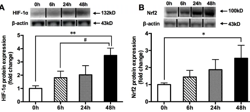
Compared with the 0h hypoxia group, the 48h hypoxia significantly increased the protein expressions of HIF-1αand Nrf2, but the 6h and 24h hypoxia did not (Fig 1).
Effects of acute hypoxia exposure with different durations on Nrf2/ARE
binding activity and the mRNA expressions of Nrf2-mediated antioxidative
genes
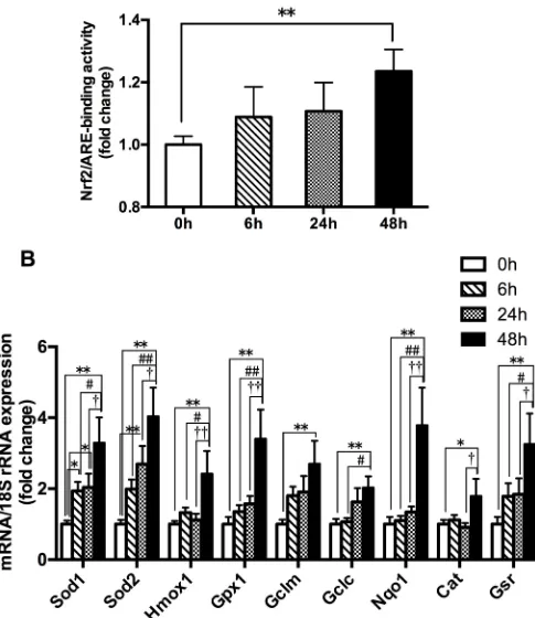
The 48h hypoxia group produced a significant increase in the Nrf2-ARE binding activity and the mRNA expressions of Nrf2-mediated antioxidative genes (Gclm,Gclc,Sod1,Sod2,Cat,
Alterations of HIF-1
α
protein expression after acute hypoxia exposure and
inhibition of the HIF-1
α
impaired hypoxia exposure (HE)-mediated
responses through Nrf2 activation in C2C12 cells
In order to identify the effects of hypoxia exposure (5% O2) with different durations on HIF-1α
protein contents in C2C12 cells, the protein expression was measured after hypoxia exposure for 24h, 48h and 72h, respectively. We found that at least 72 hours were needed to obtain the signifi-cant HIF-1αprotein expression (Fig 3A). Therefore, in the following cell experiments, we used 72 hours of hypoxia as the basic treatment. The ‘HE’ inFig 3B and 3C, andFig 4represented 72 hours of hypoxia exposure. There were four groups in the cellular studies: normoxia, HE, HE+-siControl, and HE+siHIF-1α. After the 72h hypoxia treatment, the cells were harvested. Protein expression levels of relevant molecules were analyzed by western blotting and Nrf2 nuclear trans-location was detected by immunofluorescence staining and confocal microscopy.
The results shown that the protein levels of HIF-1αand Nrf2 were markedly increased and the Nrf2 located primarily in a cytosolic location was translocated to the nucleus in the C2C12 cells in the HE group in contrast with the normoxia group, implying that the HIF-1αand Nrf2 was activated after the 72h hypoxia treatment (Fig 3B and 3C,Fig 4). Moreover, the molecule of HIF-1αwas blocked to validate its regulatory role in HE-mediated effects on Nrf2. Anti-HIF-1αsiRNA (si-HIF-1α) and its control siRNAs (siControl) were transiently transfected into C2C12 cells before the 72h hypoxia treatment. The data also revealed that the HE+si-HIF-1αgroup exhibited a significant reduction in HIF-1αand Nrf2 protein contents, and that the cytosol-to-nuclear translocation of Nrf2 was prevented, compared with the HE and HE+-siControl groups (Fig 3B and 3C,Fig 4). Collectively, the aforementioned results supported that HE-mediated effects on Nrf2 could be partially ascribed to activation of the HIF-1α.
Protein expressions of HIF-1
α
and Nrf2, Nrf2/ARE-binding activity and
the mRNA expressions of Nrf2-mediated antioxidative genes in skeletal
muscle of WT and HIF-1
α
TG mice
To determine the effects of HIF-1αon the Nrf2-mediated antioxidant responses in mouse skel-etal muscle, the HIF-1αTG and WT mice were also used. The protein content levels of
HIF-Fig 1. The changes in HIF-1α(A) and Nrf2 (B) total protein contents in mouse quadriceps femoris. Muscles were collected following acute exposure to hypoxia with different durations (n = 9 mice/group). Protein expression was measured by western blotting. The values are expressed as the means±SEM.��p<0.01 or�p<0.05 vs. 0h group; #p<0.05 vs. 6h group.
[image:6.612.135.569.481.676.2]Fig 2. The changes in the Nrf2-ARE binding activity (A) and the mRNA expressions of Nrf2-mediated antioxidative gene (B) in mouse quadriceps femoris. Muscles were collected following acute exposure to hypoxia with different durations (n = 9 mice/group). Nrf2-ARE binding activity was measured by trans-activation assay. The mRNA expressions were measured by RT-PCR. The values are expressed as the means±SEM.��p<0.01 or�p<0.05 vs. 0h group; ##p<0.01 or #p<0.05 vs. 6h group; ††p<0.01 or †p<0.05 vs. 24h group.
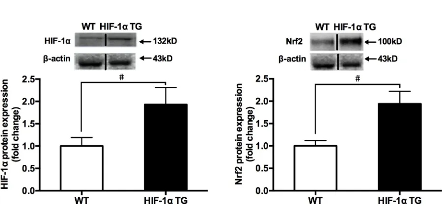
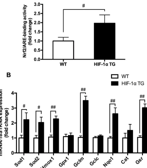
1αand Nrf2 measured by western blotting were compared in between HIF-1αTG and WT mice. The results showed that the levels of HIF-1αand Nrf2 protein contents (Fig 5), Nrf2/ ARE-binding activity, and the mRNA expressions of Nrf2-mediated antioxidative genes (Fig 6) in skeletal muscle of HIF-1αTG mice were significantly higher, compared with those in WT mice, suggesting that HIF-1αcould be involved in hypoxia exposure-induced Nrf2-me-diated antioxidant responses.
Discussion
The main findings of the present study were that the 48h hypoxia significantly increased Nrf2-ARE binding activity, Nrf2 protein, and mRNA expressions of Nrf2-mediated antioxi-dant genes in mouse skeletal muscle, longer duration associated with higher responses, in which the modulation of HIF-1αwould be involved; while the 6h and 24h hypoxia did not achieve these changes. The overall results supported the hypotheses of the present study. As far as we know, this is the first study to examine the changes in activation of Nrf2-ARE pathway in skeletal muscle after acute hypoxic exposure with different durations.
Hypoxia can stimulate Nrf2 signaling in animal or human tissues [10] or cells [11–13,22]; however, the activation process is complex. Several stress-mediated cytosolic kinases: phos-phoinositide 3-kinase (PI3K), protein kinase C (PKC), and mitogen-activated protein kinase (MAPK), have been reported to modify Nrf2 and affect its transcription activity [23–25]. For example, hypoxia activated PI3K/Akt pathway to mediate hypoxia-induced Nrf2 activation in EPCs [13] and enhanced the phosphorylation of serine residues of Nrf2 by PKC [18]. In addi-tion, dissociation of Nrf2 from Kelch-like ECH-associated protein 1 (Keap1) is the first step for nuclear translocation and subsequent DNA binding of Nrf2 to regulate the inducible expression of cytoprotective genes [26]. Before the activation, Nrf2 is seized in the cytosol as an inactive complex with its repressor Keap1 [27]. Thus, these evidences imply that the activa-tion of Nrf2-ARE pathway may need a certain time. In the present study, hypoxia exposure for 6h and 24h did not change Nrf2 protein expression, Nrf2/ARE binding activation and Nrf2’s downstream target gene mRNA expression; while hypoxia exposure for 48h promoted the rele-vant changes in skeletal muscle of WT mice (Fig 1BandFig 2). These results suggested that acute hypoxia exposure as a non-pharmacological approach, like exercise [28], could also acti-vate Nrf2/ARE signaling pathway in skeletal muscle. Under the current experimental condi-tion, 48 hours of hypoxic stress might be required for the break of Nrf2 from Keap1 and then start the Nrf2-dependent antioxidant response.
Higher expression of HIF-1αin mouse skeletal muscle in response to hypoxic stimuli is consistent with the previous reports [29,30]. Interestingly, the highest level of HIF-1α expres-sion was found in the 48h group, with same pattern of change as Nrf2 protein expresexpres-sion and the mRNA levels of antioxidative genes involved in Nrf2/ARE binding activity. Therefore, these evidences seemed to indicate that HIF-1αmight involve in the hypoxia-mediated regula-tion of Nrf2 activaregula-tion. In order to get a deeper understanding of the effects of HIF-1αon the hypoxia-induced Nrf2 responses in skeletal muscle, we employed C2C12 myoblasts with HIF-1α-specific siRNA to determine whether HIF-1αplays an important role in Nrf2 signaling pathway under hypoxia. It was shown that HIF-1αknockdown cells displayed a remarkable inhibition of Nrf2 protein along with a concomitant translocation of Nrf2 into the nuclei in
Fig 3. Alterations of the protein expressions of HIF-1αat different time points after hypoxia exposure (A); siRNA-mediated knockdown of HIF-1αinhibited HIF-1α(B) and Nrf2 (C) protein contents in C2C12 cells. Anti-HIF-1αsiRNA (si-HIF-1α,) and its control siRNAs (siControl) were transiently transfected into C2C12 cells before HE. Cells were incubated in normoxia (21% O2) or hypoxia (5% O2) and protein content levels of relevant molecules were measured by western blotting. The values are expressed as the means±SEM.��p<0.01 or�p<0.05 vs. HE or HE+siControl group; ##p<0.01 or #p<0.05 vs. normoxia group. HE = 72h hypoxia exposure at 5% O2.
HE. We noticed a methodological issue about hypoxic condition in C2C12 cell culture, other research teams used 1% O2to treat the cells [31–33]; however, with the current equipment in
our laboratory, we could only conduct the hypoxia exposure at 5% O2. The results,
neverthe-less, have achieved the experimental design of the present study. In the present study, C2C12 cells treated at 5% O2needed to take 72 hours to significantly increase the protein expression
of HIF-1α. Furthermore, the different changes in Nrf2 protein contents, Nrf2/ARE-binding activity and the mRNA expressions of Nrf2-mediated antioxidative genes were compared between HIF-1αTG and WT mice. The data indicated that with the increase of skeletal muscle HIF-1αprotein expression in the HIF-1αTG mice, their Nrf2 protein content, Nrf2/ARE-binding activity and the mRNA expressions of Nrf2-mediated antioxidative genes were also upregulated. Taken together, these evidences support that HIF-1αmay play an important part in the Nrf2-mediated antioxidant responses in mouse skeletal muscle.
We should admit the limitations in this study. We only focused on the effects of acute hyp-oxia exposure on the activation of Nrf2/ARE antioxidant signaling and whether HIF-1α
expression involved in promoting the Nrf2-mediated antioxidant responses in mouse skeletal muscle, but did not measure the change levels of reactive oxygen species (ROS) and oxidative stress products, such as malondialdehyde (MDA). It is worth to mention, besides Nrf2, nuclear factor-κB (NF-κB) is also a key transcription factor that may regulate cellular responses to oxi-dative stress and inflammation, and there is functional cross-talk between these two important pathways [30]. Future researches are required to elucidate the changes of redox homeostasis in
Fig 4. Effects of HE and HE+si HIF-1αon the nuclear distribution of Nrf2 in C2C12 cells. Cells were incubated in normoxia (21% O2) or HE (5% O2) and then localization of Nrf2 was visualized with a fluorescence microscope after immunofluorescence staining with anti-Nrf2 antibody and rabbit anti-mouse IgG second antibody coupled to Alexa Fluor 555. The nuclei were counterstained with DAPI (blue). Images of Nrf2 staining (Orange red) and DAPI staining (blue) of the same area were merged together to locate the cells with nuclear Nrf2 accumulation. HE = 72h hypoxia exposure at 5% O2.
[image:11.612.126.576.456.668.2]https://doi.org/10.1371/journal.pone.0208474.g004
Fig 5. The changes in HIF-1α(A) and Nrf2 (B) total protein contents between HIF-1αTG and WT mice groups. Mouse quadriceps femoris muscles were collected (n = 9 mice/group). Protein expression was measured by western blotting. The values are expressed as the means±SEM. #p<0.05 vs. WT mice group.
Fig 6. The changes in Nrf2-ARE binding activity (A) and the mRNA expressions of Nrf2-mediated antioxidative genes (B) between HIF-1αTG and WT mice groups. Mouse quadriceps femoris muscle was collected (n = 9 mice/group). Nrf2-ARE binding activity was evaluated in the muscle nuclear extracts using a Trans AM Nrf2 transcription factor assay. The level of mRNA expression was measured by RT-PCR. The values are expressed as the means±SEM. #p<0.05 or ##p<0.01 vs. WT mice group.
the molecular mechanisms that link the Nrf2 and NF-κB pathways after hypoxia exposure by using Nrf2 knockout mice.
In conclusion, our data demonstrated with molecular evidence that Nrf2/ARE antioxidant signaling activation in mouse skeletal muscle is dependent on hypoxia duration. Exposure to hypoxia for 48h triggers the activation of Nrf2 and is a positive regulator. HIF-1αprotein expression may be involved in promoting the Nrf2-mediated antioxidant responses, which partly underlies the beneficial effect of hypoxia in promoting Nrf2 and upregulating ARE-dependent antioxidant transcription.
Supporting information
S1 Data. Data file. (XLSX)
Acknowledgments
This study was supported by the National Natural Science Foundation of China (31471134) and the project of Beijing Sport University (2018GJ007).
Author Contributions
Conceptualization: Weixiu Ji, Siwang Yu, Ying Zhang.
Data curation: Weixiu Ji, Linjia Wang, Shiyi He, Lu Yan, Tieying Li.
Funding acquisition: Ying Zhang.
Investigation: Weixiu Ji, Linjia Wang, Shiyi He, Lu Yan, Tieying Li.
Methodology: Weixiu Ji.
Project administration: Ying Zhang.
Resources: Ying Zhang.
Supervision: Siwang Yu, Ying Zhang.
Visualization: Weixiu Ji.
Writing – original draft: Jianxiong Wang, Ying Zhang.
Writing – review & editing: Jianxiong Wang, Ah-Ng Tony Kong, Siwang Yu, Ying Zhang.
References
1. Ponsot E, Dufour SP, Zoll J, Doutrelau S, N’Guessan B, Geny B, et al. (2006) Exercise training in nor-mobaric hypoxia in endurance runners. II. Improvement of mitochondrial properties in skeletal muscle. J Appl Physiol (1985) 100: 1249–1257.https://doi.org/10.1152/japplphysiol.00361.2005PMID:
16339351
2. Faiss R, Leger B, Vesin JM, Fournier PE, Eggel Y, Deriaz O, et al. (2013) Significant molecular and sys-temic adaptations after repeated sprint training in hypoxia. PLoS One 8: e56522.https://doi.org/10. 1371/journal.pone.0056522PMID:23437154
3. Bailey DM, Davies B, Young IS (2001) Intermittent hypoxic training: implications for lipid peroxidation induced by acute normoxic exercise in active men. Clin Sci (Lond) 101: 465–475.
4. Gonchar O (2005) Muscle fiber specific antioxidative system adaptation to swim training in rats: influ-ence of intermittent hypoxia. J Sports Sci Med 4: 160–169. PMID:24431972
6. Itoh K, Wakabayashi N, Katoh Y, Ishii T, Igarashi K, Engel JD, et al. (1999) Keap1 represses nuclear activation of antioxidant responsive elements by Nrf2 through binding to the amino-terminal Neh2 domain. Genes Dev 13: 76–86. PMID:9887101
7. Suzuki T, Motohashi H, Yamamoto M (2013) Toward clinical application of the Keap1-Nrf2 pathway. Trends Pharmacol Sci 34: 340–346.https://doi.org/10.1016/j.tips.2013.04.005PMID:23664668 8. Itoh K, Ishii T, Wakabayashi N, Yamamoto M (1999) Regulatory mechanisms of cellular response to
oxi-dative stress. Free Radic Res 31: 319–324. PMID:10517536
9. Taguchi K, Maher JM, Suzuki T, Kawatani Y, Motohashi H, Yamamoto M (2010) Genetic analysis of cytoprotective functions supported by graded expression of Keap1. Mol Cell Biol 30: 3016–3026. https://doi.org/10.1128/MCB.01591-09PMID:20404090
10. Shu L, Wang C, Wang J, Zhang Y, Zhang X, Yang Y, et al. (2016) The neuroprotection of hypoxic pre-conditioning on rat brain against traumatic brain injury by up-regulated transcription factor Nrf2 and HO-1 expression. Neurosci Lett 6HO-1HO-1: 74–80.https://doi.org/10.1016/j.neulet.2015.11.012PMID:26590328 11. Huang XS, Chen HP, Yu HH, Yan YF, Liao ZP, Huang QR (2014) Nrf2-dependent upregulation of
anti-oxidative enzymes: a novel pathway for hypoxic preconditioning-mediated delayed cardioprotection. Mol Cell Biochem 385: 33–41.https://doi.org/10.1007/s11010-013-1812-6PMID:24048861 12. Yan YF, Chen HP, Huang XS, Qiu LY, Liao ZP, Huang QR (2015) DJ-1 Mediates the Delayed
Cardio-protection of Hypoxic Preconditioning Through Activation of Nrf2 and Subsequent Upregulation of Anti-oxidative Enzymes. J Cardiovasc Pharmacol 66: 148–158.https://doi.org/10.1097/FJC.
0000000000000257PMID:25915512
13. Zhao R, Feng J, He G (2016) Hypoxia increases Nrf2-induced HO-1 expression via the PI3K/Akt path-way. Front Biosci (Landmark Ed) 21: 385–396.
14. Semenza GL (1999) Regulation of mammalian O2 homeostasis by hypoxia-inducible factor 1. Annu Rev Cell Dev Biol 15: 551–578.https://doi.org/10.1146/annurev.cellbio.15.1.551PMID:10611972 15. Siren AL, Fratelli M, Brines M, Goemans C, Casagrande S, Lewczuk P, et al. (2001) Erythropoietin
pre-vents neuronal apoptosis after cerebral ischemia and metabolic stress. Proc Natl Acad Sci U S A 98: 4044–4049.https://doi.org/10.1073/pnas.051606598PMID:11259643
16. Jin W, Ming X, Hou X, Zhu T, Yuan B, Wang J, et al. (2014) Protective effects of erythropoietin in trau-matic spinal cord injury by inducing the Nrf2 signaling pathway activation. J Trauma Acute Care Surg 76: 1228–1234.https://doi.org/10.1097/TA.0000000000000211PMID:24747453
17. Genc K, Egrilmez MY, Genc S (2010) Erythropoietin induces nuclear translocation of Nrf2 and heme oxygenase-1 expression in SH-SY5Y cells. Cell Biochem Funct 28: 197–201.https://doi.org/10.1002/ cbf.1639PMID:20229611
18. Baba K, Morimoto H, Imaoka S (2013) Seven in absentia homolog 2 (Siah2) protein is a regulator of NF-E2-related factor 2 (Nrf2). J Biol Chem 288: 18393–18405.https://doi.org/10.1074/jbc.M112. 438762PMID:23645672
19. Li G, Wang J, Ye J, Zhang Y, Zhang Y (2015) PPARalpha Protein Expression Was Increased by Four Weeks of Intermittent Hypoxic Training via AMPKalpha2-Dependent Manner in Mouse Skeletal Muscle. PLoS One 10: e0122593.https://doi.org/10.1371/journal.pone.0122593PMID:25923694
20. Zhang Y, Ji W, Zhang L, Liu S, Liu G, Wang J (2015) Effects of HIF-1αon ERRα/γprotein expression in mouse skeletal muscle. Edorium Journal of Biomedical Technology 1: 4–10.
21. Livak KJ, Schmittgen TD (2001) Analysis of relative gene expression data using real-time quantitative PCR and the 2(-Delta Delta C(T)) Method. Methods 25: 402–408.https://doi.org/10.1006/meth.2001. 1262PMID:11846609
22. Polotsky VY, Savransky V, Bevans-Fonti S, Reinke C, Li J, Grigoryev DN, et al. (2010) Intermittent and sustained hypoxia induce a similar gene expression profile in human aortic endothelial cells. Physiol Genomics 41: 306–314.https://doi.org/10.1152/physiolgenomics.00091.2009PMID:20197421 23. Huang HC, Nguyen T, Pickett CB (2002) Phosphorylation of Nrf2 at Ser-40 by protein kinase C
regu-lates antioxidant response element-mediated transcription. J Biol Chem 277: 42769–42774.https://doi. org/10.1074/jbc.M206911200PMID:12198130
24. Kang KW, Choi SH, Kim SG (2002) Peroxynitrite activates NF-E2-related factor 2/antioxidant response element through the pathway of phosphatidylinositol 3-kinase: the role of nitric oxide synthase in rat glu-tathione S-transferase A2 induction. Nitric Oxide 7: 244–253. PMID:12446173
25. Yu R, Lei W, Mandlekar S, Weber MJ, Der CJ, Wu J, et al. (1999) Role of a mitogen-activated protein kinase pathway in the induction of phase II detoxifying enzymes by chemicals. J Biol Chem 274: 27545–27552. PMID:10488090
27. McMahon M, Itoh K, Yamamoto M, Hayes JD (2003) Keap1-dependent proteasomal degradation of transcription factor Nrf2 contributes to the negative regulation of antioxidant response element-driven gene expression. J Biol Chem 278: 21592–21600.https://doi.org/10.1074/jbc.M300931200PMID: 12682069
28. Li T, He S, Liu S, Kong Z, Wang J, Zhang Y (2015) Effects of different exercise durations on Keap1-Nrf2-ARE pathway activation in mouse skeletal muscle. Free Radic Res 49: 1269–1274.https://doi. org/10.3109/10715762.2015.1066784PMID:26118597
29. Xie HC, He JP, Zhu JF, Li JG (2014) Expression of HIF-1alpha and VEGF in skeletal muscle of plateau animals in response to hypoxic stress. Physiol Res 63: 801–805. PMID:25535647
30. Wardyn JD, Ponsford AH, Sanderson CM (2015) Dissecting molecular cross-talk between Nrf2 and NF-κB response pathways. Biochem Soc Tran 43: 621–626.
31. Li W, Hu ZF, Chen B, Ni GX (2013) Response of C2C12 myoblasts to hypoxia: the relative roles of glu-cose and oxygen in adaptive cellular metabolism. Biomed Res Int 2013: 326346.https://doi.org/10. 1155/2013/326346PMID:24294605
32. Cicchillitti L, Di Stefano V, Isaia E, Crimaldi L, Fasanaro P, Ambrosino V, et al. (2012) Hypoxia-inducible factor 1-alpha induces miR-210 in normoxic differentiating myoblasts. J Biol Chem 287: 44761–44771. https://doi.org/10.1074/jbc.M112.421255PMID:23148210