Identification of a Functionally Relevant Adeno-Associated Virus
Rep68 Oligomeric Interface
Martino Bardelli,aFrancisco Zárate-Pérez,bLeticia Agúndez,a*R. Michael Linden,a*Carlos R. Escalante,bEls Henckaertsa
Department of Infectious Diseases, King’s College London, London, United Kingdoma; Department of Physiology and Biophysics, Virginia Commonwealth University School of Medicine, Richmond, Virginia, USAb
ABSTRACT
The life cycle of the human parvovirus adeno-associated virus (AAV) is orchestrated by four Rep proteins. The large Rep pro-teins, Rep78 and Rep68, are remarkably multifunctional and display a range of biochemical activities, including DNA binding, nicking, and unwinding. Functionally, Rep78 and Rep68 are involved in transcriptional regulation, DNA replication, and
genomic integration. Structurally, the Rep proteins share an AAAⴙdomain characteristic of superfamily 3 helicases, with the
large Rep proteins additionally containing an N-terminal origin-binding domain (OBD) that specifically binds and nicks DNA. The combination of these domains, coupled with dynamic oligomerization properties, is the basis for the remarkable multifunc-tionality displayed by Rep68 and Rep78 during the AAV life cycle. In this report, we describe an oligomeric interface formed by Rep68 and demonstrate how disruption of this interface has drastic effects on both the oligomerization and functionality of the Rep proteins. Our results support a role for the four-helix bundle in the helicase domain of Rep68 as a bona fide oligomerization domain (OD). We have identified key residues in the OD that are critical for the stabilization of the Rep68-Rep68 interface; mu-tation of these key residues disrupts the enzymatic activities of Rep68, including DNA binding and nicking, and compromises viral DNA replication and transcriptional regulation of the viral promoters. Taken together, our data contribute to our under-standing of the dynamic and substrate-responsive Rep78/68 oligomerization that is instrumental in the regulation of the DNA transitions that take place during the AAV life cycle.
IMPORTANCE
The limited genome size of small viruses has driven the evolution of highly multifunctional proteins that integrate different do-mains and enzymatic activities within a single polypeptide. The Rep68 protein from adeno-associated virus (AAV) combines a DNA binding and endonuclease domain with a helicase-ATPase domain, which together support DNA replication, transcrip-tional regulation, and site-specific integration. The coordination of the enzymatic activities of Rep68 remains poorly under-stood; however, Rep68 oligomerization and Rep68-DNA interactions have been suggested to play a crucial role. We investigated the determinants of Rep68 oligomerization and identified a hydrophobic interface necessary for Rep68 activity during the AAV life cycle. Our results provide new insights into the molecular mechanisms underlying the regulation of the versatile Rep pro-teins. Efficient production of AAV-based gene therapy vectors requires optimal Rep expression levels, and studies such as the one presented here could contribute to further optimization of AAV production schemes.
A
deno-associated virus (AAV) is a human DNA virus of thefamilyParvoviridaewith a unique dependence on helper
vi-ruses, such as adenovirus (Ad) or herpesvirus, for productive
rep-lication (1). In order to take advantage of host pathways and
helper virus for productive replication with only a limited number of viral gene products at hand, AAV has evolved to combine mul-tiple functions into single proteins. More specifically, a single open reading frame (ORF) generates the nonstructural Rep pro-teins that orchestrate the different aspects of the AAV life cycle, including transcriptional regulation, replication, packaging, and Rep-mediated integration. The four multidomain Rep proteins Rep40, Rep52, Rep68, and Rep78 are generated through the use of
two promoters and alternative splicing (2, 3). All Rep isoforms
share a superfamily 3 (SF3) helicase domain (HD) that combines
ATPase and helicase activities (4, 5). The large Rep proteins,
Rep68 and Rep78, further contain an N-terminal origin-binding domain (OBD) that specifically binds and nicks the AAV origin
(5–7). Rep78 and Rep52 have an additional zinc finger domain
that is involved in interactions with cellular proteins (8,9).
Dif-ferences in the domain composition of the Rep isoforms confer specific functionalities to the large and the small Rep proteins.
Rep52 and Rep40 are necessary for efficient packaging of the viral DNA into preformed capsids but are dispensable for viral
replica-tion and integrareplica-tion (10). The DNA binding and nicking activities
of the OBD of the large Rep proteins, on the other hand, are the
basis for AAV DNA replication and integration (11,12). More
specifically, Rep78/68 bind to the Rep binding site (RBS) in the
Received25 February 2016 Accepted1 May 2016
Accepted manuscript posted online11 May 2016
CitationBardelli M, Zárate-Pérez F, Agúndez L, Linden RM, Escalante CR, Henckaerts E. 2016. Identification of a functionally relevant adeno-associated virus Rep68 oligomeric interface. J Virol 90:6612–6624.doi:10.1128/JVI.00356-16.
Editor:L. Banks, International Centre for Genetic Engineering and Biotechnology Address correspondence to Carlos R. Escalante, [email protected], or Els Henckaerts, [email protected].
*Present address: Leticia Agúndez, Department of Genetics, University College London Institute of Ophthalmology, London, United Kingdom; R. Michael Linden, Genetic Medicine Institute, Pfizer Inc., London, United Kingdom.
Copyright © 2016 Bardelli et al. This is an open-access article distributed under the terms of theCreative Commons Attribution 4.0 International license.
on November 7, 2019 by guest
http://jvi.asm.org/
inverted terminal repeats (ITRs) and execute a site- and
strand-specific nick at the nearby terminal resolution site (trs). This
pro-cess is nepro-cessary for resolution of the ITRs and completion of the viral DNA replication cycle with the assistance of the host cell
machinery (12,13). Similarly, the DNA binding and endonuclease
activities are required for mediation of integration at
chromo-somal target loci that contain RBS/trs, such as the integration hot
spotAAVS1(14–16). In addition, efficient nicking of the trsat both the viral and cellular origins requires ATP-dependent heli-case activity for the generation of an optimal single-stranded
sub-strate (7,17). Finally, both the OBD and the helicase domain have
the ability to mediate the transcriptional regulation of viral and cellular promoters by two independent mechanisms conferring
regulatory functions to both small and large Rep proteins (18–20).
Structurally, the Rep proteins belong to the SF3 of helicases, a group of multifunctional viral proteins combining a characteristic
AAA⫹motor domain, which couples ATP hydrolysis and DNA
unwinding, with an origin-binding domain to achieve rapid
ori-gin melting (21). Other members of this family include the simian
virus 40 (SV40) large T antigen (LTag) and papillomavirus (PV) E1. In contrast to the OBD of Rep, SV40 LTag and PV E1 lack endonuclease activity. The AAV large Rep proteins are also related to HUH endonucleases, which catalyze rolling-circle replication (RCR) in bacteriophages and geminiviruses, as well as in bacteria
(22,23), indicating a significant evolutionary conservation. The
large AAV Rep proteins have a complex and dynamic oligomeric behavior that can adapt to the different DNA substrates present during the AAV life cycle, varying from the RBS-containing dou-ble-stranded DNA encountered during initial origin binding to
the single-stranded DNA encountered after origin melting (24,
25). In contrast to the AAA⫹domains of SV40 LTag and PV E1,
which readily form hexameric rings (26,27), Rep40 and Rep52 are
monomeric, due to the absence of a complete oligomerization
domain (OD) at the N terminus of the SF3 helicase domain (28).
Mutation of the corresponding OD in SV40 LTag and PV E1 has been shown to prevent their oligomerization and to disrupt the
replication of SV40 and PV, respectively (29,30). Intriguingly, a
similar OD was also found in oligomeric HUH endonucleases, such as RepB from the pMV158 streptococcal RCR plasmid,
de-spite the absence of a helicase domain (31). In the large Rep
pro-teins, the linker connecting the OBD and the helicase domain provides the residues necessary to complete the OD and plays a
crucial role in the oligomerization of the large Rep proteins (28,
32). Thus, the OBD, linker, and helicase domain effectively
inter-act cooperatively to promote oligomerization.
While recent findings have significantly contributed to the
un-derstanding of the determinants of Rep oligomerization (25,28,
32), its relevance in the context of the AAV life cycle remains to be
elucidated. In order to gain an understanding of how oligomer-ization contributes to the multiple enzymatic functions of the Rep proteins, we took advantage of our previous findings, which showed that the linker domain and, in particular, the N-terminal
linker residue Y224 are essential for Rep oligomerization (28).
This residue was found in a position equivalent to that of the residues in SV40 LTag and PV E1 known to be crucial for the
formation and maintenance of the oligomeric interface (28). Here
we describe an oligomeric interface for Rep, identified in a dimeric complex modeled using the Rep40 structure with a predicted
ex-tended N-terminal␣-helix to complete the OD (33). This model
highlights a potential role for Y224 and I251 in the formation of
the oligomeric interface; site-directed mutagenesis confirmed that oligomerization is indeed hampered when Y224 and I251 are al-tered. Moreover, we could demonstrate that mutations that lead to a disruption of the interface result in defects in DNA binding
andtrsnicking and alter the expression levels of the viral proteins,
with severe consequences on viral DNA replication and produc-tion of infectious virus.
MATERIALS AND METHODS
Protein production and purification.All mutations were generated in the pHisRep68/15b plasmid, which contains the AAV serotype 2 (AAV2) Rep68 ORF subcloned in the vector pET-15b (Novagen), using a QuikChange mutagenesis kit (Agilent Technologies Inc.). All proteins were expressed inEscherichia coliBL21(DE3) cells (Novagen) and purified as described previously (28). In brief, cell pellets were lysed in Ni-buffer A (20 mM Tris-HCl [pH 7.9 at 4°C], 500 mM NaCl, 5 mM imidazole, 10% glycerol, 0.2% CHAPS {3-[(3-cholamidopropyl)-dimethylammonio]-1-propanesulfonate}, 1 mM TCEP [Tris(2-carboxyethyl)phosphine hydro-chloride]) and purified using an Ni column. The hexahistidine tag was removed using PreScission protease, and Rep68 was further purified by gel filtration chromatography using a HiLoad Superdex 200 16/60 column (GE Healthcare) and size exclusion buffer (25 mM Tris-HCl [pH 8.0], 200 mM NaCl, 2 mM TCEP). Rep68 wild-type (WT) and mutant proteins were concentrated to 10 mg/ml, flash-frozen in liquid N2, and kept at
⫺80°C.
Sedimentation velocity experiments.Analytical ultracentrifugation experiments were carried out using a Beckman Optima XL-I analytical ultracentrifuge (Beckman Coulter Inc.) equipped with both four- and eight-position rotors. Protein samples (420l; final concentration, 10
M) were loaded in the cells, and in all cases, buffer containing 25 mM Tris-HCl, pH 8.0, and 200 mM NaCl was used. Samples were centrifuged in 2-sector carbon-filled Epon centerpieces at 25,000 rpm at 20°C. At least 200 scans were collected at 5-min intervals at 25,000 rpm. Sedimentation velocity-concentration profiles were collected using both UV absorption (280 nm) and Rayleigh interference scanning optics. Results were ana-lyzed using the SEDFIT program (34,35).
AAV infectious particle assay.293T cells were triple transfected with an AAV2 inverted terminal repeat (ITR)-containing plasmid carrying a CAG-controlled green fluorescent protein (GFP) gene (pTRUF11), a helper plasmid expressing AAV2 Rep (WT or mutants) and Cap, and a third construct containing the adenovirus helper functions (HGTI plas-mid) (36,37). The mutations in Rep were confirmed in all plasmids by sequencing (Eurofins). After 72 h, the supernatant was harvested and spun to clear the cellular debris, and increasing volumes of superna-tant were used to transduce HeLa cells. The percentage of GFP-positive HeLa cells was determined at 48 h postransduction by flow cytometry (FACSCanto; BD Biosciences).
qPCR-based replication assay.293T cells were transfected with poly-ethylenimine (Polysciences, Inc.) and the infectious AAV plasmid pAV2 (38) or its mutant versions and superinfected 4 h later with adenovirus serotype 5 (Ad5) at a multiplicity of infection (MOI) of 5. After 72 h, cells were harvested in phosphate-buffered saline (PBS) and pelleted, and the pellet was divided into 4. One-fourth was used for RNA extraction, one-fourth was used for protein extraction, one-one-fourth was used for total DNA extraction, and the last quarter was used for Hirt extraction of low-mo-lecular-weight DNA.
Total DNA was extracted using a Qiagen DNeasy blood and tissue DNA extraction kit. Viral DNA was quantified by real-time PCR using the SYBR green JumpStart Taq ReadyMix for quantitative PCR (qPCR; Sigma-Aldrich) and an ABI Prism system (Applied Biosystems). Cap primers (forward [fw] primer, TTCTCAGATGCTGCGTACCGGAAA; reverse [rv] primer, TCTGCCATTGAGGTGGTACTTGGT) and a pAV2-based standard curve were used for absolute quantification; the signal was normalized to that of cyclophilin (fw primer, TGCTGGACCCAACACA AATG; rv primer, TGCCATCCAACCACTCAGTCT).
on November 7, 2019 by guest
http://jvi.asm.org/
Analysis of replicative intermediates.293T cells were treated as de-scribed above for the qPCR-based replication assay. Low-molecular-weight DNA was extracted using a modified version of the Hirt extract procedure (39). Briefly, cells were lysed in Hirt lysis buffer (0.6% SDS, 10 mM Tris, pH 7.5, 10 mM EDTA) and treated with proteinase K (Thermo Fisher) to digest proteins. The high-molecular-weight DNA was precipi-tated and discarded. The low-molecular-weight DNA was then purified by phenol extraction, followed by sodium acetate and isopropanol precipi-tation. The precipitated DNA was washed and resuspended in DNase-free water. The extracts were digested with the restriction enzyme DpnI (New England BioLabs) to digest input DNA. Samples were run on a 0.8% agarose gel at 30 V overnight and transferred to a nitrocellulose mem-brane by the Southern blotting method. The memmem-branes were hybridized overnight in 0.75 nylon wash buffer (40.6 g Na2HPO4, 18.65 g EDTA, and
500 g SDS in 3.58 liters of double-distilled H2O, pH 7.2) at 65°C with a
radiolabeled Rep probe (fw primer, 5=-AACTGGACCAATGAGAACTTT CC-3=; rv primer, 5=-A AAAAGTCTTTGACTTCCTGCTT-3=) or an am-picillin probe (fw primer, 5=-AATCAGTGAGGCACCTATCTCAGC-3=; rv primer, 5=-AACTCGGTCGCCGCATACACTATT-3=) to control for DpnI digestion. The probes were labeled with a Prime-It RmT random primer labeling kit from Stratagene and [32P]dCTP (PerkinElmer). The
membranes were exposed to a PhosphorImager screen overnight. Images were acquired using a Typhoon PhosphorImager (Molecular Dynamics) and analyzed with ImageQuant TL software (GE Healthcare Life Sci-ences).
Fluorescence anisotropy DNA binding assay.Binding assays were performed using a fluorescein-labeled 41-mer containingAAVS1or p5 RBS sequences. The sequences used were 5=-TGGCGGCGGTTGGGGCT CGGCGCTCGCTCGCTCGCTGGGCG-3=(AAVS1) and 5=-ACCGGGC AAAATGGAGACCCTGCGTGCTCACTCGGGCTTAA-3= (p5), where theAAVS1and p5 sequences are in italics (40,41). Rep68 WT and mutant proteins at concentrations ranging from 5 nM to 3M were mixed with DNA (5 nM) in a final volume of 300l using the following buffer: 25 mM HEPES (pH 7.0), 100 mM NaCl, 1 mM TCEP. Fluorescence readings were taken on a PC1 fluorimeter (ISS, Inc.) with excitation and emission filters at 490 and 520 nm, respectively. The tubes were equilibrated at 20°C for 20 min before measurement. Each anisotropy point is the average of 10 mea-surements. Anisotropy is calculated as the ratio of the difference between the vertical and horizontal emission intensities to the total normalized intensity. The fraction of DNA bound (B) was calculated using the follow-ing equation:B⫽([A]x⫺[A]DNA)/([A]final– [A]DNA), where [A]x
rep-resents the anisotropy measured at protein concentrationx, [A]DNAis the
anisotropy of free fluorescent DNA, and [A]finalis the anisotropy at satu-ration. The data were fit to a single binding site model by use of the Hill coefficient and the program Origin (Origin Labs). Each experiment was done in triplicate.
Helicase assay.The substrate used in the helicase assay is a heterodu-plex DNA consisting of an 18-bp duheterodu-plex region with a 10-nucleotide 3=tail at the bottom strand, referred to as 18ADT10A. The top strand (trap DNA) is labeled at the 5=end with fluorescein and is released upon unwinding. The sequences used were 5=-F-CATATGGAGCAGAACA GA-3= for the trap DNA and 5=-AGACAAGACGAGGTATACA AAAAAAAAA-3=for the complementary strand.
All reactions were performed in a buffer containing 25 mM HEPES, 50 mM NaCl (pH 7.0) at a total volume of 50l. Protein (1 mM) was mixed with 0.5 mM double-stranded fluorescein-labeled DNA (18ADT10A) and 2.5M single-stranded DNA (18-nucleotide sense DNA), and the mix-ture was then added to the mix of buffer described above containing 5 mM ATP and 5 mM MgCl2. The reaction mixture was incubated at 25°C for 1 h. EDTA was used at a final concentration of 20M to stop the reaction. Aliquots of 10l were loaded in a 12% bisacrylamide gel (30%) (19:1) using 6⫻loading dye (0.25% xylene cyanol FF, 30% glycerol). For the densitometry and analysis of the bands, a Gel Doc EZ imager was used, together with the automatic lane and band detection tool. Lane
back-ground subtraction, white illumination, and an activation time of 300 s were used for the analysis.
scDNA nicking assay.Supercoiled DNA (scDNA) nicking activity for Rep68 was assayed as described previously (14). Briefly, assays were per-formed in 30-l reaction mixtures containing 30 mM HEPES-KOH (pH 7.5), 7 mM MgCl2, 0.5 mM dithiothreitol, 4 mM ATP, 40 mM creatine
phosphate (Sigma), and 1g creatine phosphokinase (Sigma) in 15 mM (final concentration) NaCl. One hundred nanograms of supercoiled plas-mid DNA and 200 ng of purified His-Rep68 (or mutants) were added to the reaction mixtures. All samples were incubated for 1 h at 37°C; the reaction was terminated by adding 10l of stop reaction mixture (pro-teinase K [1.2g/l], 0.5% SDS, 30 mM EDTA, pH 7.5) and incubating for 1 h at 37°C. Samples were resolved in a 1% agarose gel (1⫻ Tris-acetate-EDTA [TAE]), which was subsequently stained with ethidium bromide (0.3g/ml) in 1⫻TAE. The plasmids carrying scDNA used in this assay were pRVK (which containsAAVS1from nucleotides 1 to 3536) and a mutated version containing a mutanttrssequence (42).
Western blotting.Proteins were extracted from cells transfected and infected as described above for the replication assays. Cells were lysed in radioimmunoprecipitation assay buffer, and the cleared lysate was run on a 12% acrylamide gel. The proteins were transferred onto a nitrocellulose membrane (GE Healthcare) and immunoblotted using anti-Rep antibody (1/100 dilution; clone 303.9; Progen), anti-Cap antibody (1/500 dilution; clone B1; American Research Products), and anti-HSP90 antibody (poly-clonal, 1/5,000 dilution; Santa Cruz). All antibodies were incubated in blocking buffer (5% nonfat dried milk in PBS containing 0.1% Tween 20). Images were acquired and analyzed using an ImageQuant apparatus (GE Healthcare).
Real-time quantitative RT-PCR.293T cells were transfected and in-fected as described above for the replication assays. Total RNA was ex-tracted using an RNeasy kit (Qiagen) after DNase I (Qiagen) treatment for 15 min at 37°C. Reverse transcription (RT) was performed using a high-capacity reverse transcription kit (Applied Biosystems). cDNA was quan-tified by real-time qPCR on an ABI Prism system (Applied Biosystems) using the TaqMan Universal PCR master mix (Life Technologies) and custom-designed primer-probe mixes (Eurofins). The following primers were used: p5 fw (5=-AACAAGGTGGTGGATGAGT-3=), p5 rv (5=-CGT TTACGCTCCGTGAGATT-3=), p19 fw (5=-TCACCAAGCAGGAAGTC AAAG-3=), p19 rv (5=-CCCGTTTGGGCTCACTTATATC-3=), p40 fw (5=-GGAAGCAAGGCTCAGAGAAA-3=), and p40 rv (5=-CCTCTCTGG AGGTTGGTAGATA-3=). The following probes were used: p5 (5= -FAM-ACGTGGTTGAGGTGGAGCATGAAT-TAM-3=), p19 (5= -FAM-ACGTG-GTTGAGGTGGAGCATGAA-TAM-3=), and p40 (5=-FAM-AGGAAATCA GGACAACCAATCCCGT-TAM-3=), where FAM is 6-carboxyfluorescein and TAM is 6-carboxytetramethylrhodamine. Relative expression levels were determined by the⌬⌬CTthreshold cycle (CT) quantification method (43),
using 18S rRNA (TaqMan predeveloped assay reagents, human 18S rRNA; Applied Biosystems) as a housekeeping reference gene.
RESULTS
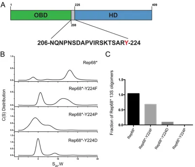
Y224 forms hydrophobic interactions necessary for Rep68
oligomerization. Previous studies using Rep68* (or Rep* for
short), a C151S Rep mutant that is functionally equivalent to WT
Rep68 but that prevents protein aggregation in solution (25),
showed that Rep68 exists as a mixture of oligomers in solution. More specifically, two major populations have been observed by sedimentation velocity experiments, including a monomer-dimer
peak that sediments at⬃3S and oligomeric rings that sediment at
13S (25). We also showed that replacement of the tyrosine
posi-tioned at the C-terminal end of the linker in Rep68 by the smaller residue alanine disrupts its oligomerization; this is presumably
because of a reduction in the surface-exposed area (Fig. 1A) (28).
To further confirm this hypothesis, we mutated the tyrosine to phenylalanine (Phe), proline (Pro), or aspartic acid (Asp) and
on November 7, 2019 by guest
http://jvi.asm.org/
performed sedimentation velocity experiments to study how these
mutations affect oligomerization.Figure 1Bshows that
replace-ment of the tyrosine with the small-side-chain amino acids Pro and Asp had a drastic effect on the sedimentation profile of Rep68. The 13S peak disappeared, and the most prominent population present in solution had a sedimentation coefficient of about 5S,
suggestive of low-molecular-weight oligomers (Fig. 1B).
Exchang-ing the tyrosine with the bulky aromatic Phe resulted in the ap-pearance of two peaks, one with a sedimentation coefficient of 5S, similar to what we observed for the other mutants, and a second peak of about 12S, which was indicative of the formation of larger oligomers. This 12S population, however, had a sedimentation coefficient smaller than what we observed with Rep68, potentially suggesting that the Y224F mutant forms different oligomeric
spe-cies.Figure 1Cshows the quantification of the 13S population that
was formed in the presence of the different mutations. Taken to-gether, these results demonstrate that the bulky aromatic charac-ter of the Y224 residue is pivotal for Rep68 oligomerization and suggest that Y224 may participate in hydrophobic interactions as part of an oligomeric interface.
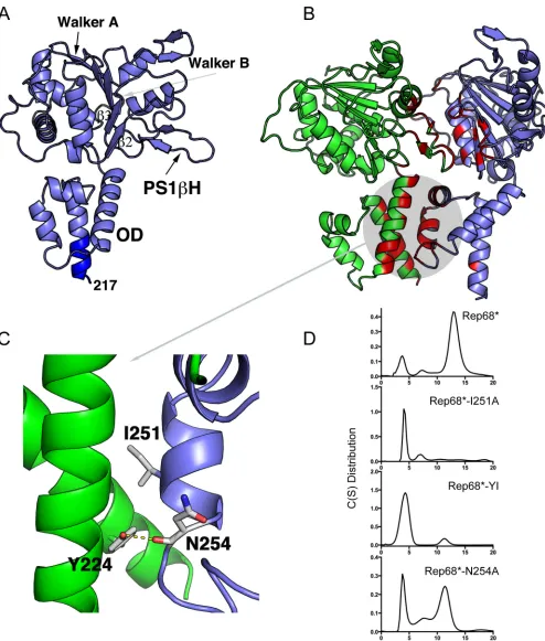
Generation of a Rep68 oligomeric interface model.To further
determine whether Y224 participates directly in forming an oligo-meric interface, we modeled an oligooligo-meric Rep dimer using the
available structure of Rep40 (PDB accession number 1S9H),
which spans residues 225 to 490 of Rep68 (33). We added the
interdomain linker residues 217 to 224 to the known Rep40
struc-ture as an extended␣-helix on the basis of secondary structure
predictions (28), resulting in a Rep molecule containing residues
217 to 490 (Fig. 2A). Two of the three molecules found in the
asymmetric unit of Rep40 crystals formed a pseudodimer. The interface formed in this dimer was similar to the oligomeric inter-face described for other SF3 helicase structures but is not optimal
to perform catalysis (44). We used this dimer with the addition of
the linker residues as our initial interface model, and we refined it by carrying out rigid body and side chain conformation
optimi-zation using the RosettaDock server (45,46). Strikingly, the top 10
models generated had almost identical interfaces, as analyzed by
the program PISA (47), suggesting that our model was robust. A
representation of the Rep interface model is shown inFig. 2B. The
interface buries a total of 1,992 Å of solvent-accessible area and includes residues from all the helices in the oligomerization
do-main (OD), the presensor 1-hairpin (PS1H), the23 loop,
and residues from the Walker A and Walker B motifs (Fig. 2B). A
closer analysis revealed that the modeled linker residues partici-pate in the interface. In particular, the conserved aromatic residue Y224, which is at the end of the linker region, is an important
FIG 1Role of Tyr224 in Rep68 oligomerization. (A) Schematic diagram of Rep68. The sequence of the linker is shown, and Y224 is highlighted in red. (B) Sedimentation velocity analysis of Rep68 and different Y224 mutants. C(S), sedimentation coefficient distribution. (C) Quantification of the amount of 13S species formed by Y224 mutants. Rep68* refers to the C151S mutant, which is functionally equivalent to WT Rep68 but prevents protein aggregation in solution (25). All mutant proteins were generated in the context of Rep68*.
on November 7, 2019 by guest
http://jvi.asm.org/
[image:4.585.93.494.64.407.2]FIG 2Model of a Rep-Rep interface. (A) Ribbon representation of Rep40 extended to residue 217 as an␣-helix highlighted in dark blue. The Walker A and Walker B motifs, PS1H, the23 loop, and OD are indicated. (B) Model of a dimeric Rep complex. The structures participating in the interface are highlighted in red and magenta. (C) Close-up of the interactions formed by residue Y224, including the hydrophobic interaction with I251 and the hydrogen bond with the backbone of N254. (D) Sedimentation velocity analysis of the Rep68 I251A, Y224A-I251A (YI), and N254A mutants. All proteins were analyzed at a concentra-tion of 1 mg/ml, as described in Materials and Methods.
on November 7, 2019 by guest
http://jvi.asm.org/
[image:5.585.43.537.63.644.2]component of the oligomeric interface. In agreement with the
results shown inFig. 1, it participates in the formation of a
hydro-phobic pocket. Among the residues from the neighboring subunit interacting with Y224, residue I251, at a distance of 3.6 Å, takes part in a hydrophobic interaction, whereas N254 contributes to a
hydrogen bond via its main chain carbonyl oxygen (Fig. 2C).
Mutations leading to disruption of the interface affect
oligo-merization of Rep68.Based on these observations, we generated a
Rep68* mutant with the I251A substitution (Rep68*-I251A mu-tant) and we assessed the consequences of mutating this residue alone or in combination with Y224 on Rep68* oligomerization.
Figure 2Dshows sedimentation velocity profiles illustrating that these mutants are mostly monomeric, thus validating the predic-tion from our model. A single N254A mutant was also evaluated to confirm that the side chain of this residue does not contribute to the oligomeric interface. As predicted, because N254 forms a hy-drogen bond through its main chain carbonyl oxygen rather than participates directly in the hydrophobic interface, the N254A mu-tation showed only a mild effect on sedimenmu-tation. Furthermore, the oligomerization defects which we observed when testing the
different Y224 mutants (Fig. 1C) could also be explained by our
modeled interface: smaller hydrophobic residues increase the sol-vent-accessible area and destabilize the interface, while a larger bulky residue maintains the hydrophobic pocket and possibly af-fects only the formation of the hydrogen bond between Y224 and N254, thus causing a milder defect. Finally, the Y224P mutation had the strongest effect, as it probably disrupts the helical charac-ter of this region and may affect the overall OD structure. To investigate whether the interface that we identified is biologically relevant and thus involved in Rep functions, we assessed how the observed disruptions in the oligomerization profile of Rep68 af-fect the AAV life cycle.
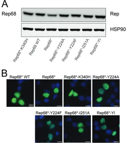
Rep68 oligomerization is necessary to support the AAV life
cycle.First, we verified if the aforementioned mutant Rep68
pro-teins were stable and localized correctly to the nucleus, where they support AAV replication. We transfected 293T cells with con-structs expressing Rep68 or the oligomerization mutants under the control of the cytomegalovirus promoter and assessed Rep68
protein stability and localization at 8 h posttransfection.Figure 3
demonstrates that all the mutants were expressed at levels compa-rable to those observed for WT Rep68 and translocated to the nucleus, as expected.
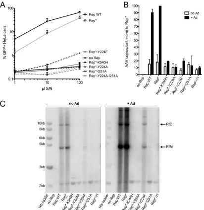
Next, we assessed how disruptions in the oligomerization pro-file of Rep68 affect the AAV life cycle. We first compared the ability of the Rep oligomerization mutants to produce infectious
AAV particles to that of WT Rep68 and Rep68* (Fig. 4A). As a
negative control, we used the nucleoside triphosphate-binding mutant K340H, which is deficient in ATPase and helicase activity
and does not support AAV replication (48,49). The K340H
mu-tant, however, was still able to oligomerize and has been shown to
have a dominant negative phenotype (48–50). Recombinant
AAV2-GFP was produced in 293T cells by transfection of an ITR-containing plasmid carrying a GFP expression cassette together with plasmids encoding the adenovirus helper functions, AAV2 Cap, and WT Rep68, Rep68*, or the interface mutants. Increasing volumes of supernatant collected from the cultures of AAV-pro-ducing cells were added to HeLa cells in order to assess the
infec-tivity of the produced virus.Figure 4Ashows that the Y224A Rep
mutant did not support the production of infectious AAV
parti-cles, as was previously reported by us (28). The mutant with the
more conservative mutation, Y224F, which retained the potential
to partially oligomerize (Fig. 1C), was severely impaired but was
not entirely deficient in producing infectious AAV. Mutating I251 to alanine on the opposite side of the predicted interface, however, reproduced the phenotype observed with the Y224A mutant. Not surprisingly, the Y224A-I251A double mutant also failed to
pro-duce infectious AAV particles (Fig. 4A).
To evaluate if the failure to produce infectious particles was due to a defect in AAV DNA replication, we determined the
num-ber of AAV genomes in the 293T producer cells by qPCR (Fig. 4B)
and studied the replicative intermediates formed during AAV
rep-lication by Southern blotting (Fig. 4C). Both assays confirmed that
the Y224A and I251A interface mutants and the Y224A-I251A double mutant all failed to support AAV DNA replication. Simi-larly to what we observed in the infectious particle production assay, the Y224F mutant supported AAV replication but did so at levels significantly lower than those observed for Rep WT or Rep*.
In addition,Fig. 4Cshows that replication in the presence of the
Y224F mutant resulted in the formation of the expected replica-tive intermediates. Background replication could be observed in the absence of adenovirus due to the presence of E1A and E1B in
293T cells (51). Altogether, these results suggest that the
oligomer-ization interface mutants fail to sustain AAV DNA replication and therefore cannot support the production of infectious AAV par-ticles.
Rep68 oligomerization mutants are deficient in RBS-specific DNA binding and site- and strand-specific nicking but maintain
the ability to unwind unspecific DNA substrates.In order to
FIG 3The interface mutants are stable and localize to the nucleus. (A) WT Rep68 or Rep68 mutants were transfected in 293T cells and tested for expres-sion levels by Western blotting at 36 h posttransfection. Anti-HSP90 antibody was used as loading control to ensure that a similar amount of protein was used under each condition. (B) 293T cells transfected with WT Rep68 or Rep68 mutants were assessed at 48 h posttransfection by immunostaining. Merged images showing green (Rep) and blue (DAPI [4= ,6-diamidino-2-phenylin-dole]-stained nuclei) channels are presented. Rep68*-K340H was used as a control for nuclear localization of a well-characterized nonfunctional Rep mutant.
on November 7, 2019 by guest
http://jvi.asm.org/
[image:6.585.317.524.68.302.2]determine the cause of the replication defect of the oligomeriza-tion-deficient mutants, we assessed various biochemical activities
in vitro. Rep has three well-characterized enzymatic functions—
RBS-specific DNA binding,trsnicking, and ATP-dependent DNA
unwinding—all of which are necessary for AAV DNA replication
and targeted genome integration.Table 1shows the binding
con-stants of Rep68* and mutant Rep68* proteins on p5 andAAVS1
RBS-containing double-stranded DNA substrates. As expected, both Rep68* and the control, Rep68*-K340H, efficiently bound
the specific DNA substrates (25,52). The mutant that retained
some residual replication potential, Rep68*-Y224F, also
effi-ciently bound the p5 andAAVS1DNA substrates. All other
mu-tants, however, lost the ability to bind both DNA substrates, with the exception of Rep68*-I251, which maintained its ability to
[image:7.585.88.500.65.488.2]FIG 4Interface mutants do not support the AAV life cycle. (A) Increasing volumes of supernatant from 293T cells producing recombinant AAV-GFP in the presence of WT Rep68 or Rep68 mutants were used to infect HeLa cells, and the percentage of GFP-positive (GFP⫹) cells was determined by fluorescence-activated cell sorting analysis. S/N, supernatant. Data are from three independent experiments and are represented as the mean⫾SEM. (B) AAV DNA replication under permissive (⫹Ad) and nonpermissive (no Ad) conditions was quantified by quantitative PCR. Data from three experiments were normalized (norm) to those obtained under permissive (Rep*⫹Ad) conditions and are represented as the mean⫾SEM. (C) AAV replicative intermediates generated under the same conditions shown in panel B were visualized by Southern blotting using a Rep-specific probe. RfM, monomeric replicative form; RfD, dimeric replicative form.
TABLE 1Binding constants of Rep68* and interface mutants onAAVS1
and p5 RBS-containing DNA
Protein
Binding constant (nM)
AAVS1-41 p5-41
Rep68* 128 203
Rep68*-K340H 123 136
Rep68*-Y224A NDa ND
Rep68*-Y224F 221 311
Rep68*-I251A 1,438 ND
Rep68*-YI ND ND
aND, not determined due to poor binding.
on November 7, 2019 by guest
http://jvi.asm.org/
[image:7.585.299.545.624.715.2]bindAAVS1-containing DNA, albeit with a 10-fold lower af-finity than its WT counterpart. These results suggest that some level of oligomerization is necessary for efficient RBS-specific DNA binding by Rep68 and that the oligomeric properties of the mutant Rep68*-Y224F are sufficient for DNA binding.
To test the ability of the Rep68 mutants to unwind nonspecific DNA, we performed a fluorescence-based helicase assay. Some-what surprisingly, all mutants except the control K340H mutant exhibited similar helicase activity on a heteroduplex nonspecific
DNA substrate (Fig. 5). These results suggest that under these
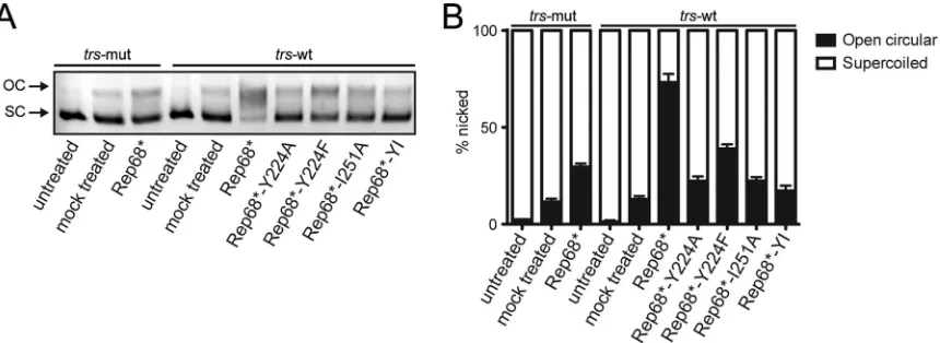
experimental conditions Rep68 can unwind DNA even in the ab-sence of large complexes or, alternatively, that an oligomeric com-plex that is stabilized by a different interface is necessary for Rep-mediated DNA unwinding. Strand- and site-specific nicking activity, however, appeared to diminish strongly when oligomer-ization was disrupted in Rep68. As expected, the oligomeroligomer-ization-
oligomerization-deficient Rep mutants that failed to bind specific DNA also failed
to nick supercoiled plasmid DNA containing RBS and trs
se-quences (Fig. 6AandB). The Rep68*-Y224F mutant, despite
re-taining the ability to bind specific DNA substrates, showed only some residual nicking activity.
Rep oligomerization is important for transcriptional
regula-tion of AAV genes.In addition to their role in AAV DNA
replica-tion, the Rep proteins coordinate the temporal regulation of tran-scription of the viral genome during the AAV life cycle. In the absence of helper virus, the Rep proteins participate in repressing transcription from the three viral promoters, p5, p19, and p40, ensuring minute levels of expression of the viral proteins. In the presence of helper virus, i.e., during a productive infection, re-pression of the p5 promoter is lifted by the adenoviral E1A protein
(53), and binding of Rep to the p5 promoter or the ITRs leads to
transactivation of the p19 and p40 promoters (54–56). The p5
promoter itself is also controlled by Rep, which can act both as a repressor and as an activator through binding at the p5 or ITR
RBS, respectively (56,57). The net result is a self-regulatory loop
that generates protein levels that are tightly controlled and are
optimal for AAV replication and packaging (58). Two
mecha-nisms of Rep-mediated repression have been identified: direct re-pression through binding at the RBS in the p5 promoter and
in-direct repression that requires the ATPase activity of Rep (19).
In light of the dependence of transcriptional regulation on Rep binding to the p5 promoter and ITR, we assessed whether the oligomerization mutants also displayed defects in transcriptional activity resulting in altered protein expression levels. As a control, we again used the K340H ATPase mutant, which has been shown to lead to the expression of exceedingly high Rep protein levels
under conditions permissive for AAV replication (48). Cells were
transfected with the various AAV infectious plasmids, and Rep and Cap protein levels were determined in the presence and ab-sence of adenovirus coinfection. In the preab-sence of WT Rep and the absence of adenovirus, we expected low levels of both Rep and Cap proteins. In the presence of adenovirus, Rep protein levels peaked at about 30 h after infection and then slowly decreased,
while Cap levels increased with viral DNA replication (56).
Be-cause we harvested the cells at 72 h posttransfection, we expected
FIG 5Comparison of the helicase activity of the interface mutants. The ability of the Rep68 interface mutants to unwind a fluorescein-labeled heteroduplex DNA substrate was assayed. Data are from three independent experiments and are represented as the mean⫾SEM.
FIG 6Rep-mediated nicking of supercoiled plasmid. Supercoiled (SC) plasmid DNA containing an RBS and atrswas mixed with Rep68* or the interface mutants. If the endonuclease activity is intact, Rep nicks and relaxes the plasmid conformation to an open circular (OC) form, which can be readily distinguished by agarose gel electrophoresis. Atrsmutant (trs-mut) plasmid that was not nicked by Rep was used as a control. Untreated DNA was left untouched, while mock treated plasmid was incubated in reaction buffer for 1 h at 37°C in the absence of protein. (A) Representative agarose gel electrophoresis image. (B) Quantification of nicking from four independent nicking experiments. Data are represented as the mean⫾SEM.
on November 7, 2019 by guest
http://jvi.asm.org/
[image:8.585.61.266.64.253.2] [image:8.585.79.509.517.674.2]to see only slightly higher levels of the Rep proteins but signifi-cantly higher Cap expression levels compared to those in cells that
were not coinfected with adenovirus. As shown inFig. 7A, we
observed strikingly high Rep protein levels in cells transfected with the mutated Rep proteins, including the control K340H mutant. Once more, we observed that the Y224F mutant showed an inter-mediate phenotype represented by a very modest increase in Rep
protein expression levels (Fig. 7A). The Cap protein levels, on the
other hand, were found to be lower in cells expressing the mutant proteins; we detected high Cap levels only in the presence of Rep proteins that support the AAV life cycle, with the sole exception
being the K340H mutant (Fig. 7A). The same trend was observed
both in the presence and in the absence of adenovirus infection, although the Cap levels were significantly lower in the absence of helper virus. These results suggest that the oligomerization mu-tants failed to regulate the expression levels of the AAV proteins, most likely by failing to autoregulate the p5 promoter through RBS binding. In view of these important differences in protein amounts, we assessed the levels of AAV transcripts by RT-qPCR (Fig. 6C). Because all AAV RNAs use the same polyadenylation
signal, we were not able to quantify the p19 and p40 transcripts separately from the p5 transcripts. As expected, with all primer
sets used, which targeted p5, p5⫹p19, and p5⫹p19⫹p40, we
ob-served an increase in mRNA levels in response to adenovirus coin-fection in the presence of Rep proteins that support AAV replica-tion. In the presence of the Y224F mutant, the response to adenovirus was still present but was nevertheless reduced com-pared to that in the presence of WT Rep. The oligomerization-deficient Y224, I251, and Y224-I251 mutants, which were unable to bind the RBS at the p5 promoter, had higher basal mRNA levels, varying between 2- and 10-fold compared with those of the WT, and did not respond to adenovirus infection. In the context of adenovirus coinfection, however, the differences in mRNA levels did not correlate with those observed for the protein levels, sug-gesting that changes in posttranscriptional regulation also con-tribute to the altered protein expression levels. Rep-mediated posttranscriptional regulation has been observed before, but its
mechanism remains unknown (59). The K340H mutant, which
oligomerized but failed to support AAV replication, had consid-erably higher basal mRNA levels than WT Rep, possibly
explain-FIG 7Rep oligomerization is important for transcriptional regulation of AAV genes. (A) Western blot showing Rep and Cap protein levels under conditions permissive (⫹adenovirus) and nonpermissive for AAV replication. The first lane in the left panel is equivalent to the third lane in the right panel. (B) The transcription levels of AAV genes were analyzed under the same conditions shown in panel A. RNA levels were measured by RT-qPCR using three primer-probe mixes detecting RNA from the p5 promoter only, from p5⫹p19, and from p5⫹p19⫹p40. The fold change was calculated relative to the mRNA levels in the presence of WT Rep but in the absence of adenovirus. Data are from three independent experiments and are represented as the mean⫾SEM.
on November 7, 2019 by guest
http://jvi.asm.org/
[image:9.585.78.502.65.434.2]ing the very high Rep and Cap protein amounts observed, and the presence of adenovirus did not lead to a clear change in mRNA levels. Taken together, our data support a model in which Rep oligomerization is important for the gene regulatory function of Rep, potentially through p5 RBS binding, which is necessary to achieve an appropriate transcription profile.
DISCUSSION
The limited genome capacity of small viruses, such as adeno-as-sociated virus, has driven the evolution of highly multifunctional nonstructural proteins that combine several enzymatic functions necessary to support the viral life cycle. In AAV, the Rep proteins are responsible for orchestrating the entire viral life cycle, from transcriptional regulation to replication and packaging as well as Rep-mediated integration. The combination of several enzymatic functions, including DNA binding, nicking, and unwinding, and the ability to interact with a multitude of DNA substrates and proteins allow the Rep proteins to support replication. However, the coordination of all these functions would require a tightly controlled system, which we envision could be provided by the different oligomeric states that the protein assumes. During the AAV life cycle, Rep has to catalyze reactions on different DNA substrates, including initiation of DNA replication, recognition
and nicking of thetrs, and binding to the p5 promoter, in order to
provide transcriptional regulation. It has been shown that Rep can
form different oligomeric speciesin vitroboth in the absence of
DNA and in the presence of different DNA substrates (24,25),
allowing an additional layer of regulation of the Rep activities during the AAV life cycle. To fully understand the mechanism of action of Rep on its different substrates, it is essential to identify the oligomeric complexes formed with the different DNA mole-cules. For example, it has been shown that Rep68 forms a double octameric ring in the presence of single-stranded DNA as well as
on forked helicase substrates (24), whereas other reports have
sug-gested that Rep68 forms hexamers when it is bound to
double-stranded DNA (50,60). However, the importance of these
com-plexes for the viral life cycle has not been formally addressed, and while it is clear that Rep oligomerization is functionally relevant, data on possible oligomerization interfaces remain scarce. Smith and colleagues identified two regions—residues 151 to 188 and residues 334 to 347—that, when deleted, disrupt Rep oligomer-ization; however, they did not investigate the functional
conse-quences of these deletions (50). Intriguingly, residues 334 to 346
include the ATP binding site that has been shown to be part of the oligomeric interface in PV E1 and SV40 LTag hexamers. A more recent report showed that one residue, R107, which was initially identified for its role in integration, origin binding, and nicking and which was shown to be in direct contact with origin DNA, is
also essential for oligomerization (24,61). Finally, we and others
have shown that the interdomain linker of Rep78/68 and, in
particu-lar, the Y224 residue are critical for Rep oligomerization (28,32).
Building on our previous studies, we further characterized the role of residue Y224 in Rep oligomerization. Replacement of Y224 with residues with different properties had various consequences on Rep68* oligomerization. More specifically, replacement of Y224 with small hydrophobic residues severely impaired oligo-merization, while the Y224F mutant with the more conservative
mutation retained the ability to oligomerize (Fig. 1), suggesting
that Y224 participates in the formation and the stabilization of a hydrophobic interface. This hypothesis was supported by a model
of a dimeric Rep-Rep interaction built from the pseudodimer ob-served in the crystal structure of Rep40 using an extended Rep40
molecule (33). In this model, a large interface that resembled the
interface formed by the PV E1 protein and included residues from
the Walker A and Walker B motifs, PS1H, and the23 loop was
formed (Fig. 2). Furthermore, all the helices in the OD also
par-ticipated in the interface and formed a hydrophobic pocket, em-phasizing the relevance of this subdomain in Rep oligomerization.
More specifically, linker residue Y224 on the extended␣-helix 1 of
one Rep molecule interacted with residue I251 and with the main
chain carbonyl oxygen of residue N254 on␣-helix 3 of the other
Rep molecule (Fig. 2C). Mutating I251 to alanine in Rep68 alone
or in combination with Y224A confirmed that this residue is im-portant for Rep68 oligomerization. Imim-portantly, because none of the residues located in the OD that we identified to be participants in the oligomeric interface are part of the catalytic sites described within the Rep proteins, the consequences of these mutations on the functions of the Rep proteins are likely to be due to oligomer-ization defects. We showed that the oligomeroligomer-ization-deficient Y224A, I251A, and Y224A-I251A mutants were unable to repli-cate AAV DNA and failed to support the production of recombi-nant AAV. Our data suggest that these defects are caused by the loss of DNA binding and origin nicking activities by the mutant Rep proteins and confirm that Rep oligomerization is critical for its function in support of the AAV life cycle. We also assessed the consequences of a more conservative mutation, Y224F, on Rep function. This substitution maintained the bulky aromatic char-acter of the residue, a feature that is conserved in the OD of SF3
helicases and other related proteins (28). The Rep68*-Y224F
mu-tant retained the ability to oligomerize but formed the large 13S
complexes less efficiently than Rep68* did (Fig. 1). Interestingly,
the binding of RBS-containing DNA did not appear to be com-promised by this mutation. The Rep68*-Y224F nicking activity, on the other hand, was severely impaired, possibly explaining the
low levels of viral replication observed (Fig. 6). In view of the
oligomeric behavior of the Y224F mutant, these results suggest that this mutant retains the ability to form an oligomeric complex sufficient for RBS-mediated DNA binding but fails to promote the subsequent DNA nicking step. How this transition is affected, however, is not clear. One intriguing possibility is that the initial Rep binding to the RBS and melting of the origin promote the recruitment of further Rep78/68 molecules and the assembly of a second, larger Rep-DNA complex that is necessary for the nicking reaction. Residue Y224 and, more generally, the OD could help stabilize the formation of this complex, allowing a shift in the
interaction with the origin DNA to allow thetrsnicking reaction
to take place. The Y224F mutation was previously identified in a
study by Walker et al. to be important for Rep function (62). In
contrast to our findings, however, those authors reported that the Y224F mutant was deficient in ITR binding, endonuclease, DNA helicase, and ATPase activities. The cause of this difference may be due to a different experimental strategy or could possibly be ex-plained by the presence of a maltose-binding protein (MBP) tag
(62), which may affect the already weakened oligomerization
po-tential of Rep68-Y224F.
SF3 helicases are thought to function as oligomeric complexes, as is the case for PV E1 and SV40 LTag, which form active hexa-meric complexes. Surprisingly, all the oligomerization-deficient mutants described here were still able to unwind a heteroduplex
DNA substrate (Fig. 5). This suggests that interaction with and
on November 7, 2019 by guest
http://jvi.asm.org/
unwinding of 3=-tailed substrates do not require the formation of large Rep oligomers, consistent with the helicase activity of Rep40, or, alternatively, that the presence of heteroduplex DNA and ATP stabilizes the formation of an oligomeric complex independently from the oligomeric interface described here. Rep40 is monomeric in solution, forms transient dimers in the presence of ATP, and retains helicase activity, albeit at a level lower than that observed
with the large Rep proteins (28,63). Therefore, although the
mu-tants presented here do not form a complex with theAAVS1site,
they could form transient oligomers in the presence of ATP that are able to unwind DNA. In our previous report, we introduced the possibility that AAV Rep proteins have evolved two distinct
helicase modes (28): one that parallels the helicase activity of other
SF3 helicases, requires oligomeric rings, and is performed by the large Rep proteins, and one that requires only a transient dimerization and that is characteristic for the activity of the small Rep isoforms. Thus, it is plausible that the mutants described here are still able to unwind DNA through the same mechanism used by Rep52/40, but they would not support the unwinding of a substrate that requires the helicase activity from oligomeric rings. On the basis of the different functions of the large and small Rep proteins in the AAV life cycle, it is tempting to suggest that melting of the AAV origin, which is mediated by the large Rep proteins, requires the formation of a stable oligomer that unwinds DNA by a mechanism analogous to that described for other SF3 helicases, while packaging AAV genomes into the viral capsids, which is efficiently carried out by the small Rep proteins, may proceed through a different helicase mode.
In addition to their role in supporting AAV DNA replication, the enzymatic activities of the Rep proteins are also essential for the correct transcriptional regulation of viral and cellular tran-scripts. Because it is known that the levels of Rep proteins are tightly regulated and not simply maximized to achieve efficient
AAV DNA replication (58,64), we assessed whether the
expres-sion levels of AAV proteins were affected by the oligomerization mutants. The K340H Rep mutant has been shown to fail in appro-priately regulating the expression of the AAV genes, suggesting that the ATPase/helicase activity of Rep is involved in
transcrip-tional regulation (48). A different mechanism of transcriptional
repression that is dependent on the RBS binding activity of
Rep78/68 has also been demonstrated (20). Thus, two
mecha-nisms of repression— one that is RBS binding dependent and one that is helicase domain dependent— exist and likely act in concert to precisely regulate the levels of expression of the Rep proteins. In this study, we show that regulation of AAV gene expression is impaired in the presence of Rep oligomerization mutants that do
not bind p5 orAAVS1DNA, indicating that at least one of two
mechanisms of repression is impaired. Our results suggest that oligomerization of the large Rep proteins is necessary for the
cor-rect regulation of the transcription of all AAV promoters (Fig. 7).
More specifically, the oligomerization-deficient mutants fail to induce transcription of the viral promoters upon infection with the helper virus adenovirus, and, in addition, both large and small Rep protein levels increase substantially in the presence of oligomerization mutants. The presence of the Y224A mutation creates a Kozak sequence at the p19 promoter stronger than that achieved with WT Rep68, but this is not sufficient to explain the differences in protein levels observed, in particular for Rep78. In addition, the increase in Rep52 expression in the presence of the Y224A-I251A mutations is modest compared to that observed in
the presence of Y224A alone. The differences in protein levels observed in the presence of adenovirus, however, cannot be ex-plained by the RNA levels alone, suggesting that there is some level of posttranscriptional control that may also be Rep dependent. A function for AAV Rep in this context was previously suggested in a study by Trempe and Carter, where it was observed that the regulation of gene expression at a transcriptional level alone was
not sufficient to explain differences in protein levels (59). Our data
also support a role for an oligomeric complex of Rep in regulating protein levels posttranscriptionally. Understanding the mecha-nism behind this potential uncharacterized function of AAV Rep proteins may reveal a new layer of complexity in the role that the Rep proteins play in coordinating the AAV life cycle.
In conclusion, our study identifies and describes an essential Rep-Rep protein interface that is involved in the formation of Rep complexes and demonstrates its functional relevance throughout the AAV life cycle. Our study focuses on residues that are part of
the␣-helical bundle located upstream of the helicase domain and
strengthens the suggestion that this subdomain of Rep plays a role as a bona fide oligomerization domain. The identification of the oligomeric interfaces of AAV Rep like the one described here and further structural and functional characterization of Rep oligo-meric complexes, particularly in the presence of different DNA substrates, will provide additional insights into the molecular mechanisms of Rep-mediated transcriptional regulation and AAV DNA replication, as well as Rep-mediated integration.
FUNDING INFORMATION
This work, including the efforts of Els Henckaerts, was funded by Pfizer Rare Diseases Consortium (Pfizer Rare Diseases Consortium Award). This work, including the efforts of Carlos R. Escalante, was funded by HHS | National Institutes of Health (NIH) (RO1-GM092854). This work, including the efforts of R. Michael Linden, was funded by Medical Re-search Council (MRC) (1001764).
REFERENCES
1. Weitzman MD, Linden RM. 2011. Adeno-associated virus biology. Methods Mol Biol807:1–23.http://dx.doi.org/10.1007/978-1-61779-370 -7_1.
2.Green MR, Roeder RG.1980. Transcripts of the adeno-associated virus genome: mapping of the major RNAs. J Virol36:79 –92.
3.Srivastava A, Lusby EW, Berns KI. 1983. Nucleotide sequence and organization of the adeno-associated virus 2 genome. J Virol45:555–564. 4.Smith RH, Kotin RM.1998. The Rep52 gene product of adeno-associated
virus is a DNA helicase with 3=-to-5=polarity. J Virol72:4874 – 4881. 5.Im DS, Muzyczka N.1990. The AAV origin binding protein Rep68 is an
ATP-dependent site-specific endonuclease with DNA helicase activity. Cell61:447– 457.http://dx.doi.org/10.1016/0092-8674(90)90526-K. 6.Owens RA, Weitzman MD, Kyostio SR, Carter BJ.1993. Identification
of a DNA-binding domain in the amino terminus of adeno-associated virus Rep proteins. J Virol67:997–1005.
7.Davis MD, Wu J, Owens RA. 2000. Mutational analysis of adeno-associated virus type 2 Rep68 protein endonuclease activity on partially single-stranded substrates. J Virol 74:2936 –2942. http://dx.doi.org/10 .1128/JVI.74.6.2936-2942.2000.
8.Di Pasquale G, Stacey SN.1998. Adeno-associated virus Rep78 protein interacts with protein kinase A and its homolog PRKX and inhibits CREB-dependent transcriptional activation. J Virol72:7916 –7925.
9.Berthet C, Raj K, Saudan P, Beard P.2005. How adeno-associated virus Rep78 protein arrests cells completely in S phase. Proc Natl Acad Sci U S A
102:13634 –13639.http://dx.doi.org/10.1073/pnas.0504583102. 10. King JA, Dubielzig R, Grimm D, Kleinschmidt JA.2001. DNA
helicase-mediated packaging of adeno-associated virus type 2 genomes into pre-formed capsids. EMBO J20:3282–3291.http://dx.doi.org/10.1093/emboj /20.12.3282.
11. Dutheil N, Linden RM. 2006. Site-specific integration by
on November 7, 2019 by guest
http://jvi.asm.org/
associated virus, p 214 –236.InKerr JR, Cotmore SF, Bloom ME, Linden RM, Parrish CR (ed), Parvoviruses. Hodder Arnold, London, United Kingdom.
12. Ward P.2006. Replication of adeno-associated virus DNA, p 189 –211.In
Kerr JR, Cotmore SF, Bloom ME, Linden RM, Parrish CR (ed), Parvovi-ruses. Hodder Arnold, London, United Kingdom.
13. Brister JR, Muzyczka N. 2000. Mechanism of Rep-mediated adeno-associated virus origin nicking. J Virol74:7762–7771.http://dx.doi.org/10 .1128/JVI.74.17.7762-7771.2000.
14. Petri K, Gabriel R, Agundez L, Fronza R, Afzal S, Kaeppel C, Linden RM, Henckaerts E, Schmidt M.2015. Presence of a trs-like motif pro-motes Rep-mediated wild-type adeno-associated virus type 2 integration. J Virol89:7428 –7432.http://dx.doi.org/10.1128/JVI.00426-15. 15. Weitzman MD, Kyostio SR, Kotin RM, Owens RA. 1994.
Adeno-associated virus (AAV) Rep proteins mediate complex formation between AAV DNA and its integration site in human DNA. Proc Natl Acad Sci U S A
91:5808 –5812.http://dx.doi.org/10.1073/pnas.91.13.5808.
16. Urcelay E, Ward P, Wiener SM, Safer B, Kotin RM.1995. Asymmetric replication in vitro from a human sequence element is dependent on ad-eno-associated virus Rep protein. J Virol69:2038 –2046.
17. Brister JR, Muzyczka N. 1999. Rep-mediated nicking of the adeno-associated virus origin requires two biochemical activities, DNA helicase activity and transesterification. J Virol73:9325–9336.
18. Horer M, Weger S, Butz K, Hoppe-Seyler F, Geisen C, Kleinschmidt JA.
1995. Mutational analysis of adeno-associated virus Rep protein-mediated inhibition of heterologous and homologous promoters. J Virol
69:5485–5496.
19. Kyostio SR, Wonderling RS, Owens RA.1995. Negative regulation of the adeno-associated virus (AAV) P5 promoter involves both the P5 Rep binding site and the consensus ATP-binding motif of the AAV Rep68 protein. J Virol69:6787– 6796.
20. Dutheil N, Smith SC, Agundez L, Vincent-Mistiaen ZI, Escalante CR, Linden RM, Henckaerts E.2014. Adeno-associated virus Rep represses the human integration site promoter by two pathways that are similar to those required for the regulation of the viral p5 promoter. J Virol88:8227– 8241.http://dx.doi.org/10.1128/JVI.00412-14.
21. Hickman AB, Dyda F.2005. Binding and unwinding: SF3 viral helicases. Curr Opin Struct Biol15:77– 85.http://dx.doi.org/10.1016/j.sbi.2004.12 .001.
22. Chandler M, de la Cruz F, Dyda F, Hickman AB, Moncalian G, Ton-Hoang B.2013. Breaking and joining single-stranded DNA: the HUH endonuclease superfamily. Nat Rev Microbiol11:525–538.http: //dx.doi.org/10.1038/nrmicro3067.
23. Gonzalez-Prieto C, Agundez L, Linden RM, Llosa M. 2013. HUH site-specific recombinases for targeted modification of the human ge-nome. Trends Biotechnol31:305–312.http://dx.doi.org/10.1016/j.tibtech .2013.02.002.
24. Mansilla-Soto J, Yoon-Robarts M, Rice WJ, Arya S, Escalante CR, Linden RM.2009. DNA structure modulates the oligomerization prop-erties of the AAV initiator protein Rep68. PLoS Pathog5:e1000513.http: //dx.doi.org/10.1371/journal.ppat.1000513.
25. Zarate-Perez F, Mansilla-Soto J, Bardelli M, Burgner JW, II, Vil-lamil-Jarauta M, Kekilli D, Samso M, Linden RM, Escalante CR.
2013. Oligomeric properties of adeno-associated virus Rep68 reflect its multifunctionality. J Virol 87:1232–1241. http://dx.doi.org/10.1128 /JVI.02441-12.
26. Sedman J, Stenlund A.1998. The papillomavirus E1 protein forms a DNA-dependent hexameric complex with ATPase and DNA helicase ac-tivities. J Virol72:6893– 6897.
27. Li D, Zhao R, Lilyestrom W, Gai D, Zhang R, DeCaprio JA, Fanning E, Jochimiak A, Szakonyi G, Chen XS.2003. Structure of the replicative helicase of the oncoprotein SV40 large tumour antigen. Nature423:512– 518.http://dx.doi.org/10.1038/nature01691.
28. Zarate-Perez F, Bardelli M, Burgner JW, II, Villamil-Jarauta M, Das K, Kekilli D, Mansilla-Soto J, Linden RM, Escalante CR.2012. The inter-domain linker of AAV-2 Rep68 is an integral part of its oligomerization domain: role of a conserved SF3 helicase residue in oligomerization. PLoS Pathog8:e1002764.http://dx.doi.org/10.1371/journal.ppat.1002764. 29. Titolo S, Pelletier A, Pulichino AM, Brault K, Wardrop E, White PW,
Cordingley MG, Archambault J.2000. Identification of domains of the human papillomavirus type 11 E1 helicase involved in oligomerization and binding to the viral origin. J Virol74:7349 –7361.http://dx.doi.org/10 .1128/JVI.74.16.7349-7361.2000.
30. Loeber G, Stenger JE, Ray S, Parsons RE, Anderson ME, Tegtmeyer P.
1991. The zinc finger region of simian virus 40 large T antigen is needed for hexamer assembly and origin melting. J Virol65:3167–3174.
31. Boer DR, Ruiz-Maso JA, Lopez-Blanco JR, Blanco AG, Vives-Llacer M, Chacon P, Uson I, Gomis-Ruth FX, Espinosa M, Llorca O, del Solar G, Coll M.2009. Plasmid replication initiator RepB forms a hexamer remi-niscent of ring helicases and has mobile nuclease domains. EMBO J28:
1666 –1678.http://dx.doi.org/10.1038/emboj.2009.125.
32. Maggin JE, James JA, Chappie JS, Dyda F, Hickman AB.2012. The amino acid linker between the endonuclease and helicase domains of ad-eno-associated virus type 5 Rep plays a critical role in DNA-dependent oligomerization. J Virol86:3337–3346. http://dx.doi.org/10.1128/JVI .06775-11.
33. James JA, Escalante CR, Yoon-Robarts M, Edwards TA, Linden RM, Aggarwal AK.2003. Crystal structure of the SF3 helicase from adeno-associated virus type 2. Structure11:1025–1035.http://dx.doi.org/10.1016 /S0969-2126(03)00152-7.
34. Schuck P.2003. On the analysis of protein self-association by sedimenta-tion velocity analytical ultracentrifugasedimenta-tion. Anal Biochem320:104 –124.
http://dx.doi.org/10.1016/S0003-2697(03)00289-6.
35. Vistica J, Dam J, Balbo A, Yikilmaz E, Mariuzza RA, Rouault TA, Schuck P.2004. Sedimentation equilibrium analysis of protein interac-tions with global implicit mass conservation constraints and systematic noise decomposition. Anal Biochem326:234 –256.http://dx.doi.org/10 .1016/j.ab.2003.12.014.
36. Zolotukhin S, Potter M, Hauswirth WW, Guy J, Muzyczka N.1996. A “humanized” green fluorescent protein cDNA adapted for high-level ex-pression in mammalian cells. J Virol70:4646 – 4654.
37. Streck CJ, Dickson PV, Ng CY, Zhou J, Hall MM, Gray JT, Nathwani AC, Davidoff AM.2006. Antitumor efficacy of AAV-mediated systemic delivery of interferon-beta. Cancer Gene Ther13:99 –106.http://dx.doi .org/10.1038/sj.cgt.7700878.
38. Laughlin CA, Tratschin JD, Coon H, Carter BJ.1983. Cloning of infec-tious adeno-associated virus genomes in bacterial plasmids. Gene23:65– 73.http://dx.doi.org/10.1016/0378-1119(83)90217-2.
39. Hirt B.1969. Replicating molecules of polyoma virus DNA. J Mol Biol
40:141–144.http://dx.doi.org/10.1016/0022-2836(69)90302-7. 40. McCarty DM, Pereira DJ, Zolotukhin I, Zhou X, Ryan JH, Muzyczka N.
1994. Identification of linear DNA sequences that specifically bind the adeno-associated virus Rep protein. J Virol68:4988 – 4997.
41. Linden RM, Winocour E, Berns KI.1996. The recombination signals for adeno-associated virus site-specific integration. Proc Natl Acad Sci U S A
93:7966 –7972.http://dx.doi.org/10.1073/pnas.93.15.7966.
42. Lamartina S, Ciliberto G, Toniatti C.2000. Selective cleavage of AAVS1 substrates by the adeno-associated virus type 2 Rep68 protein is depen-dent on topological and sequence constraints. J Virol74:8831– 8842.http: //dx.doi.org/10.1128/JVI.74.19.8831-8842.2000.
43. Schmittgen TD, Livak KJ.2008. Analyzing real-time PCR data by the comparative C(T) method. Nat Protoc3:1101–1108.http://dx.doi.org/10 .1038/nprot.2008.73.
44. James JA, Aggarwal AK, Linden RM, Escalante CR.2004. Structure of adeno-associated virus type 2 Rep40-ADP complex: insight into nucleo-tide recognition and catalysis by superfamily 3 helicases. Proc Natl Acad Sci U S A101:12455–12460.http://dx.doi.org/10.1073/pnas.0403454101. 45. Gray JJ, Moughon S, Wang C, Schueler-Furman O, Kuhlman B, Rohl CA, Baker D.2003. Protein-protein docking with simultaneous optimi-zation of rigid-body displacement and side-chain conformations. J Mol Biol331:281–299.http://dx.doi.org/10.1016/S0022-2836(03)00670-3. 46. Lyskov S, Gray JJ.2008. The RosettaDock server for local protein-protein
docking. Nucleic Acids Res36:W233–W238.http://dx.doi.org/10.1093 /nar/gkn216.
47. Krissinel E, Henrick K.2007. Inference of macromolecular assemblies from crystalline state. J Mol Biol372:774 –797.http://dx.doi.org/10.1016 /j.jmb.2007.05.022.
48. Chejanovsky N, Carter BJ.1990. Mutation of a consensus purine nucle-otide binding site in the adeno-associated virus rep gene generates a dom-inant negative phenotype for DNA replication. J Virol64:1764 –1770. 49. Kyostio SR, Owens RA.1996. Identification of mutant adeno-associated
virus Rep proteins which are dominant-negative for DNA helicase activ-ity. Biochem Biophys Res Commun220:294 –299.http://dx.doi.org/10 .1006/bbrc.1996.0399.
50. Smith RH, Spano AJ, Kotin RM.1997. The Rep78 gene product of
on November 7, 2019 by guest
http://jvi.asm.org/
adeno-associated virus (AAV) self-associates to form a hexameric com-plex in the presence of AAV ori sequences. J Virol71:4461– 4471. 51. Wang XS, Srivastava A.1998. Rescue and autonomous replication of
adeno-associated virus type 2 genomes containing Rep-binding site mu-tations in the viral p5 promoter. J Virol72:4811– 4818.
52.Owens RA, Trempe JP, Chejanovsky N, Carter BJ. 1991. Adeno-associated virus Rep proteins produced in insect and mammalian expres-sion systems: wild-type and dominant-negative mutant proteins bind to the viral replication origin. Virology184:14 –22.http://dx.doi.org/10.1016 /0042-6822(91)90817-U.
53. Shi Y, Seto E, Chang LS, Shenk T.1991. Transcriptional repression by YY1, a human GLI-Kruppel-related protein, and relief of repression by adenovirus E1A protein. Cell67:377–388.http://dx.doi.org/10.1016/0092 -8674(91)90189-6.
54. Pereira DJ, Muzyczka N.1997. The adeno-associated virus type 2 p40 promoter requires a proximal Sp1 interaction and a p19 CArG-like ele-ment to facilitate Rep transactivation. J Virol71:4300 – 4309.
55. Pereira DJ, Muzyczka N.1997. The cellular transcription factor SP1 and an unknown cellular protein are required to mediate Rep protein activation of the adeno-associated virus p19 promoter. J Virol 71:
1747–1756.
56. Weger S, Wistuba A, Grimm D, Kleinschmidt JA.1997. Control of adeno-associated virus type 2 cap gene expression: relative influence of helper virus, terminal repeats, and Rep proteins. J Virol71:8437– 8447. 57. Labow MA, Hermonat PL, Berns KI.1986. Positive and negative
autoreg-ulation of the adeno-associated virus type 2 genome. J Virol60:251–258.
58. Li J, Samulski RJ, Xiao X.1997. Role for highly regulated rep gene expression in adeno-associated virus vector production. J Virol71:5236 – 5243.
59. Trempe JP, Carter BJ.1988. Regulation of adeno-associated virus gene expression in 293 cells: control of mRNA abundance and translation. J Virol62:68 –74.
60. Dignam SS, Correia JJ, Nada SE, Trempe JP, Dignam JD.2007. Acti-vation of the ATPase activity of adeno-associated virus Rep68 and Rep78. Biochemistry46:6364 – 6374.http://dx.doi.org/10.1021/bi602412r. 61. Urabe M, Hasumi Y, Kume A, Surosky RT, Kurtzman GJ, Tobita K,
Ozawa K.1999. Charged-to-alanine scanning mutagenesis of the N-ter-minal half of adeno-associated virus type 2 Rep78 protein. J Virol73:
2682–2693.
62. Walker SL, Wonderling RS, Owens RA.1997. Mutational analysis of the adeno-associated virus Rep68 protein: identification of critical res-idues necessary for site-specific endonuclease activity. J Virol71:2722– 2730.
63. Collaco RF, Kalman-Maltese V, Smith AD, Dignam JD, Trempe JP.
2003. A biochemical characterization of the adeno-associated virus Rep40 helicase. J Biol Chem 278:34011–34017. http://dx.doi.org/10.1074/jbc .M301537200.
64. Pereira DJ, McCarty DM, Muzyczka N. 1997. The adeno-associated virus (AAV) Rep protein acts as both a repressor and an activator to reg-ulate AAV transcription during a productive infection. J Virol71:1079 – 1088.