Changes in HIV-1 Capsid Stability Induced by Common
Cytotoxic-T-Lymphocyte-Driven Viral Sequence Mutations
P. Schommers,a,b,cG. Martrus,aU. Matschl,aM. Sirignano,a*M. Lütgehetmann,dL. Richert,a,eT. J. Hope,fG. Fätkenheuer,b,c M. Altfelda,g
Department of Virus Immunology, Heinrich Pette Institute, Leibniz Institute for Experimental Virology, Hamburg, Germanya
; Department I of Internal Medicine, University Hospital Cologne, Cologne, Germanyb
; German Center for Infection Research (DZIF), Cologne, Germanyc
; Department of Microbiology, Virology and Hygiene, University Medical Center Hamburg-Eppendorf (UKE), Hamburg, Germanyd
; INSERM U1219, INRIA SISTM, Bordeaux University, Bordeaux, Francee
; Department of Cell and Molecular Biology, Feinberg School of Medicine, Northwestern University, Chicago, Illinois, USAf
; Ragon Institute of MGH, MIT, and Harvard, Cambridge, Massachusetts, USAg
ABSTRACT
HIV-1-infected individuals with protective HLA class I alleles exhibit better control of viremia and slower disease progression.
Virus control in these individuals has been associated with strong and potent HIV-1-specific cytotoxic-T-lymphocyte (CTL)
re-sponses restricted by protective HLA alleles, but control of viremia also occurs in the presence of selected CTL escape mutations.
CTL escape mutations restricted by protective HLA class I molecules are frequently located in the conserved p24 Gag sequence of
HIV-1 that encodes the conical capsid core and have been suggested to reduce viral replication capacity. In this study, the
conse-quences of well-described CTL-associated p24 Gag sequence mutations for HIV-1 capsid stability were assessed using a
cyclospo-rine (CsA) washout assay. The frequently occurring HLA-B57- and HLA-B27-associated CTL escape mutations T242N and
R264K resulted in delayed capsid uncoating, suggesting modulation of capsid stability. The described compensatory mutations
L268M and S173A observed in R264K viruses reconstituted the capsid-uncoating half-time. Interestingly, capsid stability was
correlated with infectivity. Taken together, these data demonstrate that CTL-driven escape mutations within p24 Gag restricted
by protective HLA class I alleles have a significant impact on capsid stability that might contribute to the persistent control of
viral replication observed despite viral escape from CTL responses.
IMPORTANCE
Sequence mutations within p24 Gag selected by CTL responses restricted by protective HLA class I alleles have been associated
with reduced viral fitness. However, the precise mechanisms underlying the reduced viral replication capacity and lower viral
loads associated with these mutations remain unclear. Here, we demonstrate that dominant HLA-B27-associated CTL escape
mutations within HIV-1 capsid lead to enhanced capsid rigidity, providing a possible mechanism for the reduced viral fitness of
these variants.
S
ome HIV-1-infected individuals with specific HLA class I
al-leles, including B27 and B57, exhibit better control of viremia
and slower HIV-1 disease progression (
1
,
2
). This has been
asso-ciated with strong and highly potent HIV-1-specific
cytotoxic-T-lymphocyte (CTL) responses restricted by these HLA class I
mol-ecules, forcing the virus to evade immune pressure through the
selection of CTL escape mutations (
3
,
4
). However, once mutated
viruses are transmitted to HLA-B27/57-negative individuals, the
viruses revert to wild-type (WT) sequences, indicating a
replica-tive fitness cost of these CTL escape mutations (
2–4
). Importantly,
several of the HLA-B27- and HLA-B57-associated mutations are
located in areas that have been suggested to be critical for capsid
structure (
2
,
4
,
5
), and
in vitro
experiments have shown that some
mutations can reduce viral replication capacity (
5
,
6
). However,
the precise mechanisms underlying the reduced viral replication
capacity and lower viral loads associated with these mutations
remain unknown.
HLA-B27- and HLA-57-restricted CTL escape mutations with
in vitro
impact on viral fitness are located within the HIV-1 p24
Gag sequence that encodes the conical capsid core consisting of
viral capsid (CA) protein, which houses the viral genome (
7
).
Af-ter fusion of the HIV-1 particle with human cells, the capsid is
released into the cytoplasm, and a process referred to as
“uncoat-ing” is initiated. Early biochemical data supported a model
sug-gesting that core disassembly occurred immediately after capsid is
released into the cytoplasm (
8
,
9
). However, more recent data
favor a model in which capsid shields the viral genome during
cytoplasmatic travel toward the nuclear pore complex (NUC)
(
10–12
), and some data even suggest that capsid docks to the NUC
before the preintegration complex (PIC) containing the viral
cDNA is released (
13
,
14
). Taken together, these data suggest that
HIV-1 capsid can both provide an enclosed environment for
re-verse transcription of HIV-1 RNA and shield the viral genome
during this process from innate immune sensing by cytoplasmatic
host restriction factors (
13
,
15–17
).
Modeling studies have suggested that some of the
CTL-re-Received3 May 2016Accepted3 June 2016
Accepted manuscript posted online8 June 2016
CitationSchommers P, Martrus G, Matschl U, Sirignano M, Lütgehetmann M, Richert L, Hope TJ, Fätkenheuer G, Altfeld M. 2016. Changes in HIV-1 capsid stability induced by common cytotoxic-T-lymphocyte-driven viral sequence mutations. J Virol 90:7579 –7586.doi:10.1128/JVI.00867-16.
Editor:F. Kirchhoff, Ulm University Medical Center
Address correspondence to Marcus Altfeld, [email protected].
*Present address: M. Sirignano, University of Cincinnati College of Medicine, Cincinnati, Ohio, USA.
Copyright © 2016, American Society for Microbiology. All Rights Reserved.
on November 7, 2019 by guest
http://jvi.asm.org/
USA) and were kept in Dulbecco’s modified Eagle medium (Sigma-Al-drich, Munich, Germany), 10% inactivated fetal bovine serum (Bio-chrom, Berlin, Germany), 1% penicillin-streptomycin (Sigma-Aldrich, Munich, Germany), and 1%L-glutamine (Sigma-Aldrich, Munich, Ger-many) (DMEM10 plus 2). Owl monkey kidney (OMK) cells were pro-vided by the Heinrich Pette Institute (Hamburg, Germany) and were kept in DMEM10 plus 2. For passaging, OMK cells were detached using tryp-sin-EDTA (0.25%; ThermoFisher Scientific, Carlsbad, CA, USA). CsA (Sigma-Aldrich, Munich, Germany) was prepared in ethanol (EtOH) at 5 mM and used at a final concentration of 2.5M. Peripheral blood was obtained from healthy donors of the University Medical Center Ham-burg-Eppendorf (UKE), Hamburg, Germany, cohort of healthy donors (HHCH) after informed consent was obtained, and peripheral blood mononuclear cells (PBMCs) were isolated using the Ficoll technique. The PBMCs were then kept in RPMI 1640 medium (Gibco, Carlsbad, CA, USA), 10% fetal bovine serum (Biochrom, Berlin, Germany), and 1% penicillin-streptomycin (Sigma-Aldrich, Munich, Germany) (RPMI10 plus 1). On the same day, PBMCs were activated with Dynabeads Human T-Activator CD3/CD28 (ThermoFisher Scientific, Carlsbad, CA, USA) at a ratio of 1:1, and interleukin 2 (IL-2) was added (PepRoTech, Rocky Hill, NJ, USA; 100 U per 1⫻106PBMCs). Polybrene (hexadimethrine
bro-mide; Sigma-Aldrich, Munich, Germany) was prepared in double-dis-tilled water (ddH2O).
Construction of viruses containing single or combined sequence polymorphisms within HIV-1 capsid.A plasmid encoding an HIV-1 NL4-3⌬Env strain expressing green fluorescent protein (GFP) in place of Nef (HIV-GFP) was used. The HIV-GFP plasmid (21) was modified using the QuikChange II XL site-directed mutagenesis kit (Agilent, Santa Clara, CA, USA) to encode the Gag protein mutations T242N (TN), R264K (RK), L268M (LM), G221V (GV), R264K plus L268M (RK plus LM), and S173A plus R264K plus L268M (SA plus RK plus LM). Mutagenesis was performed using the following 5=oligonucleotide primers: T242N-Fw (Gag nucleotide [Gnt] c725a) (CTATTTGTTCCTGAAGGTTACTA GTAGTTCCTGCTATGTCAC), T242N-Rv (Gnt c725a) (GTGACATAG CAGGAACTACTAGTAACCTTCAGGAACAAATAG), R264K-Fw (Gnt g791a) (CCCAGGATTATCCATTTTTTATAGATTTCTCCTACTGGGA TAGG), R264K-Rv (Gnt g791a) (CCTATCCCAGTAGGAGAAATCT ATAAAAAATGGATAATCCTGGG), L268M-Fw (Gnt c802a) (CTTACT ATTTTATTTAATCCCATGATTATCCATCTTTTATAGATTTCTCCT ACTG), L268M-Rv (Gnt c802a) (CAGTAGGAGAAATCTATAAAAGAT GGATAATCATGGGATTAAATAAAATAGTAAG), G221V-Fw (Gnt g662t) (CCTGGTGCAATAGGCACTGCATGCACTGGAT), G221V-Rv (Gnt g662t) (ATCCAGTGCATGCAGTGCCTATTGCACCAGG), R264K in L268M-Fw (Gnt g791a) (CCCATGATTATCCATTTTTTATAGATTT CTCCTACTGGGATAGG), R264K in L268M-Rv (Gnt g791a) (CCTATC CCAGTAGGAGAAATCTATAAAAAATGGATAATCATGGG), S173A-Fw (Gnt t517g) (CTTCTGATAATGCTGCAAACATGGGTATTACTTCTG GGC), and S173A-Rv (Gnt t517g) (GCCCAGAAGTAATACCCATGTTT GCAGCATTATCAGAAG) (all from Integrated DNA Technologies, Cor-alville, USA). The primer positions of the mutations are according to the wild-type NL4-3 sequence (GenBank accession no.AF324493.2). After mutagenesis, several colonies of XL10-Gold-Ultracompetent cells
(Agi-DMEM10 plus 2, and at 48 h posttransfection, supernatants were har-vested and filtered, and aliquots of viral stocks were frozen at⫺80°C. All transfections were performed using the same split of HEK293T cells on the same day. The p24 concentration of the viral stocks was quantified by p24 enzyme-linked immunosorbent assay (ELISA) (Genscreen HIV-1 Ag assay; Bio-Rad, Munich, Germany). Since the point mutations in p24 Gag that were investigated in this study might affect the detection of p24 by ELISA, the viral stocks were additionally analyzed by real-time PCR. HIV-RNA in cell culture supernatants was quantified using the fully automated Cobas 6800 system (Cobas HIV-1; Roche Molecular Diagnostics, Pleas-anton, CA, USA). The HIV-1 PCR assay quantifies between 20 copies/ml and 10,000,000 copies/ml by targeting two highly conserved regions in the viral long terminal repeat (LTR) andgagregion to overcome possible mutations in one primer target site. The results of the measurement of copies per milliliter by PCR and p24 (picograms per milliliter) as quanti-fied by ELISA in the seven viral stocks that were used in this study showed a strong and significant correlation (r⫽0.93;P⫽0.0025). p24 ELISA was subsequently used for normalization of viral stocks.
Assessment of infectivity.PBMCs were isolated and activated as de-scribed above. Forty-eight hours after activation, 1⫻105PBMCs were
plated in a 96-well plate and kept in RPMI10 plus 1; 450 ng of p24, as determined by ELISA (see above), from each viral stock was added to the appropriate wells following spinoculation for 90 min (1,200⫻g; 16°C). After spinoculation, the plates were incubated for 30 min at 37°C before the supernatant was discarded and 200l of new RPMI10 plus 1 was added. Forty-eight hours later, the cells were stained with phycoerythrin (PE)/Cy5 anti-human CD3 antibody, allophycocyanin (APC) anti-hu-man CD4 antibody, and Brilliant Violet 605 anti-huanti-hu-man CD8a antibody (all from BioLegend, San Diego, CA, USA). After fixation in phosphate-buffered saline (PBS) (Sigma-Aldrich, Munich, Germany) with 4% para-formaldehyde (Sigma-Aldrich, Munich, Germany), the percentage of GFP⫹cells, as a marker for infectivity, was determined using fluores-cence-activated cell sorter (FACS) analysis on the BD LSR Fortessa (BD, Franklin Lakes, USA). The assay was performed with PBMCs from three different healthy donors. For each donor, the viral variants were tested on one 96-well plate in quintuplicate. As the distribution across each quin-tuplicate was symmetric, the mean of each quinquin-tuplicate was used for further calculations. The results of each experiment were then calculated as the fold change over the wild type for each individual experiment. In an initial experiment, activated PBMCs were infected in the same way as described above with increasing amounts of wild-type HIV-GFP (from 20 ng to 4,000 ng). A plateau of the infection rate was observed around 20 to 25% GFP⫹PBMCs beginning from approximately 1,000 ng of virus (data not shown). Thus, 450 ng was chosen as the final amount of virus for the subsequent experiments in order to detect differences between the viral variants and to avoid saturation with virus.
Cyclosporine washout assay.The CsA washout assay was performed as described previously (19,23). In short, OMK cells were plated on a 96-well plate 1 day prior to starting the CsA washout assay. On day 2, wild-type HIV-GFP or HIV-GFP containing the indicated point muta-tions, together with CsA and Polybrene, was added to OMK cells
on November 7, 2019 by guest
http://jvi.asm.org/
ing spinoculation (90 min; 1,200⫻g; 16°C). After spinoculation, medium containing HIV-GFP, CsA, and Polybrene was replaced by warmed DMEM10 plus 2 with CsA. At time points 0, 15, 30, and 45 min and 1, 2, 3, 4, and 5 h, the medium was replaced by DMEM10 plus 2 that did not contain CsA, and thus, CsA was washed out of the cell culture. Addition-ally, an aliquot of medium with HIV-GFP, CsA, and Polybrene was left after spinoculation as a positive control. Two days after infection, cells were harvested with trypsin, and the percentage of GFP⫹cells was deter-mined by FACS analysis. As an increasing number of capsids uncoated over time and uncoated particles were not inhibited any longer by the endogenous Trim-CypA of OMK cells, increasing amounts of GFP⫹ OMK cells were observed over time when cells were harvested and ana-lyzed after 2 days. At each time point, the CsA washout assay was per-formed in triplicate for each virus, and two viruses were directly compared on a single plate, resulting in 60 wells used per plate. All experiments comparing 2 specific viral strains were performed using the same passage of cells to minimize any differences in infectibility. Each 96-well plate was considered an individual experiment, and each comparison was tested in multiple experiments (96-well plates). The number of experiments per-formed per comparison of 2 viruses was as stated (see the legend toFig. 3) and ranged from six to nine. Data were normalized by defining the per-centage of GFP⫹cells at 4 h or 5 h (as indicated) after infection as 100%. In order to determine the half-life of capsid uncoating, the best-fit line was drawn between the 2 time points within which 50% of the cells were infected.
Statistical analysis.Flow data were analyzed using FlowJo (FlowJo, Ashland, OR, USA). All data are presented as median and range. All anal-yses were performed using Prism 6 (GraphPad Software, La Jolla, CA, USA). Differences between 2 uncoating half-times were analyzed using the Mann-Whitney U test. The one-sample Studentttest was used to compare the relative infectivity rate and the uncoating half-times to the reference value of 1. The comparisons to the reference value were per-formed for each viral variant, with a Bonferroni correction of thePvalues to adjust for test multiplicity (due to the number of viral variants tested). All statistical tests were two tailed, and statistical significance was set at aPvalue of⬍0.05.
RESULTS
Impact of escape mutations within HIV-1 capsid on infectivity
in primary cells.
Sequence mutations within p24 Gag/capsid
might have an effect on the kinetics by which new viral proteins
are produced by impacting the kinetics of viral RNA/cDNA
shut-tling through the cytoplasm to the nucleus (
14
,
21
,
24
) or by
af-fecting the kinetics and location of viral cDNA integration in the
host cell genome (
12
). We therefore initially assessed the impact of
p24 Gag CTL escape mutations observed in HIV-1-infected
HLA-B27
⫹and HLA-57
⫹individuals on the production of new
virus-encoded proteins using a VSV-G-pseudotyped NL4-3
⌬
Env
HIV-1 encoding GFP. Use of the VSV-G-pseudotyped virus
en-sured that GFP production was affected only by viral replication
steps following standardized VSV-G-mediated fusion (
25
),
in-cluding uncoating of the capsid, transport of the preintegration
complex (PIC) to the nuclear pore, PIC release, and HIV-1 cDNA
integration into the host genome. Furthermore, the
⌬Env feature
of the HIV-GFP strain used ensured a single-round infection and
eliminated any alteration by postintegration steps, such as virus
assembly or extracellular maturation. The characteristics of the
virus strain used in the assay, therefore, linked the amount of
GFP
⫹cells infected by different viral strains directly to differences
of cytoplasmic travel to the nucleus and PIC release and
integra-tion, features that are at least partially dependent on capsid
func-tion.
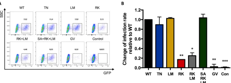
Compared to virus strains containing the wild-type HIV-1
NL4-3 sequence, viruses containing the HLA-B27-associated RK
mutation in p24 Gag showed a significant (82,8%) reduction of
GFP
⫹CD4
⫹T cells (
P
⫽
0.001) (
Fig. 1B
). A modest but
nonsig-nificant reduction in GFP
⫹CD4
⫹T cells was also observed with
viruses containing the HLA-B57-associated TN mutation (10.5%;
not significant [n.s.]), while no difference was observed with the
LM mutation (2.5%; n.s.) (
Fig. 1B
). The observed significant
de-crease in GFP
⫹CD4
⫹T cells using the VSV-G-pseudotyped
NL4-3
⌬Env HIV-1 containing the R264K mutation, the merely
modest decrease in the presence of the T242N mutation, and the
lack of any effect in the presence of the L268M mutation observed
here are in line with previous data from other groups using
differ-ent systems, such as reporter cell lines (
5
,
6
,
26
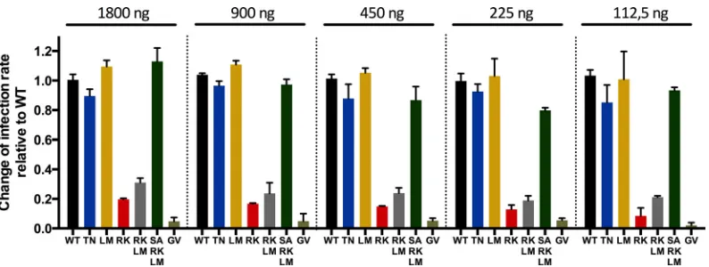
). To ensure that the
differences in infectivity of the investigated viral strains were not
due to differences in particle-to-infectivity ratios, we performed
an analysis of infectivity over a range of different p24
concentra-tions. Therefore, we repeated the assay described above in PBMCs
with different amounts of p24 (1,800 ng, 900 ng, 450 ng, 225 ng,
and 112.5 ng) (
Fig. 2
). The results showed that the amount of p24
FIG 1GFP expression in CD4⫹T cells following infection with viruses containing escape mutations and compensatory mutations. Shown is an assessment of the infectivity of escape mutations (T242N mutation in the TW10 epitope [TN] and R264K in the KK10 epitope [RK] in p24 Gag) and compensatory mutations of RK (LM, RK plus LM, and SA plus RK plus LM) in p24 Gag. PBMCs were infected with a standardized amount of p24 of HIV-GFP. (A) Dot blots showing GFP expression of CD3⫹CD4⫹CD8⫺lymphocytes. GFP positivity of the cells indicates infected cells. The numbers show the percentages of GFP⫹/infected cells. SSC, side scatter. (B) Fold change of infected cells over WT, which was set to 1 (n⫽3). ***,P⬍0.0001 versus WT; **,P⬍0.01 versus WT; *,P⬍0.05 versus WT. The error bars indicate ranges.
on November 7, 2019 by guest
http://jvi.asm.org/
[image:3.585.97.491.65.207.2]input did not alter the observed differences between the mutated
virus strains.
In addition, we assessed the impact of the G221V mutation in
p24 Gag, which alters the catalytic center of the CypA-binding site
and thereby inhibits CypA binding (
12
), on the percentage of
GFP
⫹cells. Interestingly, with a 97.8% decrease in GFP
⫹CD4
⫹T
cells, the G221V mutation showed the strongest alteration
com-pared to wild-type virus (
P
⫽
0.003), indicating the necessity for
CypA binding during capsid trafficking and viral cDNA
integra-tion in primary human cells (
Fig. 1
). Taken together, these data
show that some mutations within p24 Gag, and in particular the
HLA-B27-associated R264K mutation, as well as the G221V
mu-tation at the catalytic center of the CypA-binding site, can have a
strong impact on the viral life cycle.
The p24 Gag L268M and S173A mutations restore the
infec-tivity of the R264K virus.
After observing a significant reduction
in GFP
⫹CD4
⫹T cells in the context of the HLA-B27-associated
R264K mutation, we next determined whether the described
com-pensatory mutations of the R264K mutation (
6
) were able to
re-store GFP expression. It was previously shown that the L268M
mutation in p24 Gag appeared before the development of the
R264K mutation in HLA-B27
⫹HIV-1-infected individuals (
6
).
Subsequently, a third mutation (S173A) developed and almost
completely restored viral fitness to levels similar to those observed
using wild-type sequences (
6
). Therefore, VSV-G-pseudotyped
NL4-3
⌬
Env HIV-1 encoding both the R264K and L268M
muta-tions (referred to as RK plus LM) or all three mutamuta-tions (S173A,
R264K, and L268M, referred to as SA plus RK plus LM) were
constructed. Viruses containing the RK plus LM mutations
showed a 74.9% reduction of infectivity compared to the wild type
(
P
⫽
0.011), already slightly recovering the infectivity of the RK
mutation as described above. Furthermore, with a modest
in-crease of 3.8% in GFP
⫹cells compared to the wild-type virus
(n.s.), viruses containing the SA plus RK plus LM variants were
able to fully restore the viral fitness of the R264K variant, as
pre-viously reported (
6
). Overall, these data show that the
compensa-tory mutations of R264K in HIV-1 p24 Gag were able to restore
GFP expression in infected CD4
⫹T cells to wild-type levels, as
previously reported using reporter cell lines (
6
,
26
).
Modulation of the kinetics of HIV-1 capsid uncoating by
HLA-B27- and HLA-B57-associated sequence mutations.
The
above-mentioned data demonstrate a significant impact of the
HLA-B27-associated CTL escape mutation R264K in p24 Gag on the
ability of GFP-encoding viruses to infect CD4
⫹T cells and to
express GFP and also demonstrate that compensatory
muta-tions can rescue this effect. However, it remains unclear
pre-cisely which steps involved in viral cDNA trafficking to the
nucleus, cDNA integration, and subsequent GFP production
were affected by these mutations. As modeling studies have
suggested that HLA-B27- and HLA-B57-associated mutations
in p24 Gag might affect sites critical for capsid formation and
stability, we subsequently employed a kinetic functional CsA
washout assay to determine the consequences of mutations
within p24 Gag for capsid stability.
Using the CsA washout assay, viruses containing the
HLA-B27- and HLA-B57-associated CTL escape mutations T242N,
L268M, and R264K were compared to the NL4-3 WT virus. The
assay uses cyclosporine to inhibit Trim-CypA of OMK cells, which
restricts viral particles with an intact capsid. Thus, by washing out
CsA in a time-dependent manner, increasing amounts of
un-coated particles are not restricted by Trim-CypA any longer and
can infect the OMK cell. The final readout is the quantification of
GFP-positive OMK cells via flow cytometry (
Fig. 3A
). The GV
mutation inhibits Trim-CypA binding, and thus, viruses
contain-ing the GV mutation resulted in high percentages of GFP-positive
OMK cells at any time point independent of CsA washout (
Fig.
3B
). Raw mean data demonstrating the percentage of
GFP-posi-tive cells at each time point following CsA washout (normalized to
the highest percentage of GFP
⫹cells, defined as 100%) are shown
in
Fig. 3C
to
E
. The median capsid-uncoating half-time was longer
than that of the HIV-GFP WT for viruses containing the
muta-tions T242N (WT, 110 min [range {RNG}, 93 to 137 min] versus
T242N, 133 min [RNG, 106 to 151 min];
P
⫽
0.05) and R264K
(WT, 118 min [RNG, 97 to 153 min] versus R264K, 166 min
[RNG, 143 to 179 min];
P
⬍
0.001). A modestly but not
signifi-cantly longer uncoating half-time was observed for viruses
con-taining the L268M mutation (WT, 105 min [RNG, 76 to 122 min]
versus L268M, 113 min [RNG, 91 to 126 min]; n.s.). For a better
evaluation of the differences between WT virus and viruses
con-taining the respective escape mutations,
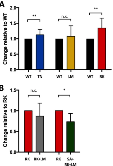
Fig. 4A
shows each pair of
comparative assays with the results of the WT virus normalized to
1. These data demonstrate that the HLA-B57- and
HLA-B27-as-FIG 2GFP expression in CD4⫹T cells following infection with different amounts of p24 of viruses containing escape mutations and compensatory mutations. The infectivities of the escape mutations (TN and RK in p24 Gag) and compensatory mutations of RK (LM, RK plus LM, and SA plus RK plus LM) in p24 Gag were assessed with the use of different amounts of p24 (1,800 ng, 900 ng, 450 ng, 225 ng, and 112.5 ng). The patterns of the infectivities of the different viral strains remained similar over a wide range of p24 input (PBMCs from one healthy donor; three wells per virus for each dose of p24). The error bars indicate ranges.
on November 7, 2019 by guest
http://jvi.asm.org/
[image:4.585.95.492.64.214.2]sociated point mutations T242N and R264K extended the
capsid-uncoating half-time, favoring a model of enhanced rigidity of the
capsid structure in these mutated viral strains.
Since the above-described compensatory mutations RK plus
LM and SA plus RK plus LM were able to restore the fitness of
viruses containing the R264K mutation, we next investigated
whether the combined mutations RK plus LM and SA plus RK
plus LM were also able to restore the longer uncoating half-time
FIG 3GFP⫹OMK cells following infection with viruses containing escape mutations and compensatory mutations after CsA washout in a time-dependent manner. (A) Dot plots showing original data from the CsA washout assay. (Top row) CsA washout of HIV-GFP WT with increasing amounts of GFP⫹OMK cells over time. (Middle row) Same assay with HIV-GFP WT but with EtOH instead of CsA as a control. Since EtOH does not inhibit the HIV-1 restriction factor Trim-Cyp, expressed in OMK cells, only very few infectious cells could be detected, and there was no time-related effect. (Bottom row) CsA washout assay with the HIV-GFP R264K mutation. The amount of GFP⫹OMK cells also increased over time, as seen with WT virus, but a substantial delay could be observed, especially in the first 2 h. (B) CsA and EtOH washout assay of HIV-GFP WT and HIV-GFP G221V. The amount of GFP⫹cells increased over time in the CsA washout assay using HIV-GFP WT, whereas this effect was not observed in the EtOH washout, as described for panel A. Neither CsA nor EtOH washout showed a time-dependent effect using the HIV-GFP G221V mutant, since the G221V mutation altered the CypA catalytic binding site, preventing Trim-CypA from binding to HIV-1 capsid. (C to E) CsA washout assays comparing the TN (n⫽9), LM (n⫽8), and RK (n⫽8) mutations, respectively, to the HIV-GFP WT. (F and G) CsA washout assays comparing compensatory mutations RK plus LM and SA plus RK plus LM, respectively, to HIV-GFP RK (bothn⫽6). The error bars indicate ranges.
on November 7, 2019 by guest
http://jvi.asm.org/
observed for the R264K-containing strain. Therefore, each of
these compensatory mutations was directly compared to the
R264K mutation using the CsA washout assay. The raw mean data
for each time point of the washout assay of R264K versus RK plus
LM (
n
⫽
6) and R264K versus SA plus RK plus LM (
n
⫽
6) are
shown in
Fig. 3F
and
G
. A slightly shorter but not significantly
different capsid-uncoating half-time compared to the R264K
mu-tation was observed for the RK plus LM variant (R264K, 137 min
[RNG, 109 to 150 min] versus RK plus LM, 128 min [RNG, 102 to
134 min]; n.s.). A significantly shorter uncoating half-time was
seen with the SA plus RK plus LM variant compared to R264K
(R264K, 153 min [RNG, 109 to 175 min] versus SA plus RK plus
LM, 104 min {lsqb]confidence interval {CI}, 95 to 138 min];
P
⫽
0.01). For a better evaluation of the comparisons between the
R264K mutations alone and in the presence of the compensatory
mutations,
Fig. 4B
shows each pair with the R264K variant set to 1.
Taken together, the described compensatory mutations for the
HLA-B27-associated R264K mutation also restored HIV-1
cap-sid-uncoating half-times.
Correlation between HIV capsid-uncoating half-time and
vi-ral GFP production in CD4
ⴙT cells.
The viral replication assay
using PBMCs and the CsA washout assay using OMK cells both
measure the impact of p24 Gag sequence mutations on viral
rep-lication steps following viral entry and prior to
de novo
assembly of
viruses using different approaches. We were therefore interested
in assessing whether the results of the two assays showed any
cor-relation. The half-time of capsid uncoating of the WT virus was set
to 1, and the fold changes of average capsid-uncoating half-times
in OMK cells and the fold changes of GFP
⫹CD4
⫹T cells of the
respective viral variants were correlated. A correlation was
ob-served for the initial viral escape mutations detectable in
HLA-B27/57
⫹HIV-infected individuals (Pearson
r
⫽ ⫺0.95;
P
⫽
0.048) (
Fig. 5A
). The same analysis was performed for the R264K
variant and its compensatory mutations. The results from the CsA
assay and GFP
⫹CD4
⫹T cells again showed a correlation;
how-ever, no statistical significance was reached, likely due to the
lim-ited data points available (Pearson
r
⫽ ⫺
0.97;
P
⫽
0.16) (
Fig. 5B
).
These correlations between the results from the two assays suggest
that the impact of CTL-induced viral sequence mutations within
HIV-1 capsid on GFP expression is directly linked to the capsid
stability of the respective strains.
FIG 4Half-life of uncoating of viruses containing escape mutations and com-pensatory mutations as calculated from the CsA washout assay. The uncoating half-time was calculated from original data from the CsA washout assay, as described in Materials and Methods. (A) The WT was set to 1, and the escape mutations were calculated as fold change over the WT. While viruses contain-ing the LM and TN mutations showed only a moderately longer uncoatcontain-ing half-time, with increases of 13% and 8%, respectively, viruses containing the RK mutation showed the most impaired uncoating, with an increase of 35%. Only viruses containing the mutations T242N (n⫽9) and R264K (n⫽8) proved to have a significant impact on the capsid-uncoating half-time com-pared to the WT (P⫽0.007 andP⫽0.001, respectively). (B) RK was set to 1, and the compensatory escape mutations were calculated as the fold change over RK. RK plus LM showed an ameliorated (13% faster; n.s.;n⫽6) and SA plus RK plus LM showed a significantly shorter (27%;P⫽0.011;n⫽6) uncoating time compared to RK. The error bars indicate ranges.
FIG 5Correlation of capsid-uncoating half-time and viral GFP expression in CD4⫹T cells. (A) The fold changes in the uncoating half-time and viral fitness of viruses containing the escape mutations over those of the WT were plotted against each other and showed a significant correlation (Pearsonr⫽ ⫺0.95; P⫽0.048). (B) The fold changes in the uncoating half-time and viral fitness of the compensatory mutations over the R264K mutation in the KK10 epitope of p24 Gag (RK) were plotted against each other and showed a correlation that did not reach statistical significance (Pearsonr⫽ ⫺0.97; n.s.).
on November 7, 2019 by guest
http://jvi.asm.org/
[image:6.585.69.262.66.348.2] [image:6.585.324.519.66.367.2]DISCUSSION
Large genome-wide association studies (GWAS) performed in
co-horts of HIV-1-infected individuals with spontaneous control of
HIV-1 viremia (HIV elite controllers) demonstrated that all single
nucleotide polymorphisms (SNPs) associated with viral control
were located in HLA class I genes (
27
,
28
). Multiple studies have
identified HLA-B57 and HLA-B27 as the two HLA class I alleles
most strongly associated with HIV-1 control (
27
). While
immu-nodominant virus-specific CTLs in HLA-B57
⫹individuals
in-fected with HIV-1 are directed against the TW10 (TSTLQEQ
IGW) epitope within p24 Gag, CTLs in HLA-B27
⫹individuals are
directed against the KK10 (KRWIILGLNK) p24 Gag epitope,
re-sulting in frequent CTL escape mutations within these epitopes
(B57
⫹, T242N; B27
⫹, R264K and L268M) (
2
,
6
,
26
,
29
).
Addi-tional studies linked the low HIV-1 viremia observed in HLA-B27/
57
⫹individuals to reduced viral fitness of HIV-1 strains
contain-ing the selected CTL escape mutations (
5
,
6
,
29
). The significant
impact of single point mutations within the p24 Gag region on
viral replicative fitness is believed to be due to the high sequence
conservation of the region (
18
), with sequence mutations leading
to structural changes (
30
). Additionally, it has been suggested that
some of these point mutations in p24 Gag can destabilize specific
helixes of the CA protein (
4
), which might lead to an alteration of
the stability of the HIV-1 capsid structure. Here, we investigated
the impact of escape, as well as compensatory, mutations seen in
HIV-1-infected HLA-B57/27
⫹individuals on viral fitness and the
contribution of capsid stability to this process, using a functional
CsA washout assay.
Several studies have assessed the impact of point mutations on
viral replication fitness using assays that are based on reporter cell
lines (
5
,
6
,
26
). In line with findings from these studies but using
human primary PBMCs, we observed that the
HLA-B27-re-stricted escape mutation R264K in the KK10 epitope, and to a
lesser extent the HLA-B57-restricted T242N mutation in the
TW10 epitope, in p24 Gag resulted in a reduction of GFP
⫹CD4
⫹T cells and that the described compensatory mutations for the
R264K mutation were able to partially reconstitute this effect.
In the current study, we used a VSV-G-pseudotyped HIV-1
NL4-3
⌬
Env strain expressing GFP in place of Nef, enabling flow
cytometric quantification of infected cells producing new viral
proteins by measuring GFP
⫹cells. As VSV pseudotyping was used
for all the virus strains tested, viral infection of cells was
standard-ized, and changes in GFP
⫹cells were a result of postfusion
differ-ences, including capsid uncoating, reverse transcription, forming
of the PIC, integration of cDNA into the host genome, and GFP
transcription and translation. VSV-G pseudotyping was
em-ployed for all the viruses that were used in this study, since the CsA
washout assay is based on the use of OMK cells, which lack a CD4
receptor. In order to correlate capsid uncoating and infectivity,
VSV-G-pseudotyped viral strains were used in all the assays, even
though VSV-G pseudotyping has been reported to alter entry
pathways compared to viruses with a native HIV-1 envelope (
31
,
32
). Viruses containing the HLA-B27-associated R264K
muta-tion, and also the G221V mutation affecting the catalytic center of
the CypA-binding site, showed significantly fewer GFP
⫹cells
fol-lowing infection of PBMCs than the wild-type virus, suggesting
that viruses containing these mutations had reduced infectivity
(
12
).
Given that the HLA-B27- and HLA-B57-associated sequence
mutations used in this study were all located in HIV-1 p24 Gag,
which encodes capsid, we hypothesized that the impact of these
mutations on infectivity was affected by the process of capsid
un-coating following infection. Indeed, we observed a correlation
be-tween the percentage of GFP
⫹cells and the half-time of capsid
uncoating as measured using the CsA washout assay. The R264K
mutation had the strongest negative effect on the percentage of
GFP
⫹cells compared to NL4-3 wild-type sequences and was also
the mutation that resulted in the most prolonged
capsid-uncoat-ing half-time. Furthermore, the compensatory mutations
de-scribed for R264K, RK plus LM and SA plus RK plus LM, were able
to both shorten the half-time of capsid uncoating and enhance the
percentage of GFP
⫹PBMCs compared to viruses containing the
R264K mutation alone. While other steps of the virus life cycle
following capsid uncoating can also contribute to reduced
infec-tivity, including integration of cDNA into the host genome,
tran-scription, and translation, these data demonstrate that modulated
capsid stability is a major determinant of the reduced infectivity of
HIV-1 variants containing frequent HLA-B27- and
HLA-B57-as-sociated sequence mutations within HIV-1 capsid.
ACKNOWLEDGMENTS
OMK cells were provided by the laboratory of Adam Grundhoff. We thank the UKE cohort of healthy donors for their contributions to this study.
This work was funded by the Deutsches Zentrum für Infektionsforsc-hung (DZIF) through a clinical leave fellowship to P.S. and by the pro-gram area Molecular Mechanisms of Viral Pathogenesis of the Heinrich Pette Institute, a Leibniz Institute for Experimental Virology.
FUNDING INFORMATION
This work, including the efforts of Philipp Schommers, was funded by Deutsches Zentrum für Infektionsforschung (DZIF) (TI 07.001).
REFERENCES
1.Goulder PJ, Brander C, Tang Y, Tremblay C, Colbert RA, Addo MM, Rosenberg ES, Nguyen T, Allen R, Trocha A, Altfeld M, He S, Bunce M, Funkhouser R, Pelton SI, Burchett SK, McIntosh K, Korber BT, Walker BD.2001. Evolution and transmission of stable CTL escape mutations in HIV infection. Nature412:334 –338.http://dx.doi.org/10.1038/35085576. 2.Leslie AJ, Pfafferott KJ, Chetty P, Draenert R, Addo MM, Feeney M, Tang Y, Holmes EC, Allen T, Prado JG, Altfeld M, Brander C, Dixon C, Ramduth D, Jeena P, Thomas SA, St John A, Roach TA, Kupfer B, Luzzi G, Edwards A, Taylor G, Lyall H, Tudor-Williams G, Novelli V, Martinez-Picado J, Kiepiela P, Walker BD, Goulder PJ. 2004. HIV evolution: CTL escape mutation and reversion after transmission. Nat Med10:282–289.http://dx.doi.org/10.1038/nm992.
3.Streeck H, Lichterfeld M, Alter G, Meier A, Teigen N, Yassine-Diab B, Sidhu HK, Little S, Kelleher A, Routy JP, Rosenberg ES, Sekaly RP, Walker BD, Altfeld M.2007. Recognition of a defined region within p24 gag by CD8⫹T cells during primary human immunodeficiency virus type 1 infection in individuals expressing protective HLA class I alleles. J Virol
81:7725–7731.http://dx.doi.org/10.1128/JVI.00708-07.
4.Martinez-Picado J, Prado JG, Fry EE, Pfafferott K, Leslie A, Chetty S, Thobakgale C, Honeyborne I, Crawford H, Matthews P, Pillay T, Rousseau C, Mullins JI, Brander C, Walker BD, Stuart DI, Kiepiela P, Goulder P.2006. Fitness cost of escape mutations in p24 Gag in associa-tion with control of human immunodeficiency virus type 1. J Virol80:
3617–3623.http://dx.doi.org/10.1128/JVI.80.7.3617-3623.2006. 5.Brockman MA, Schneidewind A, Lahaie M, Schmidt A, Miura T,
Desouza I, Ryvkin F, Derdeyn CA, Allen S, Hunter E, Mulenga J, Goepfert PA, Walker BD, Allen TM.2007. Escape and compensation from early HLA-B57-mediated cytotoxic T-lymphocyte pressure on hu-man immunodeficiency virus type 1 Gag alter capsid interactions with cyclophilin A. J Virol 81:12608 –12618. http://dx.doi.org/10.1128/JVI .01369-07.
on November 7, 2019 by guest
http://jvi.asm.org/
position. J Virol71:5382–5390.
10. Hulme AE, Perez O, Hope TJ.2011. Complementary assays reveal a relationship between HIV-1 uncoating and reverse transcription. Proc Natl Acad Sci U S A 108:9975–9980. http://dx.doi.org/10.1073/pnas .1014522108.
11. Xu H, Franks T, Gibson G, Huber K, Rahm N, Strambio De Castillia C, Luban J, Aiken C, Watkins S, Sluis-Cremer N, Ambrose Z. 2013. Evidence for biphasic uncoating during HIV-1 infection from a novel imaging assay. Retrovirology10:70.http://dx.doi.org/10.1186/1742-4690 -10-70.
12. Schaller T, Ocwieja KE, Rasaiyaah J, Price AJ, Brady TL, Roth SL, Hue S, Fletcher AJ, Lee K, KewalRamani VN, Noursadeghi M, Jenner RG, James LC, Bushman FD, Towers GJ.2011. HIV-1 capsid-cyclophilin interactions determine nuclear import pathway, integration targeting and replication efficiency. PLoS Pathog7:e1002439.http://dx.doi.org/10.1371 /journal.ppat.1002439.
13. Lahaye X, Satoh T, Gentili M, Cerboni S, Conrad C, Hurbain I, El Marjou A, Lacabaratz C, Lelievre JD, Manel N.2013. The capsids of HIV-1 and HIV-2 determine immune detection of the viral cDNA by the innate sensor cGAS in dendritic cells. Immunity39:1132–1142.http://dx .doi.org/10.1016/j.immuni.2013.11.002.
14. Rasaiyaah J, Tan CP, Fletcher AJ, Price AJ, Blondeau C, Hilditch L, Jacques DA, Selwood DL, James LC, Noursadeghi M, Towers GJ.2013. HIV-1 evades innate immune recognition through specific cofactor re-cruitment. Nature503:402– 405.http://dx.doi.org/10.1038/nature12769. 15. Gao D, Wu J, Wu YT, Du F, Aroh C, Yan N, Sun L, Chen ZJ.2013. Cyclic GMP-AMP synthase is an innate immune sensor of HIV and other retroviruses. Science 341:903–906. http://dx.doi.org/10.1126 /science.1240933.
16. Jakobsen MR, Bak RO, Andersen A, Berg RK, Jensen SB, Tengchuan J, Laustsen A, Hansen K, Ostergaard L, Fitzgerald KA, Xiao TS, Mikkelsen JG, Mogensen TH, Paludan SR.2013. IFI16 senses DNA forms of the lentiviral replication cycle and controls HIV-1 replication. Proc Natl Acad Sci U S A110:E4571–E4580.http://dx.doi.org/10.1073 /pnas.1311669110.
17. Kawai T, Akira S.2006. Innate immune recognition of viral infection. Nat Immunol7:131–137.
18. Dahirel V, Shekhar K, Pereyra F, Miura T, Artyomov M, Talsania S, Allen TM, Altfeld M, Carrington M, Irvine DJ, Walker BD, Chakraborty AK.2011. Coordinate linkage of HIV evolution reveals re-gions of immunological vulnerability. Proc Natl Acad Sci U S A108:
11530 –11535.http://dx.doi.org/10.1073/pnas.1105315108.
19. Hulme AE, Hope TJ.2014. The cyclosporin A washout assay to detect HIV-1 uncoating in infected cells. Methods Mol Biol1087:37– 46.http: //dx.doi.org/10.1007/978-1-62703-670-2_4.
20. Perez-Caballero D, Hatziioannou T, Zhang F, Cowan S, Bieniasz PD.
2005. Restriction of human immunodeficiency virus type 1 by
TRIM-A cryptic sensor for HIV-1 activates antiviral innate immunity in dendritic cells. Nature467:214 –217.http://dx.doi.org/10.1038/nature09337. 25. Cronin J, Zhang XY, Reiser J.2005. Altering the tropism of lentiviral
vectors through pseudotyping. Curr Gene Ther5:387–398.http://dx.doi .org/10.2174/1566523054546224.
26. Miura T, Brockman MA, Schneidewind A, Lobritz M, Pereyra F, Rathod A, Block BL, Brumme ZL, Brumme CJ, Baker B, Rothchild AC, Li B, Trocha A, Cutrell E, Frahm N, Brander C, Toth I, Arts EJ, Allen TM, Walker BD.2009. HLA-B57/B*5801 human immunodeficiency vi-rus type 1 elite controllers select for rare gag variants associated with re-duced viral replication capacity and strong cytotoxic T-lymphocyte rec-ognition. J Virol83:2743–2755.http://dx.doi.org/10.1128/JVI.02265-08. 27. Pereyra F, Jia X, McLaren PJ, Telenti A, de Bakker PI, Walker BD,
Ripke S, Brumme CJ, Pulit SL, Carrington M, Kadie CM, Carlson JM, Heckerman D, Graham RR, Plenge RM, Deeks SG, Gianniny L, Craw-ford G, Sullivan J, Gonzalez E, Davies L, Camargo A, Moore JM, Beattie N, Gupta S, Crenshaw A, Burtt NP, Guiducci C, Gupta N, Gao X, Qi Y, Yuki Y, Piechocka-Trocha A, Cutrell E, Rosenberg R, Moss KL, Lemay P, O’Leary J, Schaefer T, Verma P, Toth I, Block B, Baker B, Rothchild A, Lian J, Proudfoot J, Alvino DM, Vine S, Addo MM, Allen TM, et al.
2010. The major genetic determinants of HIV-1 control affect HLA class I peptide presentation. Science330:1551–1557.http://dx.doi.org/10.1126 /science.1195271.
28. Carlson JM, Listgarten J, Pfeifer N, Tan V, Kadie C, Walker BD, Ndung’u T, Shapiro R, Frater J, Brumme ZL, Goulder PJ, Heckerman D.2012. Widespread impact of HLA restriction on immune control and escape pathways of HIV-1. J Virol86:5230 –5243.http://dx.doi.org/10 .1128/JVI.06728-11.
29. Schneidewind A, Brockman MA, Sidney J, Wang YE, Chen H, Susco-vich TJ, Li B, Adam RI, Allgaier RL, Mothe BR, Kuntzen T, Oniangue-Ndza C, Trocha A, Yu XG, Brander C, Sette A, Walker BD, Allen TM.
2008. Structural and functional constraints limit options for cytotoxic T-lymphocyte escape in the immunodominant HLA-B27-restricted epitope in human immunodeficiency virus type 1 capsid. J Virol82:5594 – 5605.http://dx.doi.org/10.1128/JVI.02356-07.
30. Liu Y, Rao U, McClure J, Konopa P, Manocheewa S, Kim M, Chen L, Troyer RM, Tebit DM, Holte S, Arts EJ, Mullins JI.2014. Impact of mutations in highly conserved amino acids of the HIV-1 Gag-p24 and Env-gp120 proteins on viral replication in different genetic backgrounds. PLoS One9:e94240.http://dx.doi.org/10.1371/journal.pone.0094240. 31. Aiken C.1997. Pseudotyping human immunodeficiency virus type 1
(HIV-1) by the glycoprotein of vesicular stomatitis virus targets HIV-1 entry to an endocytic pathway and suppresses both the requirement for Nef and the sensitivity to cyclosporin A. J Virol71:5871–5877.
32. Melikyan GB.2014. HIV entry: a game of hide-and-fuse? Curr Opin Virol
4:1–7.http://dx.doi.org/10.1016/j.coviro.2013.09.004.
on November 7, 2019 by guest
http://jvi.asm.org/