SMEAR LAYER REMOVAL BY ULTRASONICALLY
ACTIVATED AND DIODE LASER ACTIVATED - EDTA AND
CHITOSAN - AN INVITRO STUDY
Dissertation submitted to
THE TAMILNADU Dr. M.G.R. MEDICAL UNIVERSITY
In partial fulfilment for the Degree of
MASTER OF DENTAL SURGERY
BRANCH - IV
CONSERVATIVE DENTISTRY AND ENDODONTICS
This is to certify that the dissertation titled “ELECTRON MICROSCOPIC
COMPARATIVE ANALYSIS OF SMEAR LAYER REMOVAL BY
ULTRASONICALLY ACTIVATED AND DIODE LASER ACTIVATED -
EDTA AND CHITOSAN - AN INVITRO STUDY” is a bonafide work done by
Dr. VINEETHA. C. S, Post graduate student, during the course of the study for the degree of MASTER OF DENTAL SURGERY in the specialty of BRANCH-IV, CONSERVATIVE DENTISTRY AND ENDODONTICS, Vivekanandha Dental College for Women,Tiruchengode, during the period of 2016-2019.
Signature of the H.O.D and Guide
Dr. Vaiyapuri Ravi, M.D.S.,
AND HEAD OF THE INSTITUTION
This is to certify that Dr. VINEETHA.C. S, Post graduate student (2016-2019) in the DEPARTMENT OF CONSERVATIVE DENTISTRY AND ENDODONTICS,
Vivekanandha Dental College for Women, has done this dissertation titled
“ELECTRON MICROSCOPIC COMPARATIVE ANALYSIS OF SMEAR
LAYER REMOVAL BY ULTRASONICALLY ACTIVATED AND DIODE
I hereby declare that no part of the dissertation will be utilized for gaining financial assistance for research or other promotions without obtaining prior permission of the Principal, Vivekanandha Dental College for Women, Tiruchengode. In addition, I declare that no part of this work will be published either in print or electronic format without the permission of the Guide who has been actively involved in the dissertation. The author has the right to reserve publishing of work solely with prior permission of the Principal, Vivekanandha Dental College for Women, Tiruchengode.
TITLE OF DISSERTATION
ELECTRON MICROSCOPIC COMPARATIVE ANALYSIS OF SMEAR LAYER REMOVAL BY
ULTRASONICALLY ACTIVATED AND DIODE LASER ACTIVATED - EDTA AND CHITOSAN -
AN INVITRO STUDY
PLACE OF STUDY VIVEKANANDHA DENTAL COLLEGE FOR
WOMEN
DURATION OF THE
COURSE 3 YEARS (2016-2019)
HEAD OF THE DEPARTMENT AND GUIDE
This is to certify that this dissertation work titled “ELECTRON
MICROSCOPIC COMPARATIVE ANALYSIS OF SMEAR LAYER REMOVAL
BY ULTRASONICALLY ACTIVATED AND DIODE LASER ACTIVATED
First of all, my sincere thanks and deep sense of gratitude to
Dr.Capt.S.Gokulanathan, B.Sc, M.D.S., (Dean) and Dr. N.Balan, M.D.S., (Principal), Vivekanandha Dental College for Women, for permitting me to pursue this work.
Without the encouragement of my Professor and Head of the Department, Dr.Vaiyapuri Ravi, M.D.S., Department of Conservative Dentistry and Endodontics, Vivekanandha Dental College for Women, who is also my guide for this dissertation, this project would not have materialised. I convey my heartfelt thanks for his innovative ideas, invaluable counsel and immeasurable encouragement throughout the course of the work. His care, matchless theoretical and clinical skills, coupled with ideals and unwavering guidance and constant support during my postgraduate tenure has enabled me to successfully conclude this effort.
Dr.A.Andamuthu Sivakumar, M.D.S., Dr.AjiMarkose, M.D.S., Dr. P.V.Syamala,
M.D.S., Readers Dr.J.S.Sivakumar, M.D.S., Dr.A.S.Prasad, M.D.S., Senior lecturer,
Dr.M.Chittrarasu, M.D.S., Department of Conservative Dentistry and Endodontics, for their support and encouragement.
I sincerely thank Dr.Sashidharan Nair, HOD of Department of Applied Sciences, PSG Institute of Technology for all the help he has provided me in using the magnetic stirrer. I am thankful to Dr.Anil Mathew M.D., Department of Community Medicine, PSG Institute of Medical Sciences for his guidance in the statistical works of this study.
I am grateful to my seniors Dr.Iswariya.R, Dr.Karthipriya.G, Dr.Vishnuvardhini.S, for their kind help and support during this period of study. It would not be justifiable on my part if I do not acknowledge the help of my batch mates
Dr.Chandrika.R.P, Dr.Sowmiya.T and juniors Dr.Anuradha.R.S, Dr.Brindha.L, Dr.Pushpalatha.K, Dr.Annabelle Primola, Dr.Ragavi.S and Dr.Sasmitha.C for their continued support and encouragement throughout my postgraduate programme.
CONTENTS
S.NO CONTENT PAGE. NO
1. INTRODUCTION 1-4
2. AIM AND OBJECTIVE 5
3. REVIEW OF LITERATURE 6-15
4 MATERIALS AND METHODS 16-36
5. RESULTS 37-39
6. DISCUSSION 40-45
7. SUMMARY AND CONCLUSION 46-47
1
INTRODUCTION
Root canal treatment is an essential procedure carried out in various clinical situations which include teeth with deep caries and irreversible pulpitis, following trauma, attrition, resorption and in certain clinical conditions such as prosthetic rehabilitation of missing teeth, when the tooth/teeth need to be taken as an abutment.
The success of an endodontic procedure majorly lies on 3 important factors that include – Creating a straight line access, proper cleaning and shaping of the canals and producing a 3 Dimensional obturation with a good seal.
Cleaning and Shaping previously described as Biomechanical preparation is one of the most essential step in achieving a successful clinical outcome. It refers to shaping the canal to produce a smooth, continuously tapering canal from the orifice to the apex and disinfecting the canal walls by removal of the dentinal debris, infected pulpal tissue, bacteria and its by-products.
Shaping of the canal wall is carried out using endodontic hand files or rotary instruments by cutting the root dentin along the canal walls. This leads to the production of an irregular amorphous smear layer.
Erick et al identified smear layer using Scanning Electron microscope in 1970s. He stated that “Smear layer contains inorganic debris and organic material.
The organic material includes vital and necrotic pulpal tissues, microorganisms and their metabolic products and odontoblastic processes”. The smear layer is divided
2 This was discussed by Cameron et al1 and Mader et al2. Brannstrom and Johnson estimated the thickness of smear layer to be 2-5 µm in 19743.
The controversy regarding retaining or removing smear layer still prevails. Some investigators support the concept of not removing the smear layer because it would seal the tubules thus preventing the entry of bacteria and their by-products, whereas others support the concept of its removal because they believed that the smear layer could act as a scaffold for harbouring bacteria and it would interfere in production of proper seal.
Cleaning the canal refers to the removal of smear layer, which can be done using chemical agents, ultrasonics and/or by the use of laser. Chemical agents such as sodium hypochlorite, EDTA and organic acids in combination with ultrasonics and laser agitation have also been used.
Ethylenediaminetetraacetic acid (EDTA) is the most commonly used irrigant/chelating agent for removal of smear layer4, 5, 6. It promotes decalcification by chelating the calcium ions in dentine at approximate depths of 20–30 µm within 5 min7. EDTA has harmful effect on periapical tissues and this has led researchers to seek more biocompatible material as an alternative. Weak acids, like apple cider vinegar and citric acid have also been studied 6, 8, 9. Citric acid reacts rapidly with calcium ions low cytotoxicity10 and antimicrobial properties11.
3 Application of chitosan has been seen in the field of medicine and pharmaceuticals (antibacterial and anti-tumour agent, drug carrier, wound healing accelerator), biotechnology (enzyme and cell carrier, chromatography resin), environment (water treatment), agriculture (seed preparation), cosmetics and food (iron and calcium absorption accelerator, fibre source)15. The use of Chitosan as a chelating agent has not been widely discussed. Recently, studies are being carried out to assess the efficacy of Chitosan as an irrigant for its chelating and antibacterial property.
Syringe irrigation is the conventional and still widely used irrigation technique. Various techniques such as moving gutta-percha (GP) cones up and down in the root canal (manual dynamic activation [MDA]) to instruments energized by (ultra)sonic or laser devices have been used for the activation of irrigants. Chemical chelating agents in combination with Ultrasonic agitation and lasers have shown to produce better results than conventional syringe irrigation.
The use of ultrasonics has been proposed as a possible solution to the problem of debriding and disinfecting the root canal system. The use of ultrasound after completion of hand or rotary instrumentation has been shown to reduce the number of bacteria. The cavitational effect produced on the canal walls by Ultrasonic agitation was found to have improved effects in combination with various irrigating solutions.1
4 disinfection of the deep radicular dentin. The type of irrigating solution used and the laser wavelength determines the quantity of irrigant absorbed into the canal walls.
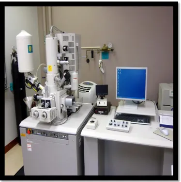
One of the effectively used methods to determine the ability of smear layer removal is Scanning electron microscopy. Among the various scoring systems for quantifying the remaining smear layer, Gutmann’s scoring criteria was followed in the
present study.
5
AIM AND OBJECTIVES
AIM OF THE STUDY
The aim of this study is
To compare the efficacy of Ultrasonically activated and Laser activated
EDTA and Chitosan on smear layer removal by using Scanning Electron Microscope
OBJECTIVES OF THE STUDY:
To evaluate the efficacy of Chitosan on smear layer removal
To compare the efficacy of Chitosan and EDTA
6
REVIEW OF LITERATURE
Cameron et al1 in 1983, studied the smear layer removal efficacy of ultrasonics by scanning electron microscopy.
Yamada et al16 in 1983, compared EDTA and NaOCl solutions as a final flush with various other solutions, or in combinations, using SEM. He concluded that 10 ml of 17% EDTA buffered to pH 7.7 followed by 10ml 5.25% NaOCI solution had the highest ability of smear layer removal.
Bystrom et al17 in 1985, compared the efficacy of 0.5% and 5 % NaOCl sodium solutions and concluded that there no significant difference between the antibacterial effects. When combined with EDTA it showed more efficacy.
Baumgartner et al18 in 1987, evaluated the ability of four irrigation regimens on debridement of root canals. They stated that combination of EDTA and NaOCl removed the pulpal remnants as well as the smear layer.
Ciucchi et al19 in 1989, compared smear layer removal efficacy by different procedures and concluded that consistently smeared surfaces were produced by NaOCl and the smear layer was moderately removed when NaOCl was stirred using ultrasonics. EDTA almost fully removed the smear layer and the chelating ability was not enhanced when EDTA was combined with ultrasonics.
Aktener et al20 in 1993, tested the effect of EDTA and ethylenediamine mixtures and concluded that 10 ml of a 4:3 by volume mixture of EDTA and ethylenediamine completely removed the smear layer.
7 preparation. This layer was partially removed during ultrasonic preparation in the apical two-thirds. A greater removal of the smear layer was achieved with the citric acid rinse (P<0.05). Coronally, root-end preparations were contaminated with moderate to heavy mounts of debris with all techniques.
Prati et al22 in 1994, manual endodontic instruments - an ultrasonic and an endosonic system were studied with a view to evaluating the morphology of the smear layer and the amount of debris and pulpal residues in the apical third of human extracted straight teeth from 55 to 75 year old patients. The manual instruments were K files, Ergoflex files used with the step-back technique, Canal Master with its own technique, and Flex-R with the Roane technique. The ultrasonic system was Suprasson Piezo and the endosonic was Excalibur. Human extracted teeth with straight canals were used and were examined under a scanning electron microscope. All manual instrumentations showed a homogeneous compact smear layer and no pulp residues. No statistical differences were observed among the four manual techniques. Ultrasonic technique showed the complete removal of the smear layer, leaving small amounts of pulp debris at the apical third, while the Excalibur showed an almost complete elimination of the smear layer, leaving a homogeneous layer of pulpal residues along the canal.
Behrend et al23 in 1996, determined the effect of smear layer on obturation and stated that the sealing ability and resistance to bacterial penetration was enhanced in the presence of smear layer.
8
Takeda et al25 in 1999, compared the ability of three acidic irrigants and two lasers on smear layers formed after hand instrumentation and stated that these irrigating solutions cause demineralisation of inter tubular dentine and Er:YAG laser was the most effective in the removal of the smear layer.
Sabins et al26 in 2003, compared sonic and passive ultrasonic irrigation and found that ultrasonic passive irrigation produced cleaner canals than hand filing and passive sonic irrigation.
Crumpton et al27 in 2005, used 17% EDTA with rotary instrumentation to evaluate the effect of additional irrigation on smear layer removal and concluded that a final rinse with 1ml of 17% EDTA for 1 minute produced effective removal of smear layer.
Wang et al28 in 2005, investigated temperature rise of canal wall during and after irradiation with diode laser and the results showed that diode laser can be effectively used in clinical practice.
Carver et al29 in 2007, compared the in vivo antibacterial efficacy of a hand/rotary technique versus a hand/rotary/ultrasound technique in mesial root canals of necrotic mandibular molars. The addition of 1 minute of ultrasonic irrigation resulted in significant reduction in CFU count and positive cultures. Logistic regression analysis indicated the addition of ultrasonic irrigation was 7 times more likely to yield a negative culture.
Mozayeni et al31 in 2009, compared MTAD with 17% EDTA and showed that MTAD as the final rinse was more effective in the apical third, while 17% EDTA was more effective in the middle and coronal third.
9 using sonic and ultrasonic activation. They observed that irrigant penetration into the lateral canals was not enhanced by the addition of EDTA.
Kuah et al33 in 2009, studied the effect of 17% EDTA with and without ultrasonics. They stated that, a combination of EDTA and ultrasonics effectively removed smear layer from the apical third.
Gu et al34 in 2009, compared various irrigating solutions after post space preparation to determine the effect of combining ultrasonics and concluded that EDTA was better than NaCl and NaOCl in the removal of smear layer, but addition ultrasonics did not show any significance difference.
Hmud et al30 in 2009, examined whether near infrared 940 and 980 nm diode lasers (Biolase Ezlase and Sirona Sirolaser, respectively) could induce cavitations in aqueous media. They concluded that both diode laser systems could induce cavitation in water-based media by the formation and implosion of water vapour. Laser power played a more important role than pulse frequency or pulse interval. Optimal laser-initiated cavitation occurred when weak (3%) peroxide solutions were used as the target irrigant, rather than water.
10
Rodig et al36 in 2010, compared the efficiency of a sonic device (Vibringe), syringe irrigation, and passive ultrasonic irrigation in the removal of debris from simulated root canal irregularities. They concluded that passive ultrasonic irrigation is more effective than the Vibringe System or syringe irrigation in removing debris. The sonic device demonstrated significantly better results than syringe irrigation in the apical root canal third.
Jiang et al37 in 2010, evaluated the effect of pulsed ultrasound on passive ultrasonic irrigation (PUI) in its ability to remove artificially placed dentin debris from a simulated apical oval extension within standardized root canals. They concluded that PUI with a pulsation pattern of 400 milliseconds on/400 milliseconds off and a duty cycle of 50% is more effective in removing dentin debris from a simulated apical oval extension in standardized root canals than continuous ultrasonic activation. Duty cycles of 13% and 88% showed no difference compared with continuous oscillation.
Caron et al38 in 2010, examined the effect of different final irrigation regimens. He concluded that root canal cleanliness benefits from solutions activation (especially sonic activation and manual-dynamic activation) in comparison with no activation during the final irrigation regimen.
Pagonis et al39 in 2010, studied the in vitro effect of PLGA nanoparticles with photosensitiser against E.fecalis and concluded that these particles encapsulated with photoactive drugs may have antimicrobial properties.
11
Jiang et al41 in 2011, evaluated the effect of the ultrasonic intensity on PUI to remove dentin debris and whether there was any lateral effect beyond the ultrasonic tip. The results showed that higher ultrasonic intensity resulted in higher amplitude of the oscillating file and, consequently, enhanced the cleaning efficacy of PUI.
Ulusoy et al42 in 2011, evaluated the root dentine micro hardness smear layer removal efficacy and erosion caused by various irrigants such as maleic acid, EDTA, MTAD and Smear Clear and showed that maleic acid was the most efficacious in the apical third of the canal. It also showed the greatest reduction in microhardness.
Andrabi et al43 in 2012, compared the smear layer removal efficacies of 3% sodium hypochlorite (NaOCl), 17% Ethylenediaminetetraacetic acid (EDTA), SmearClear and BioPure MTAD using a common irrigation protocol and stated that, in the apical third, MTAD had the highest efficacy.
Pimenta et al44 in 2012, conducted a study to evaluate the micro harness of root dentine following the use of 0.2% chitosan, 15% EDTA and 10% citric acid and concluded that there were no significant differences between them.
Stojicic et al45 in 2012, evaluated the ability of QMiX against E.fecalis and showed that a combination of QMiX and NaOCl in killing E.Fecalis was better than combination of chlorhexidine and MTAD. Smear layer removal efficacy of QMiX was similar to that of EDTA.
12
Jiang et al47in 2012, evaluated the removal of dentin debris from artificially made grooves in standardized root canals by 6 different final irrigation techniques i.e. Conventional syringe irrigation, manual dynamic activation (MDA) with tapered or non-tapered gutta-percha (GP) cones, the Safety Irrigator system, continuous ultrasonic irrigation (CUI), and apical negative pressure (ANP).They concluded that CUI was the most effective technique in dentin debris removal from the apical irregularities, and syringe irrigation alone was the least effective. MDA technique was more effective with a tapered GP cone than with a non-tapered one.
Silva et al48 in 2012, assessed the smear layer removal efficacy of different concentrations of chitosan and studied the dentin structure after application for 3 and 5 min. They concluded that, 3 minute application of 0.2% chitosan produced efficient smear layer removal with little dentine erosion.
Mancini et al49 in 2013, evaluated the effectiveness of different irrigating methods (EndoActivator, EndoVac, and Passive Ultrasonic Irrigation) in the removal of smear layer at various lengths from the apex of root canals. The results showed that the EndoActivator and EndoVac showed the best results at 3, 5, and 8 mm (EndoActivator) and 1, 3, 5, and 8 mm (EndoVac) from the apex.
Arslan et al50 in 2013, showed that effective smear layer removal was achieved from the apical third of the root canal, when 15% EDTA was agitated using an 808-nm diode laser for 20seconds.
13
Kim et al52 in 2013, investigated the relative efficacies of the flowable gel-type and liquid-gel-type EDTA solutions for removal of the smear layer and inorganic debris. They also evaluated the effects of manual dynamic activation (MDA). The results suggested that gel-type EDTA might be an acceptable irrigant for removing the smear layer and inorganic debris present on the root canal wall.
Gusiyska et al53in 2013, demonstrated by SEM analysis, that a 0.6% solution of chitosan in 1% citric acid was very effective at removing the smear layer.
Darrag et al54 in 2014, compared 4 different irrigating solutions as final rinse on smear layer removal and concluded that 0.2% chitosan solution was more effective than EDTA, citric acid and MTAD.
Persadmehr et al55 in 2014, evaluated the ability of photodynamic therapy (PDT), chitosan nanoparticles (CSnp), or their combination, to inhibit bacterial collagenase-mediated degradation of collagen. This study showed that collagen treated with CSnp, PDT, or a combination of CSnp and PDT, exhibited less degradation than controls. The abundance of post-treatment residual collagen correlated with the extent of degradation. Fourier transform infrared (FTIR) spectroscopy analysis showed that PDT treatment enhanced collagen cross-linking .Immunoblotting of sedimented CSnp indicated that CSnp and collagenase bound with low affinity. However, CSnp-bound collagenase showed a significant reduction in collagenolytic activity compared with controls.
14
Perochena et al57 in 2015 studied the use of bioactive CNPs on smear layer removal and inhibition of bacterial recolonization. He concluded that CNPs were effective on both inhibiting bacterial recolonization and removal of smear layer.
Amin et al58 in 2016, evaluated the efficacy of diode laser and ultrasonics with or without EDTA and concluded that Diode laser alone performed significantly better than ultrasonics in the removal of smear layer.
Afkhami et al59 in 2017, compared the efficacy of silver nanoparticles (AgNPs), an 810-nm diode laser (DL), conventional photodynamic therapy(PDT) with the use of indocyanine green (ICG) photosensitizer, and modified PDT with the use of AgNPs for the disinfection of root canals inoculated with Enterococcus faecalis. The results showed that PDT with ICG, an 810-nm diode laser, and AgNPs have the potential to be used as an adjunct for disinfection of the root canal system.
Machado et al60 in 2017, compared the efficacy of EDTA and citric acid and concluded that sealer penetration into the dentinal tubules was increased significantly throughout the entire length of the canal whereas the smear layer removal ability of the chelating solution was restricted to middle and coronal third.
Perochena et al61 in 2017, evaluated the efficacy of chitosan nanoparticles (CNPs) and ethanolic propolis extract (EPE) incorporated into a calcium hydroxide paste (Ca[OH]2) to kill bacterial biofilms. They concluded that incorporating CNPs into pastes of Ca(OH)2 could potentially be beneficial when using inter appointment intracanal medications because of their ability to kill bacteria in short- and long-term exposure.
15 environmental scanning electron microscopy. They concluded that the final irrigation techniques tested were equivalent in relation to the degree of erosion caused to the dentinal surface.
16
MATERIALS AND METHODS
SOURCES OF SAMPLES:
75 mandibular premolars with single canal (Department of Oral and Maxillofacial Surgery, Vivekanandha Dental College for Women).
Materials used:
Normal saline (Claris Otsuka LTD, Ahemedabad, India)
3 % NaOCl irrigating solution (VensonsIndia, Bengaluru,India)
Low molecular weight Chitosan ( Sigma Aldrich, Missouri, United States)
17% EDTA (Desmear, Anabond Stedman Pharma, Chennai, India)
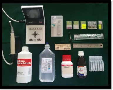
Armamentarium:
1. Diamond Disc
2. Straight Handpiece (NSK, Japan)
3. Scale 4. Endoblock
5. Electronic weighing device 6. Hot Plate Magnetic Stirrer
7. Disposable 2ml Syringe (Unolok Syringes and Medical Devices LTS, Faridabad, India)
8. X-Smart Plus Endomotor and Handpiece (Dentsply Maillefer, Ballaigues, Switzerland)
9. K files – ISO size 10, 15 and 20 (Mani Inc., Japan)
10.Protaper files SX, S1,S2, F1, F2, F3 (Dentsply Maillefer, Ballaigues, Switzerland)
11.U File 33mm , 20 ISO (Mani Inc., Japan)
12.Ultrasonic unit (Woodpecker)
13.Diode Laser (Zolar Photon plus)
17
METHODOLOGY:
SAMPLE COLLECTION
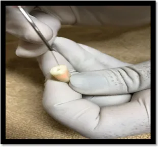
The Department of Oral and Maxillofacial surgery, Vivekanandha Dental College for Women sourced 75 freshly extracted mandibular premolars with single canal [Fig 1]. These were extracted due to poor periodontal prognosis and orthodontic reasons. X-ray were taken in both buccolingual [Fig 2] and mesiodistal [Fig3] directions to confirm the presence of single canal.
Infection Control protocol:
Occupational Safety and Health Administration (OSHA) and Centre for disease Control and Prevention (CDC) recommendations and guidelines64, 65 were followed for handling the extracted teeth. Handling of teeth was always done using gloves, mask and protective eyewear.
1. Teeth were cleaned of any visible blood and gross debris.
2. Distilled water was used in wide mouth plastic jars for initial collection. 3. Teeth were immersed in 10% formalin for 7 days, following which the liquid
was discarded and the teeth were transferred into separate jars containing distilled water.
4. The initial collection jars, lids and the gloves employed were discarded into biohazard waste receptacles.
18
INCLUSION AND EXCLUSION CRITERIA:
Inclusion Criteria:
Completely formed teeth with intact apices
Teeth without anatomical variations
Teeth without caries and root canal fillings
Teeth with single canal which are fully patent.
Exclusion Criteria:
Fractured roots
Teeth with multiple roots
Open apices
Calcifications in the canal
Root resorption and cracks on the surface
PROCEDURE:
Removal of external residual tissues:
The residual tissues on the surface of the teeth were removed and were stored in 2.5% NaOCl solution for 10 minutes. Calculi were removed using hand scalers from the external surfaces and they were again stored in distilled water.
Root canal therapy:
19 was inserted in the root canal till it was visible at the apical end of the root. Working length determination was done by reducing 1 mm from this measurement.
Pro Taper Universal rotary file was used for canal preparation. To simulate the clinical conditions the apices were sealed with sticky wax. A #20 K file was used for instrumentation of the canal after which it was instrumented up to size F3 ProTaper universal rotary files. 2ml of 3% NaOCl was used for irrigation after using each file. The irrigating solutions were delivered using a 27 gauge needle which was placed 1mm short of the measured working length. Finally, for the flushing out of debris 3ml of 3% NaOCl was used followed by a final rinse with distilled water.
0.2% Chitosan preparation:
20
Grouping of Samples:
Group A (Control) – Normal Saline
1ml of Normal saline was used to flush the canals for 1 minute followed by flushing the canal with 3 ml of 3% NaOCl.
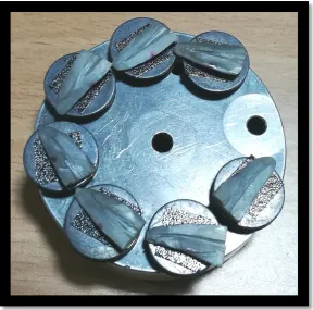
Group B1 – Ultrasonically activated EDTA
1ml of EDTA was used as a final flush to irrigate the canals and passive ultrasonic activation was done with #20 U file [Fig8, 10] followed by flushing the canal with 3ml of 3% NaOCl.
Mandibular premolar with single canal
n=75
Group A
Control - Normal Saline n= 15
Group B
Ultrasonically Activated (n=30)
Sub Group (B1)
17% EDTA (n=15)
SubGroup (B2)
0.2% Chitosan (n=15)
Group C
Diode laser Activated (n=30)
Subgroup (C1)
17% EDTA (n=15)
Subgroup 2 (C2)
21
Group B2 – Ultrasonically activated Chitosan
Final flush of 1ml of 0.2% Chitosan was used to irrigate the canals and then passive ultrasonic activation was done using #20 U file [Fig 8, 10] for 1 minute, followed by flushing the canal with 3ml of 3% NaOCl.
In groups B1 and B2 the U file was placed into the canal so that it was 1mm short of the measured working length.
Group C1 – Diode laser activated EDTA
0.8ml of 17% EDTA was used to irrigate the canal for 40 seconds and diode laser [Fig 9, 11] was used to activate the remaining 0.2ml for 20 seconds. The treatment was undertaken for four passes of each 5 seconds. Each pass was done at a fibre withdrawal rate of 1mm/second. A fiberoptic tip measuring 200-300µm, 970±15nm, with a power of 2W was used for laser activation of the canal up to the working length. In a helicoid movement the tip was withdrawn to the coronal region and reintroduced to the apical region for an irradiation cycle of 20 seconds, followed by 3ml of 3% NaOCl.
Group C2 – Diode laser activated Chitosan
22 and reintroduced to the apical region for an irradiation cycle of 20 seconds, followed by 3ml of 3% NaOCl.
5ml of distilled water was used as a final flush in all the samples to terminate the action of the other irrigants used. Scanning electron microscopic examination [Fig 12, 13] was carried out after the samples were dried and prepared.
SEM Analysis:
24
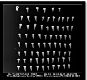
Fig 1: 75 Single rooted mandibular premolar
[image:37.595.176.456.432.688.2]25
Fig 3: Verification of single canal: X- ray image (Mesiodistal)
[image:38.595.170.460.437.712.2]26
27
Fig 6: Chitosan and Acetic acid
[image:40.595.136.497.446.728.2]28
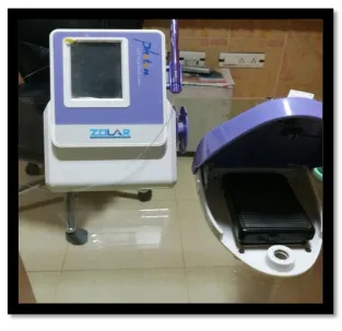
Fig 8: Ultrasonic unit
[image:41.595.157.471.440.743.2]29
Fig 10: U File used for agitation of irrigant
[image:42.595.160.471.452.739.2]30
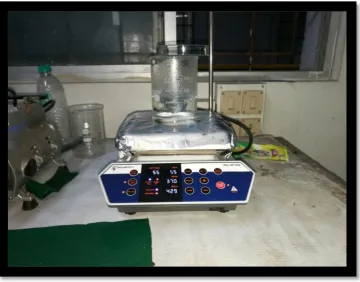
Fig 12: Magnetron Sputtering Coater
[image:43.595.171.460.452.737.2]31
32
GROUP A (NORMAL SALINE)
2000 X 5000X
CORONAL
MIDDLE
33
GROUP B1 (ULTRASONICALLY ACTIVATED –EDTA)
2000 X 5000X
CORONAL
MIDDLE
34
GROUP B2 (ULTRASONICALLY ACTIVATED – CHITOSAN)
2000 X 5000X
CORONAL
MIDDLE
35
GROUP C1 (DIODE LASER ACTIVATED – EDTA)
2000 X 5000X
CORONAL
MIDDLE
36
GROUP C2 (DIODE LASER ACTIVATED – CHITOSAN)
2000 X 5000X
CORONAL
MIDDLE
37
RESULTS
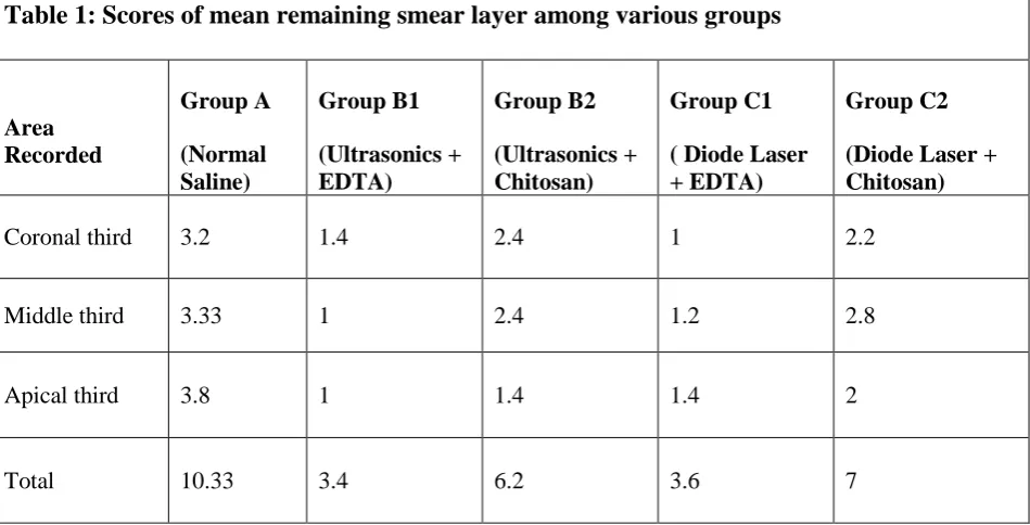
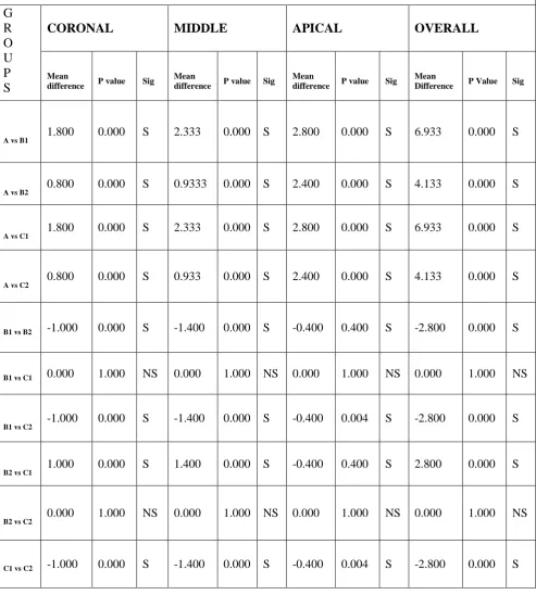
The mean values of the remaining smear layer scores were tabulated (Table 1). Analysis of the data was done using One-way analysis of variance (ANOVA), using the SPSS version 20. The values were considered statistically significant when P value < 0.05.
[image:51.595.89.564.378.620.2]There was statistically significant difference among all the tested groups except among Group B2 and C1 in the apical third, which had no significant difference (Table 2).
Table 1: Scores of mean remaining smear layer among various groups
Area Recorded Group A (Normal Saline) Group B1 (Ultrasonics + EDTA) Group B2 (Ultrasonics + Chitosan) Group C1 ( Diode Laser + EDTA)
Group C2 (Diode Laser + Chitosan) Coronal third 3.2 1.4 2.4 1 2.2 Middle third 3.33 1 2.4 1.2 2.8
Apical third 3.8 1 1.4 1.4 2
38
Table 2 : Intra group comparison of remaining smear layer scores at various levels
G R O U P S
CORONAL MIDDLE APICAL OVERALL
Mean
difference P value Sig
Mean
difference P value Sig
Mean
difference P value Sig
Mean
Difference P Value Sig
A vs B1 1.800 0.000 S 2.333 0.000 S 2.800 0.000 S 6.933 0.000 S
A vs B2 0.800 0.000 S 0.9333 0.000 S 2.400 0.000 S 4.133 0.000 S
A vs C1 1.800 0.000 S 2.333 0.000 S 2.800 0.000 S 6.933 0.000 S
A vs C2 0.800 0.000 S 0.933 0.000 S 2.400 0.000 S 4.133 0.000 S
B1 vs B2 -1.000 0.000 S -1.400 0.000 S -0.400 0.400 S -2.800 0.000 S
B1 vs C1 0.000 1.000 NS 0.000 1.000 NS 0.000 1.000 NS 0.000 1.000 NS
B1 vs C2 -1.000 0.000 S -1.400 0.000 S -0.400 0.004 S -2.800 0.000 S
B2 vs C1 1.000 0.000 S 1.400 0.000 S -0.400 0.400 S 2.800 0.000 S
B2 vs C2 0.000 1.000 NS 0.000 1.000 NS 0.000 1.000 NS 0.000 1.000 NS
39
Coronal third – C1 > B1 > C2 > B2 > A
Middle third – B1 > C1 > B2 > C2 > A
Apical third – B1 > B2 = C1 > C2 > A
Overall – B1 > C1 > B2 > C2 > A
40
DISCUSSION
The disinfection of dentin walls using irrigants is adversely affected in the presence of smear layer by blocking them from entering dentinal tubules2. It also adversely affects sealer penetration and increases microleakage following obturation leading to increasedintra-canal microflora68, 69.
Hence, to enhance sealer penetration and a fluid tight seal it is necessary to remove the smear layer. There has been an increasing interest in developing new irrigating solutions due to the limitations of the presently available ones.
Chitosan is a natural, cationic aminopolysaccharide copolymer of glucosamine and N-acetylglucosamine obtained by the alkaline, partial deacetylation of chitin. It is obtained from shells of crustaceans and shrimps44. It has biocompatibility, biodegradability, bioadhesion as well as antimicrobial activity70. It can also chelate various metal ions such as Fe2+, Co2+, Mg2+, Zn2+ and Cu2+ respectively) in acid conditions71. As it is hydrophilic, it is adsorbed on the root canal wall72.
In this study, mandibular premolars with single canal were decoronated at the CEJ for standardisation. ProTaper universal system up to size F3 was used for preparation of the root canal since it provides a uniform preparation. Specimens were randomly divided into 3 groups and 2 subgroups – Group A- Normal Saline (Control), Group B – Ultrasonic activation, Group C – Diode Laser activation, Group B and C were subdivided into (B1 and C1) 17% EDTA and (B2 and C2) 0.2% Chitosan.
41 Chitosan citrate produces more amount of dentinal erosion when compared to chitosan acetate when used as an irrigant for smear layer removal 48.
A.M.Darrag54 studied the ability of ability of 17% EDTA, 10% CA, MTAD, and 0.2% chitosan solutions to remove the smear layer and concluded that 0.2% chitosan was better, but there is no significant difference among them.
The effect of smear layer removal by Chitosan was compared to that of EDTA because EDTA is has been accepted as a gold standard for removal of smear layer2, 48
A combination of NaOCl and EDTA has been successfully used in debridement and for enlarging narrow and obstructed canals. Fraser in 197473, found that in the apical third the chelating ability of EDTA was minimal and it also caused root dentine erosion. EDTA also has limited antimicrobial activity compared to NaOCl. To minimise the harmful effects of EDTA the search continues for a newer material which is more biocompatible with enhanced antimicrobial effect.
Chitosan removes the inorganic portion of the smear layer due to its chelating ability, but its effect for endodontic application has not been widely explored. But this property has been used for the recovery of metal ions and purification of drinking water74.
In the present study, Chitosan solution preparation was done using 1% acetic acid. According to Silva et al48, it was attributed that the chelating ability of Chitosan was because of its own properties and not by 1% acetic acid. Thus, we could deduce that the chelating behaviour of Chitosan favoured its smear layer removal.
42 increased the smear layer removing by enhancing the penetration of irrigating solution into the narrow apical regions of the root canals1, 75, 76, 77.
When endodontic files are used in the handpiece, the files oscillate along the longitudinal axis of the instrument, with maximal amplitude occurring at the antinodes and minimal oscillation at the nodes78. The file oscillations are primarily responsible for the production of acoustic streaming (vortex like motion). Acoustic streaming may also be associated with the occurrence of cavitation which enhances smear layer removal79, 80.
Laser activation of irrigating solutions enhances the efficacy of the irrigating solution by the absorption of laser energy. This causes formation of vapour bubbles followed by collapse of these bubbles, acoustic streaming, which finally leads to cavitation50, 56.
According to Walmsley et al, the smear layer removal at the apical third was found to be the least because of the constriction in the root canal, which restricted the oscillation of the ultrasonic tip. The apical part is the most affected due to attenuation of oscillation because the amplitude is greatest at the tip of the instrument78. This was in accordance with the current study in which EDTA and Chitosan which showed effective smear layer removal from coronal third.
43 In Group A (Normal Saline) there was thick smear layer all through the length of the root canal which is in accordance to a study conducted by Mensudar Rathakrishnan et al81.
In the coronal third, a combination of EDTA with diode laser had the least remaining smear layer score. This was similar to the study Neelakantan et al, in which diode laser was found to be better than ultrasonics in the disinfection of dentinal tubules56.
Arslan et al evaluated the activation of 15% EDTA using 808-nm diode laser and concluded that on the removal of smear layer removal and concluded that agitation with diode laser was effective in the removal of smear layer. This was in accordance with the present study where EDTA activated with Diode laser had the greatest efficacy of smear layer removal 50.
44 Chitosan to be more effective than 17% EDTA and 10% Citric acid. This can be due to the use of Ultrasonics as adjunct in the present study, which shows better results when used in combination with EDTA.
Comparing the overall efficacy of various combinations used in this study, Group B1 (EDTA+ Ultrasonics) produced better smear layer removal than Group B2 (EDTA+Diode Layer), which in turn was better than Group C1 and C2 i.e., a combination of Chitosan with Ultrasonics and diode laser respectively. Group A had the least efficacy in the removal of smear layer.
According to a several recent studies, a combination of Chitosan- EDTA (1:1) can perform as a root canal disinfectant and can also be used in the removal of smear layer 82, 83, 84. EDTA potentiates the antibacterial activity of Chitosan and facilitates the entry of Chitosan into bacterial cell, this combination is known to restrain the growth of microorganisms by enzyme inhibition83, 84.
45
Limitations:
Being an in-vitro study, the results cannot be directly correlated to the clinical
situations.
Further studies are needed to evaluate the concentration and efficacy of
chitosan required for smear layer removal.
In the present study the ability of smear layer removal was only evaluated,
46
SUMMARY AND CONCLUSION
This study was to compared the ability of Ultrasonically activated and Laser activated EDTA and Chitosan on smear layer removal by using Scanning Electron Microscope.
Seventy five mandibular premolars with single canal were collected. These were decoronated at the level of cementoenamel junction. Cleaning and shaping was carried out using Protaper rotary file system up to F3 and the samples were divided randomly into 3 groups and 2 subgroups based on the irrigation protocol.
Group A (Normal saline), Group B1 (ultrasonically activated –EDTA), B2 (ultrasonically activated Chitosan), Group C1 (Diode laser activated – EDTA) and C2 (Diode laser activated – Chitosan).
After following the irrigation protocol for each group, the samples were sectioned longitudinally using a diamond disc and Scanning electron Microscopic analysis was carried out to study the surface morphology of root dentin.
The remaining smear layer scores were obtained based on Gutmann’s criteria and the data were tabulated. Statistical analysis was carried out using one- way analysis of variance using SPSS software version 20 and results were obtained.
48
BIBLIOGRAPHY
1. Cameron JA. The use of ultrasonics in the removal of the smear layer: A scanning electron microscope study. J Endod 1983;9:289-92.
2. Baumgartner JC, Brown CM, Mader CL, Peters DD, Shulman JD. A scanning electron microscopic evaluation of root canal debridement using saline, sodium hypochlorite, and citric acid. J Endod. 1984;10(11):525-31.
3. Brännström M, Johnson G. Effects of various conditioners and cleaning agents on prepared dentin surfaces: a scanning electron microscopic investigation. J Prosthet Dent. 1974;31(4):422-30.
4. Marques AA, Marchesan MA, Sousa-Filho CB, Silva-Sousa YT, Sousa-Neto MD, Cruz-Filho AM. Smear layer removal and chelated calcium ion quantification of three irrigating solutions. Braz Dent J. 2006;17(4):306-9.
5. Estrela C, Lopes HP, Elias CN, Leles CR, Pécora JD. Cleanliness of the surface of the root canal of apple vinegar, sodium hypochlorite, chlorhexidine and EDTA.
Rev Assoc Paul Cir Dent 2007;61:177-82.
6. Spanó JC, Silva RG, Guedes DF, Sousa-Neto MD, Estrela C, Pécora JD. Atomic absorption spectrometry and scanning electron microscopy evaluation of concentration of calcium ions and smear layer removal with root canal chelators. J Endod 2009;35:727-30.
49 8. Haznedaroğlu F. Efficacy of various concentrations of citric acid at different pH values for smear layer removal. Oral Surg Oral Med Oral Pathol Oral Radiol Endod. 2003;96:340–344.
9. Prado M, Gusman H, Gomes BP, Simão RA. Scanning electron microscopic investigation of the effectiveness of phosphoric acid in smear layer removal when compared with EDTA and citric acid. J Endod. 2011;37(2):255-8.
10.Papagianni M. Advances in citric acid fermentation by Aspergillus niger: biochemical aspects, membrane transport and modeling. Biotechnol Adv. 2007;25(3):244-63.
11.Yamaguchi M1, Yoshida K, Suzuki R, Nakamura H. Root canal irrigation with citric acid solution. J Endod. 1996;22(1):27-9.
12.Senel S, Kremer MJ, Kaş S, Wertz PW, Hincal AA, Squier CA. Enhancing effect of chitosan on peptide drug delivery across buccal mucosa. Biomaterials. 2000;21(20):2067-71.
13.Akncbay H1, Senel S, Ay ZY. Application of chitosan gel in the treatment of chronic periodontitis. J Biomed Mater Res B Appl Biomater. 2007;80(2):290-6. 14.Kurita K. Chemistry and application of chitin and chitosan. Polymer Degradation
and Stability. 1998;59(1-3):117-120.
15.Jeon YJ, Shahidi F, Kim S.-J. Preparation of chitin and chitosan oligomers and their applications in physiological functional foods. Food rev int. 2000;16(2):159-176.
16.Yamada RS, Armas A, Goldman M, Lin PS. A scanning electron microscopic comparison of a high volume final flush with several irrigating solutions: Part 3.
50 17.Byström A, Sundqvist G.The antibacterial action of sodium hypochlorite and
EDTA in 60 cases of endodontic therapy. Int Endod Jl. 1985;(18):35–40.
18.Baumgartner JC, Mader CL. A scanning electron microscopic evaluation of four root canal irrigation regimens. J Endod. 1987;13:147–57.
19.Ciucchi B, Khettabi M, Holz J. The effectiveness of different endodontic irrigation procedures on the removal of the smear layer: a scanning electron microscopic study. Int EndodJ. 1989;22(1):21-8.
20.Aktener BO, Bilkay U. Smear layer removal with different concentrations of EDTA-ethylenediamine mixtures. J Endod. 1993;19(5):228-31.
21.Gutmann JL, Saunders WP, Nguyen L, Guo IY, Saunders EM. Ultrasonic root-end preparation. Part 1. SEM analysis. Int Endod J. 1994;27(6):318-24.
22.Prati C, Selighini M, Ferrieri P, Mongiorgi R. Scanning electron microscopic evaluation of different endodontic procedures on dentin morphology of human teeth. J Endod. 1994;20(4):174-9.
23.Behrend GD, Cutler CW, Gutmann JL. An in-vitro study of smear layer removal and microbial leakage along root-canal fillings. Int Endod J. 1996;29(2):99-107.
24.Chailertvanitkul P1, Saunders WP, MacKenzie D. The effect of smear layer on microbial coronal leakage of gutta-percha root fillings. Int Endod J.
1996;29(4):242-8.
51 26.Sabins RA, Johnson JD, Hellstein JW. A comparison of the cleaning efficacy of short-term sonic and ultrasonic passive irrigation after hand instrumentation in molar root canals. JEndod.2003;29(10):674-8.
27.Crumpton BJ, Goodell GG, McClanahan SB. Effects on smear layer and debris removal with varying volumes of 17% REDTA after rotary instrumentation. J Endod. 2005;31(7):536-8.
28.Wang X, Sun Y, Kimura Y, Kinoshita J, Ishizaki NT, Matsumoto K. Effects of diode laser irradiation on smear layer removal from root canal walls and apical leakage after obturation. Photomed Laser Surg. 2005;23(6):575-81.
29.Carver K, Nusstein J, Reader A, Beck M. In vivo antibacterial efficacy of ultrasound after hand and rotary instrumentation in human mandibular molars. J Endod. 2007;33(9):1038-43.
30.Hmud R, Kahler WA, George R, Walsh LJ. Cavitational effects in aqueous endodontic irrigants generated by near-infrared lasers. J Endod. 2010;36(2):275-8.
31.Mozayeni MA, Javaheri GH, Poorroosta P, Ashari MA, Javaheri HH. Effect of 17% EDTA and MTAD on intracanal smear layer removal: a scanning electron microscopic study. Aust Endod J. 2009;35(1):13-7.
32.de Gregorio C, Estevez R, Cisneros R, Heilborn C, Cohenca N. Effect of EDTA, sonic, and ultrasonic activation on the penetration of sodium hypochlorite into simulated lateral canals: an in vitro study. J Endod. 2009;35(6):891-5.
52 34.Gu XH, Mao CY, Kern M. Effect of different irrigation on smear layer removal
after post space preparation. J Endod. 2009;35(4):583-6.
35.Desai P, Himel V. Comparative safety of various intracanal irrigation systems. J Endod. 2009;35(4):545-9.
36.Rödig T, Döllmann S, Konietschke F, Drebenstedt S, Hülsmann M. Effectiveness of different irrigant agitation techniques on debris and smear layer removal in curved root canals: a scanning electron microscopy study. J Endod. 2010;36(12):1983-7.
37.Jiang LM, Verhaagen B, Versluis M, Zangrillo C, Cuckovic D, van der Sluis LW. An evaluation of the effect of pulsed ultrasound on the cleaning efficacy of passive ultrasonic irrigation. J Endod. 2010;36(11):1887-91.
38.Caron G, Nham K, Bronnec F, Machtou P. Effectiveness of different final irrigant activation protocols on smear layer removal in curved canals. J Endod. 2010;36(8):1361-6.
39.Pagonis TC, Chen J, Fontana CR, Devalapally H, Ruggiero K, Song X, Foschi F, Dunham J, Skobe Z, Yamazaki H, Kent R, Tanner AC, Amiji MM, Soukos NS. Nanoparticle-based endodontic antimicrobial photodynamic therapy. J Endod. 2010;36(2):322-8.
53 41.Jiang LM, Verhaagen B, Versluis M, Langedijk J, Wesselink P, van der Sluis LW. The influence of the ultrasonic intensity on the cleaning efficacy of passive ultrasonic irrigation. J Endod. 2011 ;37(5):688-92.
42.Ulusoy Öİ, Görgül G. Effects of different irrigation solutions on root dentine microhardness, smear layer removal and erosion. Aust Endod J. 2013 Aug;39(2):66-72.
43.Andrabi SM, Kumar A, Kumar Tewari R, Kumar Mishra S, Iftekhar H. An In Vitro SEM Study on the Effectiveness of Smear Layer Removal of Four Different Irrigations. Iran Endod J. 2012l;7(4):171-6.
44.Pimenta JA, Zaparolli D, Pécora JD, Cruz-Filho AM. Chitosan: effect of a new chelating agent on the microhardness of root dentin. Braz Dent J. 2012;23(3):212-7.
45.Stojicic S, Shen Y, Qian W, Johnson B, Haapasalo M. Antibacterial and smear layer removal ability of a novel irrigant, QMiX. Int Endod J. 2012;45(4):363-71.
46.Castelo-Baz P, Martín-Biedma B, Cantatore G, Ruíz-Piñón M, Bahillo J, Rivas-Mundiña B, Varela-Patiño P. In vitro comparison of passive and continuous ultrasonic irrigation in simulated lateral canals of extracted teeth. J Endod.
2012;38(5):688-91.
54 48.Silva PV, Guedes DF, Pécora JD, da Cruz-Filho AM. Time-dependent effects of
chitosan on dentin structures. Braz Dent J. 2012;23(4):357-61.
49.Mancini M1, Cerroni L, Iorio L, Armellin E, Conte G, Cianconi L. Smear layer removal and canal cleanliness using different irrigation systems (EndoActivator, EndoVac, and passive ultrasonic irrigation): field emission scanning electron microscopic evaluation in an in vitro study. JEndod. 2013;39(11):1456-60.
50.Arslan H, Ayrancı LB, Karatas E, Topçuoğlu HS, Yavuz MS, Kesim B. Effect of agitation of EDTA with 808-nanometer diode laser on removal of smear layer. J Endod. 2013;39(12):1589-92
51.Silva PV, Guedes DF, Nakadi FV, Pécora JD, Cruz-Filho AM. Chitosan: a new solution for removal of smear layer after root canal instrumentation. Int Endod J. 2013;46(4):332-8.
52.Kim HJ, Park SJ, Park SH, Hwang YC, Yu MK, Min KS. Efficacy of flowable gel-type EDTA at removing the smear layer and inorganic debris under manual dynamic activation. J Endod. 2013;39(7):910-4.
53.Gusiyska A, Dyulgerova E2, Vassileva R3, Gyulbenkiyan E. The Effectiveness of a Chitosan-Citrate Solution toRemove the Smear Layer in Root Canal Treatment- An in-vitro study. Intl J of Science and Research.2016;5(9):1169-73.
54. A.M.Darrag. Effectiveness of different final irrigation solutions on smear layer removal in intraradicular dentin. Tanta Dental Journal. 2014:11(2);93-99.
55 photodynamic therapy inhibit collagen degradation in vitro. J Endod. 2014;40(5):703-9.
56.Neelakantan P, Cheng CQ, Mohanraj R, Sriraman P, Subbarao C, Sharma S. Antibiofilm activity of three irrigation protocols activated by ultrasonic, diode laser or Er:YAG laser in vitro. Int Endod J. 2015;48(6):602-10.
57.Del Carpio-Perochena A, Bramante CM, Duarte MA, de Moura MR, Aouada FA, Kishen A. Chelating and antibacterial properties of chitosan nanoparticles on dentin. Restor Dent Endod. 2015;40(3):195-201.
58.Amin K, Masoodi A, Nabi S, Ahmad P, Farooq R, Purra AR, Ahangar FA. Effect of diode laser and ultrasonics with and without ethylenediaminetetraacetic acid on smear layer removal from the root canals: A scanning electron microscope study. J Conserv Dent. 2016;19(5):424-7.
59.Afkhami F, Akbari S, Chiniforush N. Entrococcus faecalis Elimination in Root Canals Using Silver Nanoparticles, Photodynamic Therapy, Diode Laser, or Laser-activated Nanoparticles: An In Vitro Study. J Endod. 2017;43(2):279-282. 60.Machado R, Garcia LDFR, da Silva Neto UX, Cruz Filho AMD, Silva RG,
Vansan LP. Evaluation of 17% EDTA and 10% citric acid in smear layer removal and tubular dentin sealer penetration. Microsc Res Tech. 2018;81(3):275-282. 61.Del Carpio-Perochena A, Kishen A, Felitti R, Bhagirath AY, Medapati MR, Lai 6,
Cunha RS. Antibacterial Properties of Chitosan Nanoparticles and Propolis Associated with Calcium Hydroxide against Single- and Multispecies Biofilms: An In Vitro and In Situ Study. J Endod. 2017;43(8):1332-1336.
56 Ultrasonic Irrigation versus Irrigation with Reciprocating Activation: An Environmental Scanning Electron Study. J Endod. 2017;43(1):141-146.
63.Zhou H, Li Q, Wei L, Huang S, Zhao S. A comparative scanning electron microscopy evaluation of smear layer removal with chitosan and MTAD. Niger J Clin Pract. 2018;21(1):76-80.
64.Kumar M, Sequeira PS, Peter S, Bhat GK. Sterilisation of Extracted Human Teeth For Educational Use. Indian Journal of Medical Microbiology. 2005;23:256-258.
65.Dominici JT, Eleazer PD, Clark SJ, Staat RH, Scheetz. Disinfection/sterilization of extracted teeth for dental use. J Dent Educ. 2001;65:1278-1280.
66.De-Deus G, Souza EM, Marins JR, Reis C, Paciornik S, Zehnder M. Smear layer dissolution by peracetic acid of low concentration. Inter Endod J. 2011;44:485e90.
67.Gopikrishna V1, Venkateshbabu N, Krithikadatta J, Kandaswamy D. Evaluation of the effect of MTAD in comparison with EDTA when employed as the final rinse on the shear bond strength of three endodontic sealers to dentine. Aust Endod J. 2011;37(1):12-7.
68.Economides N, Liolios E, Kolokuris I, Beltes P. Long-term evaluation of the influence of smear layer removal on the sealing ability of different sealers. J Endod. 1999;25:123–5.
57 70.Shenoy A, AhmaduddinBolla N, Raj S, Mandava P, Nayak S. Effect of final irrigating solution on smear layer removal and penetrability of the root canal sealer. J Conserv Dent. 2014;17:40e4.
71.Campos-Ibarra P, La Fuente-Hern_andez J, Tenorio-Rocha F, Acosta-Torres L. Biocompatible antimicrobial irrigants and nanoparticles-sealers for endodontics.
Entresciencias. 2013;1:9e28.
72.Zhang J, Xia Z, Liu P, Cheng Q, Tahirou T, Gu W, et al. Chitosan modification and pharmaceutical/biomedical applications. Mar Drugs. 2010;8:1962e87
73. Fraser JG. Chelating agents: Their softening effect on root canal dentin. Oral Surg 1974; 37 (5): 803-11.
74.Onsøyen E, Skaugrud O. Metal recovery using chitosan. J Chem Technol Biotechnol. 1990;49(4):395-404.
75.Baumgartner JC1, Cuenin PR. Efficacy of several concentrations of sodium hypochlorite for root canal irrigation. J Endod. 1992;18(12):605-12.
76.Cameron JA. The use of ultrasound for the removal of the smear layer. The effect of sodium hypochlorite concentration; SEM study. Aust Dent J. 1988;33(3):193-200.
77.Lui JN, Kuah HG, Chen NN. Effect of EDTA with and without surfactants or ultrasonics on removal of smear layer. J Endod. 2007;33(4):472-5.
78.Walmsley AD. Ultrasound and root canal treatment: the need for scientific evaluation. Int Endod J. 1987: 20: 105–111.
58 80.Ahmad M, Pitt Ford TR, Crum LA. Ultrasonic debridement of root canals: an
insight into the mechanisms involved. J Endod. 1987: 13: 93–101.
81.Rathakrishnan M, Sukumar VG, Subbiya A. Evaluate the efficacy of an Innovative Irrigant on Smear layer Removal. J Clin Diagn Res. 2016;10:ZC104-6 82.Geethapriya N, Subbya A, Padmavathy K, Mahalakshmi K, Vivekanandan P;
Sukumaran V G. Effect Of Chitosan-Ethylenediamine Tetraacetic Acid On Enterococcus Faecalis Dentinal Biofilm And Smear Layer Removal. J Conserv Dent.,2016: 19: 472-477
83.Raafat D, von Bargen K, Haas A, Sahl HG. Insights into the mode of action of chitosan as an antibacterial compound. Appl Environ Microbiol 2008;74:3764-73.
84.Banin E, Brady KM, Greenberg EP. Chelator-induced dispersal and killing of Pseudomonas aeruginosa cells in a biofilm. Appl Environ Microbiol