The Variable Region of Pneumococcal Pathogenicity Island 1 Is
Responsible for Unusually High Virulence of a Serotype 1 Isolate
Richard M. Harvey, Claudia Trappetti, Layla K. Mahdi, Hui Wang, Lauren J. McAllister, Alexandra Scalvini, Adrienne W. Paton, James C. Paton
Research Centre for Infectious Diseases, Department of Molecular and Cellular Biology, University of Adelaide, S.A., Australia
Streptococcus pneumoniae
is the leading infectious cause of death in children in the world. However, the mechanisms that drive
the progression from asymptomatic colonization to disease are poorly understood. Two virulence-associated genomic accessory
regions (ARs) were deleted in a highly virulent serotype 1 clinical isolate (strain 4496) and examined for their contribution to
pathogenesis. Deletion of a prophage encoding a platelet-binding protein (PblB) resulted in reduced adherence, biofilm
forma-tion, reduced initial infection within the lungs, and a reduction in the number of circulating platelets in infected mice. However,
the region’s overall contribution to the survival of mice was not significant. In contrast, deletion of the variable region of
pneu-mococcal pathogenicity island 1 (vPPI1) was also responsible for a reduction in adherence and biofilm formation but also
re-duced survival and invasion of the pleural cavity, blood, and lungs. While the 4496
⌬
PPI1 strain induced higher expression of the
genes encoding interleukin-10 (IL-10) and CD11b in the lungs of challenged mice than the wild-type strain, very few other genes
exhibited altered expression. Moreover, while the level of IL-10 protein was increased in the lungs of 4496
⌬
PPI1 mutant-infected
mice compared to strain 4496-infected mice, the levels of gamma interferon (IFN-
␥
), CXCL10, CCL2, and CCL4 were not
differ-ent in the two groups. However, the 4496
⌬
PPI1 mutant was found to be more susceptible than the wild type to phagocytic killing
by a macrophage-like cell line. Therefore, our data suggest that vPPI1 may be a major contributing factor to the heightened
viru-lence of certain serotype 1 strains, possibly by influencing resistance to phagocytic killing.
S
treptococcus pneumoniae
(the pneumococcus) is a significant
cause of human morbidity and mortality and is a leading cause
of pneumonia, bacteremia, meningitis, and otitis media (
1
).
How-ever, the mechanism by which the pathogen progresses from
asymptomatic colonization to disease is poorly understood.
Moreover, significant variations in virulence exist even between
strains of the same serotype (
2–5
). For example, outbreaks of
un-usually severe invasive pneumococcal disease (IPD) caused by
se-rotype 1 strains belonging to clonal lineage group B have been
reported in parts of Africa (
6
,
7
), while clustered cases of carriage
without disease caused by serotype 1 strains belonging to clonal
lineage A, which include the common clonal complex (CC)
CC227, have been reported within Australia (
8
). Furthermore,
disease caused by invasive lineage A strains has been reported to be
of relatively low severity (
9
). These naturally occurring differences
in virulence provide opportunities to investigate the molecular
processes that drive progression to disease by comparing closely
related invasive and noninvasive strains. In previous work, the
pathogenesis of noninvasive and invasive serotype 1 clinical
iso-lates was characterized in mice (
10
,
11
). Highly virulent human
strains 1861 (sequence type 3079 [ST3079] [CC217]) and 4496
(ST3018 [CC615]), belonging to serotype 1 lineages B and C,
re-spectively, were found to rapidly invade and survive in the lungs,
pleural cavity, and blood of mice. In contrast, human carriage
strains, including strain 1 (ST304), were able to colonize the
na-sopharynx to an extent similar to that seen with the highly virulent
strains but were rapidly cleared from the lungs and were not
de-tected in either the pleural cavity or the blood. In addition, highly
virulent strain 1861 induces a stronger type I interferon (IFN-1)
response in the lungs shortly after challenge that facilitates the
early stages of invasion of the pleural cavity and blood (
10
).
Genomic comparisons identified 8 accessory regions (AR)
⬎
1 kb
in length that were present in both highly virulent strains but were
absent from the less virulent lineage A strains (
11
). These ARs
include a region encoding a prophage (AR1), a specific variant of
the variable region of pneumococcal pathogenicity island 1
(vPPI1) (AR3 and AR4), a region encoding a putative iron
per-mease and DyP-type peroxidase (AR6), a region encoding an
ABC-type transporter and ArsR family transcriptional regulator
(AR7), and other regions consisting of sequences encoding
puta-tive metabolic enzymes and transcriptional regulators (AR2, AR5,
and AR8) (
11
). In particular, the content of vPPI1 was shown to
influence the relative levels of competitive fitness of D39 mutants
in niches of the mouse associated with disease (
11
). In this study,
a serotype 1-derived variant of vPPI1 encoding the PezAT
toxin-antitoxin system, and a number of genes annotated as encoding
metabolic enzymes such as neopullulanase, 3-hydroxyisobutyrate
dehydrogenase, UDP-glucose 4-epimerase, prephenate
dehydra-tase, and biotin carboxylase, was associated with increased
inva-sive potential. In an earlier study, mice challenged with a TIGR4
mutant lacking
pezT
survived longer following intraperitoneal
challenge than the wild type (
12
). Bioinformatic analyses show
that, while the accessory component of vPPI1 is harbored by other
strains and serotypes in publicly available databases, such as G54,
Received26 November 2015Accepted2 January 2016 Accepted manuscript posted online11 January 2016
CitationHarvey RM, Trappetti C, Mahdi LK, Wang H, McAllister LJ, Scalvini A, Paton AW, Paton JC. 2016. The variable region of pneumococcal pathogenicity island 1 is responsible for unusually high virulence of a serotype 1 isolate. Infect Immun 84:822–832.doi:10.1128/IAI.01454-15.
Editor:L. Pirofski
MLV-016, and 11-BS70, these strains are all PezAT negative (
11
).
Therefore, the configuration of vPPI1 under study appears to be
more specific to lineage B and C serotype 1 strains, such as P1031
(ST303), PNI0373 (ST618), and NCTC7465 (ST615).
BLAST analysis of available
S. pneumoniae
genome sequences
indicates that, as described above, the complete prophage of AR1
is present in the same lineage B and C serotype 1 strains as vPPI1,
although portions of the phage are more widely distributed. The
prophage includes a gene with sequence similarity to the
platelet-binding protein gene (pblB) and a gene encoding an endolysin,
both of which were shown to be required for the virulence of
Streptococcus mitis
in an animal model of infective endocarditis
(
13
). In addition, the product of
pblB
has been shown to promote
persistence within the murine nasopharynx and lungs of the
sero-type 14 ST46 clone as well as increased adherence to the lung and
nasopharyngeal epithelium (
14
). However, the role that the phage
plays in more highly virulent strains, such as serotype 1 clinical
isolates, is not clear. Moreover, efforts to study the contributions
that these ARs make to pathogenesis have to date been hampered
by the genetically intractable nature of invasive serotype 1 isolates.
In previous work, our laboratory was the first to construct isogenic
mutants in the serotype 1 ST306 background (
15
). In the present
study, we developed a technique to construct mutants in
non-lineage A serotype 1 isolates and used it to remove the prophage
and vPPI1 to determine the relative levels of phenotypic impact of
these regions on the highly virulent serotype 1 4496 strain. This
work documents some of the first evidence for a single region of
the accessory genome contributing almost the entire difference in
virulence between noninvasive and highly invasive isolates of the
same serotype.
MATERIALS AND METHODS
Ethics statement.This study was conducted in compliance with the Aus-tralian Code of Practice for the Care and Use of Animals for Scientific Pur-poses(7th edition, 2004) and the South Australian Animal Welfare Act of 1985. All animal experiments were approved by the Animal Ethics Com-mittee of the University of Adelaide.
Bacterial strains and media.Serotype 2 strain D39 (NCTC 7466) has been described previously (16). Strain 4496 is a highly virulent serotype 1 clinical isolate that has also been described previously (11,17). Opaque-phase variants of all strains selected on Todd-Hewitt broth supplemented with 1% yeast extract (THY broth)-catalase plates (18) were used in all animal experiments. Strains were routinely grown in THY broth or C⫹Y medium (19) or on blood agar (BA). For animal inoculation, the bacteria were grown in serum broth (SB) (nutrient broth [10 g/liter peptone {Oxoid}, 10 g/liter Lab Lemco powder {Oxoid}, 5 g/liter NaCl] plus 10% [vol/vol] donor horse serum) to an absorbance at 600 nm (A600) of 0.16,
which approximates 1⫻108CFU/ml.
Construction of mutants in highly virulent serotype 1 strain 4496. Transformation of 4496 requires chromosomal DNA as the template. Therefore, in order to construct the 4496⌬⌽and 4496⌬PPI1 mutants, the relevant mutations were first constructed in the more tractableS. pneu-moniaeD39 strain (i.e., D39⌬⌽and D39⌬PPI1 mutants). Theerm -con-taining construct used to make the D39⌬PPI1 mutant was generated by overlap extension PCR, as previously described (20), using the relevant primers listed inTable 1to amplify the flanking products from strain 4496 template DNA (primers 5 to 8) and primers J214 to J215 to amplifyerm
from pVA831. D39 was transformed with the product of overlap exten-sion PCR, as previously described (21,22), and selected on erythromycin-containing blood agar plates. In the case of the D39⌬⌽mutant, the wild-type strain does not carry the prophage, so theermcassette was instead inserted into the position in the D39 chromosome that corresponded to the location of the prophage in strain 4496. The construct used to make
the 4496⌬⌽mutant was generated by PCR amplification of flanking products from strain 4496 template DNA using primers 1 to 4 and theerm
cassette as described above but using primers J293a and J215b (2). The amplified flanking products anderm cassette subsequently underwent restriction digestion using EagI-HF and XhoI-HF (NEB), followed by ligation. D39 was transformed with the resultant ligation product as de-scribed above. 4496 mutants were constructed in a 2-step process, com-mencing with transformation with genomic DNA (gDNA) from the ap-propriate D39 construct. After selection on BA plus 2 g/ml erythromycin, genomic DNA was extracted from one of the transfor-mants and used as the donor in a second round of transformation of strain 4496. The transformation procedure itself was a modification of that de-veloped previously for type 1 ST306 (15). Strain 4496 was grown over-night on BA and then inoculated into a 1:1 mixture of C⫹Y medium and Dulbecco’s modified Eagle’s medium (DMEM; Gibco, Grand Island, NY) supplemented with 10% heat-inactivated fetal calf serum (FCS). After 1 h at 37°C, the culture was diluted into fresh medium and incubated for a further 2 h at 37°C. Competence-stimulating peptide 1 (CSP-1) was added to achieve a concentration of 50 ng/ml, followed 15 min later by donor DNA (approximately 1g). After a further 2 h of incubation at 37°C, transformation mixes were plated on BA plus erythromycin.
A549 and Detroit 562 adherence assays.A549 (human type II pneu-mocyte) and Detroit 562 (human nasopharyngeal carcinoma) cells were grown in DMEM and in a 1:1 mix of DMEM and Ham’s F-12 nutrient mixture (Gibco), respectively, supplemented in both cases with 5% fetal bovine serum, 2 mML-glutamine, 50 IU/ml penicillin, and 50g/ml streptomycin. Confluent monolayers in 24-well plates were washed with phosphate-buffered saline (PBS) and infected with pneumococci (ap-proximately 5⫻105CFU per well) in a 1:1 mixture of the respective
culture medium (without antibiotics) and C⫹Y (pH 7.4). Plates were centrifuged at 500⫻gfor 5 min and then incubated at 37°C in 5% CO2for
2.5 h. Monolayers were washed 3 times in PBS, and adherent bacteria were released by treatment with 100l trypsin–EDTA, followed by 400l 0.025% Triton X-100. Lysates were serially diluted and plated on BA to enumerate adherent bacteria.
Biofilm assays.Biofilm formation was performed on immobilized A549 cells as previously described (23). Briefly, cells were grown until confluent in 12-well polystyrene plates and immobilized by fixation with 2% paraformaldehyde (Sigma). Immobilized cells were then washed thor-oughly with sterile deionized water and supplemented with C⫹Y media containing 1⫻107CFU/ml of each bacterial strain. Following incubation
for 6 h, plates were sonicated for 3 s at 35 kHz in a Soniclean ultrasonic water bath (Soniclean, Thebarton, S.A., Australia). Biofilm cell counts (quantified as CFU per milliliter) were determined by dilution and plating of the resulting dispersed biofilm-derived bacterial suspensions.
[image:2.585.298.547.76.224.2]Quantitation of CPS.Capsular polysaccharide (CPS) samples were prepared by resuspending pneumococci grown in C⫹Y medium–PBS to
TABLE 1Primers used in this study
Primer Sequence (5=–3=)
anA600of 0.5. Subsequent preparation and quantification of CPS were
carried out using a uronic acid assay as described previously (24). Quantitative Western blot analysis.Strain 4496 and the 4496⌬PPI1 mutant were grown to anA600of 0.2 in C⫹Y. Cultures were concentrated
10-fold and incubated at 37°C for 30 min in 1⫻PBS– 0.1% sodium de-oxycholate (Sigma-Aldrich). Protein concentrations were determined by the use of a Pierce bicinchoninic acid (BCA) protein assay kit (Thermo Fisher Scientific), according to the manufacturer’s instructions. Lysates containing 20g total protein were subjected to SDS-PAGE before trans-fer to nitrocellulose using an iBlot system (Life Technologies). Blots were probed with antigen-specific antisera, as described previously (25), prior to detection by the use of anti-mouse IRDye 800 and analysis using an Odyssey infrared imaging system (Li-COR). Band intensities were quan-titated according to the manufacturer’s instructions.
Animal studies.Outbred 5-to-6-week-old female CD1 (Swiss) mice were used in all animal experiments. For intranasal (i.n.) challenge, mice were anesthetized by intraperitoneal (i.p.) injection of pentobarbital so-dium (Nembutal; Rhone-Merieux) at a dose of 66g per g of body weight, followed by i.n. challenge with 50l of bacterial suspension containing approximately 1⫻107CFU bacteria and SB. The challenge dose was
confirmed retrospectively by serial dilution and plating on BA. For sur-vival experiments, mice were monitored regularly for signs of illness and euthanized once moribund. All surviving mice were euthanized at 336 h postchallenge.
For the pathogenesis experiments, mice were euthanized by CO2
as-phyxiation at the indicated time points. Blood was collected by syringe from the posterior vena cava. The pleural cavity was lavaged with 1 ml sterile PBS containing 2 mM EDTA introduced through the diaphragm. Pulmonary vasculature was perfused by infusion of sterile PBS through the heart. Lungs were subsequently excised into 2-ml vials containing 1 ml sterile PBS and 2.8-mm-diameter ceramic beads for CFU counts. To ob-tain unattached pneumococci, the nasopharynx was subjected to lavage by insertion of a 26-gauge needle sheathed in tubing into the tracheal end of the upper respiratory tract and injection of 1 ml 0.5% trypsin–1⫻PBS through the nasopharynx and collection from the nares. Additionally, the upper palate and nasopharynx were excised and placed into 2-ml vials containing 1 ml sterile PBS and 2.8-mm-diameter ceramic beads to obtain attached pneumococci. CFU counts for both the nasal wash and nasal tissue samples were combined to determine the total number of bacteria in the nasopharynx. Lung and nasopharyngeal tissues were homogenized using a Precellys 24 tissue homogenizer (Bertin Technologies) at 3 cycles of 30 s and 5,000 rpm. At each time point, a 40-l aliquot of homogenate was serially diluted in SB and plated on BA to determine the number of CFU present. Aliquots (20l) of blood and pleural lavage samples were serially diluted and plated on BA to determine the number of CFU in these niches. Data were analyzed in GraphPad Prism using unpairedttests, as described in figure legends.
Isolation of RNA.Lungs were excised from resting (mock-infected) andS. pneumoniae-challenged mice (n⫽4 per group), following perfu-sion as described above, and transferred to 2-ml vials containing 1 ml TRIzol reagent (Life Technologies) and 2.8-mm-diameter ceramic beads for immediate homogenization as described above. RNA was isolated from TRIzol-treated samples per the manufacturer’s instructions. All RNA samples were purified using an RNeasy RNA minikit (Qiagen) per the manufacturer’s instructions, including on-column DNase treatment (Qiagen) performed to remove any contaminating genomic DNA (gDNA) before use in quantitative arrays. Removal of gDNA was con-firmed by PCR amplification of the gene encoding GAPDH (glyceralde-hyde-3-phosphate dehydrogenase) with and without reverse transcrip-tase.
PCR arrays.cDNA synthesis was carried out on the RNA extracted as described above using a RT2First Strand kit (Qiagen). RNA was analyzed
using a LightCycler 480 II system (Roche) by quantitative reverse tran-scription-PCR (qRT-PCR) and a RT2Profiler PCR Array Mouse Innate
and Adaptive Immune Responses kit (Qiagen), according to the
instruc-tions of the manufacturers. Data were analyzed using PCR Array data analysis software provided by the manufacturer.
Cytokine quantification by ELISA.The concentrations of IFN-␥, IL-10, CXCLIL-10, CCL2, and CCL4 were determined in lung homogenates (n⫽5) using an enzyme-linked immunosorbent assay (ELISA) and a Quantikine ELISA system (R&D Systems), according to the manufactur-er’s instructions. Homogenized lung tissue at 6 h post-intranasal chal-lenge was subjected to centrifugation at 21,000⫻gfor 5 min at 4°C. Supernatants were then assayed by ELISA. Absorbance readings were per-formed on a PHERAstar FS microplate reader (BMG Lab Tech). Calcula-tions to determine the concentration of each cytokine were performed by regression analysis according to the kit manufacturer’s instructions.
Cell culture and differentiation of THP-1 cells into a macrophage-like cell type.All tissue culture media and reagents were obtained from Gibco. THP-1 cells (ATCC TIB-202) were grown in 95% air and 5% CO2
at 37°C in complete RPMI medium (RPMI medium with phenol red, supplemented with 10% FCS, 10 mM HEPES, 50 IU/ml penicillin, and 50 g/ml streptomycin). Flasks (25 cm2) were seeded with 1⫻105THP-1
cells and differentiated by adding phorbol 12-myristate 13-acetate (PMA) to reach a final concentration of 100 ng/ml and incubating for 3 days at 37°C in 95% air and 5% CO2. The differentiated cells attached to the
plastic surface. Following the 3-day incubation, cells were washed twice with complete RPMI medium and then rested at 37°C in 95% air and 5% CO2and complete RPMI medium for a further 3 days.
Macrophage killing assay.Resting differentiated THP-1 cells were detached by using StemPro Accutase cell dissociation reagent (Gibco) and resuspended in fresh Hanks’ balanced salt solution (HBSS).S. pneu-moniaestrains were freshly grown to mid-log phase and added to the macrophage culture (bacterium/macrophage ratio, 10:1) in a 1.5-ml tube. Survival of the internalized bacteria was determined after incubation of bacteria and macrophages for 2 h followed by incubation with antibiotics for different time periods. To evaluate the internalized bacteria, antibiot-ics (10g/ml penicillin and 200g/ml gentamicin) in HBSS were applied for 30 min at 37°C to kill extracellular bacteria. Samples at each time point were lysed with 0.025% trypsin, and bacterial CFU counts were deter-mined on the blood agar plates. The survival rate was calculated from comparisons of the CFU count at each time point to the CFU count obtained at 30 min post-antibiotic treatment (assumed to be 100%). All the assays were performed in biological triplicates, and statistical signifi-cance was analyzed using a two-tailed unpairedttest.
RESULTS
Adherence and biofilm formation by 4496
⌬⌽
and 4496
⌬
PPI1
mutants.
In order to assess the contribution that the
PblB-encod-ing prophage and vPPI1 make to the virulence of strain 4496,
isogenic mutants were constructed as described in Materials and
Methods, generating 4496
⌬⌽
and 4496
⌬
PPI1 mutants,
respec-tively. Mutant constructs were confirmed by PCR and sequence
analysis (results not shown).
In vitro
growth rates of strain 4496
and the 4496
⌬⌽
and 4496
⌬
PPI1 mutant strains were compared;
the results showed no differences in growth rates in either C
⫹
Y or
SB media (data not shown). The relative capacities of the two
mutants and the wild-type 4496 strain to adhere to A549 and
Detroit 562 cells were then assayed (
Fig. 1
). Both mutants
exhib-ited significantly lower adherence to both cell types than the wild
type. For the 4496
⌬⌽
mutant strain, adherence to A549 cells was
20.9% of that of 4496, while adherence to Detroit 562 cells was
44.9% of that of the wild-type strain (P
⬍
0.001 in both cases). For
the 4496
⌬
PPI1 mutant strain, adherence to A549 cells was 34.2%
of that of 4496, while adherence to Detroit 562 cells was only
10.4% of that of the wild-type strain (P
⬍
0.01 and
P
⬍
0.001,
respectively).
biofilm on fixed A549 cells was also compared with the
biofilm-forming ability of the wild type (
Fig. 2
). Again, both mutants
ex-hibited significantly lower biofilm formation capacity than 4496.
Biofilm formation by the 4496
⌬⌽
and 4496
⌬
PPI1 mutants was
only 30% and 10% of that of the wild type, respectively (P
⬍
0.01
and
P
⬍
0.001, respectively).
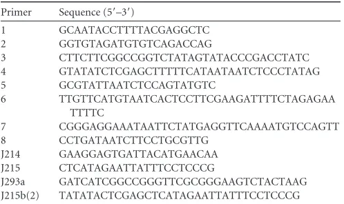
Virulence of the 4496
⌬⌽
mutant.
The survival times of mice
following i.n. challenge with either the 4496 or 4496
⌬⌽
strain
were initially compared (
Fig. 3
). However, there was no significant
difference in survival rates or median survival times between the
two groups. A more detailed comparison of the pathogenic
pro-files of the two strains was performed by quantifying the number
of pneumococci present in samples from the nasopharynx, lungs,
pleural lavage fluid, and blood at 12 h, 24 h, or 36 h post-i.n.
challenge. The 4496
⌬⌽
strain colonized the nasopharynx to an
extent similar to that shown by the wild type at both 12 h and 36 h.
However, there was a small but significant reduction in nasal
col-onization by the 4496
⌬⌽
mutant at 24 h compared to the wild
type (P
⬍
0.05) (
Fig. 4A
). The number of mutant bacteria in the
lungs was significantly lower than the number of wild-type
bacte-ria at both 12 h and 24 h (P
⬍
0.001 and
P
⬍
0.05, respectively)
(
Fig. 4B
). However, numbers of the mutant bacteria in the lungs
were not significantly different from those of the wild type by
36 h, suggesting that the pathogenic impact of the prophage is
greatest during the initial stages of lung infection. There was no
statistically significant difference between the numbers of
mu-tant and wild-type bacteria in the pleural cavity or the blood at
the three time points that were tested (
Fig. 4C
and
D
).
Never-theless, only 2 of the 8 mice infected with the 4496
⌬⌽
strain
had detectable bacteria in either the pleural cavity or blood at
FIG 1The capacity of the 4496 strain and the 4496⌬⌽and 4496⌬PPI1 mutants to adhere to A549 and Detroit 562 cell monolayers was examined as described in Materials and Methods. Data are means⫾standard errors of the means (SEM) of CFU/well for quadruplicate assays. Significant differences in adherence relative to that for the respective 4496 control are indicated as follows: **,P⬍0.01; ***,P⬍0.001 (Student’sttest, two-tailed).
FIG 2Biofilm formation by the 4496⌬⌽and 4496⌬PPI1 mutants relative to the 4496 strain. Biofilm formation on fixed A549 cells was determined after 6 h of incubation, as described in Materials and Methods. Data are expressed as means (⫾SEM) of percentages of that for wild type 4496. Statistical differences were analyzed by two-tailed unpairedttests (****,P⬍0.0001).
[image:4.585.59.522.62.294.2] [image:4.585.65.259.530.674.2] [image:4.585.323.518.551.676.2]12 h compared with 5/8 and 6/8 for the respective
compart-ments in mice infected with 4496. These findings may suggest
that the 4496
⌬⌽
strain invades the blood more slowly than the
wild-type strain.
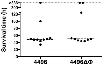
Impact of the PblB-encoding phage on circulating platelets.
Previous work has shown that the protein encoded by other
pblB
genes mediates attachment to platelets (
14
,
26
). Therefore, we
investigated whether the
⌬⌽
mutation would lead to any change
[image:5.585.59.523.71.602.2]in the number of platelets in the peripheral circulation. The data
presented are representative of the results of two separately
con-ducted experiments. Blood was taken at 12 h, 20 h, and 36 h
post-i.n. challenge. Platelet counts revealed a significant reduction in
the number of circulating platelets in mice challenged with the
wild type compared to the mutant at 12 h (P
⬍
0.001), 20 h (P
⬍
0.01), and 36 h (P
⬍
0.05) (
Fig. 5A
). There was no change in the
number of circulating platelets between time points for either
challenge group, which suggests that the progression of
bactere-mia has no impact on the number of circulating platelets. No
difference in the amount of platelet aggregation was observed in
blood smears between strain 4496-infected and 4496
⌬⌽
mutant-infected mice (data not shown). Neutrophil counts were also
per-formed at each time point. However, while overall neutrophil
numbers as a proportion of white blood cells were elevated to an
extent consistent with infection at all of the time points, there was
no difference between the mutant and wild type (
Fig. 5B
). The
proportion of neutrophils was significantly reduced in both
chal-lenge groups once mice had reached the fulminant stage of disease
at 36 h compared to the two earlier time points (P
⬍
0.001).
However, the
⌬⌽
mutation had no detectable impact on the
num-ber of neutrophils as a proportion of total white blood cells during
bacteremia.
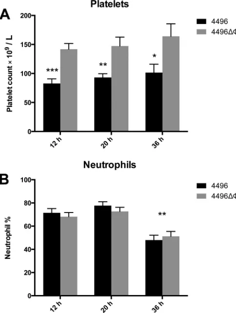
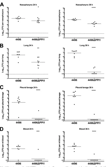
PPI1 is important for the invasiveness of strain 4496.
The
pathogenic profiles of strain 4496 and the 4496
⌬
PPI1 mutant
were compared by quantifying the number of pneumococci in the
nasopharynx, lungs, pleural cavity, and blood at 24 h and 36 h
post-i.n. challenge. A small but significant difference in
nasopha-ryngeal colonization results was observed at both 24 h and 36 h
(P
⬍
0.05 and
P
⬍
0.00001, respectively), with 0.51- and 1-log
10lower geometric mean (GM) CFU counts for the mutant strain at
the respective time points (
Fig. 6A
). However, differences in
bac-terial loads in the lungs were much more pronounced, with the
GM CFU count for the 4496
⌬
PPI1 mutant at 24 h 1.7-log
10lower
than for mice infected with wild-type 4496 (P
⬍
0.01) (
Fig. 6B
). By
36 h, numbers of 4496
⌬
PPI1 mutant bacteria in the lungs had
diminished further and were below the limit of detection in 5 of 8
mice, whereas lung bacterial loads in 4496-infected mice
re-mained at approximately 10
7CFU (P
⬍
0.0001) (
Fig. 6B
).
How-ever, the differences in bacterial loads between strain 4496- and
4496
⌬
PPI1 mutant-infected mice were greatest in the pleural
cav-ity and blood. In the former compartment, GM CFU counts for
strain 4496 were approximately 10
7and 10
9at 24 h and 36 h,
respectively. In stark contrast, only 1 of 8 4496
⌬
PPI1
mutant-infected mice had detectable CFU in the lungs at each of the two
time points, and even in these 2 animals, bacterial loads were
2-and 5-log
10-fold lower at 24 h and 36 h, respectively (P
⬍
0.0001 at
both time points) (
Fig. 6C
). Similarly, in the blood, 7 of 8 and 8 of
8 4496-infected mice were bacteremic at both 24 h and 36 h,
re-spectively (GM CFU/ml, approximately 10
5and 5
⫻
10
5,
respec-tively), compared with 1 of 8 4496
⌬
PPI1 mutant-infected mice at
both time points (P
⬍
0.001 and 0.0001, respectively) (
Fig. 6D
).
Therefore, we conclude that vPPI1 plays a critical role in the
vir-ulence of 4496 by enabling the strain to survive and proliferate in
the lungs and then to invade the pleural cavity and blood.
Expression of known pneumococcal virulence factors by the
4496
⌬
PPI1 mutant strain.
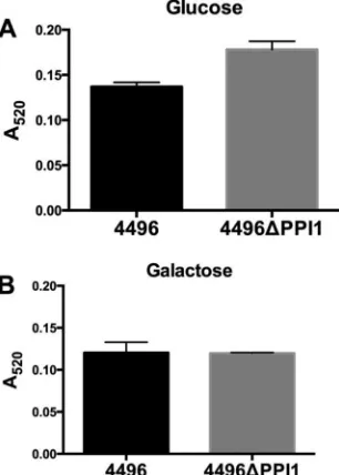
A number of the genes present within
vPPI1 of strain 4496 exhibit sequence similarity to metabolic
pathway genes (
11
). One such example is the UDP-glucose
4-epi-merase gene
galE1, which is responsible for the interconversion of
UDP-glucose and UDP-galactose. Since UDP-glucose is a
precur-sor for CPS synthesis, we investigated whether the loss of vPPI1
impacted
in vitro
CPS production by strain 4496 with either
glu-cose or galactose as the carbon source. Using the uronic acid assay,
we found no significant difference in the amount of capsule
be-tween the 4496 and 4496
⌬
PPI1 strains with either carbon source
(
Fig. 7
). In order to determine whether the loss of these putative
metabolic enzymes could have a secondary effect on the
expres-sion of other virulence factors, total levels of choline-binding
pro-tein A (CbpA), neuraminidase A (NanA), and pneumolysin (Ply)
were compared between the 4496 strain and the 4496
⌬
PPI1
mu-tant strain by quantitative Western blot analysis (
Fig. 8
). These
proteins were chosen because of their well-known roles in
adher-ence to respiratory epithelial cells, nasopharyngeal colonization,
and generation of inflammatory responses, particularly in the
lung. No significant differences between the 4496 and 4496
⌬
PPI1
strains in the relative expression levels of CbpA, NanA, and Ply
were detected.
Immune response to strain 4496 and the 4496
⌬
PPI1 mutant
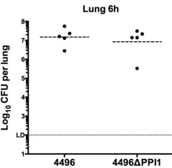
in the lungs.
In previous work, the IFN-1 response was found to
facilitate the early stages of invasion of the pleural cavity and
rep-resented the primary difference in the response to a noninvasive
serotype 1 strain versus the response to a highly virulent serotype
FIG 5Platelet and neutrophil counts in the peripheral blood of strain 4496 and 4496⌬⌽mutant-infected mice. (A) Data are platelet counts⫾SEM (n⫽
[image:6.585.41.283.68.389.2]FIG 6Pathogenesis of the 4496 strain versus the 4496⌬PPI1 mutant in mice. Two groups of 8 mice were infected intranasally with 107CFU of the 4496 or 4496⌬PPI1 strain. At 24 h and 36 h postchallenge, numbers of pneumococci in the nasopharynx (A), lung (B), pleural cavity (C), and blood (D) were determined. Horizontal broken lines indicate the geometric mean bacterial load for each group. The horizontal dotted line indicates the limit of detection (LD). At the 12-h time point, this equates to 102CFU in all niches; at 24 h and 36 h, this equates to 102CFU in nasopharynx and lung and to 2⫻102CFU in the pleural lavage fluid and blood. Statistical differences were analyzed by two-tailed unpairedttests performed on log-transformed data (*,P⬍0.05; **,P⬍0.01; ***,P⬍0.001; ****,
[image:7.585.112.478.60.634.2]1 strain (
10
). Therefore, we investigated the possibility that
ex-pression of vPPI1-carried genes might increase the strength of the
IFN-1 response. The immune responses (at the transcriptional
level) to the two strains were compared in the lungs at 6 h
post-challenge, a time at which bacterial loads in the lungs of strain
4496-infected and 4496
⌬
PPI1 mutant-infected mice are similar
(
Fig. 9
). RNA extracted from infected lungs was examined by
real-time qRT-PCR analysis to compare transcription levels of 84
genes that represent the major pathways of the murine innate and
adaptive immune response to microbial pathogens (data not
shown). Six genes with significantly altered expression in the
pres-ence of the 4496
⌬
PPI1 mutant relative to strain 4496 were
de-tected (
Table 2
). The genes encoding IL-10 (Il10), CD11b (Itgam),
lymphocyte antigen 96 (Ly96), and Toll-like receptor 4 (Tlr4)
ex-hibited increased expression in the presence of the 4496
⌬
PPI1
mutant compared to the wild type. The genes encoding lysozyme
2 (Lyz2) and retinoic acid receptor (RAR)-related orphan receptor
gamma (RorC) exhibited decreased expression in the presence of
the 4496
⌬
PPI1 mutant relative to the wild type. Therefore, the
presence or absence of vPPI1 does not appear to contribute to any
change in the IFN-1 response at the level of transcription. In
ad-dition to the transcriptional analyses, IFN-
␥
, IL-10, CXCL10,
CCL2, and CCL4 were compared at the protein level in the lungs
of infected mice at 6 h postchallenge (
Fig. 10
). These targets were
chosen because they were differentially expressed in noninvasive
versus invasive serotype 1 isolates in previous work (
10
). IL-10
expression was significantly increased in the lungs of mice
chal-lenged with the 4496
⌬
PPI1 mutant compared to those challenged
with strain 4496 (P
⬍
0.05). The higher level of IL-10 in the lungs
of 4496
⌬
PPI1-infected mice is consistent with the increased
Il10
transcription observed above. However, there were no significant
differences in the levels of IFN-
␥
, CXCL10, CCL2, or CCL4 in the
lungs between the two challenge groups.
The 4496
⌬
PPI1 mutant is more susceptible than 4496 to
kill-ing by macrophage-like cells.
Levels of susceptibility to
phago-cytic killing by a macrophage-like cell line were compared
between the 4496 and 4496
⌬
PPI1 strains. Following a 2-h
coincu-bation, extracellular pneumococci were removed by antibiotic
treatment for 30 min, and the number of viable intracellular
pneu-mococci was quantified after a further 0, 30, 60, or 90 min of
incubation. Data at 30, 60, and 90 min are expressed as a
percent-age of the data determined for intracellular bacteria at 0 min (
Fig.
11
). At 30 min, 62.7% of the strain 4496 bacteria remained viable
FIG 7Capsular polysaccharide production by strain 4496 and the 4496⌬PPI1 mutant with either glucose or galactose as the carbon source. Total CPS pro-duction was quantitated colorimetrically using a uronic acid assay, as de-scribed in Materials and Methods. Data are means⫾SEM of the results ofA520 determinations for biological duplicates. Cells were grown in C⫹Y medium with either glucose (A) or galactose (B) as the carbon source. Differences between meanA520values were analyzed using the two-tailed unpairedttest.
[image:8.585.87.240.70.284.2] [image:8.585.316.525.74.505.2]compared to 48.1% of the 4496
⌬
PPI1 mutant bacteria (P
⬍
0.01).
At 60 min, 14.1% of the strain 4496 bacteria remained viable
com-pared to 6.7% of the 4496
⌬
PPI1 mutant bacteria (P
⬍
0.001), and
at 90 min, 0.9% remained viable compared to 0.2% of the
4496
⌬
PPI1 mutant bacteria (P
⬍
0.05). Therefore, the 4496
⌬
PPI1
mutant appeared to be more susceptible to phagocytic killing.
DISCUSSION
To date, very little is known about the association between the
accessory genome of the pneumococcus and the various
propen-sities of different strains to cause disease. In previous work,
inva-sive and noninvainva-sive serotype 1 clinical isolates were compared at
the genomic level, and in terms of virulence profiles and the early
immune response in mice, to help understand why some strains
cause disease more readily than others. In the present study, a
prophage encoding a putative platelet-binding protein and a
vari-ant of the vPPI1 were analyzed to determine their contribution to
the heightened virulence of a highly invasive serotype 1 strain. The
⌬⌽
mutation was found to have a modest impact on the ability of
strain 4496 to establish early infection within the lungs. However,
this deficiency appeared to diminish with time. Moreover, no
dif-ferences were observed in bacterial loads in the pleural cavity or
blood at any of the time points tested. The reduced ability of the
4496
⌬⌽
mutant to adhere to live A549 cells and its reduced
ca-pacity to form a biofilm on fixed A549 monolayers are consistent
with the reduced capacity of the mutant to establish infection
within the lungs which was observed following intranasal
chal-lenge in mice. The impact of the
⌬⌽
mutation on adherence to
Detroit 562 nasopharyngeal cells was less pronounced, which is
consistent with the lesser, but still statistically significant, effect on
colonization of the murine nasopharynx at 24 h. These findings
are also consistent with the recently published report that
phage-encoded PblB plays a role in adherence to the lung epithelium in
serotype 14 pneumococci (
14
). The fact that a difference was
ob-served in the present study only at early time points, whereas the
mutant serotype 14 strain was reduced in numbers in the lung
over longer time periods, may be a function of the distinct
viru-lence profiles. Typically, serotype 14 strains are not highly virulent
in mouse models, whereas type 1 strains such as 4496 may cause
fulminant sepsis within 2 days of challenge. Interestingly, in our
study significantly fewer platelets were found in the circulation of
mice challenged with the wild type than in those challenged with
the phage-deficient strain. While differences in platelet
aggrega-tion were not observed in blood smears, the PblB-encoding phage
clearly has some impact on the number of circulating platelets,
perhaps through its recently reported ability to mediate
attach-ment to platelets (
14
). However, while the AR1 phage appears to
have some impact on the early stages of lung infection by the
serotype 1 strain, the mutant and the wild type progress to
fulmi-nant infection at similar rates. Therefore, AR1 on its own is
un-likely to be responsible for the vast differences in virulence
be-tween the highly invasive strains and the less virulent lineage A
serotype 1 strains.
In contrast to the 4496
⌬⌽
mutant, the 4496
⌬
PPI1 mutant was
severely attenuated in its ability to persist within the lungs and
invade and survive in either the pleural cavity or blood. However,
the 4496
⌬
PPI1 mutant was attenuated only modestly in its ability
to colonize the nasopharynx. It is interesting that the pathogenic
profile of the 4496
⌬
PPI1 strain closely mirrored that of the
natu-rally noninvasive type 1 lineage A strain studied in previously
pub-lished work (
10
,
11
). Thus, it appears that the particular variant of
vPPI1 carried by the highly invasive serotype 1 strains may be a
significant factor contributing to their heightened virulence in
mice and perhaps to the unusually severe disease caused by closely
related strains in humans. Notwithstanding the facts noted above,
it is interesting that, while the IFN-1 response was found to play a
role in the earliest stages of invasion of the pleural cavity by a
closely related serotype 1 strain in previous work, vPPI1 plays no
role in inducing this response. Instead, there were very few
im-mune function genes that were differentially expressed between
the two groups, which suggests that the contribution of vPPI1 to
virulence has little to do with inducing a particular type of host
response at the level of transcription. However, while the
expres-sion of IL-10 was also increased at the protein level, the levels of
other important cytokines such as CXCL10, IFN-
␥
, CCL2, and
CCL4 were unaltered by the loss of vPPI1. Increased expression of
Il10
and
Itgam
in the presence of the 4496
⌬
PPI1 mutant
com-pared to the wild type suggests possible alterations in neutrophil
[image:9.585.73.250.63.236.2]FIG 9Numbers of strain 4496 and 4496⌬PPI1 mutant pneumococci in the lungs at 6 h postchallenge. Two groups of 5 mice were infected intranasally with 107CFU of the 4496 or 4496⌬PPI1 strain. At 6 h, numbers of pneumococci in the lungs were determined. Horizontal lines indicate the geometric mean bacterial load for each group. The horizontal dotted line indicates the limit of detection (LD), which equates to 102CFU. Statistical differences were analyzed by two-tailed unpairedt tests performed on log-transformed data (P⬍0.05).
TABLE 2Differentially expressed immune response genesa
Gene
4496⌬PPI1 mutant vs strain 4496
Fold change Significance
Il10 3.5 ***
Itgam 3.4 *
Ly96 2.5 *
Lyz2 ⫺2.45 *
RorC ⫺2.08 **
Tlr4 2.33 *
a
[image:9.585.299.544.560.651.2]and macrophage recruitment into the lungs (
27–29
). In
particu-lar, increased
Il10
expression suggests greater suppression of the
inflammatory response to the 4496
⌬
PPI1 mutant, which may be a
reflection of the fact the mutant is more readily cleared from the
lungs than the wild type. This is consistent with the finding that
the 4496
⌬
PPI1 mutant appeared to be significantly more
suscep-tible than the wild type to
in vitro
phagocytic killing. However,
what is apparent is that, while the 4496
⌬
PPI1 mutant may be
more susceptible to phagocytosis, the differences, if any, in the
immune responses to strain 4496 versus the 4496
⌬
PPI1 mutant
appear to be more subtle than could be detected by the methods
employed in this study. This hypothesis does not discount the
importance of host factors in the development of disease caused
by
S. pneumoniae
strains more characteristically associated with
opportunistic infections (
9
). However, those strains that tend to
behave as primary pathogens may drive much more of the invasive
process by direct mechanisms unrelated to modulation of host
innate or adaptive immune responses.
In this study, we have provided the first evidence linking
un-usually severe disease caused by non-lineage A serotype 1
S.
pneu-moniae
isolates to a specific region of its accessory genome. While
we showed that the PblB-encoding prophage within AR1 does
appear to play some role in the earliest stages of infection, its
FIG 10Concentrations of specific cytokines in lung homogenates. The concentrations of IFN-␥, IL-10, CXCL10, CCL2, and CCL4 were determined in the homogenized lung supernatants by ELISA at 6 h postchallenge. Data are the means⫾SEM of the results determined for 5 mice per group in technical duplicates. The concentrations were calculated according to the instructions of the manufacturers by using regression analysis to determine a line of best fit from the relevant standards. Statistical differences were analyzed by two-tailed unpairedttests (*,P⬍0.05).
[image:10.585.140.452.61.395.2] [image:10.585.61.266.490.619.2]contribution is modest compared to that of vPPI1. Thus, future
study of the gene products encoded on vPPI1 in strain 4496 will
provide important information on the mechanisms that drive the
progression from colonization to severe invasive disease.
ACKNOWLEDGMENTS
J.C.P. is a National Health and Medical Research Council (NHMRC) Senior Principal Research Fellow; C.T. is an Australian Research Council DECRA Fellow.
FUNDING INFORMATION
Department of Health | National Health and Medical Research Council (NHMRC) provided funding to James C Paton and Adrienne W Paton under grant number 565526. Department of Health | National Health and Medical Research Council (NHMRC) provided funding to James C Paton under grant number 1071659.
REFERENCES
1.O’Brien KL, Wolfson LJ, Watt JP, Henkle E, Deloria-Knoll M, McCall N, Lee E, Mulholland K, Levine OS, Cherian T; Hib and Pneumococcal Global Burden of Disease Study Team.2009. Burden of disease caused by
Streptococcus pneumoniaein children younger than 5 years: global esti-mates. Lancet 374:893–902.http://dx.doi.org/10.1016/S0140-6736 (09)61204-6.
2.Brueggemann AB, Griffiths DT, Meats E, Peto T, Crook DW, Spratt BG.2003. Clonal relationships between invasive and carriage Streptococ-cus pneumoniaeand serotype- and clone-specific differences in invasive disease potential. J Infect Dis187:1424 –1432.http://dx.doi.org/10.1086 /374624.
3.Brueggemann AB, Peto TE, Crook DW, Butler JC, Kristinsson KG, Spratt BG.2004. Temporal and geographic stability of the serogroup-specific invasive disease potential ofStreptococcus pneumoniaein children. J Infect Dis190:1203–1211.http://dx.doi.org/10.1086/423820.
4.Sandgren A, Sjostrom K, Olsson-Liljequist B, Christensson B, Samu-elsson A, Kronvall G, Henriques Normark B.2004. Effect of clonal and serotype-specific properties on the invasive capacity ofStreptococcus pneu-moniae. J Infect Dis189:785–796.http://dx.doi.org/10.1086/381686. 5.Henriques-Normark B, Blomberg C, Dagerhamn J, Battig P, Normark
S.2008. The rise and fall of bacterial clones:Streptococcus pneumoniae. Nat Rev Microbiol6:827– 837.http://dx.doi.org/10.1038/nrmicro2011. 6.Leimkugel J, Adams Forgor A, Gagneux S, Pfluger V, Flierl C, Awine E,
Naegeli M, Dangy JP, Smith T, Hodgson A, Pluschke G. 2005. An outbreak of serotype 1Streptococcus pneumoniaemeningitis in northern Ghana with features that are characteristic ofNeisseria meningitidis men-ingitis epidemics. J Infect Dis 192:192–199. http://dx.doi.org/10.1086 /431151.
7.Yaro S, Lourd M, Traore Y, Njanpop-Lafourcade BM, Sawadogo A, Sangare L, Hien A, Ouedraogo MS, Sanou O, Parent du Chatelet I, Koeck JL, Gessner BD.2006. Epidemiological and molecular character-istics of a highly lethal pneumococcal meningitis epidemic in Burkina Faso. Clin Infect Dis43:693–700.http://dx.doi.org/10.1086/506940. 8.Smith-Vaughan H, Marsh R, Mackenzie G, Fisher J, Morris PS, Hare K,
McCallum G, Binks M, Murphy D, Lum G, Cook H, Krause V, Jacups S, Leach AJ.2009. Age-specific cluster of cases of serotype 1Streptococcus pneumoniaecarriage in remote indigenous communities in Australia. Clin Vaccine Immunol16:218 –221.http://dx.doi.org/10.1128/CVI.00283-08. 9.Sjöström K, Spindler C, Ortqvist A, Kalin M, Sandgren A, Kühlmann-Berenzon S, Henriques-Normark B.2006. Clonal and capsular types decide whether pneumococci will act as a primary or opportunistic patho-gen. Clin Infect Dis42:451– 459.http://dx.doi.org/10.1086/499242. 10. Hughes CE, Harvey RM, Plumptre CD, Paton JC.2014. Development of
primary invasive pneumococcal disease caused by serotype 1 pneumo-cocci is driven by early increased type I interferon response in the lung. Infect Immun82:3919 –3926.http://dx.doi.org/10.1128/IAI.02067-14. 11. Harvey RM, Stroeher UH, Ogunniyi AD, Smith-Vaughan HC, Leach
AJ, Paton JC.2011. A variable region within the genome ofStreptococcus pneumoniaecontributes to strain-strain variation in virulence. PLoS One
6:e19650.http://dx.doi.org/10.1371/journal.pone.0019650.
12. Brown JS, Gilliland SM, Spratt BG, Holden DW.2004. A locus
con-tained within a variable region of pneumococcal pathogenicity island 1 contributes to virulence in mice. Infect Immun72:1587–1593.http://dx .doi.org/10.1128/IAI.72.3.1587-1593.2004.
13. Mitchell J, Siboo IR, Takamatsu D, Chambers HF, Sullam PM.2007. Mechanism of cell surface expression of theStreptococcus mitisplatelet binding proteins PblA and PblB. Mol Microbiol64:844 – 857.http://dx .doi.org/10.1111/j.1365-2958.2007.05703.x.
14. Hsieh YC, Lin TL, Lin CM, Wang JT. 2015. Identification of PblB mediating galactose-specific adhesion in a successfulStreptococcus pneu-moniaeclone. Sci Rep5:12265.http://dx.doi.org/10.1038/srep12265. 15. Harvey RM, Hughes CE, Paton AW, Trappetti C, Tweten RK, Paton JC.
2014. The impact of pneumolysin on the macrophage response to Strep-tococcus pneumoniaeis strain-dependent. PLoS One9:e103625.http://dx .doi.org/10.1371/journal.pone.0103625.
16. Avery OT, Macleod CM, McCarty M.1944. Studies on the chemical nature of the substance inducing transformation of pneumococcal types: induction of transformation by a desoxyribonucleic acid fraction isolated from pneumococcus type III. J Exp Med79:137–158.http://dx.doi.org/10 .1084/jem.79.2.137.
17. Harvey RM, Ogunniyi AD, Chen AY, Paton JC.2011. Pneumolysin with low hemolytic activity confers an early growth advantage toStreptococcus pneumoniaein the blood. Infect Immun79:4122– 4130.http://dx.doi.org /10.1128/IAI.05418-11.
18. Weiser JN, Austrian R, Sreenivasan PK, Masure HR.1994. Phase vari-ation in pneumococcal opacity: relvari-ationship between colonial morphol-ogy and nasopharyngeal colonization. Infect Immun62:2582–2589. 19. Lacks S, Hotchkiss RD.1960. A study of the genetic material determining
an enzyme in Pneumococcus. Biochim Biophys Acta39:508 –518.http: //dx.doi.org/10.1016/0006-3002(60)90205-5.
20. Morona JK, Paton JC, Miller DC, Morona R.2000. Tyrosine phosphor-ylation of CpsD negatively regulates capsular polysaccharide biosynthesis inStreptococcus pneumoniae. Mol Microbiol35:1431–1442.
21. Giammarinaro P, Paton JC.2002. Role of RegM, a homologue of the catabolite repressor protein CcpA, in the virulence ofStreptococcus pneu-moniae. Infect Immun70:5454 –5461.http://dx.doi.org/10.1128/IAI.70 .10.5454-5461.2002.
22. Martin B, Garcia P, Castanie MP, Glise B, Claverys JP.1995. TherecA
gene ofStreptococcus pneumoniaeis part of a competence-induced operon and controls an SOS regulon. Dev Biol Stand85:293–300.
23. Vidal JE, Howery KE, Ludewick HP, Nava P, Klugman KP. 2013. Quorum-sensing systems LuxS/autoinducer 2 and Com regulate Strepto-coccus pneumoniaebiofilms in a bioreactor with living cultures of human respiratory cells. Infect Immun81:1341–1353.http://dx.doi.org/10.1128 /IAI.01096-12.
24. Morona JK, Morona R, Paton JC.2006. Attachment of capsular poly-saccharide to the cell wall ofStreptococcus pneumoniaetype 2 is required for invasive disease. Proc Natl Acad Sci U S A103:8505– 8510.http://dx .doi.org/10.1073/pnas.0602148103.
25. McAllister LJ, Ogunniyi AD, Stroeher UH, Leach AJ, Paton JC.2011. Contribution of serotype and genetic background to virulence of serotype 3 and serogroup 11 pneumococcal isolates. Infect Immun79:4839 – 4849. http://dx.doi.org/10.1128/IAI.05663-11.
26. Bensing BA, Siboo IR, Sullam PM.2001. Proteins PblA and PblB of Streptococcus mitis, which promote binding to human platelets, are en-coded within a lysogenic bacteriophage. Infect Immun69:6186 – 6192. http://dx.doi.org/10.1128/IAI.69.10.6186-6192.2001.
27. Kadioglu A, De Filippo K, Bangert M, Fernandes VE, Richards L, Jones K, Andrew PW, Hogg N.2011. The integrins Mac-1 and alpha4beta1 perform crucial roles in neutrophil and T cell recruitment to lungs during
Streptococcus pneumoniaeinfection. J Immunol186:5907–5915.http://dx .doi.org/10.4049/jimmunol.1001533.
28. Kirby AC, Raynes JG, Kaye PM.2006. CD11b regulates recruitment of alveolar macrophages but not pulmonary dendritic cells after pneumo-coccal challenge. J Infect Dis 193:205–213. http://dx.doi.org/10.1086 /498874.