Novel Permissive Cell Lines for Complete Propagation of Hepatitis C
Virus
Mai Shiokawa,aTakasuke Fukuhara,aChikako Ono,aSatomi Yamamoto,aToru Okamoto,aNoriyuki Watanabe,bTakaji Wakita,b Yoshiharu Matsuuraa
Department of Molecular Virology, Research Institute for Microbial Diseases, Osaka University, Osaka, Japana
; Department of Virology II, National Institute of Infectious Diseases, Tokyo, Japanb
ABSTRACT
Hepatitis C virus (HCV) is a major etiologic agent of chronic liver diseases. Although the HCV life cycle has been clarified by
studying laboratory strains of HCV derived from the genotype 2a JFH-1 strain (cell culture-adapted HCV [HCVcc]), the
mecha-nisms of particle formation have not been elucidated. Recently, we showed that exogenous expression of a liver-specific microRNA,
miR-122, in nonhepatic cell lines facilitates efficient replication but not particle production of HCVcc, suggesting that
liver-spe-cific host factors are required for infectious particle formation. In this study, we screened human cancer cell lines for expression
of the liver-specific
␣
-fetoprotein by using a cDNA array database and identified liver-derived JHH-4 cells and stomach-derived
FU97 cells, which express liver-specific host factors comparable to Huh7 cells. These cell lines permit not only replication of
HCV RNA but also particle formation upon infection with HCVcc, suggesting that hepatic differentiation participates in the
ex-pression of liver-specific host factors required for HCV propagation. HCV inhibitors targeting host and viral factors exhibited
different antiviral efficacies between Huh7 and FU97 cells. Furthermore, FU97 cells exhibited higher susceptibility for
propaga-tion of HCVcc derived from the JFH-2 strain than Huh7 cells. These results suggest that hepatic differentiapropaga-tion participates in
the expression of liver-specific host factors required for complete propagation of HCV.
IMPORTANCE
Previous studies have shown that liver-specific host factors are required for efficient replication of HCV RNA and formation of
infectious particles. In this study, we screened human cancer cell lines for expression of the liver-specific
␣
-fetoprotein by using
a cDNA array database and identified novel permissive cell lines for complete propagation of HCVcc without any artificial
ma-nipulation. In particular, gastric cancer-derived FU97 cells exhibited a much higher susceptibility to HCVcc/JFH-2 infection
than observed in Huh7 cells, suggesting that FU97 cells would be useful for further investigation of the HCV life cycle, as well as
the development of therapeutic agents for chronic hepatitis C.
M
ore than 170 million individuals worldwide are infected with
hepatitis C virus (HCV), and the cirrhosis and
hepatocellu-lar carcinoma induced by HCV infection are life-threatening
dis-eases (
1
). Current standard therapy combining
pegylated-inter-feron (peg-IFN) and ribavirin (RBV) has achieved a sustained
virological response (SVR) in 50% of individuals infected with
HCV genotype 1 (
2
). Recently, directly acting antiviral (DAA)
agents have been applied in a clinical setting (
3
). An SVR rate of
over 80% has been realized by combination therapy with peg-IFN,
RBV, and NS3/4A inhibitors in genotype 1 patients (
4
,
5
). In
ad-dition, several DAAs, including inhibitors for NS3/4A protease,
NS5A, and NS5B polymerase, are currently in clinical trials.
Sev-eral reports have shown that
in vitro
replication of HCV RNA is
significantly inhibited by treatment with daclatasvir (NS5A
inhib-itor) and asunaprevir (NS3 protease inhibinhib-itor), and these two
DAAs are also effective for patients infected with genotype 1 HCV
who showed no response to previous therapy with peg–IFN-
␣
and
RBV (
6–8
). On the other hand, it has been shown that
drug-resis-tant breakthrough viruses emerge during treatment with DAAs
(
9–12
). Therefore, identification of host factors crucial for the
propagation of HCV is an important task for the development of
novel therapeutics for chronic hepatitis C with a low frequency of
emergence of drug-resistant viruses.
The establishment of an
in vivo
infection model has been
ham-pered by the narrow host range and tissue tropism of HCV.
Al-though chimpanzees are the only experimental animals
suscepti-ble to HCV infection, it is difficult to use a chimpanzee model of
experimental infection due to ethical concerns (
13
,
14
). In
addi-tion,
in vitro
infection models have also been restricted to the
combination of cell culture-adapted clones based on the genotype
2a JFH-1 strain (HCVcc) and human hepatoma cell lines,
includ-ing Huh7 (
15
). Recently, several reports have shown that the
ex-ogenous expression of microRNA-122 (miR-122) facilitates the
efficient propagation of HCVcc in HepG2 and Hep3B cells, which
are nonpermissive for propagation of HCVcc (
16
,
17
).
Further-more, we reported that nonhepatic cell lines, including Hec1B
cells derived from uterine endometrial adenocarcinoma, also
per-mit replication of HCV RNA by exogenous expression of miR-122
(
18
). These reports indicate that miR-122 is one of the most
im-portant determinants for liver tropism of HCV infection.
Inter-estingly, formation of infectious particles was not observed in
Received26 December 2013Accepted25 February 2014 Published ahead of print5 March 2014
Editor:T. S. Dermody
Address correspondence to Yoshiharu Matsuura, [email protected]. Copyright © 2014, American Society for Microbiology. All Rights Reserved. doi:10.1128/JVI.03839-13
on November 7, 2019 by guest
http://jvi.asm.org/
spite of efficient replication of HCV RNA in nonhepatic cells,
suggesting that liver-specific factors other than miR-122 are
in-volved in HCV assembly. Previous reports suggested that
very-low-density lipoprotein (VLDL)-associated proteins, including
apolipoprotein B (ApoB), apolipoprotein E (ApoE), and
micro-somal triglyceride transfer protein (MTTP), play important roles
in infectious particle production of HCV (
19–23
). In addition,
Miyanari et al. indicated that lipid droplets (LDs) are crucial
or-ganelles for HCV particle assembly (
24
). These reports suggest
that liver-specific lipid metabolism and liver-specific host factors
closely participate in assembly of HCV.
Cancer cells are classified into well-differentiated and
interme-diately and poorly differentiated stages, and these stages have been
shown to be strongly related to cancer behaviors, with an
imma-ture tumor generally being more aggressive than its more
differ-entiated counterpart. Thus, it is believed that well-differdiffer-entiated
cancer cells maintain the tissue-specific cellular functions and
ex-hibit morphology similar to that of normal cells (
25
). Permissive
cell lines for HCV propagation, including Huh7, HepG2, and
Hep3B cells, are derived from well-differentiated hepatocellular
carcinoma (HCC) (
26
,
27
). In addition, recent reports indicated
that hepatocyte-like cells derived from induced pluripotent stem
cells (iPS cells/iPSCs) express high levels of miR-122 and
VLDL-associated proteins and support propagation of HCVcc (
28–30
).
These results suggest that the hepatic differentiation required for
hepatic functions is involved in HCV propagation.
In this study, we identified novel cell lines supporting complete
propagation of HCVcc by screening cancer cell lines expressing
␣
-fetoprotein (AFP), which is highly expressed in
well-differenti-ated hepatocellular carcinoma and evaluwell-differenti-ated in most cancer cell
lines (
31
). These cells exhibit high levels of expression of
liver-specific host factors and permit complete propagation of HCVcc
without any exogenous expression of liver-specific factors,
includ-ing receptor molecules, miR-122, and apolipoproteins. Our
cur-rent study suggests that hepatic diffecur-rentiation participates in the
expression of liver-specific host factors required for complete
propagation of HCV.
MATERIALS AND METHODS
NextBio Body Atlas.The NextBio Body Atlas application allows the ag-gregated analysis of gene expression across various normal tissues, nor-mal cell types, and cancer cell lines. It enables us to investigate the expression of individual genes as well as gene sets. Samples for Body Atlas data are obtained from publicly available studies that are inter-nally curated, annotated, and processed (32). Body Atlas measure-ments are generated from all available RNA expression studies that used Affymetrix U133 Plus or U133A Genechip arrays for human stud-ies. The results for 128 human tissue samples from 1,067 arrays, those for 157 human cell types from 1,474 arrays, and those for 359 human cancer cell lines from 376 arrays are incorporated. In this study, we screened cell lines expressing a high level of AFP. The details of the analysis protocol developed by NextBio were described previously (32). The raw data used in this application are derived from the GSK Cancer Cell Line data deposited at the National Cancer Institute web-site (https://array.nci.nih.gov/caarray/project/woost-00041).
Plasmids.The cDNA clones of pri-miR-122 andAequorea coerulescens green fluorescent protein (AcGFP) were inserted between the XhoI and XbaI sites of lentiviral vector pCSII-EF-RfA, which was provided by M. Hijikata, and the resulting plasmids were designated pCSII-EF-miR-122 and pCSII-EF-AcGFP, respectively. Plasmids pHH-JFH1-E2p7NS2mt, encoding a cDNA of a full-length RNA of the JFH-1 strain, and pJFH2/ AS/mtT4, encoding a cDNA of a full-length RNA of the JFH-2 strain, were
described previously (33,34). pSGR-Con1, which encodes a subgenomic replicon (SGR) of the Con1 strain, was provided by R. Bartenschlager. pIFN--Luc and pISRE-Luc carrying a firefly luciferase (Luc) gene under the control of the IFN-- and IFN-sensitive response element (ISRE) promoters, respectively, were provided by S. Akira. The internal control plasmid encoding aRenillaluciferase (pRL-SV40) was purchased from Promega (Madison, WI). The plasmids used in this study were confirmed by sequencing with an ABI Prism 3130 genetic analyzer (Applied Biosys-tems, Tokyo, Japan).
Cell lines.All cell lines were cultured at 37°C under the conditions of a humidified atmosphere and 5% CO2. Human hepatocellular carcino-ma-derived Huh7, Hep3B, HepG2, and JHH-4 (JCRB0435) cells, embry-onic kidney-derived 293T cells, gastric cancer-derived FU97 (JCRB1074) cells, and ovarian adenocarcinoma-derived OV-90 cells were maintained in Dulbecco’s modified Eagle’s medium (DMEM; Sigma-Aldrich, St. Louis, MO) supplemented with 100 U/ml penicillin, 100g/ml strepto-mycin, and 10% fetal calf serum (FCS). JHH-4 and FU97 cells were ob-tained from the Japanese Collection of Research Bioresources (JCRB) Cell Bank. OV-90 cells were obtained from the American Type Culture Col-lection (ATCC). 293T-CLDN cells stably expressing claudin-1 (CLDN1) were established by the introduction of expression plasmid pCAG-pm3 encoding CLDN1 under the control of the CAG promoter. The Huh7-derived cell line Huh7.5.1 was provided by F. Chisari. Huh7 and FU97 cells harboring the SGR of the Con1 strain (Con1-SGR) were prepared as described previously (35) and maintained in DMEM containing 1 mg/ml and 400g/ml of G418 (Nacalai Tesque, Kyoto, Japan), respectively.
Preparation of viruses.HCVs derived from the genotype 2a JFH-1 strain (HCVcc) were prepared after serial passages of the culture super-natants of Huh7.5.1 cells transfected with pHH-JFH1-E2p7NS2mt into Huh7.5.1 cells (33). HCVs derived from the genotype 2a JFH-2 strain (HCVcc/JFH-2) were prepared by several passages of the culture super-natants of Huh7.5.1 cells electroporated with JFH-2 RNA transcribedin vitro. Infectious titers were determined by a focus-forming assay and ex-pressed as focus-forming units (FFU) (15). The vesicular stomatitis virus (VSV) variant NCP12.1 derived from the Indiana strain was provided by M. Whitt. Pseudotype VSVs bearing HCV E1 and E2 glycoproteins (HCVpv), were prepared as described previously (36), and infectivity was assessed by a luciferase assay system (Promega) according to a protocol provided by the manufacturer and expressed in relative light units (RLU). Antibodies and drugs.Mouse monoclonal antibodies to HCV non-structural protein 5A (NS5A) and-actin were purchased from Austral Biologicals (San Ramon, CA) and Sigma-Aldrich, respectively. Rabbit anti-HCV core protein was prepared as described previously (37). Mouse anti-E2 polyclonal antibody was also prepared (unpublished data). Anti-human CD81 (hCD81) monoclonal antibody (JS-81) and rabbit anti-scavenger receptor class B type 1 (SR-BI) antibody were purchased from BD Biosciences (Franklin Lakes, NJ) and Novus Biologicals (Littleton, CO), respectively. Rabbit CLDN1 and occludin (OCLN) anti-bodies, Alexa Fluor 488 (AF488)-conjugated anti-rabbit and -mouse IgG antibodies, and AF594-conjugated anti-rabbit IgG antibodies were pur-chased from Life Technologies (Carlsbad, CA). Rabbit anti-signal trans-ducer and activators of transcription 2 (STAT2) antibody and anti-cal-regulin antibody were purchased from Santa Cruz (Santa Cruz, CA). Rabbit anti-IFN regulatory factor 3 (IRF3) antibody was purchased from Abcam (Cambridge, United Kingdom). Phycoerythrin (PE)-conjugated anti-hCD81 and anti-mouse IgG antibodies were purchased from BD Biosciences. Anti-ApoB horseradish peroxidase (HRP)-conjugated anti-body was purchased from ALerCHEK (Springvale, ME). The HCV NS3/4A protease inhibitor was purchased from Acme Bioscience (Salt Lake City, UT). Human recombinant alpha IFN (IFN-␣) was purchased from PBL Biomedical Laboratories (Piscataway, NJ). BODIPY558/568 lipid probe and 4=, 6-diamidino-2-phenylindole (DAPI) were purchased from Life Technologies and Vector Laboratories, Inc. (Burlingame, CA), respectively. Cyclosporine and CP-346086 were purchased from Sigma-Aldrich. Ribavirin (RBV) was purchased from Tokyo Chemical Industry
on November 7, 2019 by guest
http://jvi.asm.org/
(Tokyo, Japan). BMS-790052 and PSI-7977 were purchased from Shang-hai Haoyuan Chemexpress (ShangShang-hai, China). BMS-200150 was pur-chased from ChemStep (Martillac, France). The locked nucleic acid (LNA) targeted to miR-122, miR-122–LNA (5=-CcAttGTcaCaCtCC-3=), and its negative control, control-LNA (Ctrl-LNA) (5=-CcAttCTgaCcCtA C-3=), were purchased from Gene Design (Osaka, Japan); LNAs and DNAs are indicated in capital and lowercase letters, respectively. Sulfur atoms in oligonucleotide phosphorothioates are substituted for non-bridging oxygen atoms. The capital C indicates LNA methylcytosine.
Transfection and lentiviral gene transduction.Cells were transfected with the plasmids by using TransIT LT-1 transfection reagents (Mirus, Madison, WI) according to the manufacturer’s protocol. LNAs were in-troduced into cells by Lipofectamine RNAi MAX (Life Technologies). The lentiviral vectors and ViraPower lentiviral packaging mix (Life Technol-ogies) were cotransfected into 293T cells, and the supernatants were re-covered at 48 h posttransfection. The lentivirus titer was determined by a lenti-XTM quantitative reverse transcription-PCR (qRT-PCR) titration kit (Clontech, Mountain View, CA), and the expression levels of miR-122 and AcGFP were determined at 48 h postinoculation.
Quantitative RT-PCR. HCV RNA levels were determined by a method described previously (38). Total RNA was extracted from cells by using an RNeasy minikit (Qiagen, Valencia, CA), and the first-strand cDNA synthesis and qRT-PCR were performed with TaqMan EZ RT-PCR core reagents and a ViiA7 system (Life Technologies), respectively, ac-cording to the manufacturer’s protocol. The primers for TaqMan PCR targeted to the noncoding region of HCV RNA were synthesized as pre-viously reported (39). To determine the expression of miR-122, total miRNAs were prepared by using an miReasy minikit (Qiagen), and miR-122 was determined by using the fully processed miR-miR-122-specific RT and PCR primers provided in the TaqMan microRNA assays (Life Technolo-gies) according to the manufacturer’s protocol. U6 small nuclear RNA was used as an internal control. Fluorescent signals were analyzed with the ViiA7 system.
Immunoblotting.Cells were lysed on ice in lysis buffer (20 mM Tris-HCl [pH 7.4], 135 mM NaCl, 1% Triton X-100, 10% glycerol) supple-mented with a protease inhibitor mix (Nacalai Tesque). Culture superna-tants of cells incubated for 3 days were used for detection of ApoB. The samples were boiled in loading buffer and subjected to a 5% to 20% gradient SDS-PAGE or 3 to 8% Tris-acetate gel (Thermo Scientific, Wal-tham, MA). The proteins were transferred to polyvinylidene difluoride membranes (Millipore, Bedford, MA) and reacted with the appropriate antibodies. The immune complexes were visualized with SuperSignal West Femto substrate (Pierce, Rockford, IL) and detected with an LAS-3000 image analyzer system (Fujifilm, Tokyo, Japan).
Immunofluorescence assay.Cells cultured on glass slides were fixed with 4% paraformaldehyde (PFA) in phosphate-buffered saline (PBS) at room temperature for 30 min, permeabilized for 20 min at room temper-ature with PBS containing 0.2% Triton X-100, washed three times with PBS, and blocked with PBS containing 2% FCS for 1 h at room tempera-ture. Then cells were incubated with PBS containing appropriate primary antibodies at room temperature for 1 h, washed three times with PBS, and incubated with PBS containing AF488- or AF594-conjugated secondary antibodies at room temperature for 45 min. For lipid droplet staining, cells were incubated in medium containing 20g/ml BODIPY for 20 min at 37°C, washed with prewarmed fresh medium, and incubated for 20 min at 37°C. The stained cells were covered with Vectashield Mounting Me-dium containing DAPI (Vector Laboratories Inc., Burlingame, CA) and observed with a FluoView FV1000 laser scanning confocal microscope (Olympus, Tokyo, Japan).
In vitrotranscription, RNA transfection, and colony formation.The plasmid pSGR-Con1 was linearized with ScaI and transcribedin vitroby using a MEGAscript T7 kit (Life Technologies) according to the manu-facturer’s protocol. Thein vitro-transcribed RNA (10g) was electropo-rated into FU97 cells at 107cells/0.4 ml under conditions of 210 V and 960 F using a Gene Pulser apparatus (Bio-Rad, Hercules, CA) and plated on
DMEM containing 10% FCS. The medium of FU97 cells was replaced with fresh DMEM containing 10% FCS and 400g/ml G418 at 24 h postelectroporation. The remaining colonies were cloned by using a clon-ing rclon-ing (Asahi Glass, Tokyo, Japan) or fixed with 4% PFA in PBS and stained with crystal violet at 5 weeks postelectroporation.
Flow cytometry.Cultured cells were detached with 0.25% trypsin-EDTA, incubated with PE-conjugated hCD81 antibody or anti-mouse IgG antibody for 1 h at 4°C, washed twice with PBS containing 1% bovine serum albumin (BSA), and analyzed by using a flow cytometry system (FACSCalibur; BD Biosciences).
Gene silencing. A commercially available small interfering RNA (siRNA) pool targeting ApoB and ApoE (siGENOME SMARTpool hu-man ApoB and ApoE) and a control nontargeting siRNA were purchased from Dharmacon (Buckinghamshire, United Kingdom) and transfected into JHH-4 and FU97 cells using Lipofectamine RNAi MAX (Life Tech-nologies) according to the manufacturer’s protocol.
Luciferase assay.Cells seeded onto 24-well plates at a concentration of 5⫻104cells/well were transfected with 250 ng of each of the plasmids,
stimulated with the appropriate ligands for 24 h at 24 h posttransfection, and lysed in 100l of passive lysis buffer (Promega). Luciferase activity was measured in 20-l aliquots of the cell lysates using a dual-luciferase reporter assay system (Promega). Firefly luciferase activity was standard-ized with that ofRenillaluciferase cotransfected with the internal control plasmid pRL-SV40 and was expressed in RLU.
Neutralization assay.Huh7, JHH-4, and FU97 cells were pretreated with 10g/ml of anti-hCD81 (JS-81) monoclonal antibody for 1 h at 37°C and then inoculated with HCVcc (1⫻106FFU/ml). Anti-E2 monoclonal
antibody (10g/ml) was incubated with HCVcc (1⫻106FFU/ml) for 1 h
and then inoculated into cells. Intracellular HCV RNA levels at 12, 24, 48, and 72 h postinfection were determined by qRT-PCR.
Buoyant density gradient analysis.Culture supernatants of Huh7.5.1 and FU97 cells infected with HCVcc at 72 h postinfection were passed through 0.45-m-pore-size filters and concentrated by a Spin-X Concen-trator (100,000-molecular-weight cutoff column; Corning, Lowell, MA). One milliliter of concentrated sample was layered onto the top of a linear gradient formed from 10% to 40% of OptiPrep (Axis-Shield PoC, Oslo, Norway) in PBS and spun at 32,000 rpm for 16 h at 4°C by using an SW41-Ti rotor (Beckman Coulter, Fullerton, CA). Each fraction collected from the top was analyzed by qRT-PCR, focus-forming assay, and immu-noblotting.
Statistical analysis.The data for statistical analyses are the averages of three independent experiments. Results were expressed as the means⫾ standard deviations. The significance of differences in the means was de-termined by Student’sttest.
RESULTS
JHH-4 and FU97 cells express high levels of the liver-specific
host factors required for HCV propagation.
AFP is known as a
marker for not only well-differentiated HCC (
31
) but also the
early stage of differentiation to hepatocytes in embryonic stem
(ES)/iPS cells (
29
,
40
,
41
). Generally, well-differentiated cancer
cells show better maintenance of their cell-specific functions than
poorly differentiated cancer cells. Therefore, we hypothesized that
cancer cell lines with high levels of AFP expression retain sufficient
hepatic function for HCV propagation. To examine this
hypoth-esis, we first screened cancer cell lines by using the NextBio Body
Atlas application and identified the following cell lines that
ex-pressed high levels of AFP: Takigawa and FU97 cells derived from
gastric cancer, HepG2 and Hep3B cells from hepatocellular
carci-noma, Caco-2 cells from colon cancer, and OV-90 cells from
ovar-ian cancer. To evaluate the correlation of AFP expression with the
hepatic functions in these cell lines, we examined the expression of
liver-specific host factors, including albumin (ALB), ApoB, and
ApoE, by using the Web-based NextBio search engine and found
Shiokawa et al.
on November 7, 2019 by guest
http://jvi.asm.org/
that these cell lines expressed higher levels of the liver-specific
genes than the HEK293 cells used as negative controls (
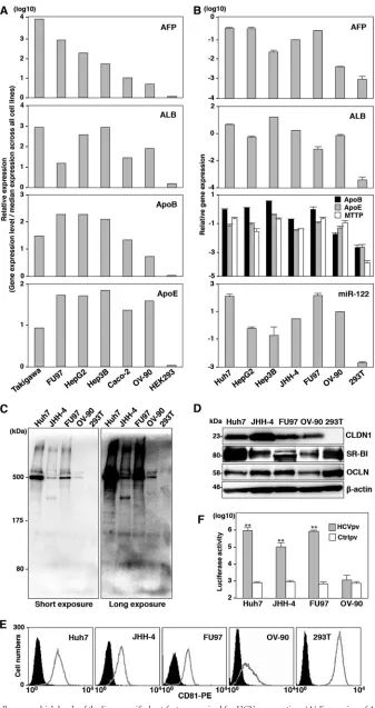
Fig. 1A
).
These results suggested that the expression of AFP was correlated
with that of the examined liver-specific host factors in cancer cell
lines. Next, to confirm this correlation, the expression levels of
ALB, ApoB, ApoE, MTTP, and miR-122 were determined by
qPCR in AFP-expressing cell lines, including FU97, OV-90, and
HCC-derived Huh7, HepG2, Hep3B, and JHH-4 cells (
Fig. 1B
).
Takigawa cells were difficult to culture, and Caco-2 cells have
pre-viously been reported to permit entry and replication of HCV (
42
,
43
); therefore, we excluded these cell lines for further analyses.
JHH-4 cells were previously shown to permit a partial
propaga-tion of HCV in a three-dimensional cultivapropaga-tion by using a
radial-flow bioreactor system upon inoculation with plasma from an
HCV carrier (
44
). In contrast to 293T cells, these AFP-expressing
cell lines express high levels of the examined liver-specific host
factors, suggesting that these cell lines maintain their hepatic
func-tions. Because previous studies have shown that Huh7, HepG2,
and Hep3B cells are susceptible to HCVcc infection, we selected
JHH-4, FU97, and OV-90 cells for further investigation as new cell
line candidates for HCV propagation. It is well known that
hepa-tocytes and intestinal cells produce ApoB100 and ApoB48,
respec-tively. ApoB100 was detected in the culture supernatants of Huh7,
JHH-4, FU97, and OV-90 cells but not in those of 293T cells by
immunoblotting (
Fig. 1C
). These results suggest that FU97 and
OV-90 cells are differentiated into hepatocyte-like cells and
pos-sess liver-specific functions. The expression of entry receptor
mol-ecules for HCV, including hCD81, CLDN1, OCLN, and SR-BI
(
45–48
), in these cell lines was confirmed by immunoblotting and
fluorescence-activated cell sorting (FACS) analyses (
Fig. 1D
and
E
). To further determine the authenticity of the receptor
candi-dates for HCV entry, HCVpv was inoculated into these cell lines.
Although the infectivity of HCVpv to JHH-4 and FU97 cells was
comparable to that in Huh7 cells, OV-90 cells did not show any
susceptibility to HCVpv infection (
Fig. 1F
). Cell surface
expres-sion of CLDN1 and OCLN was detected in OV-90 cells (data not
shown); therefore, lack of susceptibility of OV-90 cells to HCVpv
infection might be attributable to lower expression of SR-B1 and
hCD81 in OV-90 cells than in other cell lines (
Fig. 1D
and
E
).
Thus, we selected JHH-4 and FU97 cells for further investigation
of HCV propagation.
JHH-4 and FU97 cells permit HCV propagation.
To examine
the susceptibility of JHH-4 and FU97 cells to HCV propagation,
HCVcc was inoculated into these cells at a multiplicity of infection
(MOI) of 1, and intracellular HCV RNA and infectious titers in
the culture supernatants were determined by qRT-PCR and
focus-forming assay, respectively. FU97 cells exhibited higher levels of
HCV gene expression than JHH-4 cells, and these levels increased
continuously until 48 h postinfection; treatment with IFN-
␣
sig-nificantly inhibited HCV gene expression in both JHH-4 and
FU97 cells (
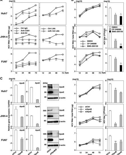
Fig. 2A
, left panels). In addition, substantial amounts
of infectious particles were detected in the culture supernatants of
JHH-4 and FU97 cells infected with HCVcc, in contrast to the lack
of infectious particles in the culture supernatants of 293T-CLDN/
miR-122 cells infected with HCVcc (
Fig. 2A
, bar graph). Recent
reports have shown that exogenous expression of miR-122
en-hances HCV RNA abundances in several hepatic or nonhepatic
cell lines (
16–18
). Therefore, we examined the effect of miR-122
overexpression on HCV RNA abundances in both JHH-4 and
FU97 cells. miR-122 was introduced in these cells by a lentiviral
vector encoding pri-122, an unprocessed 122, and
miR-122 expression was confirmed by quantitative PCR (qPCR)
anal-ysis (
Fig. 2B
, bar graph). In contrast to the slight increase of HCV
RNA in FU97 cells, JHH-4 cells exhibited a significant increase of
HCV RNA, suggesting that the expression level of miR-122 is a key
determinant for the efficient propagation of HCV (
Fig. 2B
, right
panels). A previous study has shown that NS5A proteins were
localized around the endoplasmic reticulum (ER) membrane, and
accumulation of core protein around lipid droplets (LDs)
facili-tates efficient assembly of infectious particles in Huh7 cells (
24
).
Immunofluorescence microscopy observation revealed that core
and NS5A proteins in JHH-4 and FU97 cells infected with HCVcc
were detected around LDs and in the ER together with
double-stranded RNA (dsRNA), respectively (
Fig. 2C
). These results
sug-gest that expression of liver-specific factors permits complete
propagation of HCVcc in JHH-4 and FU97 cells and that hepatic
characteristics play crucial roles in HCV propagation.
JHH-4 and FU97 cells permit complete propagation of
HCVcc without any exogenous expression of host factors
cru-cial for propagation of HCV.
To further characterize the
propa-gation of HCV in JHH-4 and FU97 cells, we examined the effects
of the HCV inhibitors on the replication of HCV RNA.
Preincu-bation with anti-HCV E2 antibody and pretreatment of cells with
anti-hCD81 monoclonal antibody significantly inhibited HCVcc
infection not only in Huh7 cells but also in JHH-4 and FU97 cells,
suggesting that hCD81 is also involved in HCV entry into JHH-4
and FU97 cells (
Fig. 3A
, left panels). To examine the effect of
miR-122 expression on HCV RNA abundances, cells were treated
with LNA specific to either miR-122 (miR-122–LNA) or a
non-specific LNA (Ctrl-LNA) at 6 h before infection with HCVcc.
Treatment with miR-122–LNA but not with Ctrl-LNA
signifi-cantly reduced the HCV RNA abundances in these cell lines,
sug-gesting that miR-122 also plays a crucial role in the efficient
prop-agation of HCVcc in JHH-4 and FU97 cells (
Fig. 3A
, right panels).
Previous reports showed that treatment with MTTP inhibitors
inhibited the production of infectious particles of HCVcc in Huh7
cells (
20
,
22
). Although intracellular HCV RNA levels in Huh7,
JHH-4, and FU97 cells were not inhibited by the treatment with
MTTP inhibitors, including CP-346086 and BMS-200150 (
Fig.
3B
, left panels), the production of infectious particles was
signifi-cantly decreased in these cells (
Fig. 3B
, right panels). These results
suggest that the VLDL secretion pathway also participates in the
propagation of HCV in JHH-4 and FU97 cells. Furthermore, it
was shown that ApoB and ApoE are involved in the production of
HCV particles in Huh7 cells (
20–22
). To confirm the role of ApoB
and ApoE in HCV propagation in JHH-4 and FU97 cells, the
expression of ApoB and ApoE was suppressed by siRNAs (
Fig. 3C
,
left panels). The suppression of ApoB and ApoE expression
sig-nificantly reduced HCV RNA levels in cells infected with HCVcc
at an MOI of 1 (
Fig. 3C
, middle panels) and significantly reduced
the infectious titers in the supernatants (
Fig. 3C
, right panels) at 72
h postinfection. Collectively, these results suggest that the JHH-4
and FU97 cells permit complete propagation of HCVcc without
any exogenous expression of the host factors crucial for
propaga-tion of HCV, including receptor molecules, miR-122, and
VLDL-associated proteins. FU97 cells exhibited higher susceptibility to
HCVcc propagation than JHH-4 cells (
Fig. 2A
), and thus we
char-acterized the FU97 cells in greater detail.
Establishment of HCV RNA replicon and cured cells by using
FU97 cells.
To further examine the characteristics of FU97 cells
on November 7, 2019 by guest
http://jvi.asm.org/
FIG 1JHH-4 and FU97 cells express high levels of the liver-specific host factors required for HCV propagation. (A) Expression of AFP, ALB, ApoB, and ApoE in cancer cell lines screened by the NextBio Body Atlas application. The expression levels were standardized by the median expression across all cell lines. (B) Expression of AFP, ALB, ApoB, ApoE, MTTP, and miR-122 in AFP-expressing cell lines including HepG2, Hep3B, FU97, and OV-90 cells identified by NextBio Shiokawa et al.
on November 7, 2019 by guest
http://jvi.asm.org/
[image:5.585.124.461.64.701.2]with respect to HCV RNA replication,
in vitro
-transcribed
sub-genomic HCV RNA of the Con1 strain was electroporated into
Huh7 and FU97 cells and cultured in medium containing G418
for a month, and then subgenomic replicon (SGR) cells of the
Con1 strain were established (
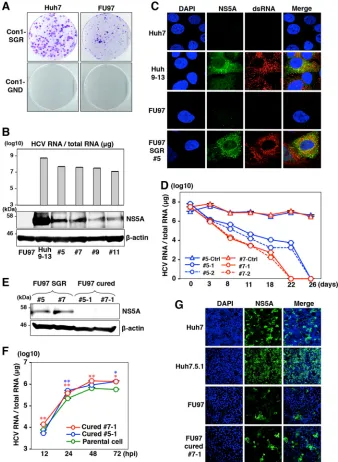
Fig. 4A
). Replication of HCV RNA
in four clones of the FU97 replicon cells was examined by
qRT-PCR and immunoblotting. All clones contained a high level of
HCV RNA (3
⫻
10
7to 7
⫻
10
7copies/
g total RNA) (
Fig. 4B
,
upper panel), and the NS5A protein was also detected (
Fig. 4B
,
lower panel). We examined the localization of NS5A and dsRNA
in clone 5 of FU97 SGR cells by immunofluorescence analysis.
Colocalization of NS5A with dsRNA was observed in clone 5,
sug-gesting that the replication complex required for viral RNA
repli-cation was generated in the FU97 SGR cells (
Fig. 4C
). It has been
shown that the infectivity of HCVcc in the cured cells that were
established by elimination of the viral genome by treatment with
antivirals from Huh7 replicon cells is significantly higher than that
in parental Huh7 cells (
49
). To establish FU97 cured cells, two
clones of FU97 replicon cells (clones 5 and 7) were treated with a
combination of either 100 IU/ml of IFN-
␣
and 100 nM BILN 2061
(clones 5-1 and 7-1) or 10 pM BMS-790052 and 100 nM BILN
2061 (clones 5-2 and 7-2) to eliminate viral RNA. Viral RNA was
gradually decreased and was completely eliminated at 26 days
posttreatment in four clones (
Fig. 4D
), and elimination of NS5A
expression in cured cells was confirmed by immunoblot analysis
(
Fig. 4E
). Next, to examine the susceptibility of the cured cells to
the propagation of HCVcc, FU97 cured cell clones (clones 5-1 and
7-1) and parental FU97 cells were infected with HCVcc at an MOI
of 1. The cured cells are more permissive to HCV infection,
result-ing in increased HCV RNA (
Fig. 4F
) and NS5A abundances (
Fig.
4G
) compared to the parental cells. These results suggest that
sus-ceptibility of the cured FU97 cells to the propagation of HCVcc is
higher than that of parental cells, as seen in previous studies using
hepatic and nonhepatic cells (
17
,
18
,
49
).
Cured FU97 cells exhibit normal innate immune response.
It
has been shown that one of the reasons for the high susceptibility
of the cured cell line, Huh7.5 cells, to HCVcc infection is the
impairment of the innate immune responses caused by mutation
in RIG-I, a key sensor for viral RNA (
50
). To examine the
involve-ment of the innate immune response in the enhanceinvolve-ment of
HCVcc propagation in the cured FU97 cells, the expression levels
of IFN-stimulated gene 15 (ISG15) were determined upon
stimu-lation with IFN-
␣
or infection with VSV. Expression of ISG15 was
significantly increased in both parental and cured FU97 cells by
treatment with IFN-
␣
or infection with VSV (
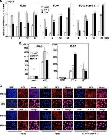
Fig. 5A
). To further
confirm the innate immune responses in the cured FU97 cells,
reporter plasmids encoding the luciferase gene under the control
of either the IFN-

(
Fig. 5B
, left) or ISRE (
Fig. 5B
, right) promoter
were transfected into both parental and cured FU97 cells and
treated with IFN-
␣
or inoculated with VSV. Activation of these
promoters in the cured cells was comparable to that in the parental
cells. To further assess the authenticity of viral RNA recognition
and ISG induction pathways in the cured cells, nuclear
localiza-tion of IRF3 and STAT2 upon stimulalocaliza-tion was determined by
immunofluorescence analysis. IRF3 and STAT2 in both parental
and cured FU97 cells were translocated at similar levels into the
nucleus upon infection with VSV or treatment with IFN-
␣
(
Fig.
5C
). These results suggest that the efficient propagation of HCVcc
in the FU97 cured cells is attributable to reasons other than
im-pairment of innate immunity.
Expression of miR-122 is one of the determinants for HCV
RNA abundances.
We hypothesized that HCV replicon cells are
capable of surviving in the presence of G418 by amplification of
the viral genome through the enhancement of miR-122
expres-sion, and cured FU97 cells acquired the ability to propagate
HCVcc due to the high-level expression of miR-122. Our previous
study also suggested that the expression levels of miR-122 in
Huh7, Hep3B, and Hec1B cured cells were higher than those in
parental cells (
17
,
18
). To test this hypothesis, the expression levels
of miR-122 in the cured FU97 cells were compared with those in
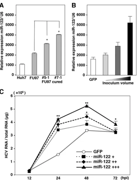
parental cells. Interestingly, the cured FU97 cell clones exhibited a
1.8-fold increase in miR-122 expression (
Fig. 6A
). These results
suggested that the efficient propagation of HCVcc in the cured
FU97 cells was attributable to enhanced expression of miR-122
rather than the impairment of the innate immunity. To further
confirm the correlation between the expression of miR-122 and
HCV RNA abundances, we established FU97 cell lines expressing
various concentrations of miR-122 by using a lentiviral vector
(
Fig. 6B
), and HCV RNA abundances in these cell lines upon
infection with HCVcc were determined by qRT-PCR (
Fig. 6C
).
HCV RNA abundances increased in accord with the expression of
miR-122, suggesting that expression of miR-122 is one of the
de-terminants for HCV RNA abundances in cells infected with
HCVcc.
HCV particles produced in FU97 cells exhibit similar
charac-teristics to those in hepatic cells.
To examine the characteristics
of viral particles produced in FU97 cells, HCV particles recovered
from the culture supernatants of Huh7.5.1 and FU97 cells infected
with HCVcc were fractionated by buoyant density gradient
anal-ysis. Previous reports indicated that viral RNA and infectious
par-ticles were broadly distributed, with peaks in fractions from 1.13
to 1.14 g/ml and from 1.09 to 1.10 g/ml, respectively (
51
,
52
). In
agreement with the previous data, major peaks of HCV RNA and
infectious particles in culture supernatants of both Huh7.5.1 and
FU97 cells were detected around 1.10 g/ml and 1.09 g/ml,
respec-tively (
Fig. 7A
and
7B
, upper panels). Furthermore, ApoE was
detected around the peak fractions of infectivity in both Huh7.5.1
and FU97 cells (
Fig. 7A
and
B
, lower panels). These results suggest
that HCV particles produced in FU97 cells exhibit characteristics
similar to those in hepatic cells.
Effects of anti-HCV drugs on the propagation of HCV in
FU97 cells.
To determine the difference in the efficacies of
antivi-rals on the HCV propagated in Huh7 and FU97 cells, three DAAs,
i.e., BMS-790052, PSI-7977, and BILN 2061 targeting NS5A,
Body Atlas and Huh7, JHH-4, and 293T cells was determined by qPCR. The relative expression of AFP, ApoB, ApoE, MTTP, and ALB mRNA was normalized to that of glyceraldehyde-3-phosphate dehydrogenase (GAPDH) mRNA, and that of miR-122 was normalized to that of U6 snRNA. (C) Secretion of ApoB in the culture supernatants of Huh7, JHH-4, FU97, OV-90, and 293T cells was determined by immunoblotting by using anti-ApoB antibody. The molecular mass of ApoB100 secreted from hepatocyte is about 500 kDa. (D) Expression of CLDN1, SR-BI, and OCLN in these cell lines was determined by immunoblotting. (E) Expression of hCD81 in the cell lines was determined by flow cytometry. (F) HCVpv-bearing HCV envelope proteins and control virus (Ctrlpv) were inoculated into the cell lines, and luciferase activities were determined at 24 h postinfection. Asterisks indicate significant differences (*,P⬍0.05; **,P⬍0.01) versus the results for control virus.
on November 7, 2019 by guest
http://jvi.asm.org/
Shiokawa et al.
on November 7, 2019 by guest
http://jvi.asm.org/
NS5B, and NS3/4A, respectively, were treated with various
con-centrations at 3 h postinfection with HCVcc, and the intracellular
HCV RNA level was determined by qRT-PCR at 48 h
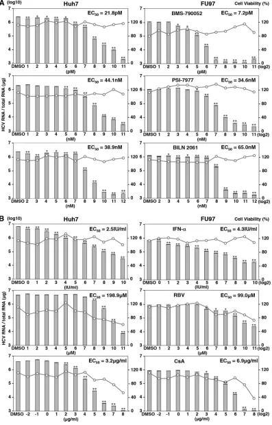
postinfec-tion. Treatment with these DAAs inhibited the HCV RNA level in
a dose-dependent manner in both Huh7 and FU97 cells (
Fig. 8A
,
bar graphs) and exhibited no cell toxicity at all even at the highest
dose (
Fig. 8A
, line graphs). The inhibitory effects of BMS-790052
(
Fig. 8A
, top graphs) on the propagation of HCVcc in FU97 cells
were higher than those in Huh7 cells, and the 50% effective
con-centration (EC
50) values of BMS-790052 against propagation of
HCVcc in FU97 and Huh7 cells were 7.2 and 21.8 pM, respectively
(
P
⬍
0.05). On the other hand, the antiviral effects of BILN 2061
(
Fig. 8A
, bottom graphs) on the propagation of HCVcc in FU97
cells were lower than those in Huh7 cells, and EC
50s of BILN 2061
against propagation of HCVcc in FU97 and Huh7 cells were 65.0
and 38.9 nM, respectively (
P
⬍
0.01). PSI-7977 showed almost
equivalent inhibitory effects to HCV propagated in FU97 and
Huh7 cells, and the EC
50s of PSI-7977 against propagation of
HCVcc in FU97 and Huh7 cells were 34.6 and 44.1 nM,
respec-tively (
Fig. 8A
, middle graphs). These results suggest that the
an-tiviral effect of DAAs on the propagation of HCVcc varied
be-tween Huh7 and FU97 cells.
Next, we examined the efficacy of IFN-
␣
, RBV, and
cyclospo-rine, which are inhibitors for HCV targeting host factors involved
in the propagation of HCVcc (
53–55
), on the propagation of
HCVcc in Huh7 and FU97 cells. Cells were treated with various
concentrations of the reagents at 3 h postinfection with HCVcc,
and the level of intracellular HCV RNA was determined by
qRT-PCR at 48 h postinfection. In contrast to the treatment with DAAs,
both Huh7 and FU97 cells exhibited cell toxicity by the treatment
with RBV and cyclosporine but not with IFN-
␣
at higher
concen-trations (
Fig. 6B
, line graphs). The inhibitory efficacies of IFN-
␣
(
Fig. 8B
, top graphs) and cyclosporine (
Fig. 8B
, bottom graphs) on
the propagation of HCVcc in FU97 cells were lower than those in
Huh7 cells, and the EC
50s of IFN-
␣
against propagation of HCVcc
in FU97 and Huh7 cells were 4.3 and 2.5 IU/ml, (
P
⬍
0.05),
re-spectively; those of cyclosporine were 6.9 and 3.2
g/ml (
P
⬍
0.01), respectively. On the other hand, the antiviral effect of RBV
on the propagation of HCVcc in FU97 cells was higher than that in
Huh7 cells, and the EC
50s of RBV against propagation of HCVcc
in FU97 and Huh7 cells were 99.0 and 198.9
M, respectively (
P
⬍
0.05) (
Fig. 8B
, middle graphs). These results suggest that the
effi-cacies of anti-HCV drugs targeting host factors involved in the
infection of HCV were also different between Huh7 and FU97
cells.
FU97 cells exhibit higher susceptibility to HCVcc/JFH-2
propagation than Huh7 cells.
HCVcc/JFH-2 was cloned from a
patient with fulminant hepatitis and exhibited efficient
propaga-tion in Huh7 cured cells (
34
).
In vitro
-transcribed RNA of pJFH2/
AS/mtT4 encoding a full-length JFH-2 strain was electroporated
into Huh7.5.1 cells, and HCVcc/JFH-2 of 1.5
⫻
10
5FFU/ml was
recovered in the supernatants after serial passages. To examine the
susceptibility of FU97 cells to the propagation of HCVcc/JFH-2,
cells were infected with HCVcc/JFH-2 at an MOI of 1, and the
intracellular HCV RNA level was determined by qRT-PCR.
Intra-cellular HCV RNA in parental and cured FU97 cells increased
until 72 h postinfection, while it reached a peak at 48 h
postinfec-tion in Huh7 cells, and the highest HCV RNA level was observed
in the cured FU97 clones upon infection with HCVcc/JFH-2 (
Fig.
9A
). Infectious titers in the culture supernatants at 72 h
postinfec-tion with HCVcc/JFH-2 were also highest in the cured FU97 7-1
cells (2.5
⫻
10
4FFU/ml), followed by parental FU97 (1.2
⫻
10
4FFU/ml) and Huh7 (9
⫻
10
3FFU/ml) cells (
Fig. 9B
). Next, we
examined the expression and subcellular localization of HCV
pro-teins in cells infected with HCVcc/JFH-2 by immunofluorescence
analysis. Expression of NS5A in cells upon infection with HCVcc/
JFH-2 was highest in the cured FU97 7-1 cells, followed by
paren-tal FU97 cells, and that in Huh7 cells was low (
Fig. 9C
, left panels).
Core protein was detected around LDs in cells infected with
HCVcc/JFH-2, as seen in those infected with the HCVcc/JFH-1
strain (
Fig. 9C
, right). To further confirm the efficient
propaga-tion of HCVcc/JFH-2 in FU97 cells,
in vitro
-transcribed viral
RNAs of the JFH-1 and JFH-2 strains of HCVcc were
electropo-rated into Huh7, FU97, and cured FU97 cells. Although the
infec-tious titers of the JFH-1 strain in FU97 cells were lower than those
in Huh7 cells, those of the JFH-2 strain in FU97 and cured FU97
cells were significantly higher than those in Huh7 cells (
Fig. 9D
).
These results suggest that FU97 cells are more susceptible to
prop-agate HCVcc/JFH-2 than Huh7 cells.
DISCUSSION
Several reports have shown that hepatic differentiation is involved
in the susceptibility of ES/iPS cells to HCVcc infection (
28
,
30
,
41
).
In addition, in hepatic cancer cell lines, including Huh7, HepG2,
and Hep3B, cells derived from not poorly but well-differentiated
HCC permit complete propagation of HCVcc (
15–17
), suggesting
that hepatic differentiation is closely related to the susceptibility of
cells to HCVcc propagation. In this study, we identified two cell
lines susceptible to HCVcc infection by the screening of cancer cell
lines expressing AFP as a marker of hepatic differentiation.
HCC-derived JHH-4 cells and gastric cancer-HCC-derived FU97 cells permit
complete propagation of HCVcc without any exogenous
expres-sion of the host factors required for HCVcc propagation,
includ-ing HCV receptor candidates, miR-122, and apolipoproteins. In
particular, FU97 cells exhibited higher susceptibility to HCVcc/
JFH-2 infection than Huh7 cells, suggesting that FU97 cells would
be useful tools for further HCV analyses.
Although HCV has been classified into seven major genotypes
and a series of subtypes (
56
,
57
), the
in vitro
infection model had
been restricted to the JFH-1 strain based on the genotype 2a until
FIG 2JHH-4 and FU97 cells permit HCV propagation. (A) Intracellular HCV RNA levels in Huh7, JHH-4, and FU97 cells inoculated with HCVcc at an MOI of 1, treated with 100 IU/ml of IFN-␣or untreated (mock), were determined by qRT-PCR at 12, 24, 48, and 72 h postinfection (hpi). Infectious titers in the culture supernatants of Huh7, JHH-4, FU97, and 293T-CLDN/miR-122 cells infected with HCVcc at an MOI of 1 were determined by a focus-forming assay at 72 h postinfection (bar graph). (B) Exogenous expression of miR-122 in Huh7, JHH-4, and FU97 cells by lentiviral vector (bar graph). Total cellular miRNA extracted from the cells was subjected to qRT-PCR. U6 was used as an internal control. Intracellular HCV RNA in Huh7, JHH-4, and FU97 cells inoculated with HCVcc at an MOI of 1 was determined by qRT-PCR at 12, 24, 48, and 72 h postinfection. Solid and broken lines indicate HCV RNA abundances in miR-122-expressing and GFP-expressing control cells, respectively. (C) Huh7, JHH-4, and FU97 cells were infected with HCVcc at an MOI of 1, fixed with 4% PFA, and subjected to immunofluorescence analyses by using antibodies against core, NS5A, dsRNA, and calregulin. Lipid droplets and cell nuclei were stained by BODIPY and DAPI, respectively. Asterisks indicate significant differences (*,P⬍0.05; **,P⬍0.01) versus the results for control cells.
on November 7, 2019 by guest
http://jvi.asm.org/
FIG 3JHH-4 and FU97 cells permit complete propagation of HCVcc without any exogenous expression of host factors crucial for propagation of HCVcc. (A) Effect of inhibitors on the propagation of HCVcc in Huh7, JHH-4, and FU97 cells. (Left panels) HCVcc was preincubated with anti-E2 antibody and inoculated into cells. Cells were preincubated with anti-hCD81 antibody or isotype control antibody (Ctrl IgG) and then infected with HCVcc. (Right panels) Cells were infected with HCVcc and treated with miR-122-LNA (30 nM) or Ctrl-LNA (30 nM) at 6 h postinfection. (B) Huh7, JHH-4, and FU97 cells infected with HCVcc at an MOI of 1 were treated with dimethyl sulfoxide (DMSO) or MTTP inhibitor, CP-346086 (5M)or BMS-200150 (10M), at 3 h postinfection. Intracellular HCV RNA in cells at 12, 24, 48, and 72 h postinfection was determined by qRT-PCR (left panels). Infectious titers in the culture supernatants of cells infected with HCVcc at an MOI of 1 and treated with 5M CP-346086, 10M BMS-200150, or dimethyl sulfoxide alone (DMSO) at 3 h postinfection were determined at 72 h postinfection by a focus-forming assay (right graphs). (C) mRNA and protein expression levels of ApoB and ApoE (left panels) in Huh7, JHH-4, and FU97 cells at 48 h posttransfection with siRNA targeting either ApoB or ApoE or a control siRNA (siApoB, siApoE, or siCtrl, respectively) were determined by qRT-PCR and immunoblotting, respectively. Huh7, JHH-4, and FU97 cells were infected with HCVcc at an MOI of 1 at 6 h posttransfection with siRNA targeting either ApoB or ApoE or a control siRNA (siApoB, siApoE, or siCtrl, respectively) (right panels). Intracellular HCV RNA at 12, 24, 48, and 72 h postinfection and infectious titers in the culture supernatants at 72 h postinfection were determined by qRT-PCR and focus-forming assay, respectively. Asterisks indicate significant differences (*,P⬍0.05; **,P⬍0.01) versus the results for control cells.
on November 7, 2019 by guest
http://jvi.asm.org/
[image:9.585.75.516.40.582.2]recently (
15
). To clarify the pathogenesis of HCV depending on
the genotypes, the establishment of cell-culture-adapted clones
derived from various genotypes is essential (
58
). Viable
JFH1-based intergenotypic recombinants, containing genotype-specific
structural proteins, p7 and the complete or partial NS2, were
gen-erated for various genotypes of HCV (
56
,
59
,
60
). Although robust
propagation systems of full-length HCV infectious clones of the
H77 strain (genotype 1a) (
61
), TN strain (1a) (
62
), JFH-2 strain
FIG 4Establishment of HCV RNA replicon and cured FU97 cells. (A) Wild-type SGR RNA (Con1-SGR) or replication-defective RNA (Con1-GND) of the HCV Con1 strain was electroporated into Huh7 and FU97 cells and replaced with medium containing 1 mg/ml and 400g/ml of G418 at 24 h postelectroporation, respectively. Colonies were stained with crystal violet at 30 days postselection. (B) Four clones derived from FU97 SGR cells (clones 5, 7, 9, and 11) were subjected to qRT-PCR after extraction of total RNA (upper panel) and to immunoblotting using anti-NS5A antibody (lower panel). Huh9-13 cells, which were Huh7-derived Con1-SGR cells, were used as a positive control. (C) Huh9-13 cells, Huh7 parental cells, FU97-Huh7-derived Con1-SGR cells (FU97 SGR, clone 5), and FU97 parental cells were fixed in 4% PFA and subjected to immunofluorescence assay using anti-NS5A and anti-dsRNA antibodies. Cell nuclei were stained by DAPI. (D) Elimination of HCV RNA from FU97-derived Con1-SGR cells. Two clones derived from FU97 SGR cells (clones 5 and 7) were treated with a combination of either 100 IU/ml of IFN-␣and 100 nM BILN 2061 (clones 5-1 and 7-1) or 10 pM of BMS-790052 and 100 nM BILN 2061 (clones 5-2 and 7-2) to eliminate the HCV genome. Clones 5-Ctrl and 7-Ctrl are negative controls, untreated with anti-HCV drugs. Intracellular HCV RNA at 3, 8, 11, 18, 22, and 26 days posttreatment was determined by qRT-PCR. (E) The expression levels of NS5A in FU97 SGR cells (clones 5 and 7) and in FU97 cured cells (clones 5-1 and 7-1) were determined by immunoblot analysis using anti-NS5A antibody. (F) FU97 cured cells (clone 5-1 and clone 7-1) and parental cells were infected with HCVcc at an MOI of 1; the levels of intracellular HCV RNA at 12, 24, 48, and 72 h postinfection were determined by qRT-PCR. (G) The expression of NS5A in Huh7, Huh7.5.1, FU97, and cured FU97 clone 7-1 was determined by immunofluorescence analysis at 72 h postinfection by using anti-NS5A antibody. Asterisks indicate significant differences (*,P⬍0.05; **,P⬍0.01) versus the results for control cells.
on November 7, 2019 by guest
http://jvi.asm.org/
[image:10.585.122.460.67.529.2](2a) (
34
), and S310 strain (3a) (
63
) were established, the
construc-tion of infectious clones of other genotypes has not succeeded yet.
Because permissive cell lines for HCVcc infection
in vitro
had
been limited to Huh7 cells due to cell tropism and the narrow host
range (
13
,
14
), the establishment of a novel cell culture system
supporting HCV propagation is needed for further HCV analyses.
Previous reports have demonstrated that HepG2, Hep3B, and
HEK293 cells permit HCVcc propagation (
16
,
17
,
64
). However,
exogenous expression of host factors is necessary for complete
propagation of HCVcc in these cell lines. In HepG2 and Hep3B
cells, overexpression of miR-122 is essential for efficient
replica-tion of HCV RNA (
16
,
17
). In HEK293 cells, the exogenous
ex-pression of CLDN1, miR-122, and ApoE was required for
infec-tious particle formation upon infection with HCVcc (
64
). On the
other hand, JHH-4 and FU97 cells permit complete propagation
of HCVcc without any exogenous expression of the host factors
required for propagation of HCVcc. JHH-4 cells grown in a
three-dimensional radial-flow bioreactor were successfully infected
fol-lowing inoculation with plasma from an HCV carrier and
trans-fection of HCV RNA transcribed from full-length cDNA (
44
). In
FIG 5Innate immune response in cured FU97 cells. (A) Huh7, parental, and cured FU97 cells (clone 7-1) were stimulated with 100 IU/ml of IFN-␣or infected with VSV. The expression of mRNA of ISG15 at 4, 8, 12, and 24 h posttreatment (hpt) was determined by qPCR and standardized by that of glyceraldehyde-3-phosphate dehydrogenase (GAPDH). (B) Huh7, parental FU97, and cured FU97 (clone 7-1) cells cotransfected with pIFN--Luc and pRL-SV40 were infected with VSV at an MOI of 1 at 24 h posttransfection (left). Cells cotransfected with pISRE-Luc and pRL-SV40 were infected with VSV at an MOI of 1 or stimulated with 100 IU/ml of IFN-␣at 24 h posttransfection (right). Luciferase activities were determined at 24 h posttreatment. (C) Huh7, parental FU97, and cured FU97 (clone 7-1) cells were infected with VSV at an MOI of 1 or stimulated with 100 IU/ml of IFN-␣, fixed with 4% PFA at 18 h posttreatment, and subjected to immunofluorescence assay using anti-IRF3 and -STAT2 antibodies. Cell nuclei were stained by DAPI. Asterisks indicate significant differences (*,P⬍0.05; **, P⬍0.01) from the results for control cells.
Shiokawa et al.
on November 7, 2019 by guest
http://jvi.asm.org/
[image:11.585.113.473.69.516.2]addition, JHH-4 cells were suggested to possess some host factors
involved in the enhanced translation of HCV RNA (
64
,
65
).
Fur-thermore, high susceptibility of FU97 cells to HCVcc/JFH-2
infec-tion compared to Huh7 cells raises the possibility of using FU97
cells for the propagation of HCVcc derived from other genotypes,
including the H77, TN, and S310 strains.
AFP-producing gastric cancer (AFPGC) cell lines, FU97 and
Takigawa cells (
66
), which were identified by using a cDNA array
database, were shown to express high levels of liver-specific
fac-tors. AFPGC is a rare case and exhibits a worse prognosis and the
characteristics of early hepatic metastasis (
67
). It is hypothesized
that production of AFP, which is suppressed in mature
hepato-cytes, is induced in HCC by the dedifferentiation of cancer cells or
the increase in oval cells in the oncogenic pathway (
68
). Oval cells
are believed to be capable of producing AFP, are candidates for
hepatic stem cells, have bipotentiality to differentiate into
hepato-cytes and bile duct epithelial cells, and play an important role in
liver regeneration (
69
,
70
). These hypotheses suggest that cancer
cells acquired a new function, such as the ability to produce AFP
through an alteration in differentiation status. Although the
mechanism of AFP production in gastric cancer remains
un-known, hepatic dedifferentiation might be induced in gastric
can-cer. Furthermore, previous reports have proposed the concept of
“hepatoid adenocarcinoma” based on the differentiation of
AFPGC into hepatocyte-like cells (
71
,
72
), suggesting that FU97
and Takigawa cells obtained the hepatocyte-like characteristics
required for HCV propagation through dedifferentiation during
the oncogenic process. In addition, recent studies demonstrated
that hepatocyte-like cells derived from induced pluripotent stem
cells (iPS cells/iPSCs) express high levels of miR-122 and
VLDL-associated proteins and support propagation of HCVcc and HCV
derived from patient serum (
28–30
). These results suggest that
hepatic differentiation required for hepatic functions plays crucial
roles in HCV propagation. In accord with these observations, our
data suggest that cancer cell lines differentiated into
hepatocyte-FIG 6Expression of miR-122 is one of the determinants for HCV RNA abun-dances. (A) Total RNA was extracted from Huh7 and parental and cured FU97 (clones 5-1 and 7-1) cells, and the relative expression of miR-122 was deter-mined by qPCR. U6 snRNA was used as an internal control. (B) Establishment of FU97 cell lines stably expressing various concentration of miR-122 by in-fection with a lentiviral vector. FU97 cells infected with lentiviral vector to express GFP were used as a control. (C) FU97 cell lines expressing various concentrations of miR-122 were infected with HCVcc at an MOI of 1, and HCV RNA abundances were determined at 12, 24, 48, and 72 h postinfection (hpi) by qRT-PCR. Asterisks indicate significant differences (*,P⬍0.05; **, P⬍0.01) versus the results for control cells.
FIG 7HCV particles produced in FU97 cells exhibit similar characteristics to those in hepatic cells. HCV particles in the culture supernatants of Huh7.5.1 and FU97 cells were harvested at 72 h postinfection with HCVcc and analyzed by using iodixanol density gradient centrifugation. HCV RNA and infectious titers of each fraction were determined by qRT-PCR and focus-forming assay, respectively. Buoyant density was plotted for each fraction (upper panels). Expression of ApoE in each fraction was detected by immunoblotting using anti-ApoE antibody (lower panels).
on November 7, 2019 by guest
http://jvi.asm.org/
[image:12.585.47.281.63.370.2] [image:12.585.84.502.491.684.2]FIG 8Effects of anti-HCV drugs on the propagation of HCVcc in FU97 cells. (A) Effect of DAAs on the propagation of HCVcc in Huh7 and FU97 cells. Cells infected with HCVcc at an MOI of 1 were treated with BMS-790052, PSI-7977, and BILN 2061 at 3 h postinfection (identifications in right-hand panels). (B) Effect of HCV inhibitors targeting host factors on the propagation of HCVcc in Huh7 and FU97 cells. Cells infected with HCVcc at an MOI of 1 were treated with IFN-␣, RBV (middle), and cyclosporine (CsA) at 3 h postinfection (identifications in right-hand panels). Intracellular HCV RNA levels were determined by qRT-PCR at 48 h postinfection (bar graphs), and cell viability was determined as a percentage of the viability of cells treated with 0.1% dimethyl sulfoxide (DMSO) at 48 h posttreatment (line graphs). From the assay results, the 50% effective concentration (EC50) of each reagent was determined. Asterisks indicate
significant differences (*,P⬍0.05; **,P⬍0.01) versus the results for control cells.
on November 7, 2019 by guest
http://jvi.asm.org/
[image:13.585.95.492.36.656.2]like cells to gain hepatic functions could permit complete
propa-gation of HCVcc.
Treatment with DAAs including BMS-790052 (NS5A
inhibi-tor) (
73
), PSI-7977 (NS5B polymerase inhibitor) (
74
), and BILN
2061 (NS3/4A protease inhibitor) (
75
) inhibited propagation of
HCV in both Huh7 and FU97 cells infected with HCVcc without
any cell toxicity. Antiviral effects of BMS-790052 and BILN 2061
were significantly different between Huh7 and FU97 cells,
sug-gesting that efficacies of DAAs are varied, depending on cell lines.
Although anti-HCV drugs targeting host factors including IFN-
␣
,
RBV, and cyclosporine also inhibited propagation of HCVcc in a
dose-dependent manner in both Huh7 and FU97 cells, treatment
with RBV and cyclosporine produced cell toxicity at higher
con-centrations than treatment with DAAs. Although the antiviral
mechanism of RBV against HCV has not been well elucidated yet
(
53
), inhibitory effects of RBV against HCV infection were
signif-icantly higher in Li23 cells than those in Huh7 cells (
76
,
77
), and
RBV also exhibited a low inhibitory effect upon infection with
HCVcc in Huh7 cells compared to that in FU97 cells. Although
adenosine kinase (ADK) was shown to be a determinant for the
sensitivity of RBV (
78
), the expression levels of ADK in Huh7 and
FU97 cell lines were comparable (data not shown).
The
IL28B
genotype is associated with the sensitivity of IFN
treatment for chronic hepatitis C patients (
79–81
), and patients
with the minor
IL28B
genotype exhibit lower susceptibility to the
treatment than those with major genotypes. Although FU97 cells
showed lower sensitivity to the IFN-
␣
treatment than Huh7 cells,
FU97 and Huh7 cells possess major and minor
IL28B
genotypes
FIG 9Propagation of HCVcc/JFH-2 in FU97 cells. (A) Huh7, FU97 parental, FU97 cured 5-1, and FU97 cured 7-1 cells were infected with HCVcc/JFH-2 at an MOI of 1, and the intracellular HCV RNA level was determined by qRT-PCR at 12, 24, 48, and 72 h postinfection. (B) Huh7, FU97, and FU97 cured 7-1 cells were infected with HCVcc/JFH-2 at an MOI of 1, and infectious titers in the culture supernatants were determined by focus-forming assay. (C) Huh7, FU97, and FU97 cured 7-1 cells were infected with HCVcc/JFH-2 at an MOI of 1, fixed with 4% PFA at 72 h postinfection, and subjected to immunofluorescence assay using antibodies against NS5A or core. Lipid droplets and cell nuclei were stained with BODIPY and DAPI, respectively. (D)In vitro-transcribed JFH-1 and JFH-2 RNAs were electroporated into Huh7, FU97, and FU97 cured 7-1 cells. The infectious titers of JFH-1 and JFH-2 in the culture supernatants from these cells were determined by focus-forming assay up to 14 days postransduction. Asterisks indicate significant differences (*,P⬍0.05; **,P⬍0.01) versus the results for control cells.
on November 7, 2019 by guest
http://jvi.asm.org/
[image:14.585.137.451.64.475.2](data not shown), respectively. Furthermore, induction of ISG15
by treatment with IFN-
␣
was almost comparable between Huh7
and FU97 cells (
Fig. 5A
), and expression levels of IFN-
␣
receptor
in the cell lines were the same (data not shown), suggesting the
involvement of other factors in the difference in the IFN responses
between FU97 and Huh7 cells.
Cyclophilins possess peptidyl-prolyl
cis
/
trans
isomerase
(PPIase) activity and are involved in protein folding and assembly.
Cyclophilin A (CypA), the most abundant cyclophilin, localizes in
the cytoplasm and interacts with the immunosuppressive drug
cyclosporine (
82
). In addition, CypA has been shown to be
in-volved in the propagation of human immunodeficiency virus (
83
,
84
), hepatitis B virus (
85
,
86
), influenza A virus (
87
), and HCV
(
88
). Replication of HCV RNA was inhibited by suppression of the
PPIase activity of CypA by treatments with cyclosporine,
muta-tion in the active site of CypA, and knockdown of CypA (
55
,
89–
91
). The same level of CypA expression in Huh7 and FU97 cells
(data not shown) suggests that the difference in inhibitory effect of
cyclosporine in the cell lines may be attributable to other reasons,
such as a difference in PPIase activity of CypA in these cell lines.
The differences in the efficacy of anti-HCV drugs between Huh7
and FU97 cells were small; however, FU97 cells have the possibility
to possess antiviral activity different from that of Huh7 cells.
In summary, we identified novel permissive cell lines for
com-plete propagation of HCVcc without any artificial manipulation.
In particular, gastric cancer-derived FU97 cells exhibited a much
higher susceptibility to HCVcc/JFH-2 infection than observed in
Huh7 cells, suggesting that FU97 cells would be useful for further
investigation of the HCV life cycle, as well as the development of
therapeutic agents for chronic hepatitis C.
ACKNOWLEDGMENTS
We thank M. Tomiyama for her secretarial work and M. Ishibashi and Y. Sugiyama for their technical assistance. We also thank M. Hijikata, R. Bartenschlager, S. Akira, F. Chisari, and M. Whitt for providing experi-mental materials.
This work was supported in part by grants-in-aid from the Japanese Ministry of Health, Labor, and Welfare (Research on Hepatitis), the Jap-anese Ministry of Education, Culture, Sports, Science, and Technology, the Naito Foundation, and the Takeda Science Foundation.
REFERENCES
1.Maasoumy B, Wedemeyer H.2012. Natural history of acute and chronic hepatitis C. Best Pract. Res. Clin. Gastroenterol.26:401– 412.http://dx.doi .org/10.1016/j.bpg.2012.09.009.
2.Poynard T, Colombo M, Bruix J, Schiff E, Terg R, Flamm S, Moreno-Otero R, Carrilho F, Schmidt W, Berg T, McGarrity T, Heathcote EJ, Gonçales F, Diago M, Craxi A, Silva M, Bedossa P, Mukhopadhyay P, Griffel L, Burroughs M, Brass C, Albrecht J, Epic Study Group.2009. Peginterferon alfa-2b and ribavirin: effective in patients with hepatitis C who failed interferon alfa/ribavirin therapy. Gastroenterology136:1618 – 1628.e2.http://dx.doi.org/10.1053/j.gastro.2009.01.039.
3.Chatel-Chaix L, Germain MA, Götte M, Lamarre D.2012. Direct-acting and host-targeting HCV inhibitors: current and future directions. Curr. Opin. Virol.2:588 –598.http://dx.doi.org/10.1016/j.coviro.2012.08.002. 4.Jazwinski AB, Muir AJ. 2011. Direct-acting antiviral medications for
chronic hepatitis C virus infection. Gastroenterol. Hepatol. (NY)7:154 – 162.http://www.ncbi.nlm.nih.gov/pmc/articles/PMC3079144/. 5.McHutchison JG, Manns MP, Muir AJ, Terrault NA, Jacobson IM,
Afdhal NH, Heathcote EJ, Zeuzem S, Reesink HW, Garg J, Bsharat M, George S, Kauffman RS, Adda N, Di Bisceglie AM, Team PS. 2010. Telaprevir for previously treated chronic HCV infection. N. Engl. J. Med.
362:1292–1303.http://dx.doi.org/10.1056/NEJMoa0908014.
6.Lok AS, Gardiner DF, Lawitz E, Martorell C, Everson GT, Ghalib R,
Reindollar R, Rustgi V, McPhee F, Wind-Rotolo M, Persson A, Zhu K, Dimitrova DI, Eley T, Guo T, Grasela DM, Pasquinelli C.2012. Pre-liminary study of two antiviral agents for hepatitis C genotype 1. N. Engl. J. Med.366:216 –224.http://dx.doi.org/10.1056/NEJMoa1104430. 7.McPhee F, Friborg J, Levine S, Chen C, Falk P, Yu F, Hernandez D,
Lee MS, Chaniewski S, Sheaffer AK, Pasquinelli C.2012. Resistance analysis of the hepatitis C virus NS3 protease inhibitor asunaprevir. Antimicrob. Agents Chemother.56:3670 –3681.http://dx.doi.org/10 .1128/AAC.00308-12.
8.Pelosi LA, Voss S, Liu M, Gao M, Lemm JA.2012. Effect on hepatitis C virus replication of combinations of direct-acting antivirals, including NS5A inhibitor daclatasvir. Antimicrob. Agents Chemother.56:5230 – 5239.http://dx.doi.org/10.1128/AAC.01209-12.
9.Fridell RA, Wang C, Sun JH, O’Boyle DR, Nower P, Valera L, Qiu D, Roberts S, Huang X, Kienzle B, Bifano M, Nettles RE, Gao M.2011. Genotypic and phenotypic analysis of variants resistant to hepatitis C virus nonstructural protein 5A replication complex inhibitor BMS-790052 in humans: in vitro and in vivo correlations. Hepatology54:1924 –1935.
http://dx.doi.org/10.1002/hep.24594.
10. Sarrazin C, Zeuzem S.2010. Resistance to direct antiviral agents in pa-tients with hepatitis C virus infection. Gastroenterology138:447– 462.
http://dx.doi.org/10.1053/j.gastro.2009.11.055.
11. Susser S, Welsch C, Wang Y, Zettler M, Domingues FS, Karey U, Hughes E, Ralston R, Tong X, Herrmann E, Zeuzem S, Sarrazin C.
2009. Characterization of resistance to the protease inhibitor boceprevir in hepatitis C virus-infected patients. Hepatology50:1709 –1718.http://dx .doi.org/10.1002/hep.23192.
12. Vermehren J, Sarrazin C.2012. The role of resistance in HCV treatment. Best Pract. Res. Clin. Gastroenterol. 26:487–503. http://dx.doi.org/10 .1016/j.bpg.2012.09.011.
13. Bukh J.2004. A critical role for the chimpanzee model in the study of hepatitis C. Hepatology 39:1469 –1475. http://dx.doi.org/10.1002/hep .20268.
14. Bukh J.2012. Animal models for the study of hepatitis C virus infection and related liver disease. Gastroenterology142:1279 –1287.e1273.http: //dx.doi.org/10.1053/j.gastro.2012.02.016.
15. Wakita T, Pietschmann T, Kato T, Date T, Miyamoto M, Zhao Z, Murthy K, Habermann A, Kräusslich HG, Mizokami M, Bartenschlager R, Liang TJ.2005. Production of infectious hepatitis C virus in tissue culture from a cloned viral genome. Nat. Med.11:791–796.http://dx.doi .org/10.1038/nm1268.
16. Narbus CM, Israelow B, Sourisseau M, Michta ML, Hopcraft SE, Zeiner GM, Evans MJ.2011. HepG2 cells expressing microRNA miR-122 sup-port the entire hepatitis C virus life cycle. J. Virol.85:12087–12092.http: //dx.doi.org/10.1128/JVI.05843-11.
17. Kambara H, Fukuhara T, Shiokawa M, Ono C, Ohara Y, Kamitani W, Matsuura Y.2012. Establishment of a novel permi