White Rose Research Online URL for this paper:
http://eprints.whiterose.ac.uk/142114/
Version: Published Version
Article:
Dehghani-Tafti, Saba, Levdikov, Vladimir, Antson, Alfred A orcid.org/0000-0002-4533-3816
et al. (2 more authors) (2019) Structural and functional analysis of the nucleotide and DNA
binding activities of the human PIF1 helicase. Nucleic Acids Research. pp. 3208-3222.
ISSN 0305-1048
https://doi.org/10.1093/nar/gkz028
[email protected] https://eprints.whiterose.ac.uk/
Reuse
This article is distributed under the terms of the Creative Commons Attribution (CC BY) licence. This licence allows you to distribute, remix, tweak, and build upon the work, even commercially, as long as you credit the authors for the original work. More information and the full terms of the licence here:
https://creativecommons.org/licenses/
Takedown
If you consider content in White Rose Research Online to be in breach of UK law, please notify us by
Structural and functional analysis of the nucleotide
and DNA binding activities of the human PIF1 helicase
Saba Dehghani-Tafti
1,†, Vladimir Levdikov
2,†, Alfred A. Antson
2,*, Ben Bax
2,3,*and Cyril
M. Sanders
1,*1Department of Oncology and Metabolism, Academic Unit of Molecular Oncology, University of Sheffield, Beech Hill
Rd., Sheffield S10 2RX, UK,2York Structural Biology Laboratory, Department of Chemistry, University of York, York
YO10 5DD, UK and3Medicines Discovery Institute, Cardiff University, Main Building, Park Place, Cardiff CF10 3AT,
UK
Received September 27, 2018; Revised December 20, 2018; Editorial Decision January 10, 2019; Accepted January 11, 2019
ABSTRACT
Pif1 is a multifunctional helicase and DNA process-ing enzyme that has roles in genome stability. The enzyme is conserved in eukaryotes and also found in some prokaryotes. The functions of human PIF1 (hPIF1) are also critical for survival of certain tumour cell lines during replication stress, making it an im-portant target for cancer therapy. Crystal structures of hPIF1 presented here explore structural events along the chemical reaction coordinate of ATP hy-drolysis at an unprecedented level of detail. The structures for the apo as well as the ground and tran-sition states reveal conformational adjustments in defined protein segments that can trigger larger do-main movements required for helicase action. Com-parisons with the structures of yeast and bacterial Pif1 reveal a conserved ssDNA binding channel in hPIF1 that we show is critical for single-stranded DNA binding during unwinding, but not the binding of G quadruplex DNA. Mutational analysis suggests that while the ssDNA-binding channel is important for helicase activity, it is not used in DNA annealing. Structural differences, in particular in the DNA strand separation wedge region, highlight significant evolu-tionary divergence of the human PIF1 protein from bacterial and yeast orthologues.
INTRODUCTION
Accurate DNA replication requires a suite of enzymes in-cluding helicases that translocate on DNA. Helicases can
catalyse protein displacement from DNA (1) but they are known primarily for their ability to remodel DNA sec-ondary structure (2) and generate single-stranded DNA (ss-DNA) during DNA replication, repair, recombination or restart (3). Most replication helicases are modular enzymes. In addition to a catalytic ‘helicase core’ auxiliary domains may provide additional enzymatic functions required for DNA processing, such as nuclease activity (4) and DNA strand annealing functions (5), or a substrate targeting ac-tivity via a structure-speciic DNA binding domain (6).
The founding member of the Pif1 protein family was
identiied in genetic screens inSaccharomyces cerevisiaeas
a gene involved in mitochondrial DNA recombination and stability (7). Later, the puriied yeast protein, ScPif1, was shown to be a helicase (8) and nuclear DNA replication functions were also identiied (9). In ission yeast it was demonstrated that the enzyme was required for the comple-tion of S phase (10). Nuclear ScPif1 has roles in Okazaki fragment maturation (11,12), telomere length regulation (13), replication through loci that normally impede the replication fork (e.g. the rRNA Replication Fork Barrier, 14,15) and the resolution of G4 DNA structures (16,17).
Puriied ScPif1 is a DNA-dependent ATPaseand 5′–3′
heli-case (8,18) that unwinds forked dsDNA substrates with ss-DNA tails and RNA-ss-DNA hybrids (19) and binds and
un-winds G4 DNA (20).Like a subset of helicases, including
RecQs (5,21), ScPif1 has a DNA strand annealing activity
(22). Genome analysis has since identiied at least onePif1
-like gene in almost all eukaryotes and, curiously, also some prokaryotes (23,24).
Pif1 proteins are monomeric enzymes and members of
helicase superfamily 1 (SF1), while the 5′–3′polarity of
un-winding place them in the SF1B subgroup. Seven conserved
*To whom correspondence should be addressed. Tel: +44 11 4215 9060; Email: [email protected]
Correspondence may also be addressed to Alfred A. Antson. Tel: +44 19 0432 8255; Email: [email protected] Correspondence may also be addressed to Ben Bax. Tel: +44 29 2251 1070; Email: [email protected]
†The authors wish it to be known that, in their opinion, the irst two authors should be regarded as Joint First Authors.
C
The Author(s) 2019. Published by Oxford University Press on behalf of Nucleic Acids Research.
This is an Open Access article distributed under the terms of the Creative Commons Attribution License (http://creativecommons.org/licenses/by/4.0/), which permits unrestricted reuse, distribution, and reproduction in any medium, provided the original work is properly cited.
D
o
w
n
lo
a
d
e
d
fro
m
h
ttp
s:
//a
ca
d
e
mi
c.
o
u
p
.co
m/
n
a
r/
a
d
va
n
ce
-a
rt
icl
e
-a
b
st
ra
ct
/d
o
i/1
0
.1
0
9
3
/n
a
r/
g
kz0
2
8
/5
3
0
4
3
3
1
b
y
U
n
ive
rsi
ty
o
f Y
o
rk
u
se
r
o
n
0
4
F
e
b
ru
a
ry
2
0
1
amino acid motifs (I, Ia, II, III, IV, V and VI) have been identiied in SF1 helicases that are located in two struc-turally related RecA-like domains, 1A and 2A. The RecA domains and two additional domains, 1B and 2B, form the helicase core (2). The nucleotide triphosphate (NTP) binding site required for helicase activity is structured by residues from both RecA domains. Here, motifs I (Walker
A), II (Walker B) and IV are directly involved in NTP/Mg2+
binding and hydrolysis while the other conserved motifs (Supplementary Figure S1) are proposed to be involved in
the energy transduction events coupling NTPaseto helicase
activity (25). Pif1 also belongs to the RecD helicase sub-family that share three additional motifs, A in domain 2A and B and C in domain 2B (23). The function of these mo-tifs has become apparent recently from studies of bacterial Pif1 proteins (26,27) and will be discussed further in this manuscript.
Although the overall biochemical activities of human PIF1 (hPIF1) are conserved relative to ScPif1 (28–30) its cellular functions are unclear. Nuclear and mitochondrial splice variants of hPIF1 exist (31) and the gene is not
es-sential, as inS. cerevisiae(32). However, siRNA mediated
depletion of the enzyme results in a delayed S-phase, indi-cating a role in the completion of DNA replication (31). Im-portantly, several studies indicated that hPIF1 may be re-quired for the maintenance of replication fork progression during tumourigenesis, especially during replication stress induced by genotoxic drugs, including those used in cancer chemotherapy (33,34). hPIF1 has therefore been proposed as a cancer therapy target.
Here, we focus on hPIF1 for which little data are avail-able due to challenges posed in producing protein suitavail-able for structural and biochemical characterisation. The full-length nuclear form of hPIF1 is 641 amino acids, with the
∼45 kDa helicase core (hPIF1HD) residing in residues 206–
620 (29). The functions of the segments N- and C-terminal of hPIF1HD are unclear, but the N-terminal residues 1-205 have a non-speciic DNA binding activity that augments the activity of helicase core (28,30). Only a low-resolution (3.6
˚
A) structure of hPIF1HD is available and crystallization of hPIF1 with DNA has thus far failed (26,27), so structural
studies initially focused on the more amenableBacteroides
spp. Pif1 (BsPif1) protein. Interestingly, the full-length 433 amino acid BsPif1 is structurally similar to hPIF1HD (26) although the proteins share only 21% sequence identity (Supplementary Figure S1).
Recent structures of Bs (26,27) and ScPif1 (35), with and without nucleotide cofactors and ssDNA bound have ad-vanced understanding of the helicase mechanism. However, the chemo-mechanical chain of events in NTP hydrolysis and DNA unwinding remain poorly deined, as does the structural basis for Pif1’s G4 DNA binding and strand an-nealing activities. Given the low level of sequence identity between microbial Pif1 and human PIF1 proteins it is also unclear whether these orthologues provide accurate tem-plates for understanding hPIF1 functions. Here, we describe the irst high-resolution crystal structures of hPIF1HD,
in-cluding an apo structure at 1.44 ˚A, and a complex with the
nucleotide analogue AMP-PNP at 1.13 ˚A. We also
deter-mined a structure with ADP and AlF4−bound, mimicking
a transition state intermediate. These structures allowed us
to model the structural events along the chemical reaction coordinates of ATP hydrolysis and how they may be linked to helicase activity. This and the crystal structures of BsPif1 (26,27) and the ScPif1 helicase domain (residues 237–780) bound to ssDNA (35), provided the basis for a model of the ssDNA bound conformation of hPIF1. We probed possible modes of DNA binding and unwinding by site-directed mu-tagenesis and biochemical analysis of variant proteins. We conclude that while hPIF1 is likely to function generally as an ATP-dependent motor protein like the previously char-acterized ScPif1 and BsPif1, the functional sites for DNA binding and unwinding have diverged in sequence and ac-tivity.
MATERIALS AND METHODS
Protein expression and puriication
Human PIF1 helicase domain proteins were expressed codon-optimized using pET system plasmids (pET15b) in
ArcticExpress™BL21(DE3) cells (Agilent Technologies) at
6◦C for 3 days. hPIF1HD (residues 206–621) was expressed
with an N-terminal hexa-histidine and thrombin cleavage
site tag (His-tag) and hPIF1HD206-END (residues 206–641)
was expressed with an N-terminal hexa-histidine-maltose binding protein (His-MBP) tag, followed by a thrombin
cleavage site. All puriication steps were at 4◦C. For both
constructs, cells were lysed by sonication in 50 mM Tris–Cl
pH 8.0 (4◦C), 550 mM NaCl, 5 mM DTT, 10% (v/v)
glyc-erol, 1 mM PMSF (3 ml/g of cells) and centrifuged at 25
000×gfor 30 min at 4◦C. The NaCl concentration of the
cleared lysate was adjusted to∼1 M and nucleic acids
re-moved by polyethylenimine P (5% w/v) precipitation. 0.226 grams of ammonium sulphate was added per ml of cleared solution and protein precipitated by centrifugation at 25 000
×gfor 20 min at 4◦C. The pellet was re-suspended in 50
mM Tris–Cl pH 8.0 (4◦C), 500 mM NaCl, 2.5 mM DTT,
20 mM imidazole, 10% (v/v) glycerol and applied to a His-Trap Ni-Sepharose column (GE Healthcare) and eluted in a gradient developed to 200 mM imidazole over 15 column volumes (CV). The His or His-MBP tags were removed by thrombin cleavage, leaving four residues of the tag, GSRM. hPIF1HD was further puriied with a second round of Ni-sepharose chromatography, size exclusion
chromatogra-phy (SEC, Superdex 200; 20 mM Tris–Cl pH 8.0 (4◦C), 300
mM NaCl, 2.5 mM DTT, 5% (v/v) glycerol, 1 mM EDTA, 1 mM PMSF) and cation exchange chromatography (Source S; 20 mM NaPhosphate pH 7.2, 0.05 mM EDTA, 10% (v/v) glycerol, 2.5 mM DTT, 0.1 mM PMSF, gradient from 100– 350 mM NaCl, 15 CV) and inally dialysed against 10 mM Tris–Cl pH 8.0, 200 mM NaCl, 2% (v/v) glycerol, 5 mM DTT, 0.1 mM PMSF, 0.1 mM EDTA.
hPIF1HD206–641was further puriied by SEC (as above),
followed by a second round of Ni-sepharose chromatogra-phy and cation exchange chromatograchromatogra-phy (buffer as above, 50–300 mM NaCl gradient, 15 CV) and inally dialysed
against 10 mM Tris–Cl pH 8 (4◦C), 300 mM NaCl, 2% (v/v)
glycerol, 5 mM DTT, 0.1 mM PMSF, 0.1 mM EDTA.
Proteins were concentrated to ∼30 mg ml−1 for
stor-age (−80◦C). Concentrations were determined from
ab-sorbance at 280 nm readings determined in 7M
guani-D
o
w
n
lo
a
d
e
d
fro
m
h
ttp
s:
//a
ca
d
e
mi
c.
o
u
p
.co
m/
n
a
r/
a
d
va
n
ce
-a
rt
icl
e
-a
b
st
ra
ct
/d
o
i/1
0
.1
0
9
3
/n
a
r/
g
kz0
2
8
/5
3
0
4
3
3
1
b
y
U
n
ive
rsi
ty
o
f Y
o
rk
u
se
r
o
n
0
4
F
e
b
ru
a
ry
2
0
1
dinium hydrochloride using the calculated molar extinction coeficients.
Crystallization and structure determination
Initial crystallization conditions were identiied at 4◦C from
screens using 150 nl of precipitant solution and 150 nl of
hPIF1 protein (10–15 mg ml−1). Crystals of the
hPIF1HD-AMP-PNP complex were grown in hanging drops (4◦C)
by mixing the protein at a inal concentration of 11 mg
ml−1 in 10 mM Tris–Cl pH 8.0, 2% (v/v) glycerol, 5 mM
MgCl2, 5 mM AMP-PNP, 2.5 mM TCEP, 0.3 M NaCl with
a reservoir solution in a 1:1 ratio. The well solution con-tained 0.3 M Na-acetate, 17–27% (w/v) PEG 2K MME in 0.1 M Tris–Cl pH 7.5. For phasing, crystals were soaked briely in mother liquid supplemented with 0.8 M KBr. All diffraction data were collected at cryo-conditions using syn-chrotron radiation at Diamond Light Source (DLS, Ox-ford) and processed with XDS (36). Data sets at three differ-ent wavelengths for Br-containing crystals (Supplemdiffer-entary Table S1) were collected for structure determination by the multiple-wavelength anomalous dispersion (MAD) method
using SHELX (37). Crystals belonged to theP212121space
group and contained two molecules in the asymmetric unit. Co-crystals of hPIF1HD-AMP-PNP in a different space
group, C2221, diffracting to 1.13 ˚A resolution and
contain-ing one molecule per asymmetric unit, were grown uscontain-ing 11
mg ml−1protein solution in 0.1 M Tris–Cl pH 7.5, 1% (v/v)
glycerol, 5 mM MgCl2, 5 mM AMP-PNP, 2.5 mM TCEP,
0.3 M NaCl, 0.3 M Na-acetate, 10% (w/v) PEG 8K and
10% (w/v) PEG 1K. The apo structure of hPIF1HD206–641
was obtained from crystals belonging to theP212121space
group with one protein molecule per asymmetric unit grown
using 15 mg ml−1 protein solution in 0.1 M MES pH, 6.0,
0.5% (v/v) glycerol, 5 mM MgCl2,2.5 mM TCEP, 150 mM
NaCl 0.2 M Li2SO4and 25% (w/v) PEG 2KMME at 4◦C.
The hPIF1HD-ADP•AlF4−co-crystals in theP3121 space
group with two molecules per asymmetric unit were grown
using 13 mg ml−1protein solution in 0.1 M MES pH 6, 1%
(v/v) glycerol, 5 mM MgCl2,5 mM ADP, 6 mM AlCl3,50
mM NaF, 150 mM NaCl, 2.5 mM TCEP, 0.2 M Ca-acetate, 8% (w/v) PEG 20K and 8% (w/v) PEG 550MME. The co-ordinates of the reined Br derivative structure were used as a molecular replacement search model using the data
ob-tained from the hPIF1HD-AMP-PNP, apo hPIFHD206–641
and ADP•AlF4−-hPIF1HD crystals. An unambiguous
so-lution was found using MOLREP (38). All atomic models were built with COOT (39) and reined using a restrained maximum likelihood approach implemented in REFMAC (40), Supplementary Table S1.
Modelling of the human PIF1 helicase core complex with ss-DNA bound
An initial model of a ssDNA bound conformation of hPIF1HD was made by superposing domains from the
1.13 ˚A human complex with AMP-PNP onto the
equiva-lent domains in the 2.0 ˚A BsPif1 complex with ssDNA and
ADP•AlF4−(pdb code: 5FHD), and using the ssDNA
co-ordinates from 5FHD. The human PIFHD-ssDNA model was then energy minimized in Maestro (41). A second
model of the ssDNA bound conformation of human PIF1HD was made when coordinates for the more closely
related ScPif1 became available (the 2.03 ˚A structure of
ScPif1 in complex with GGGTTT and ADP•AlF4− (35),
PDB code: 5O6B).
DNA binding assays
Single stranded DNA binding reactions were performed
with a poly-T(35) substrate (0.4 nM), end-labelled with32P
using polynucleotide kinase (pnk) and [␥-32P]ATP (6000
Ci/mmol) and puriied from poly-acrylamide gels as de-scribed previously (29). The binding buffer was 20 mM HEPES–NaOH pH 7.5, 135 mM NaCl, 5% (v/v) glycerol,
1 mM DTT and 1 mg ml−1BSA. Reactions were incubated
for 20 min at 20◦C before resolving complexes on 5%
poly-acrylamide gels (29:1) using 0.25×TBE running buffer. The
radiolabelled tetramolecular G4 DNA substrate used was
formed from the single-stranded precursor 5′-TTTTTTTT
TTGGGGTTTTGGGG as described previously (30). The reaction buffer (0.1 nM G4 DNA) was as described above, except glycerol was omitted from the reactions and replaced
with 2% (w/v) PEG 8000 and the reactions contained 5M
poly-T(35)competitor ssDNA. Reaction products were
visu-alized and quantiied following exposure of the dried gels to phosphorimaging plates. Data analysis was performed using the program PRISM (GraphPad), as were all other biochemical data described below.
Helicase assays
A radiolabelled partially single- and double-stranded test substrate was generated by annealing the following
oligonu-cleotides: 5′-(T)
55-CGAATTCGAGCTCGGTACCC and
5′-GGGTACCGAGCTCGAATTCG, as described
previ-ously (29). The reaction buffer was 20 mM HEPES–NaOH
pH 7.5, 20 mM NaCl, 5 mM MgCl2, 2 mM ATP, 1 mM
DTT and 0.1% (v/v) NP-40. Reactions (0.1 nM substrate)
were incubated at 20◦C for 30 min and terminated by the
ad-dition of 0.2 volumes of 120 mM EDTA, 0.6% (w/v) SDS, 60% (v/v) glycerol and 0.1% (w/v) bromophenol blue
be-fore polyacrylamide gel electrophoresis (8% (19:1), 0.25×
TBE/0.05% (w/v) SDS) and exposure of dried gels to phos-phorimaging plates.
ATPaseassays
A charcoal binding assay (42) was used to measure DNA-dependent ATP hydrolysis for 100 nM hPIF1HD/200 nM
poly-T(30)as described previously (29). The reaction buffer
used was 20 mM HEPES–NaOH pH 7.5, 75 mM NaCl,
5 mM MgCl2, 5 mM ATP, 0.0125 mM [␥-32P]ATP (6000
Ci/mmol), 1 mM DTT, 0.1 mg ml−1 BSA and 0.1%
(v/v) NP-40. Phosphate release was determined after 10
min (20◦C) when∼3% of the ATP was hydrolysed.
DNA strand annealing assay
Strand annealing assays employed two
radiola-belled oligonucleotides with a complementary
se-quence of 20 bases and non-complementary 55
D
o
w
n
lo
a
d
e
d
fro
m
h
ttp
s:
//a
ca
d
e
mi
c.
o
u
p
.co
m/
n
a
r/
a
d
va
n
ce
-a
rt
icl
e
-a
b
st
ra
ct
/d
o
i/1
0
.1
0
9
3
/n
a
r/
g
kz0
2
8
/5
3
0
4
3
3
1
b
y
U
n
ive
rsi
ty
o
f Y
o
rk
u
se
r
o
n
0
4
F
e
b
ru
a
ry
2
0
1
nucleotide 5′ or 30 nucleotide 3′ overhangs-5′
-(T)55-CGAATTCGAGCTCGGTACCC and 5′
-GGGTACCGAGCTCGAATTCG-(C)30. The reaction
buffer was 20 mM HEPES–NaOH pH 7.5, 135 mM NaCl, 1 mM DTT and 0.1% (v/v) NP-40. The annealed substrate was gel-puriied and heat denatured before addition to the reaction (0.1 nM inal concentration). Reactions were incubated and processed as described for the helicase assays.
Preparation of variant hPIF1 proteins
Variants of hPIF1HD (residues 206–620) were made by site directed mutagenesis by primer extension and puriied as described above. Protein concentrations of mutant and wild-type proteins for biochemical assays were determined using the Bradford assay (BioRad) from triplicate measure-ments performed in parallel using BSA as a standard.
RESULTS
hPIF1 structure determination and overview
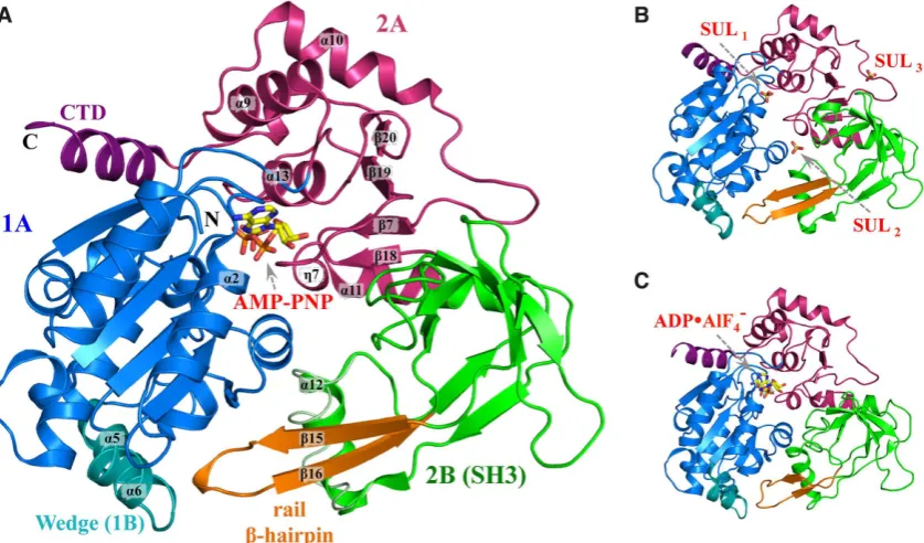
High resolution X-ray structures were determined for the
helicase domain of hPIF1 (Figure 1) corresponding to
an idle cycle of ATP hydrolysis: structures of complexes with the ground state analogue AMP-PNP in two different
crystal forms (1.13 ˚A, 1.43 ˚A; Figure 1A), an apo
struc-ture complexed with three sulphate moieties determined
at 1.44 ˚A resolution (Figure1B) and also a low-resolution
structure of a complex with the transition state analogue
ADP•AlF4−, Figure 1C (see Supplementary Table S1 for
details of the data collection and reinement statistics).
While the hPIF1-ADP•AlF4−structure (Rfactor17.9%;Rfree
= 25.3%) mirrors a previously published low resolution
(3.6 ˚A) crystal structure of the hPIF1 helicase domain
(residues 200–641, pdb code: 5FHH,Rfactor31.3%;Rfree=
35.5%, (26)), it is based on using the high-resolution struc-ture of the hPIF1-AMP-PNP complex during molecular re-placement, resulting in a more accurate model than the
pre-viously available structure (RMSCA=2.0 ˚A; RMSoverall=
2.5 ˚A). Notably, average temperature factors
(Supplemen-tary Table S1) indicate signiicant lexibility of the
hPIF1-ADP•AlF4−structure relative to all others. The structures
we report are of constructs from residues 206–641 (apo hPIF1) or 206–620 (the structures with nucleotide
ana-logues bound). In the 1.44 ˚A apo-hPIF1 crystal structure
residues after 620 are not seen in the electron density sug-gesting they are disordered. Our two constructs (which are henceforth called hPIF1) are active in helicase/DNA bind-ing assays and demonstrate characteristic DNA dependent
ATPaseactivity.
The hPIF1 helicase has the domain architecture charac-teristic of the SF1B RecD2 helicase subgroup (2,43). The overall organization of these domains is similar in all
nu-cleotide bound and free structures determined (Figure1A–
C). hPIF1 has three structural domains, two RecA-like
do-mains 1A (residues 206–381, blue in Figure 1) and 2A
(residues 382–435, red in Figure1) and domain 2B (residues
436–548, green in Figure1) with a SRC Homology 3
(SH3)-like fold, in addition to the small functional domain 1B
within domain 1A (residues 280–300, cyan in Figure1). The
ATP binding site is located between domains 1A and 2A. A
notable feature of domain 2B is a rail-like-hairpin (orange
in Figure1) with its tip proximal to domain 1B. A small
C-terminal domain (CTD, purple in Figure1), protruding
from domain 2A contains an␣-helix which packs against
and is structurally part of domain 1A. In addition, the␣11
helix (residues 429–442) is shared between domains 2A and 2B. The side-chain of the conserved N436, which is in the
middle of the␣11 helix, makes two hydrogen bonds to the
main-chain NH and CO of A551 in domain 2B, keeping the
C-terminus of the␣11-helix relatively ixed (and part of) the
SH3 domain. The domain boundaries and secondary struc-ture names we have used here broadly follow those for the
yeast andBacteroidesPif1 structures (26,27,35). However,
we note that hPIF1 and BsPif1 do not contain the addi-tional 2C domain inserted in the 2B domain of ScPif1 (see Supplementary Figure S1).
The functional domain 1B contains the␣5 and ␣6
he-lices and is stabilized by interactions with the␣-helical Pif1
family signature motif (PFSM, refs.26,27). In ScPif1 there
are also two␣-helices but in BsPif1 there is no
correspond-ing helix at the␣5 position. The 1B ‘wedge’ domain is
be-lieved to play a role in separating the incoming DNA
du-plex. In other SF1B helicases a corresponding -hairpin
serves the same role (43,44). Importantly, the amino acid sequence of the wedge is not conserved. In ScPif1 the se-quence KKVRRSRKHLRR at the C-terminal end of helix
␣5 and the N-terminal end of helix␣6 contains many
pos-itively charged residues. The equivalent sequence in hPIF1 is very different (ALAQ-RPGVRQG), and the sequence is again different in BsPif1 (ENK-FSEYKVEL).
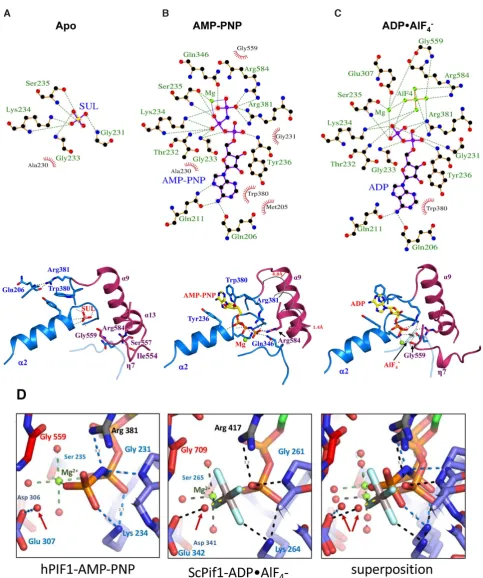
ATP binding and hydrolysis
ATP binding and hydrolysis are accompanied by signii-cant rearrangement in the hydrogen-bonding network, not
only in the ligand binding site (Figure2) but also
through-out the whole protein structure, particularly at interdomain interfaces. Only key interactions will by described further below, although all interactions are detailed using a
LIG-PLOT (45), Figure2A–C.
In the apo-hPIF1 structure the position of the 
-phosphate of ATP is occupied by a sulphate moiety, SUL1
in Figure1. SUL1is stabilized by hydrogen bonds with the
main chain atoms of the loop residues from the Walker A motif (Supplementary Figure S1) and the side chain of the
Walker A K234 residue (Figure2A) at the beginning of the
␣2 helix, situated at the 1A–2A inter-domain contact area.
Tight packing of this area is maintained by interaction of the inter-domain linker R381 (motif IV) with the carboxyl group of Q206 at the N-terminal part of the 1A domain. A second arginine, R584 (motif VI), interacts with S557 (motif V) and the carboxyl group of I554, contributing to the
rigid-ity of the inter-domain 2A–2B contact area (Figure2A).
During ATP binding, represented by the AMP-PNP
structure, R381 recognizes the␥-phosphate of ATP
(Fig-ure 2B). This interaction involves a signiicant movement
of the residue and the 1A–2A inter-domain linker relative to the apo conformation. As such, the aromatic side chain of the adjacent W380 (motif IV) then stacks with the
ade-D
o
w
n
lo
a
d
e
d
fro
m
h
ttp
s:
//a
ca
d
e
mi
c.
o
u
p
.co
m/
n
a
r/
a
d
va
n
ce
-a
rt
icl
e
-a
b
st
ra
ct
/d
o
i/1
0
.1
0
9
3
/n
a
r/
g
kz0
2
8
/5
3
0
4
3
3
1
b
y
U
n
ive
rsi
ty
o
f Y
o
rk
u
se
r
o
n
0
4
F
e
b
ru
a
ry
2
0
1
Figure 1. Structural overview of hPIF1. Cartoon representation of the hPIF1 helicase core domain. (A) The 1.13 ˚A hPIF1-AMP-PNP structure. Domains are coloured as 1A (blue), 2A (red) and 2B (green). Note the C-terminal domain (CTD, purple) and the functional wedge domain (1B, cyan) are structurally
part of domain 1A. The rail-hairpin (orange) is part of domain 2B (a SH3 domain). Secondary structure elements␣2,␣5,␣6,␣9,␣10,␣11,␣12,␣13,7,
15,16,18,19,20,7 are labeled. An alternative conformation of the␣12 helix is shown in tube representation. (B) and (C), the 1.44 ˚A hPIF1-apo
structure with the position of the three bound sulphates (SUL1–3) indicated and the low-resolution hPIF1-ADP•AlF4−structure respectively. The domains
are coloured as in (A).
nine base and together with Y236 forms a pocket for the
base. R584 also shifts in position to interact with the ATP␥
-phosphate, ixing the␣13 helix (domain 2A) relative to the
ATP-binding pocket, while Q346 from motif III coordinates
the ATP␥-phosphate. Importantly, this conformational
re-arrangement of the 1A–2A inter-domain linker (W380 and
R381) during nucleotide binding causes a signiicant∼4 ˚A
shift of the N-terminal end of the␣9 helix, which in turn
induces a structural rearrangement of the 2B domain. Structural rearrangement of the 2B domain (described in detail below) induces changes at the inter-domain 2A–2B
contact area, allowing the segment18-7-19 comprising
helicase motif V (see Supplementary Figure S1), to reach
the ␥-phosphate of ATP, represented in the ADP•AlF4−
structure by the AlF4− moiety (Figure 2C). Accordingly,
the ATP␥-phosphate and Mg2+ion interact with G559 in
motif V. At this point, the␥-phosphate attains its status as
the focal point for the ensuing chemo-mechanical reaction linked to DNA translocation and unwinding, relaying con-formational rearrangements to the whole protein and the 2B domain in particular.
Comparing the 1.13 ˚A hPIF1-AMP-PNP structure
(Fig-ure 2D, left) with the 2.0 ˚A ScPif1 ADP•AlF4− structure
(Figure 2D, centre), suggests that the water molecule
hy-drogen bonded to E307 of the Walker B motif (motif II)
and arrowed in Figure 2D, is likely the ‘attacking water’,
that makes a nucleophilic attack on the␥-phosphate to
ini-tiate ATP hydrolysis in the conventional model for ATP hydrolysis (see for example Supplementary Figure S1A in (46)). In the conventional model, E307 would act as a
base removing a proton from the ‘attacking water’ giving a more associative model for ATP-hydrolysis initiated by
converting the ‘attacking water’ to an ‘attacking OH−ion’.
However, a comparison of our 1.13 ˚A AMP-PNP
struc-ture with our 1.44 ˚A apo structure (Supplementary Figure
S2) shows that the binding of the nucleotide induces
move-ment of the Walker A motif (residues228GSAGTGKS235).
It is also possible that movement of the Walker A motif
could cause the main-chain N–H of G231 (Figure2D, left)
to directly protonate the bridging oxygen of ATP (46).
In-terestingly, the ATP-binding pocket in the 1.13 ˚A human
PIF1-AMP-PNP structure more closely resembles that in
the yeast Pif1-ADP•ALF4−-ssDNA complex and much of
the water structure is conserved (Figure 2D, right), while
there are substantive movements when the human hPIF1 apo and AMP-PNP structures are compared (Supplemen-tary Figure S2A–C). A fully dissociative ‘wellington-boot’ type model (47) for ATP hydrolysis by the Walker A mo-tif is tentatively proposed in Supplementary Figure S2D–F. This proposed mechanism has some similarities to that
pre-viously proposed for an unrelated ATPase(see Figure 5 in
(46)).
Protein conformational changes during ATP binding and hy-drolysis
ATP binding results in a 4 ˚A movement of the N-terminus
of helix ␣9 (helix ␣9 corresponds to the conserved motif
A, Supplementary Figure S1) while the C-terminus remains
relatively ixed to␣13, which is immobilized by interactions
(R584) with the bound nucleotide. The immobilization of
D
o
w
n
lo
a
d
e
d
fro
m
h
ttp
s:
//a
ca
d
e
mi
c.
o
u
p
.co
m/
n
a
r/
a
d
va
n
ce
-a
rt
icl
e
-a
b
st
ra
ct
/d
o
i/1
0
.1
0
9
3
/n
a
r/
g
kz0
2
8
/5
3
0
4
3
3
1
b
y
U
n
ive
rsi
ty
o
f Y
o
rk
u
se
r
o
n
0
4
F
e
b
ru
a
ry
2
0
1
Figure 2. Nucleotide binding and conformational changes in hPIF1. (A) LIGPLOT (45) scheme (above) and structural representation of critical inter-actions (below) at the nucleotide binding site of apo hPIF. The green dashed lines represent hydrogen bonds and the non-ligand residues involved in
hydrophobic contacts are indicated with annotations in red. Note, the sulphate ion is coordinated at the position of the ATP-phosphate. (B) and (C), as
in (A) for the AMP-PNP and ADP•AlF4−bound hPIF1 structures. In (B), centre, the conformation of the␣9 helix in apo hPIF1 is represented by a grey
tube and the relative displacement is indicated. (D) A comparison of the ATP binding site in hPIF1-AMP-PNP (left), ScPif1-ADP•AlF4−(centre) and the
superposition of the two (right). The attacking water is indicated with an arrow in each case.
D
o
w
n
lo
a
d
e
d
fro
m
h
ttp
s:
//a
ca
d
e
mi
c.
o
u
p
.co
m/
n
a
r/
a
d
va
n
ce
-a
rt
icl
e
-a
b
st
ra
ct
/d
o
i/1
0
.1
0
9
3
/n
a
r/
g
kz0
2
8
/5
3
0
4
3
3
1
b
y
U
n
ive
rsi
ty
o
f Y
o
rk
u
se
r
o
n
0
4
F
e
b
ru
a
ry
2
0
1
the ␣9 C-terminus is ensured by H-bonds between con-served residues R395 (motif A) and D343 (motif III). The
corresponding angular displacement is∼18◦ (Figure3A).
The N-terminus of␣9 pushes the␣10 helix aside, stretching
the adjacent segment (residues 412–417, domain 2A) and breaking the hydrogen bond between the side chain of K414 and the main chain carboxyl of G416 (Supplementary Fig-ure S3). Consequently, loop 412–417 becomes more lexible
and allows a distortion of the-sheet20–19–7–18 and
adjustment of its interactions with the hydrophobic core of domain 2A. Comparing the AMP-PNP structure to apo-hPIF1, the observed overall structural changes during ATP
binding are small, corresponding to a∼2.5 ˚A anti-clockwise
shift of the 2A and 2B domains, as depicted in Figure3A.
Notably, despite sequence conservation in the inter-domain area the level of sequence similarity does not allow identi-ication of conserved interactions responsible for the struc-tural rearrangements. As such, it is likely that conforma-tional changes are conditioned by integral characteristics of the structure, such as lexibility and formation of hydropho-bic clusters.
The increased lexibility of the 2A–2B interdomain area,
shaded pink in Figure3B, prepares the active site for
nu-cleotide hydrolysis. In the post hydrolysis state, represented
by the ADP•AlF4− structure, G559 of motif V interacts
with the␥-phosphate of ATP as described above. Changes
in the ATP binding site, focused on interactions with the␥
-phosphate, are relayed to signiicant clockwise movements
at the periphery of the structure, as depicted in Figure3B.
This breaks a set of H-bonds which controls the relative orientations of the 2A and 2B domains, allowing the rota-tion of the 2B domain necessary for formarota-tion of the ss-DNA binding cleft (26,27), including an H-bond between the conserved R515 and the carboxyl group of D418
(Sup-plementary Figure S3). Signiicantly, there is a∼7.1 ˚A
dis-placement of the rail -hairpin (orange in Figure3B)
to-wards the wedge domain. Supplementary Movie S1, based
on the solved apo-, AMP-PNP and ADP•AlF4− bound
hPIF1 structures shows the structural rearrangements dur-ing ATP binddur-ing and hydrolysis.
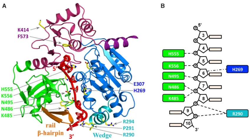
A model of hPIF1 in complex with DNA
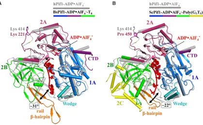
The hPIF1 structures and the yeast and Bacteroides spp.
ssDNA-bound Pif1 structures (26,27,35) are in different conformations, with a large relative movement of domain
2B (Figure4A and B). When the ADP•AlF4−and
ssDNA-bound and DNA-free conformations of BsPif1 structures are compared (26,27), it is seen that the conformational change involves a complicated rearrangement of residues in the 2A–2B inter-domain area, including a movement of
the N-terminus of the ␣11-helix relative to the SH3
do-main. Our attempts to co-crystallize hPIF1 with ssDNA, using accurately determined ratios of protein (see materials and methods, protein expression and puriication) to DNA did not result in diffracting crystals, as has been the case previously (26,27). In the absence of structural data for a hPIF1–ssDNA complex, we constructed a model. An initial
model was made by superposing domains from the 1.13 ˚A
human complex with AMP-PNP onto the equivalent
do-mains in the 2.0 ˚ABacteroidesPif1 complex with ssDNA
and ADP•AlF4−bound (Supplementary Figure S4). A
sec-ond hPIF1HD-ssDNA model (Figure5A) was made when
coordinates for the more closely related ScPif1 (sequence identity 44% for helicase core) in complex with ssDNA and
ADP•AlF4−(35) became available (Supplementary Figure
S4). The two models are essentially the same, and hence-forth referred to as ‘the model’, although there is a small shift in the DNA position and in the position of domains 2B (the SH3 domain). The model also conirms that the over-all coniguration and position of critical conserved amino
acids (Figure5B) in the ssDNA binding channel are likely
to be conserved in Pif1 family helicases.
Mutational analysis of DNA binding
The hPIF1-ssDNA model (Figure5A) along with the
pri-mary sequence alignment of Pif1 proteins (Supplementary Figure S1) identiies speciic residues that may have roles in engaging ssDNA. To test the model, we selected residues
(Figure 5 and Table 1) for functional analysis in
DNA-binding and unwinding assays. Three residues from the ‘sep-aration wedge’ (domain 1B) at the entrance to the ssDNA binding channel and conserved residues therein were mu-tated. As noted above, the structure and sequence of the Pif1 wedge region is variable. In BsPif1, ssDNA has been shown to bend sharply near the wedge domain as it enters the ss-DNA binding channel, while basic residues in the Lys-Arg rich wedge of ScPif1 have been implicated in G4 DNA bind-ing (35). We therefore mutated the two arginine residues in the hPif1 wedge, R290 and R294, as well as P291 separating
helices␣5 and␣6 for testing in functional assays.
We also mutated K485, N486 and N495 of the poorly characterised B (residues 477–486) and C (residues 494– 499) motifs within domain 2B (Supplementary Figure S1) that are characteristic of the SF1B RecD helicases sub-family (2,23). K485, N486 and N495 are in exposed loops where a sulphate ion is located in the apo-hPIF1
struc-ture (SUL2in Figure1B). In bacterial Pif1, the large
move-ments of domain 2B in response to substrate binding posi-tion residues of the B and C motifs in the ssDNA binding channel (26,27), as in our hPIF1-ssDNA model. We there-fore reasoned that a decrease in ssDNA binding and cat-alytic activity of such mutants would be consistent with a similar movement and functional activity of these motifs in hPIF1. Residue K414 was also selected for analysis and we also included the E307Q Walker B motif variant as a
con-trol. K414 is also in the vicinity of a bound sulphate (SUL3
in Figure1B) and is conserved between mammalian and
bacterial Pif1, but it is not in the ssDNA binding channel revealed in Bs and ScPif1 structures (26,27,35). Based on our analysis described above, the alanine substitution would preclude formation of a hydrogen bond and leave the linker region (residues 412–417) more lexible. All variant proteins were expressed and puriied as described for the wild-type hPIF1HD protein construct, hereafter referred to as wild-type hPIF1.
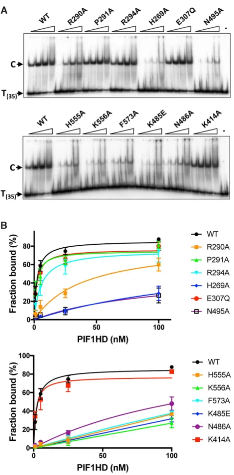
Previously, we have observed that in the absence of nu-cleotide cofactors hPIF1 forms a single discrete ssDNA complex in gel-shift assays (EMSA) with polydeoxynu-cleotide substrates greater than 30 bases (29). Here, ss-DNA binding reactions were assembled at pH 7.2 and 135
D
o
w
n
lo
a
d
e
d
fro
m
h
ttp
s:
//a
ca
d
e
mi
c.
o
u
p
.co
m/
n
a
r/
a
d
va
n
ce
-a
rt
icl
e
-a
b
st
ra
ct
/d
o
i/1
0
.1
0
9
3
/n
a
r/
g
kz0
2
8
/5
3
0
4
3
3
1
b
y
U
n
ive
rsi
ty
o
f Y
o
rk
u
se
r
o
n
0
4
F
e
b
ru
a
ry
2
0
1
Figure 3. Structural rearrangements during ATP binding and hydrolysis. Structures shown in cartoon representation were superposed via the 1A domains.
Ligand positions and secondary structural elements␣9,␣10,␣13 and7 and the-sheet20–19–7–18 are indicated. K414 and G416 are represented
as stick to illustrate their interaction. The direction and magnitude of the positional shifts in the domains, measured at the periphery of the structures, are
indicated by the arrows in domain colours. (A) Changes upon nucleotide triphosphate binding represented by the AMP-PNP ligand. Apo-hPIF1 is shown
in grey and the hPIF1-AMP-PNP structure with domains coloured as in Figure1. The∼18◦angular displacement of␣9, corresponding to conserved
motif A of the RecD helicase subfamily, is indicated. (B) Structural transitions during ATP hydrolysis modelled with the hPIF1-AMP-PNP structure in
grey and the ADP•AlF4−bound structure in domain colours. The 2A–2B interdomain area is depicted by the pink shading.
Figure 4. A comparison of the hPIF1-ADP•AlF4−structure with structures of yeast andBacteroidesPif1 bound to ADP•AlF4−and ssDNA. The
hPIF1-ADP•AlF4−structure is shown in grey and the BsPif1 (A) and ScPif1 (B) nucleotide and ssDNA bound structures with the corresponding domains
coloured (as for hPIF1, Figure1). Structures are orientated to depict the ssDNA binding channel in side view with the ssDNA shown in red. hPIF1 K414
and the corresponding residues in Bs (K221) and ScPif1 (P450) are represented as stick models. The large relative movement and angular displacement of
domain 2B (green) induced by simultaneous binding of ADP•AlF4−and ssDNA is indicated in each case. Note the absence of a helix corresponding to
hPIF1␣5 in the BsPif1 wedge domain and the insertion of the large domain 2C (yellow) in ScPif1.
D
o
w
n
lo
a
d
e
d
fro
m
h
ttp
s:
//a
ca
d
e
mi
c.
o
u
p
.co
m/
n
a
r/
a
d
va
n
ce
-a
rt
icl
e
-a
b
st
ra
ct
/d
o
i/1
0
.1
0
9
3
/n
a
r/
g
kz0
2
8
/5
3
0
4
3
3
1
b
y
U
n
ive
rsi
ty
o
f Y
o
rk
u
se
r
o
n
0
4
F
e
b
ru
a
ry
2
0
1
[image:9.612.92.512.413.672.2]Figure 5. The hPIF1 DNA model with amino acid substitutions mapped. (A) The hPIF1-ssDNA structure showing the modelled bound ssDNA in red. The
residues selected for substitution (Table1) are represented as sticks model and numbered in domain colors. (B) A schematic of the proposed interactions
with ssDNA, based on analysis of the hPIF1-ssDNA model, of the residues selected for substitution. The numbering of the bases is based on those observed
in the ADP•AlF4−and ssDNA bound BsPif1 structure (5FHD).
Table 1. Residues mutated in hPIF1HD for biochemical analysis of puriied proteins. The region or functional site within which the substitutions were made
is indicated with reference to observations made on BsPif1 (26,27). Corresponding residues in ScPif1 and BsPif1, based on primary sequence alignments
(Supplementary Figure S1), are given. Note the lack of sequence conservation in the separation wedge
hPIF1 mutation Domain Motif/functional site BsPif1 residue ScPif1 residue
1 R290A 1B Separation wedge S89 S325
2 P291A 1B Separation wedge E90 R326
3 R294A 1B Separation wedge V93 L329
4 H269A 1A ssDNA binding channel H68 H393
5 E307Q 2B Walker B, ATP hydrolysis E106 E342
6 N495A 2B ssDNA binding, C motif N296 N533
7 H555A 2A ssDNA binding channel H361 H705
8 K556A 2A ssDNA binding channel K362 K706
9 F573A 2A ssDNA binding channel F379 F723
10 K485E 2B B motif V287 K525
11 N486A 2B ssDNA binding, B motif N288 N526
12 K414A 2A SO4binding (apo hPIF1 structure) K221 P450
mM NaCl and binding of all proteins to a radiolabelled
poly T(35) substrate was assayed, Figure 6A. To quantify
the bound fraction, we included all the shifted species dis-tinguished by comparison with the control lane with
sub-strate alone. With the wild-type protein, ∼85% binding
was achieved at the highest protein concentration tested
(100 nM) and an apparent Kd of 2.3 ±0.3 nM was
de-termined from the quantiied data shown in Figure6B
us-ing GraphPad PRISM. In the separation wedge, variants
P291A bound T(35)ssDNA with similar afinity to wild-type
(1.9±0.6 nM), while the binding afinity of R294 was
re-duced∼2–3-fold (apparentKd5.7±1.6). Binding of variant
R290A was reduced ∼10–20-fold (apparentKd 40.4±14
nM), although the failure to reach near saturation binding
precludes the determination of a more accurate apparentKd
value. Variants E307Q (Walker B motif) and K414A also
bound ssDNA at close to wild-type levels (apparentKd
val-ues of 3.6±0.5 and 1.6±0.4 nM respectively). All amino
acid substitutions in the ssDNA binding channel including F573 and those from the B and C motifs (residues K485, N486 and N495A) result in large decreases in ssDNA bind-ing afinity, with variant K556A showbind-ing the greatest
de-fect. As above, insuficient binding extents (Figure6B)
pre-clude the determination of accurate comparative apparent
Kdvalues, but the data indicate at least a 50-fold decrease
in ssDNA binding afinity for all these variants.
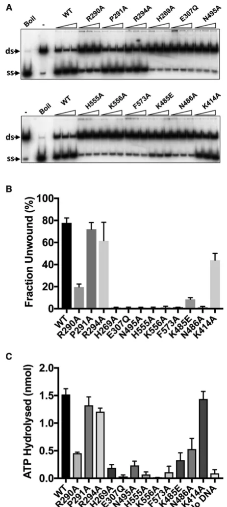
Helicase and ATPaseactivity
Wild-type and variant hPIF1 proteins were tested in an ATP-dependent helicase assays for their ability to displace a 20 base radiolabelled oligonucleotide from a partially single- and double-stranded test substrate. Unwinding re-actions were performed at low salt concentration (20 mM NaCl) as the strand displacement activity of hPIF1 is signif-icantly inhibited at higher salt concentrations and the
pro-D
o
w
n
lo
a
d
e
d
fro
m
h
ttp
s:
//a
ca
d
e
mi
c.
o
u
p
.co
m/
n
a
r/
a
d
va
n
ce
-a
rt
icl
e
-a
b
st
ra
ct
/d
o
i/1
0
.1
0
9
3
/n
a
r/
g
kz0
2
8
/5
3
0
4
3
3
1
b
y
U
n
ive
rsi
ty
o
f Y
o
rk
u
se
r
o
n
0
4
F
e
b
ru
a
ry
2
0
1
[image:10.612.55.572.393.522.2]Figure 6.Binding of hPIF1 and variant proteins to oligo T(35). (A)
Prod-ucts of ssDNA binding reactions (0.4 nM radiolabelled T(35)substrate, 1, 5,
25 and 100 nM hPIF1) were resolved by poly-acrylamide gel electrophore-sis. The primary protein–DNA complex ‘C’ is indicated as is the mobility of free DNA. The inal lane in each panel shows the mobility of free substrate
(no hPIF1 protein). (B) Binding data were quantiied following
phospho-rimaging. Graphs of fraction bound plotted against protein concentration
analysed by nonlinear regression using a single binding site model,n=4
experimental repeats, mean and standard deviation delimited by the error bars shown.
tein inactive in unwinding assays performed at ∼50 mM
NaCl. The inclusion of 0.1% NP40 was also required for optimal activity. Reaction products were separated by poly-acrylamide gel electrophoresis and quantiied by phospho-rimaging. The protein concentration range chosen was at the upper-end of the range where maximum unwinding
is observed for wild-type protein (Figure7A and
Supple-mentary Figure S5) and where further increases in protein concentration result in decreased unwinding eficiency. The quantiied data for the intermediate titration point is shown
in Figure 7B, while Supplementary Figure S5 shows the
analysis for all titration points. The magnitude of the
de-fects observed in the helicase activity (Figure 7A and B)
of hPIF1 variants R290A (25% of wild-type), P291A and R294A (>80% of wild-type) relect the defects observed in ssDNA binding. As expected, variant E307Q (Walker B
motif, Figure2D) was inactive in helicase assays. All amino
acid substitutions in the putative ssDNA binding channel, including K485E, N486A (B motif) and N495 (C motif) re-sulted in complete or near complete abolition of unwinding
activity. Variant K414 retained unwinding activity at∼60%
of the wild-type level.
hPIF1 is a ssDNA dependent ATPase (29). We asked
whether the observed defects in ssDNA binding would also
result in reduced ATPase activity. Like hPIF1 unwinding
activity, ATP hydrolysis is inhibited with increasing salt concentration. In order to measure suitable levels of ATP hydrolysis, it is necessary to perform reactions at higher pro-tein concentration relative to helicase assays (100 nM), with
excess T(35)ssDNA and at a reduced salt concentration (75
mM NaCl) relative to direct ssDNA binding assays
(Fig-ure6). ATP hydrolysis was assayed for wild-type and
vari-ant forms. As expected hPIF1 E307Q, which is directly
in-volved in ATP hydrolysis, had minimal measurable ATPase
activity. Variants P291A and R294 hydrolysed ATP at close to wild type-levels, while the extent of ATP hydrolysis by
R290A was reduced∼3-fold. Therefore, for the separation
wedge region, the defects in ATPaseactivity relect the
fect observed in ssDNA binding and helicase activity de-scribed above. All amino acid substitutions in the ssDNA binding channel resulted in variant hPIF1 forms with signif-icantly impaired ATP hydrolysis. Again, there was a strong
correlation between loss of ATPaseactivity and the
magni-tude of the reduction in apparent ssDNA binding afinity,
where variant K556A showed little or no ATPaseactivity
and N486A retained only∼30% activity compared to
wild-type. Variant K414 had wild-type ATPaseactivity, as was
the ssDNA binding activity observed in the assays described
above (Figure6).
ssDNA strand annealing activity
The hPIF1 helicase core catalyses the annealing of
com-plementary ssDNA strands in the absence of ATP/Mg2+
(29), but the mechanism of ssDNA strand annealing is un-known. We asked if the residues required for ssDNA bind-ing and unwindbind-ing activity are also required for DNA an-nealing. Like the DNA unwinding activity of hPIF1, the DNA strand annealing activity is highly salt sensitive al-though, unlike helicase activity, not completely inhibited at high salt concentrations. Here, for direct comparison with the ssDNA binding assays, we performed DNA strand an-nealing assays at pH 7.2 and 135 mM NaCl and at higher protein concentration relative to helicase assays. To assess the ssDNA strand annealing activity of wild-type and vari-ant hPIF1 proteins we puriied a fork-like duplex substrate
(20 base pairs) with ssDNA tails (5′T55 and 3′ C30) and
D
o
w
n
lo
a
d
e
d
fro
m
h
ttp
s:
//a
ca
d
e
mi
c.
o
u
p
.co
m/
n
a
r/
a
d
va
n
ce
-a
rt
icl
e
-a
b
st
ra
ct
/d
o
i/1
0
.1
0
9
3
/n
a
r/
g
kz0
2
8
/5
3
0
4
3
3
1
b
y
U
n
ive
rsi
ty
o
f Y
o
rk
u
se
r
o
n
0
4
F
e
b
ru
a
ry
2
0
1
Figure 7. Enzymatic activities of hPIF1 and variant protein forms. (A)
Displacement of a32P radiolabelled 20 base ssDNA strand (ss) from a
partially single- and double-stranded test substrate (ds). Reaction prod-ucts (0.1 nM substrate, 3.75, 7.5 and 15 nM hPIF1) were resolved by poly-acrylamide gel electrophoresis for quantiication following phosphorimag-ing. On the left of each panel the mobility of native substrate (ds, reac-tion with no hPIF1 protein) and the labelled ssDNA product generated by
heat denaturation (Boil/(ss) product) are indicated. (B) Strand
displace-ment activity (fraction unwound) for reactions with 7.5 nM hPIF1.n=4
experimental repeats, mean and standard deviation delimited by the error bars are shown. A quantitative analysis of all titration points is shown in
Supplementary Figure S5. (C) DNA dependent ATPasewas determined
at 100 nM hPIF1/200 nM T(30)ssDNA after 10 min incubation (∼3% of
substrate hydrolyse for wild type PIF1).n=3 experimental repeats, mean
and standard deviation delimited by the error bars are shown
both strands end-labelled with 32P. The duplex was heat
denatured prior to addition to the annealing reaction and reaction products were separated by polyacrylamide gel electrophoresis for visualization and quantiication. Wild-type and mutant proteins showed increasing strand anneal-ing activity with increasanneal-ing protein concentration over the
range tested, Figure 8A. Figure 8B shows the quantiied
data for the intermediate protein concentration, while Sup-plementary Figure S6 shows the quantiied data for all titra-tion points. With the exceptitra-tion of mutants N495A (C motif) and F573A all mutants catalysed strand annealing at or sig-niicantly above wild-type activity levels across the range of protein concentrations tested.
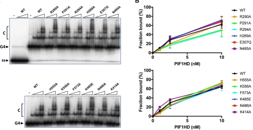
Binding of tetramolecular G4 DNA
A G4 DNA binding activity of hPIF1 resides in helicase core (29) and this activity can be readily visualized by EMSA. However, the structural determinants of G4 DNA binding have not been identiied. Here we asked whether the same residues that impact on ssDNA binding and dsDNA unwinding activity are also necessary for G4 DNA binding. In the absence of nucleotide cofactors hPIF1 demonstrates a substantially higher afinity for a synthetic
tetramolecu-lar G4 DNA substrate with 5′ ssDNA tails compared to
its single-stranded DNA precursor (ref.29and Figure9A).
Here, the reaction conditions used were identical to those
used in ssDNA binding reactions (Figure 6), except that
glycerol was omitted and replaced with 2% w/v PEG 8000, which was necessary to improve resolution of the bound products. The pattern of G4 DNA binding observed for wild-type shows predominantly a single complex at low pro-tein concentrations and then the formation of higher-order species at higher protein concentrations, indicating a bind-ing mode other than a simple bimolecular bindbind-ing reaction, as observed for the ScPif1 protein (48,49). To quantify G4
DNA binding extents (Figure9B), all complexes were
in-cluded in the bound fraction, including the minor fraction of material retained in the wells at the highest protein con-centrations.
Overall, the results of hPIF1 binding to G4 DNA were signiicantly different to the results observed for ssDNA
binding (Figure6). Only small reductions in binding extents
(less than a 2-fold at the lowest protein concentration tested, 1 nM hPIF1) were observed, which reached statistical
sig-niicance (P<0.05) only for the variant H269A, H555A and
K556A (Figure9B). Curiously, variant K414A consistently
displayed an increase in G4 DNA binding activity relative
to wild type at the lower protein concentrations tested (P=
0.01 at 1 nM hPIF1).
DISCUSSION
Our understanding of helicase mechanisms is incomplete since, for any one enzyme, structural information is only available for a limited number of conformational states. Here, we have obtained structures of hPIF1 representing three steps along the chemical reaction coordinate of ATP hydrolysis. Comparing these structures revealed the struc-tural changes associated with an idle cycle of ATP hydroly-sis and suggested how they could be involved in helicase
ac-D
o
w
n
lo
a
d
e
d
fro
m
h
ttp
s:
//a
ca
d
e
mi
c.
o
u
p
.co
m/
n
a
r/
a
d
va
n
ce
-a
rt
icl
e
-a
b
st
ra
ct
/d
o
i/1
0
.1
0
9
3
/n
a
r/
g
kz0
2
8
/5
3
0
4
3
3
1
b
y
U
n
ive
rsi
ty
o
f Y
o
rk
u
se
r
o
n
0
4
F
e
b
ru
a
ry
2
0
1
Figure 8.Strand annealing catalysed by hPIF1 and variant proteins. Reactions (50, 100, 150 nM PIF1) employed two oligonucleotides with 20 bases of complementary DNA (0.1 nM, both strands labelled). The electrophoretic mobility of the partially single- and double-stranded product (native/ds) and the
heat denatured substrate (–, no hPIF1 reaction) are indicate in the lanes to the left of each gel image. Quantiied data (150 nM hPIF1),n=4 experimental
repeats, mean and standard deviation delimited by the error bars, are shown to the right of each panel. A quantitative analysis of all titration points is shown in Supplementary Figure S6.
Figure 9. Binding of hPIF1 and variant proteins to tetramolecular G4 DNA. (A) Products of G4 DNA binding reactions (0.1 nM G4 DNA substrate; 1, 2.5, 10 nM hPIF1) were resolved by polyacrylamide gel electrophoresis and quantiied following phosphorimaging. The irst four lanes of the top panel
demonstrate minimal binding of wild-type hPIF1 to the single-stranded precursor of the G4 substrate (ss). (B) Quantiication of the binding data forn=
3 experimental repeats, mean and standard deviation delimited by the error bars.
tion. First, roles of conserved residues in SF1 motifs III-VI, as well as the Walker A (motif I) and Walker B (motif II), are clearly deined in the chemo-mechanical chain of events. Second, events at the ATP binding site and in particular
the ATP␥-phosphate are relayed to positional and
confor-mational adjustments in deined protein segments. Third, such structural changes result in an increased lexibility of the protein structure that could affect protein–DNA
inter-actions. The increased lexibility of the ADP•AlF4−bound
hPIF1 is clearly indicated in the overall atomic temperature factors of the reined models (Supplementary Table S1).
Although Pif1 proteins can bind ssDNA in the presence and absence of nucleotide cofactors, so far Pif1-ssDNA
structures have only been obtained with ADP•AlF4−
bound. Indeed, the higher afinity of BsPif1 towards ssDNA
observed with the ADP•AlF4−transition-state mimic may
have facilitated determination of the protein-DNA complex
X-ray structures (26,27). ADP•AlF4− also induces an
in-crease in hPIF1 ssDNA binding afinity, while in the
pres-D
o
w
n
lo
a
d
e
d
fro
m
h
ttp
s:
//a
ca
d
e
mi
c.
o
u
p
.co
m/
n
a
r/
a
d
va
n
ce
-a
rt
icl
e
-a
b
st
ra
ct
/d
o
i/1
0
.1
0
9
3
/n
a
r/
g
kz0
2
8
/5
3
0
4
3
3
1
b
y
U
n
ive
rsi
ty
o
f Y
o
rk
u
se
r
o
n
0
4
F
e
b
ru
a
ry
2
0
1
[image:13.612.97.512.349.563.2]ence of AMP-PNP (ground-state mimic) or ADP (prod-uct) ssDNA binding afinities are lower than those observed in the absence of cofactor (Supplementary Figure S7). Ro-tation of domain 2B is required for ssDNA binding and this is permitted due to the increased lexibility and
confor-mational adjustments observed for Pif1-ADP•AlF4−
struc-tures. In hPIF1, breaking the hydrogen bond between R515 and the main chain of D418 is an important component of this induced lexibility, for example. A likely explanation therefore for the increased ssDNA binding afinity observed
with ADP•AlF4−is the facilitation of the movement of the
ssDNA binding residues of the B and C motifs in domain 2B to become an integral part of the ssDNA binding channel.
Importantly, a ∼7.1 ˚A displacement of the rail-hairpin
(orange in Figure3B) towards the wedge domain could also
be utilized to split the DNA duplex. Although not directly visualized here, it is also likely that accompanying structural changes in the ssDNA binding channel modulated ssDNA binding events associated with translocation, as has been
observed in the 3′–5′SF1A helicase PcrA (50). Thus,
struc-tural rearrangements linked to nucleotide hydrolysis may synchronise duplex splitting with directional movement on ssDNA (Supplementary Movie S1).
Our hPIF1 structures allowed the generation and testing of a model for hPIF1 ssDNA binding. Although the model does not allow a precise structural and mechanistic interpre-tation of the data, signiicant conclusions can be made. All substitutions of conserved residues in the putative hPIF1
ssDNA binding channel resulted in a substantial∼50-fold
decrease in ssDNA binding afinity in the absence of nu-cleotide cofactors. With the exception of K485E, which
re-tained∼10% activity, all ssDNA binding channel mutants
were inactive in helicase assays and had decreased
DNA-dependent ATPase activity, mirroring the results of direct
ssDNA binding assays. However, variants K485E, N486A and N495A in the conserved B and C motifs of domain 2B
retained elevated levels of DNA-dependent ATPase
activ-ity compared to others. In the 2B domain of BsPif1, only N288 (hPIF1 N486, B motif) and N296 (hPIF1 N495, C motif) interact with DNA (26,27), forming hydrogen bonds with the phosphate backbone, as do the equivalent residues in ScPif1 (35). The residue corresponding to hPIF1 K485 in BsPif1 is V287 and K525 in ScPif1, neither of which interact with ssDNA in the structures determined (26,27,35). Lysine at this position is largely conserved in mammalian Pif1
pro-teins and RecD fromE. coliandD. radiodurans. Mutation
and deletion of the short linker region between the BsPif1 B and C motifs (27) signiicantly impairs DNA unwinding but
not ssDNA binding and ATPaseactivity. The BsPif1
sub-stitution N296A (C motif) retained∼60% ssDNA binding
activity while for the corresponding substitution in hPIF1
(N495A) binding afinity is reduced∼50 fold. Together, the
mutational data indicate that the mode of ssDNA binding is substantially conserved in Pif1 proteins. However, they also indicate that the residues in the B and C motifs may make a greater contribution to ssDNA binding in the mammalian compared to bacterial Pif1 proteins.
The separation wedge, domain 1B, is at the entrance to the ssDNA binding channel. As noted above, the sequence and structure of the wedge is variable. The apical residues of the helical turns where strand separation is assumed to
occur are87KFSEYK92in BsPif1,289QRPGVR294in hPIF1
and323RRSRKH328in ScPif1. Although several residues of
the BsPif1 segment interact with ssDNA, which in the crys-tal structure (pdb code 5FHD) is observed to bend sharply as it enters the ssDNA binding channel, alanine substitu-tion of the residues results in only modest reducsubstitu-tions in ss-DNA binding and ss-DNA unwinding activity (26). In hPIF1, while variants P291A and R294A have near wild-type ss-DNA binding and unwinding activities, R290A results in
a signiicant decrease in ssDNA binding afinity (∼10–20
fold), unwinding (∼25% of wild-type) and DNA-stimulated
ATP hydrolysis (∼30% wild-type). Our model places R290
but not P291 or R294 in contact with ssDNA. These data suggest that wedge domains of Pif1 proteins may be opti-mised differently, and hence caution should be exercised in extrapolating directly from microbial to human PIF1.
The mechanism of DNA strand annealing is unknown. Unexpectedly, all variant proteins other than F573A and N495A had annealing activities equivalent to or greater than wild-type. N495 (C motif), is on the outer surface of the DNA-free hPIF1 structures, and based on our
ssDNA-ADP•AlF4− models moves∼16 ˚A to become an integral
part of the ssDNA binding channel. F573 is at the exit of
the ssDNA binding channel (Figure5) and binds the
ter-minal 5′ residue of ssDNA in the BsPif1 structure.
With-out nucleotide cofactors high afinity hPIF1-ssDNA
bind-ing is only observed with poly-T oligonucleotides>30 bases
(29). ScPif1 can interact simultaneously with two ssDNA molecules (22), suggesting that at least two low afinity binding sites exist that could allow interactions with long oligonucleotides or facilitate DNA strand annealing. Other direct and indirect observations indicate that SF1, SF2 (51,52) and hexameric helicase (53) engage both the translo-cating (active) and displaced (passive) ssDNA strands along distinct binding paths. Whether residues N495 and F573 have direct roles in ssDNA annealing and why mutations in the active ssDNA binding channel can enhance annealing activity awaits further structural information. In particu-lar, structures of hPIF1 with forked DNA substrates bound should indicate all DNA binding surfaces involved in
an-nealing or unwinding as well as the role of the railhairpin
in duplex splitting, proposed from our structural observa-tions described above.
Although it is unknown how Pif1 proteins bind G4 DNA some parallels could be drawn from the structure of the
SF2 helicase DHX36 (RHAU, G4R1) bound to a 3′
ss-DNA tailed G4 ss-DNA substrate (54), where the G4 ss-DNA
is engaged by an␣-helical motif at the entrance to the
ss-DNA binding channel. Indirect observations indicate that in ScPif1 G4 DNA is clamped at the entrance to the ssDNA binding channel in ‘pliers’ of two sets of positively charged residues (35). However, one arm of the plier is in domain 2C and absent in hPIF1 (and BsPif1), while the second is in the positively charged wedge. Of the three wedge residues im-plicated in ScPif1 G4 DNA binding, only one (R326) is at a similar position in the hPIF1 structure, residue R290. Anal-ysis of hPIF1 G4 DNA binding demonstrated only small defects in G4 DNA binding for mutants in the ssDNA bind-ing channel and R290. As such the data do not identify a G4-speciic DNA binding segment in hPIF1 and indicate
D
o
w
n
lo
a
d
e
d
fro
m
h
ttp
s:
//a
ca
d
e
mi
c.
o
u
p
.co
m/
n
a
r/
a
d
va
n
ce
-a
rt
icl
e
-a
b
st
ra
ct
/d
o
i/1
0
.1
0
9
3
/n
a
r/
g
kz0
2
8
/5
3
0
4
3
3
1
b
y
U
n
ive
rsi
ty
o
f Y
o
rk
u
se
r
o
n
0
4
F
e
b
ru
a
ry
2
0
1