JOURNAL OFVIROLOGY, Feb. 2005, p. 1842–1852 Vol. 79, No. 3 0022-538X/05/$08.00⫹0 doi:10.1128/JVI.79.3.1842–1852.2005
Copyright © 2005, American Society for Microbiology. All Rights Reserved.
Characterization of Snakehead Rhabdovirus Infection in Zebrafish
(
Danio rerio
)†
Peter E. Phelan,
1‡ Meagan E. Pressley,
1‡ P. Eckhard Witten,
2,3Mark T. Mellon,
1Sharon Blake,
1and Carol H. Kim
1*
Department of Biochemistry, Microbiology, and Molecular Biology, University of Maine, Orono, Maine1;
Zoological Institute, University of Hamburg, Hamburg, Germany2; and Department of Biology,
Dalhousie University, Halifax, Nova Scotia, Canada3
Received 2 July 2004/Accepted 7 September 2004
The zebrafish,Danio rerio, has become recognized as a valuable model for the study of development, genetics, and toxicology. Recently, the zebrafish has been recognized as a useful model for infectious disease and immunity. In this study, the pathogenesis and antiviral immune response of zebrafish to experimental snake-head rhabdovirus (SHRV) infection was characterized. Zebrafish 24 h postfertilization to 30 days postfertil-ization were susceptible to infection by immersion in 106
50% tissue culture infective doses (TCID50) of SHRV/
ml, and adult zebrafish were susceptible to infection by intraperitoneal (i.p.) injection of 105
TCID50of SHRV/
ml. Mortalities exceeded 40% in infected fish, and clinical presentation of infection included petechial hemor-rhaging, redness of the abdomen, and erratic swim behavior. Virus reisolation and reverse transcription-PCR analysis of the viral nucleocapsid gene confirmed the presence of SHRV. Histological sections of moribund embryonic and juvenile fish revealed necrosis of the pharyngeal epithelium and liver, in addition to congestion of the swim bladder by cell debris. Histopathology in adult fish injected i.p. was confined to the site of injection. The antiviral response in zebrafish was monitored by quantitative real-time PCR analysis of zebrafish inter-feron (IFN) and Mx expression. IFN and Mx levels were elevated in zebrafish exposed to SHRV, although expression and intensity differed with age and route of infection. This study is the first to examine the patho-genesis of SHRV infection in zebrafish. Furthermore, this study is the first to describe experimental infection of zebrafish embryos with a viral pathogen, which will be important for future experiments involving targeted gene disruption and forward genetic screens.
Recently, zebrafish have been recognized for their value as a model for infectious disease. Genomic comparisons and the discovery of evolutionarily conserved host defense strategies, such as the interferon (IFN)-mediated antiviral response (3, 4) and Toll-like receptors (16, 33), suggest that studies of infec-tious disease in zebrafish will lead to a better understanding of nonspecific immunity and resistance. Few groups have been successful in identifying viral pathogens that can infect and cause disease in zebrafish. LaPatra et al. (27) reported that infectious hematopoietic necrosis virus (IHNV) and infectious pancreatic necrosis virus (IPNV) were able to replicate in adult zebrafish. Despite toxic effects in the hematopoietic tissue of the kidney in zebrafish, no mortalities occurred. Sanders et al. (40) described the susceptibility of adult zebrafish to experi-mental infection with spring viremia of carp virus (SVCV). Zebrafish infected with SVCV exhibited significant mortalities and histopathology, marked by necrosis of the gills, liver, and spleen. In a recent collaboration between our lab and the Leong lab (2), recombinant SHRV mutants were used to dem-onstrate the role of the SHRV nonvirion gene during infection in adult zebrafish; however, detailed pathogenesis and antiviral response were not reported.
Snakehead rhabdovirus (SHRV) was chosen for the present study because the virus affects warm-water fish and the optimal temperature range for SHRV replication is suitable for ze-brafish maintenance. SHRV is an enveloped, nonsegmented, negative-sense RNA virus that belongs to theRhabdoviridae family, genusNovirhabdovirus (20). The virus is chloroform, heat, and acid labile; replicates optimally between 24 and 30°C; and produces the highest titers in snakehead and carp cell lines (21, 21a). First isolated from the tissues of diseased snakehead fish (Ophicephalus striatus) during an epizootic ulcerative syn-drome (EUS) outbreak in Thailand (50), the specific role of SHRV in EUS had not been determined (21). Experimental infection with EUS-associated rhabdoviruses was successful in snakehead fry and juveniles (12, 29), although the characteris-tic ulcerative disease was not observed. In the present study, we examine the ability of SHRV to cause disease and to stimulate an inflammatory immune response in embryonic and adult zebrafish.
When a viral pathogen is able to penetrate the external bar-riers of an organism and cause infection, initially a nonspecific inflammatory response is triggered. This immediate and vigor-ous response is mediated in part by antiviral response proteins IFN and Mx. IFN induces an antiviral state in which host cells block mRNA transcription and translation in order to prevent viral replication in infected cells (22). Genes for IFN have been identified in several species of mammals, birds, and fish, in-cluding zebrafish, channel catfish, carp, and Atlantic salmon (3, 19, 31, 39, 42, 44, 47, 55). IFN production also induces the expression of antiviral proteins such as Mx, double-stranded * Corresponding author. Mailing address: Department of
Biochem-istry, Microbiology, and Molecular Biology, 5735 Hitchner Hall, Uni-versity of Maine, Orono, ME 04469. Phone: (207) 581-2803. Fax: (207) 581-2801. E-mail: carolkim@maine.edu.
† Maine Agricultural and Forest Experiment Station publication 2651.
‡ P.E.P. and M.E.P. contributed equally to this study.
1842
on November 8, 2019 by guest
http://jvi.asm.org/
RNA-dependent protein kinase, and 2⬘-5⬘oligoadenylate syn-thetase (22). Mx is a member of the GTPase family and was originally recognized for its antiviral activity against ortho-myxoviruses (15). Antiviral activity of Mx has also been re-ported after infection of rhabdoviruses (32, 45), bunyaviruses (13), togaviruses (26), and paramyxoviruses (41). Mx genes have been identified in mammals, birds, and a variety of fish, including zebrafish (4, 17, 18, 28, 46, 49).
We report here the successful infection of zebrafish embryos and adults with SHRV, resulting in the induction of disease-related immune responses and pathology. Experimental infec-tion of both embryonic and adult zebrafish with SHRV pro-duced infection kinetics and histopathology that are indicative of acute infection. Analyses of IFN and Mx mRNA expression profiles revealed evidence of upregulation of the antiviral re-sponse in zebrafish embryos and adults as a result of SHRV infection. The results presented here provide the first evalua-tion of pathogenesis and antiviral response in zebrafish after experimental SHRV infection. We also describe here the first method for infecting zebrafish embryos with a viral pathogen, which will be important for forward genetic screens and tar-geted gene disruption experiments.
MATERIALS AND METHODS
Cells and virus.SHRV was propagated in Epithelioma papulosum cyprini
(EPC) cells (11) or zebrafish embryo fibroblast (ZF4) cells (8) at 28°C. EPC cells were grown in minimal essential medium (MEM) supplemented with 10% heat-inactivated fetal bovine serum (Gibco, Carlsbad, Calif.). ZF4 cells were grown in Dulbecco modified eagle medium/F12 supplemented with 5% fetal bovine serum (Gibco). Propagation of virus was achieved by infecting 75%-80% confluent cultures at a multiplicity of infection (MOI) of 0.001. Cultures with 80 to 90% CPE were harvested and centrifuged 2000⫻gfor 10 min at 4°C, and the supernatants were stored at⫺80°C. Virus titers were quantified by determining 50% tissue culture infective doses (TCID50)/ml. EPC cells were used to determine
virus titers for virus stocks propagated in EPC cells, and ZF4 cells were used to determine virus titers for virus stocks propagated in ZF4 cells.
Zebrafish care and maintenance.AB inbred strain of zebrafish were reared in recirculating systems from Aquatic Habitats (Apopka, Fla.). The water temper-ature was maintained at 28°C with a flow rate of 150 liters/min. All adult fish (⬎3 months old) used in infection experiments were transferred to an isolated flowthrough system, a modified version of the recirculating system, and accli-mated for several days before infection. Effluent from the flowthrough system was treated with chlorine. Zebrafish embryos were maintained at 28°C in egg water (60g of Instant Ocean sea salts/ml). Zebrafish were handled according to Institutional Animal Care and Use Committee guidelines.
Exposure by immersion.At 24 h postfertilization (hpf), 3 days postfertilization (dpf), 7 dpf, and 30 dpf zebrafish embryos were exposed to 106
TCID50of SHRV/
ml by static immersion for 5 h. At 24 hpf embryos were manually dechorionated prior to infection. Control fish were exposed to phosphate-buffered saline (PBS). Exposures were carried out in triplicate with 20 fish per group per dose. Parallel groups for histology and reisolation sampling were exposed similarly. Zebrafish at 24 hpf, 3 dpf, 7 dpf, and 30 dpf were exposed to 106TCID
50of SHRV/ml in
total volumes of 2, 3, 3, and 100 ml, respectively. Adult fish ranging from 300 to 800 mg in weight (3 to 4 cm in length) were exposed to 103, 104, 105, or 106
TCID50of SHRV/ml. After 5 h, each group of adult fish was moved to a 2.75-liter
tank in the flowthrough system and maintained for 20 days. Mortalities were tallied daily. Fish from parallel groups were routinely sampled for reisolation of virus and histology.
Exposure by injection.Adult fish were anesthetized in 160g of tricaine/ml and injected intraperitoneally (i.p.) with 105
TCID50of SHRV/ml. Control groups
were similarly anesthetized and injected with PBS. Exposures were conducted in triplicate with 20 fish per group per dose. Parallel groups for histology and reisola-tion sampling were infected accordingly. Mortalities were tallied daily. The fish from the parallel groups were routinely sampled for reisolation of pathogen and histology. Virus confirmation.Randomly selected adult zebrafish, ranging in weight from 300 to 800 mg, from the parallel sampling groups infected with SHRV, were euthanized in 4 mg of tricaine/ml, immediately frozen in liquid nitrogen, and placed in a plastic stomacher bag for storage at⫺80°C. Moribund and dead adult fish were collected in a similar fashion. Adult samples were processed by addition of 1:10 (wt/vol) serum-free MEM supplemented with 50 U penicillin, 0.05 mg of streptomycin, and 0.01 mg of neomycin and then homogenized. Infected embryos and juvenile fish from the parallel 24-hpf, 3-dpf, 7-dpf, and 30-dpf sampling groups were euthanized and diluted 1:100 (wt/vol) in serum-free MEM. Embryo and juvenile samples were homogenized in 1.5-ml microcentrifuge tubes; super-natants were collected, filtered, and stored at 4°C. Filtered samples were diluted 10-fold and added to cells in culture. Infected cells were monitored for 1 week, and TCID50/milliliter concentrations were calculated.
Histology.Adult zebrafish sampled for histology were euthanized in 4 mg of tricaine/ml and placed in 10 ml of buffered 10% formalin. Fish samples were embedded in paraffin and sections were stained with hematoxylin-eosin. Ze-brafish embryos sampled for histology were euthanized in 4 mg of tricaine/ml and placed in 0.5 ml of TEM fixative (0.2 M sodium cacodylate, 25% glutaraldehyde, 15% paraformaldehyde, 0.5% calcium chloride). Embryonic samples were post-fixed with osmium tetroxide and embedded in Epon medium. Semithin sections were stained with toluidine blue.
Primer design and nucleotide sequences.Primers were designed from their respective gene sequences by using PrimerQuest (Integrated DNA Technolo-gies). Primer sequences and their expected fragment sizes for quantitative real-time PCR are listed in Table 1. Primers for the PCR analysis of the SHRV nucleocapsid gene were synthesized from the SHRV N-gene sequence (accession no. AF147498). The sense primer, 5⬘ -ATTTATCCGCTGGAGAGGGATTGG-3⬘, and the antisense primer, 5⬘-GTTGAGCCCATAGGCCTTGAAGTA-3⬘, di-rect the amplification of 829-nucleotide portion of the SHRV nucleocapsid gene. Cycling parameters were 94°C for 30s, 55°C for 30s, and 72°C for 30s for a total of 35 cycles. The 829-nucleotide amplicon was subcloned into a pGEM-T Easy vector (Promega, Madison, Wis.) and submitted for sequencing to the University of Maine Sequencing Facility with an ABI 373 DNA sequencer (Applied Bio-systems, Foster City, Calif.).
RNA extraction and cDNA synthesis.Zebrafish embryos and larvae were sampled at 6, 12, 24, 48, 72, and 96 h postinfection (hpi) for total RNA extrac-tion. All sample fish were collected and placed in 200l of RNAlater (Ambion, Austin, Tex.) for storage. Exposures were conducted in triplicate with 10 fish per sample time point. Livers from adult fish injected i.p. with virus were removed from randomly selected fish and stored in 200l of RNAlater. Exposures were conducted in triplicate with two adult livers per sample time point. Total RNA was extracted from RNAlater preserved samples by using the MasterPure RNA Purification kit, according to the manufacturer’s instructions (Epicentre, Madi-son, Wis.). Total RNA concentrations were determined by UV spectrophotom-etry. Reverse transcription (RT) reactions were performed to convert total RNA into cDNA. Briefly, ca. 1.0g of total RNA and 0.5g of random hexamers were combined and incubated at 70°C for 5 min, followed by a quick chill at 4°C for 5 min. To this mixture, Improm II 5⫻reaction buffer, 25 mM MgCl2, 10 mM
[image:2.585.41.544.80.173.2]deoxynucleoside triphosphate, 0.5 U of RNase inhibitor, and 1l of Improm II
TABLE 1. Quantitative real-time primer sequences and amplicon size
Gene Accession no. Primer sequences Product size (bp)
Zebrafish-actin AF025305 5⬘-ATGGATGAGGAAATCGCTG-3⬘ 130
5⬘-ATGCCAACCATCACTCCCTG-3⬘
Zebrafish Mx AF533769 5⬘-ATAGGAGACCAAAGCTCGGGAAAG-3⬘ 145
5⬘-ATTCTCCCATGCCACCTATCTTGG-3⬘
Zebrafish IFN AY135716 5⬘-GAATGGCTTGGCCGATACAGGATA-3⬘ 136
5⬘-TCCTCCACCTTTGACTTGTCCATC-3⬘
on November 8, 2019 by guest
http://jvi.asm.org/
reverse transcriptase was added (Promega). The reaction mixture was incubated at 25°C for 5 min, 37°C for 1 h, and 72°C for 15 min. Reactions were brought up to 40l with RNase-free water.
Quantitative real-time PCR.Quantitative real-time PCR was performed by using the iCycler iQ Detection System (Bio-Rad Laboratories, Hercules, Calif.). Gene-specific primers for quantitative real-time PCR were designed to generate single gene-specific amplicons of 125 to 200 nucleotides. The 96-well real-time PCR format included duplicate 10-fold dilutions of the linearized plasmid DNA standard ranging from 109to 102copies. Zebrafish-actin primers were used to
normalize the starting quantity of RNA. Zebrafish IFN and Mx were assayed in triplicate for each sample time point with appropriate standards. Reactions were performed in an iCycler iQ real-time PCR detection system (Bio-Rad) according to the manufacturer’s instructions. Reactions were performed in a 20-l volume comprised of 1l of cDNA reaction, 10l of 2x IQ SYBR Green Supermix (Bio-Rad), and 250 nM concentrations of each primer. The cycling parameters were 94°C for 15 min to activate the polymerase, followed by 40 cycles of 94°C for 30 s, 55°C for 30 s, and 72°C for 30 s. Fluorescence measurements were taken at each cycle during the 55°C annealing step. The copy number for each reaction was calculated by the iCycler software. Values were normalized to the
corre-sponding-actin values to determine the relative copy number. The relative copy number was then used to calculate the fold induction values of virus-induced samples over the control samples.
RESULTS
SHRV infection kinetics and gross pathology.The infectivity of SHRV grown on ZF4 cells and EPC cells was determined for 24-hpf fish exposed to 106TCID
50of SHRV/ml by
immer-sion challenge (Fig. 1A) and for adult fish injected i.p. with 105
TCID50 of SHRV/ml (Fig. 1B). The 24-hpf fish exposed to
SHRV showed average cumulative mortalities of 55% with virus isolated from EPC cells compared to mortalities of 40% with virus produced in ZF4 cells (Fig. 1A). Adult fish infected with SHRV showed average cumulative mortalities of 70% with SHRV grown on EPC cells and average cumulative mor-FIG. 1. Comparison of cumulative percent mortalities in zebrafish exposed to SHRV grown on EPC (SHRV-EPC) or ZF-4 cells (SHRV-ZF4). (A) Zebrafish embryos at 24 hpf were exposed to 106TCID
50of SHRV/ml by immersion and then monitored for 14 days. (B) Adult zebrafish were
injected i.p. with 105TCID
50of SHRV/ml and monitored for 21 days. The data are representative of multiple independent challenges run in triplicate.
1844 PHELAN ET AL. J. VIROL.
on November 8, 2019 by guest
http://jvi.asm.org/
talities of 40% with SHRV grown on ZF4 cells (Fig. 1B). Mortalities due to SHRV infection began 1 day postinfection (dpi) and continued through 10 dpi, although the majority of mortalities occurred between 2 and 8 dpi. Based upon these results, SHRV grown on EPC cells (SHRV-EPC) was more virulent and was used for all subsequent challenges.
In order to determine the effect on overall fish survival, embryonic and juvenile fish of ages 24 hpf and 3, 7, and 30 dpf were exposed by immersion to 106TCID
50of SHRV-EPC/ml
and monitored daily for 14 days (Fig. 2). The 24-hpf fish ex-posed to virus showed average cumulative mortalities of 55% during the 14-day challenge period compared to 10% mortality in control fish. The 3-dpf fish showed average cumulative mor-talities of 47% by 14 dpi after exposure to SHRV compared to 11% mortality in control fish. The 7-dpf fish exposed to virus showed average cumulative mortalities of 51% at 14 dpi com-pared to 11% mortality in control fish. The 30-dpf fish exposed to virus showed mortalities of 57% during the challenge period compared to 5% mortality in control fish. Mortalities in control fish were comparable to the proportion of mortalities in typical healthy clutches of eggs from routine breeding. Mortalities associated with immersion infection in the embryonic and ju-venile fish began as early as 1 dpi and continued through 12 dpi. Infected fish appeared lethargic prior to death, with
sub-epidermal petechial hemorrhaging observed in the fish in-fected at 30 dpf. Adult fish exposed to virus by immersion appeared to be refractory to infection and showed inconsistent mortalities of⬍20% over a period of 21 days (data not shown). In a separate experiment, adult zebrafish were i.p. injected with 105 TCID
50of SHRV/ml and monitored for 21 days to
determine whether they were susceptible to infection by this route. Injection of 105TCID
50of SHRV/ml produced average
cumulative mortalities of 69% (Fig. 1B). Injection of PBS caused no mortalities. Mortalities in adult fish after exposure to SHRV began at 1 dpi and continued through 13 dpi, after which there were no further deaths. Adult zebrafish injected with SHRV exhibited severe petechial hemorrhages on the abdomen as soon as 2 dpi (Fig. 3). Moribund fish exhibited erratic swimming patterns and lingered near the surface of the water. Control fish exhibited no abnormal behaviors or lesions.
SHRV reisolation and confirmation.Juvenile and adult fish exposed to virus by immersion or i.p. injection were randomly sampled for virus titers at intervals during infection. The 24 hpf fish yielded a virus titer that increased from 3.2⫻107TCID
50/
ml at 1 dpi to a maximum of 4.7⫻ 109 TCID
50/ml at 3 dpi
(Table 2). Virus titers in the 24-hpf fish decreased to 3.2⫻108
TCID50/ml by 6 dpi and continued to decline to 1.0 ⫻ 10 6
[image:4.585.125.459.71.264.2]TCID50/ml by 10 dpi. The 3-dpf fish showed a virus titer of
FIG. 2. Cumulative percent mortalities in zebrafish infected at 24 hpf and at 3, 7, and 30 dpf with SHRV. Zebrafish were exposed to 106TCID 50
of SHRV/ml by immersion and then monitored for 14 days. The final percent mortalities were calculated and recorded for each age group. The data are representative of multiple challenges run in triplicate.
FIG. 3. Gross pathology of adult zebrafish injected i.p. with SHRV. Fish were infected with SHRV and examined at 2 dpi for signs of viral infection and clinical disease. (A) Control fish were injected with 10l of PBS. (B) Infected fish were injected i.p. with 105TCID
50of SHRV/ml.
on November 8, 2019 by guest
http://jvi.asm.org/
FIG. 4. Histopathology of zebrafish embryos infected with SHRV by immersion. (A) Perianal region of control fish. White arrowheads indicate normal blood vessels. The black arrowhead indicates a normal pigment cell. Magnification,⫻400. (B) Perianal region of infected fish. The white arrowheads indicate a blood vessel filled with monocytes. The black arrowhead indicates an irregularly shaped pigment cell. Magnification,⫻400. (C) Branchial region of control fish, with normal mucus cells of the branchial epithelia (white arrowhead). Magnification,⫻400. The black
1846 PHELAN ET AL. J. VIROL.
on November 8, 2019 by guest
http://jvi.asm.org/
3.2 ⫻ 107 TCID
50/ml at 1 dpi that increased to 4.7 ⫻ 108
TCID50/ml by 3 dpi. The 3-dpf fish virus titer declined to 3.2⫻
107TCID
50/ml by 6 dpi and reached 3.2⫻106TCID50/ml by
10 dpi. The 7-dpf fish showed a virus titer of 3.2⫻107TCID
50/
ml at 1 dpi that increased to 4.7⫻107TCID
50/ml by 3 dpi. At
6 dpi, the virus titer had dropped to 3.2⫻107TCID
50/ml, and
by 10 dpi the virus titer had decreased to 3.2⫻106TCID
50/ml.
SHRV immersion challenges with 30-dpf fish showed a slightly different pattern in virus titer with 3.2⫻ 106 TCID
50/ml at
1 dpi and a maximum of 1.0 ⫻ 109 TCID
50/ml by 2 dpi.
However, virus titers in the 30-dpf fish decreased to 1.0⫻108
TCID50/ml by 6 dpi and to 4.2⫻10
6TCID
50/ml by 10 dpi.
Virus titers were determined for i.p.-injected adult fish and showed a virus titer of 4.7⫻ 103TCID
50/ml at 1 dpi and a
maximum virus titer at 3 dpi of 4.7⫻107TCID
50/ml (Table 2).
The levels of SHRV remained elevated at 3.2⫻107TCID
50/ml
at 6 and 8 dpi. By 10 dpi, the virus titer in the sampled fish injected i.p. had fallen to 3.2⫻106TCID
50/ml. As seen in the
immersion challenges with the young fish, the decrease in virus titers at 10 dpi corresponded to a decrease in overall cumula-tive fish mortalities.
To better understand the inconsistent mortality data ob-served when adult fish were infected with SHRV by immersion, adult fish were sampled at intervals for reisolation of virus. Adult fish immersed in SHRV showed a maximum virus titer of 1.0⫻104TCID
50/ml by 1 dpi (Table 2). The virus titers in
the adults immersed in SHRV were 3.2⫻103TCID 50/ml by
2 dpi and decreased to 1.0⫻102TCID
50/ml by 3 dpi, where it
remained through 8 dpi. By 10 dpi, there was no detectable virus titer in sampled adult fish. Due to the refractory nature of adult infection by immersion, very few adult fish died; however, fish that died were sampled and had virus titers of 3.2⫻107
TCID50/ml (data not shown). Uninfected control fish produced
no virus titers in cell culture in any of the infected age groups (Table 2).
To further confirm infection by SHRV, primers for PCR analysis were synthesized from the sequence encoding the nu-cleocapsid (N-gene) of SHRV (accession no. AF147498). An amplicon of 829 nucleotides spanning nucleotides 47 to 876 of
the SHRV N-gene was observed in all virus-infected samples by 1 dpi, confirming the cell culture data. The amplicon con-tinued to be present through 10 dpi in the juvenile fish im-mersed in SHRV and in livers of the adult fish injected i.p. In the adult fish infected by immersion in SHRV, the amplicon was undetectable in isolated fish livers at 3 dpi. The corre-sponding amplicon for the SHRV N-gene was absent in all uninfected control samples (data not shown).
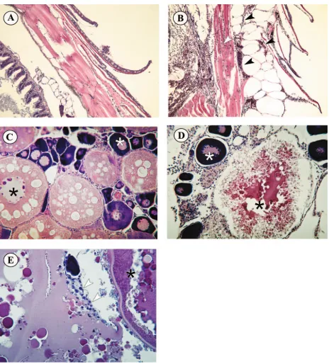
[image:6.585.45.541.81.180.2]Histopathology of SHRV infection.For each of the develop-mental stages, fish from parallel infected and control groups were sampled daily for histological examination. Similar histo-pathology was observed in all developmental stages of fish exposed to SHRV by immersion. Figure 4 shows representa-tive histopathology, comparing the blood vessels (Fig. 4A and B), branchial regions (Fig. 4C and D), and livers (Fig. 4E to 4H) of control and infected embryos and juveniles. The histo-pathology observed in zebrafish infected with SHRV included high numbers of monocytes in the blood vessels of the perianal region and a marked absence of erythrocytes (Fig. 4B, white arrows). Compared to the control (Fig. 4C), virus-infected fish displayed a higher number of mucus cells in the buccopharyn-geal epithelium (Fig. 4D, white arrow). The pharynbuccopharyn-geal epi-thelium itself became necrotic, displaying a rough structure and epithelial cells sloughing into the lumen (Fig. 4D, black arrow). The pigment cells of the infected fish also appeared to be irregular in shape (Fig. 4B, black arrow, and Fig. 4F, white arrow) compared to the pigment cells in the control fish (Fig. 4A, black arrow, and Fig. 4E, white arrow). The black asterisk in Fig. 4E indicates the lumen of the swim bladder of the control fish. In the infected fish the lumen of the swim bladder was congested with cell debris (Fig. 4F, black asterisk). The white asterisk in Fig. 4E and F indicates the liver tissue. The dark staining liver of the infected fish contained necrotic cells and fat droplets not present in control fish. Upon closer ex-amination, the liver tissue displayed cytoplasmic vacuolization and pyknosis of hepatocytic nuclei, indicating toxic conditions (Fig. 4H, black arrows). Moreover, hepatocytes were filled with fat droplets that, after the cells disintegrated, appeared in the extracellular space (Fig. 4H, white arrows). In addition, several TABLE 2. Virus titers from fish exposed to SHRV via immersion or i.p. injection
Sample
Virus titer at:
1 dpi 2 dpi 3 dpi 6 dpi 8 dpi 10 dpi
24 hpf 3.2⫻107 1.0⫻109 4.7⫻109 3.2⫻108 1.0⫻107 1.0⫻106
3 dpf 3.2⫻107 3.2⫻108 4.7⫻108 3.2⫻107 1.0⫻107 3.2⫻106
7 dpf 3.2⫻107 1.0⫻107 4.7⫻107 3.2⫻107 3.2⫻107 3.2⫻106
30 dpf 3.2⫻106 1.0⫻109 1.0⫻109 1.0⫻108 3.2⫻108 4.2⫻106
Adult (i.p. injected) 4.7⫻103 3.2⫻107 4.7⫻107 3.2⫻107 3.2⫻107 3.2⫻106
Adult (immersion) 1.0⫻104 3.2⫻103 1.0⫻102 1.0⫻102 1.0⫻102 0
Controls 0 0 0 0 0 0
arrowhead points to the upper pharyngeal epithelium. (D) Branchial region of infected fish. The upper pharyngeal epithelium has a rough structure and contains many proliferating cells (black arrowhead). The white arrowhead indicates numerous pink mucus cells. Magnification,⫻400. (E) Liver (white asterisk) and swim bladder (black asterisk) of control fish. The white arrowhead indicates normal pigment cells. Magnification, ⫻250. (F) Dark staining liver (white asterisk) and congested swim bladder (black asterisk) of infected fish. The white arrowhead indicates irregularly shaped pigment cells of infected fish. magnification,⫻250. (G) Higher magnification of liver tissue (white asterisk) from control fish. Magnification,⫻400. (H) Higher magnification of liver tissue (white asterisk) from infected fish. Black arrowheads indicate intracellular vacuoles. White arrowheads indicate glycogen vesicles in the extracellular space. Magnification,⫻1000.
on November 8, 2019 by guest
http://jvi.asm.org/
obstructed hepatic ducts were visible in the infected fish (Fig. 4H) in comparison with the liver of the control fish (Fig. 4G). Figure 5 presents comparisons of the scales and epidermis (Fig. 5A and B) and ovaries (Fig. 5C to E) of control and SHRV i.p.-injected adult zebrafish. Histopathology of SHRV-infected adult fish included subepidermal petechial
[image:7.585.64.528.63.577.2]hemor-rhages and edema near the site of injection (Fig. 5B), whereas PBS-injected control fish showed no signs of infection or in-flammation. These findings agree with the gross pathology ob-served in SHRV-infected adult fish, as seen in Fig. 3. Infected females also exhibited degeneration of secondary oocytes (Fig. 5D and E). Disruption of the yolks of secondary oocytes and FIG. 5. Histopathology of adult zebra fish infected with SHRV by i.p. injection. (A) Normal scales and epidermis of control fish. Magnification, ⫻100. (B) Scales and epidermis of infected fish. Black arrowheads indicate subdermal edema and hemorrhaging. The white arrowhead indicates hemorrhaging in the underlying muscle tissue. Magnification,⫻100. (C) Ovaries of control fish showing normal egg development, with generations of ova in different developmental stages. The white asterisk indicates a primary oocyte, the black asterisk indicates a secondary oocyte, and the black arrowhead indicates the epithelial granulosa (nursing) cells. Magnification,⫻200. (D) Degenerating secondary oocyte of SHRV-infected fish (black asterisk). Primary oocytes seem to be unaffected (white asterisk). Magnification,⫻200. (E) Epithelial granulosa cells (white arrows) reabsorbing remaining yolk from secondary oocyte.
1848 PHELAN ET AL. J. VIROL.
on November 8, 2019 by guest
http://jvi.asm.org/
subsequent reabsorption of the yolk by neighboring epithelial granulosa cells were most notable. Some of the infected fish displayed fluid and inflammatory cell accumulation in the ab-dominal cavity. All other major organs appeared to be unaf-fected by infection with SHRV.
IFN and Mx expression. The mRNA expression levels of two immune effector molecules, IFN and Mx, were examined in control and SHRV-infected zebrafish samples by using quantitative real-time PCR analysis. In order to understand the effects of SHRV infection throughout the development of the zebrafish immune system, fish were infected by immersion at 24 hpf and at 3, 7, and 30 dpf. Figure 6A compares IFN mRNA induction levels in fish at 24 hpf and at 3, 7, and 30 dpf at selected times after infection with SHRV. Expression of IFN transcripts in the 24-hpf fish remained at basal levels through 12 hpi and increased 2.5-fold over controls at 24 hpi and 2.4-fold over controls at 72 hpi (Fig. 6A). IFN induction by SHRV in the 3-dpf fish also remained at basal levels of expression through 12 hpi but increased 2.5-fold over controls at 24 hpi
and 5.0-fold over controls at 72 dpi. In contrast to the 24-hpf and 3-dpf fish, the 7- and 30-dpf fish displayed a different pattern of IFN mRNA expression. The 7-dpf fish exposed to SHRV showed basal levels of IFN mRNA expression through 24 hpi and an increase of 7.7-fold over controls at 48 hpi. IFN mRNA in the 7-dpf fish reached a maximum expression of 18.0-fold over controls at 72 hpi and decreased to 14.8-fold by 96 hpi. Similar to the 7-dpf fish, the 30-dpf fish remained at basal levels of IFN mRNA expression through 24 hpi and showed an increase of 4.0-fold over controls by 48 hpi. IFN induction increased to a maximum of 11.6-fold in the 30-dpf fish at 72 hpi and decreased to 3.8-fold induction over controls by 96 hpi. These data indicate that SHRV can induce IFN in zebrafish and demonstrates a difference in the antiviral re-sponse during zebrafish development.
SHRV-induced Mx mRNA expression closely paralleled the expression of IFN during infection and development (Fig. 6B). The 24-hpf fish infected with SHRV showed basal levels of Mx mRNA expression through 12 hpi, with increases in Mx ex-pression of 6.7-fold at 24 hpi and 5.2-fold at 72 hpi. By 96 hpi, Mx mRNA levels in the 24-hpf fish began to decrease but remained 4.1-fold greater than the control levels. In 3-dpf fish, Mx remained at basal expression through 6 hpi and increased only 2.0-fold at 12 hpi. At 24 and 72 hpi, the Mx levels in the 3-dpf fish increased by 5.2- and 3.5-fold, respectively. Similar to the 24-hpf fish, Mx levels the 3-dpf fish remained elevated at 3.3-fold through 96 hpi. As was observed in the IFN expression patterns, the Mx expression patterns for the 24-hpf and 3-dpf fish differed from the pattern observed in the 7- and 30-dpf fish. Mx expression in the 7-dpf fish remained at basal levels through 12 hpi and increased only slightly by 1.6-fold over controls at 24 hpi. By 48 hpi, Mx mRNA in the 7-dpf fish had increased by 8.8-fold and continued to increase to a maximum at 72 hpi of 48.0-fold over controls. Expression remained ele-vated in the 7-dpf fish through 96 hpi, with a 22.7-fold increase in Mx transcripts. The fish samples infected at 30 dpf also remained at basal levels of Mx expression through 12 hpi and showed only a minor increase at 24 hpi of 1.7-fold. Again, Mx expression increased 16.5-fold by 48 hpi in the 30-dpf fish and reached a maximum of 51.0-fold at 72 hpi. Levels decreased in the 30-dpf fish by 96 hpi, but remain elevated at 25.2-fold above controls. As observed with the expression of IFN, the Mx expression pattern and intensity varied with the develop-mental age of the fish. Mx transcripts increased to higher levels in fish older than 7 dpf, suggesting a more robust antiviral response.
Adult fish exposed to SHRV by i.p. injection were used to examine the antiviral response in the mature zebrafish. Injec-tion of adult fish i.p. with 105TCID
50of SHRV/ml showed a
more rapid antiviral response compared to the 24 hpf and 3-, 7-, and 30-dpf fish exposed by immersion to SHRV. IFN mRNA levels in the adult fish injected i.p. reached a maximum of 10.9-fold by 6 hpi, followed by a rapid return to basal levels of expression by 24 hpi (Fig. 7). Mx mRNA transcripts were elevated 15.4-fold by 6 hpi, and a maximum of 40.0-fold over PBS injected controls was achieved by 24 hpi. Mx expression decreased to 20.5-fold induction by 48 hpi but remained ele-vated at 16.0-fold through 72 hpi. Transcripts of Mx returned to basal levels of expression by 96 hpi.
FIG. 6. Quantitative real-time PCR analysis of zebrafish IFN (A) and Mx (B) mRNA expression after immersion for 5 h in 106TCID
50
of SHRV/ml. Zebrafish were exposed at 24 hpf and at 3, 7, and 30 dpf. Total RNA was extracted from selected samples through 96 hpi. The data are representative of three independent exposures. Each bar represents the mean fold induction of SHRV-infected samples over corresponding controls. Expression values were normalized to ze-brafish-actin.
on November 8, 2019 by guest
http://jvi.asm.org/
DISCUSSION
We describe here experimental infection of zebrafish with SHRV and the ensuing pathology and antiviral response to in-fection. Zebrafish were exposed to SHRV at various stages of development via immersion or i.p. injection. Embryonic and juvenile fish ages 24 hpf to 30 dpf were susceptible to infection by immersion with SHRV. Adult zebrafish were only suscep-tible to infection with SHRV by i.p. injection and were refrac-tory to infection by immersion. Observation of gross pathology and examination of histological sections of infected fish showed substantial evidence of viral disease and necrosis. Quantitative analysis of IFN and Mx mRNA levels in infected juvenile zebrafish showed an increase in IFN and Mx expression in response to SHRV infection compared to controls.
Natural SHRV infection has only been associated with the complex disease EUS; however, experimental infection with SHRV has been successful in snakehead fry and juveniles and in zebrafish (2, 12, 21, 29). This is the first study to examine, in detail, the pathogenesis and antiviral response in zebrafish associated with SHRV infection. In infected embryos and ju-veniles, the histopathology indicates the virus was able to in-vade and spread throughout the vasculature and adjacent tis-sues. The marked accumulation of monocytes in the blood vessels of infected embryos indicates the presence of a primary immune response in the infected fish (43). The accumulation of cell debris in the swim bladder and the necrosis of the pharyngeal epithelium and liver cells all indicate toxic condi-tions (38). Alteracondi-tions in the pharyngeal epithelium and he-patic necrosis have been observed in rainbow trout, zebrafish, and carp in connection with other acute rhabdoviral diseases, including VHSV, IHNV, and SVCV (5, 37, 38, 53, 54).
The difference in virulence of the two virus stocks derived from separate host cells is likely due to the selective pressures placed upon the virus during replication in the specific cell line. Single cell lines often prevent microvariant virus particles from replicating and select for those virus particles that will replicate most efficiently in the given cell line. This selective pressure
can affect virulence factors and the overall pathogenicity of the virus. Therefore, although the virus stocks were used at the same titer for infections, the pathogenicity of the stocks is sig-nificantly different, so that different mortalities were observed after challenge.
Pathogenesis due to SHRV infection by injection in adult fish appeared confined to the abdominal cavity at the site of viral injection. The absence of viral infection involving other major organ systems, as seen in the immersion challenges, may be due to the route of infection in the adult i.p. injection challenges. Infection by immersion may mimic a more natural route of infection and allow the virus to spread throughout the fish. However, adult zebrafish were refractory to infection by immersion in SHRV, whereas fish at 24 hpf and at 3, 7, and 30 dpf succumb to infection by immersion in SHRV. This change in susceptibility to SHRV infection by immersion may reflect an alteration in receptor-mediated entry of the virus or devel-opment of an innate immune barrier in the adult zebrafish that prevents viral penetration into the host. Neely et al. found that adult zebrafish were refractory to infection by immersion with Streptococcus iniaeandStreptococcus pyogenes(35). These au-thors observed that removal of scales and abrasion of the dermis rendered the fish susceptible to infection by immersion. A similar method of challenge was used with SHRV in adult zebrafish but did not produce mortalities (data not shown).
Real-time quantitative PCR was used to examine the anti-viral immune response to SHRV by comparing the expression levels of IFN and Mx in embryo, juvenile, and adult zebrafish infected by SHRV. Transfection of zebrafish cells with ze-brafish IFN in vitro has demonstrated that zeze-brafish IFN has antiviral activity against SHRV (3). In addition, our lab has previously shown that Mx can be induced by SHRV and poly(I-C) in cell culture (4). In the zebrafish embryos and juveniles in the present study, SHRV induces IFN and Mx expression at various levels, and patterns that appear depen-dent upon the age of the fish age and route of infection. Fish at 24 hpf and 3 dpf displayed minor increases in IFN and Mx expression at 24 and 48 hpi, whereas fish older than 7 dpf showed a single maximum increase in IFN and Mx expression at 72 hpi. Furthermore, a more robust increase in IFN and Mx expression was observed in the older fish.
The differences in cytokine response in fish at 24 hpf and 3 dpf and in fish 7 and 30 dpf may be attributable to changes in physiological or immunological development. During zebrafish development, the pharyngeal arch structures that give rise to the gills begin to develop during the 24- to 48-hpf pharyngula period, and gill slits do not form in the branchial arches until the 48- to 72-hpf hatching period (23). The primordium of the liver along the gastrointestinal tract also begins to develop during the 24- to 48-hpf pharyngula period (10, 24). Previous work conducted with the related fish rhabdoviruses IHNV, SVCV, and VHSV demonstrates the importance of the gills and gastrointestinal tract as initial routes of entry and replica-tion (1, 9, 36). Therefore, potentially important routes of entry for SHRV are not fully developed in the 24-hpf and 3-dpf fish at the time of infection. Nevertheless, the histopathology of SHRV infection clearly showed extensive involvement of the branchial regions and livers of infected zebrafish at all stages of development. The histopathology data, in conjunction with the cumulative percent mortality and virus isolation data, indicate FIG. 7. Quantitative real-time PCR analysis of zebrafish IFN and
Mx mRNA expression in the livers of adult fish injected i.p. with 105
TCID50 of SHRV/ml. The data are representative of three
inde-pendent exposures. Each bar represents the mean fold induction of SHRV-infected samples over corresponding controls. Expression val-ues were normalized to zebrafish-actin.
1850 PHELAN ET AL. J. VIROL.
on November 8, 2019 by guest
http://jvi.asm.org/
that whereas possible entry routes for SHRV are not mature in the 24-hpf and 3-dpf fish, the organ primordia for the gills and gastrointestinal tract may provide sufficient binding sites for SHRV to invade zebrafish of these ages and cause infection.
In addition to differences in physiological development, the maturity of the immune system may also be playing a role in the antiviral response. Prior reports indicate expression of ze-brafishrag1, rag2, T-cell receptor ␣, and immunoglobulin M transcripts as early as 4 dpf (6, 7, 51, 52). Experiments per-formed by Lam et al. (25) support earlier findings showing the zebrafish immune system requires 4 to 6 weeks postfertilization to become morphologically and functionally mature. The com-plement system in zebrafish may also contribute to the ob-served differences in antiviral cytokine response between fish at 24 hpf and 3 dpf versus fish at 7 and 30 dpf. Complement is produced by the liver and serves as an important mediator to amplify the immune response to pathogens by activating monocytes, macrophages, neutrophils, and dendritic cells (14, 48). In mammals, activated macrophages and dendritic cells then produce IFN-␣/to induce an antiviral state in host cells (14). Fish contain a highly developed complement system that is similar to the complement system of mammals, and zebrafish have several important C3 and factor B genes that, in mam-mals, act as an inflammatory mediator and a serine protease, respectively (48). Since the livers of zebrafish embryos begin to develop at 3 dpf, the mature liver may be enhancing the anti-viral response observed in the 7- and 30-dpf fish. Therefore, we may be detecting increased levels of IFN and Mx due to a more mature immune system in 7- and 30-dpf fish.
Livers from adults infected by i.p. injection displayed a rapid induction of IFN by 6 hpi and of Mx by 24 hpi, possibly due to the introduction of virus directly into the body cavity of the fish. Expression studies of Mx mRNA in Japanese flounder leukocytes showed approximately threefold increases in re-sponse to intramuscular injection of Hirame rhabdovirus at 72 hpi (28). At 4 dpi, Jensen et al. (17) found that livers from Atlantic salmon injected i.p. with poly(I-C) had three- to eight-fold increases in Mx expression compared to controls. Similar experiments in Atlantic halibut demonstrated that Mx mRNA expression is induced when fish are injected i.p. with either poly(I-C) or IPNV (18). Our results for the 24-hpf and 3-dpf SHRV-infected fish correlate well with previous reports de-scribing the expression of Mx after viral infection in other fish species such as Japanese flounder, Atlantic salmon, and At-lantic halibut. However, the data for the 7-dpf, 30-dpf, and adult liver samples show much more dramatic increases in Mx induction. Previous studies examined Mx mRNA expression in fish through the use of Northern blot and RT-PCR analysis (18, 28). Northern blot and RT-PCR analysis lack the sensi-tivity of quantitative real-time PCR for determining relative levels of mRNA. Therefore, the discrepancies between our Mx mRNA expression data and previously reported Mx mRNA expression data may be a result of increased sensitivity using quantitative real-time PCR experiments to quantify Mx mRNA levels.
Few zebrafish disease models for viral infection have been documented. LaPatra et al. found that IHNV and IPNV were able replicate in zebrafish (26). Although no mortalities oc-curred in fish exposed by immersion or injection of either virus, histological analysis revealed that both viruses produced
sim-ilar toxic effects in erythroid kidney cells. Recently, Sanders et al. described a viral pathogen model in adult zebrafish with SVCV (40). Histological and gross pathological evaluation of SVCV-infected fish showed branchial necrosis, hepatic and splenic necrosis, an increase in melanomacrophages, and epi-dermal petechial hemorrhages. The average percent mortali-ties were shown to increase as infected adult zebrafish were maintained at colder temperatures of 15 and 20°C, well below the optimal temperature for zebrafish. A major drawback to the zebrafish SVCV model is the length of time involved for the experimental infection. The fish needed to acclimate to colder temperatures, and the water temperature had to be gradually increased throughout the challenge. In addition, SVCV infection was only examined in adult zebrafish. LaPatra et al. and Sanders et al. did not describe the use of zebrafish embryos in any of their viral challenge experiments. The ability to infect zebrafish embryos will be important for future studies involving genetic modulation and forward genetic screening to identify immunological mutations.
Zebrafish have become an established model in biomedical research and have great potential for studies involving verte-brate immune system development and function. The prolifer-ation of mutant strains and completion of the zebrafish ge-nome project will provide new opportunities for investigating vertebrate biology and immune function. As more immune function-related genes are identified in the zebrafish, biological and functional assays will be needed to elucidate their func-tion. For example, antisense morpholino technology has been used to inhibit gene translation in developing embryos (34). This method of targeted gene disruptions can be used in con-junction with pathogen challenge to alter immunity to infec-tion. Differences in mortality rates, pathogenesis, and gene expression may provide clues about the role of genes linked to immunity. The methods and results of the present study com-prise an essential foundation on which future research on host-pathogen interactions and innate immunity in the zebrafish model can be built.
ACKNOWLEDGMENTS
We thank Steve Altmann, Con Sullivan, and Paul Millard for helpful comments on the manuscript. We also thank Nick Stasulis for excellent technical support.
This study was supported by National Institutes of Health grant R15 AI49237-01 and funds administered through the Maine Agricultural and Forest Experiment Station.
REFERENCES
1.Ahne, W.1978. Uptake and multiplication of spring viraemia of carp virus in carp,Cyprinus carpioL. J. Fish Dis.1:265–268.
2.Alonso, M., C. H. Kim, M. C. Johnson, M. Pressley, and J. A. Leong.2004. The NV gene of snakehead rhabdovirus (SHRV) is not required for patho-genesis, and a heterologous glycoprotein can be incorporated into the SHRV envelope. J. Virol.78:5875–5882.
3.Altmann, S. M., M. T. Mellon, D. L. Distel, and C. H. Kim.2003. Molecular and functional analysis of an interferon gene from the zebrafish,Danio rerio. J. Virol.77:1992–2002.
4.Altmann, S. M., M. T. Mellon, M. C. Johnson, B. H. Paw, N. S. Trede, L. I. Zon, and C. H. Kim.2004. Cloning and characterization of an Mx gene and its corresponding promoter from the zebrafish,Danio rerio. Dev. Comp. Immunol.28:295–306.
5.Amend, D. F., W. T. Yasutake, and R. W. Mead.1969. A hematopoietic virus disease of rainbow trout and sock-eye salmon. Trans. Am. Fish. Soc.98:796– 804.
6.Danilova, N., V. S. Hohman, F. Sacher, T. Ota, C. E. Willett, and L. A. Steiner.2004. T cells and the thymus in developing zebrafish. Dev. Comp. Immunol.28:755–767.
on November 8, 2019 by guest
http://jvi.asm.org/
7.Danilova, N., and L. A. Steiner.2002. B cells develop in the zebrafish pancreas. Proc. Natl. Acad. Sci. USA99:13711–13716.
8.Driever, W., and Z. Rangini.1993. Characterization of a cell line derived from zebrafish (Brachydanio rerio) embryos. In Vitro Cell Dev. Biol. Anim. 29A:749–754.
9.Drolet, B. S., J. S. Rohovec, and J. C. Leong.1994. The route of entry and progression of infectious hematopoietic necrosis virus in Oncorhynchus mykiss(Walbaum): a sequential immunohistochemical study. J. Fish Dis.17: 337–347.
10.Field, H. A., E. A. Ober, T. Roeser, and D. Y. Stainier.2003. Formation of the digestive system in zebra fish. I. Liver morphogenesis. Dev. Biol.253:279– 290.
11.Fijan, N.1983. Some properties of theEpithelioma papulosum cyprini(EPC) cell line from common carpCyprinus carpio. Ann. Virol.134E:207–220. 12.Frerichs, G. N., S. D. Millar, and S. Chinabut.1993. Clinical response of
snakeheads (Ophicephalus striatus) to experimental infection with snakehead fish rhabdovirus and snakehead cell line retrovirus. Aquaculture116:297– 301.
13.Frese, M., G. Kochs, H. Feldmann, C. Hertkorn, and O. Haller.1996. Inhibition of bunyaviruses, phleboviruses, and hantaviruses by human MxA protein. J. Virol.70:915–923.
14.Goldsby, R., T. Kindt, and B. Osborne.2001. Kuby immunology, 4th ed. W. H. Freeman & Co., New York, N.Y.
15.Horisberger, M. A., P. Staeheli, and O. Haller.1983. Interferon induces a unique protein in mouse cells bearing a gene for resistance to influenza virus. Proc. Natl. Acad. Sci. USA80:1910–1914.
16.Jault, C., L. Pichon, and J. Chluba.2004. Toll-like receptor gene family and TIR-domain adapters inDanio rerio. Mol. Immunol.40:759–771. 17.Jensen, I., A. Albuquerque, A. I. Sommer, and B. Robertsen.2002. Effect of
poly I:C on the expression of Mx proteins and resistance against infection by infectious salmon anaemia virus in Atlantic salmon. Fish Shellfish Immunol. 13:311–326.
18.Jensen, V., and B. Robertsen.2000. Cloning of an Mx cDNA from Atlantic halibut (Hippoglossus hippoglossus) and characterization of Mx mRNA ex-pression in response to double-stranded RNA or infectious pancreatic ne-crosis virus. J. Interferon Cytokine Res.20:701–710.
19.Johansen, A., B. Collet, E. Sandaker, C. J. Secombes, and J. B. Jorgensen. 2004. Quantification of Atlantic salmon type-I interferon using an Mx1 promoter reporter gene assay. Fish Shellfish Immunol.16:173–184. 20.Johnson, M. C., B. E. Simon, C. H. Kim, and J. A. Leong.2000. Production
of recombinant snakehead rhabdovirus: the NV protein is not required for viral replication. J. Virol.74:2343–2350.
21.Kasornchandra, J., C. N. Lannan, J. S. Rohovec, and J. L. Fryer.1991. Characterization of a rhabdovirus isolated from the snakehead fish ( Ophi-cephalus striatus, p. 175–182.InSecond International Symposium on Viruses of Lower Vertebrates, Corvallis, Oreg.
21a.Kasornchandra, J., H. M. Engelking, C. N. Lannan, J. S. Rohovec, and J. L. Fryer. 1992. Characteristics of three rhabdoviruses from snakehead fish
Ophicephalus striatus. Dis. Aquat. Org.13:89–94.
22.Katze, M. G., Y. He, and M. Gale, Jr.2002. Viruses and interferon: a fight for supremacy. Nat. Rev. Immunol.2:675–687.
23.Kimmel, C. B., W. W. Ballard, S. R. Kimmel, B. Ullmann, and T. F. Schill-ing.1995. Stages of embryonic development of the zebrafish. Dev. Dyn.203: 253–310.
24.Korzh, S., A. Emelyanov, and V. Korzh.2001. Developmental analysis of ceruloplasmin gene and liver formation in zebrafish. Mech. Dev.103:137– 139.
25.Lam, S. H., H. L. Chua, Z. Gong, T. J. Lam, and Y. M. Sin.2004. Develop-ment and maturation of the immune system in zebrafish,Danio rerio: a gene expression profiling, in situ hybridization and immunological study. Dev. Comp. Immunol.28:9–28.
26.Landis, H., A. Simon-Jodicke, A. Kloti, C. Di Paolo, J. J. Schnorr, S. Schnei-der-Schaulies, H. P. Hefti, and J. Pavlovic. 1998. Human MxA protein confers resistance to Semliki Forest virus and inhibits the amplification of a Semliki Forest virus-based replicon in the absence of viral structural pro-teins. J. Virol.72:1516–1522.
27.LaPatra, S. E., L. Barone, G. R. Jones, and L. I. Zon. 2000. Effects of infectious hematopoietic necrosis virus and infectious pancreatic necrosis virus infection on hematopoietic precursors of the zebrafish. Blood Cells Mol. Dis.26:445–452.
28.Lee, J. Y., I. Hirono, and T. Aoki.2000. Cloning and analysis of expression of Mx cDNA in Japanese flounder,Paralichthys olivaceus. Dev. Comp. Im-munol.24:407–415.
29.Lio-Po, G. D., L. J. Albright, G. S. Traxler, and E. M. Leano.2001.
Patho-genicity of the epizootic ulcerative syndrome (EUS)-associated rhabdovirus to snakeheadOphicephalus striatus. Fish Pathol.36:57–66.
30.Lio-Po, G. D., G. S. Traxler, L. J. Albright, and E. M. Leano.2000. Char-acterization of a virus obtained from snakeheadsOphicephalus striatuswith epizootic ulcerative syndrome (EUS) in the Philippines. Dis. Aquat. Organ. 43:191–198.
31.Long, S., M. Wilson, E. Bengten, L. Bryan, L. W. Clem, N. W. Miller, and V. G. Chinchar.2004. Identification of a cDNA encoding channel catfish interferon. Dev. Comp. Immunol.28:97–111.
32.Meier, E., G. Kunz, O. Haller, and H. Arnheiter.1990. Activity of rat Mx proteins against a rhabdovirus. J. Virol.64:6263–6269.
33.Meijer, A. H., S. F. Gabby Krens, I. A. Medina Rodriguez, S. He, W. Bitter, B. Ewa Snaar-Jagalska, and H. P. Spaink.2004. Expression analysis of the Toll-like receptor and TIR domain adaptor families of zebrafish. Mol. Im-munol.40:773–783.
34.Nasevicius, A., and S. C. Ekker.2000. Effective targeted gene “knockdown” in zebrafish. Nat. Genet.26:216–220.
35.Neely, M. N., J. D. Pfeifer, and M. Caparon.2002.Streptococcus-zebrafish model of bacterial pathogenesis. Infect. Immun.70:3904–3914.
36.Neukirch, M.1984. An experimental study of the entry and multiplication of a viral haemorrhagic septicemia virus in rainbow trout,Salmo gairdneri
Richardson, after water-borne infection. J. Fish Dis.7:231–234.
37.Noga, E. J.2000. Fish disease: diagnosis and treatment. Iowa State Univer-sity Press, Ames, Iowa.
38.Roberts, R. J. (ed.).1978. Fish pathology. Baillire Tindall, London, England. 39.Roberts, R. M., L. Liu, Q. Guo, D. Leaman, and J. Bixby.1998. The
evolu-tion of the type I interferons. J. Interferon Cytokine Res.18:805–816. 40.Sanders, G. E., W. N. Batts, and J. R. Winton.2003. Susceptibility of
zebrafish (Danio rerio) to a model pathogen, spring viremia of carp virus. Comp. Med.53:514–521.
41.Schnorr, J. J., S. Schneider-Schaulies, A. Simon-Jodicke, J. Pavlovic, M. A. Horisberger, and V. ter Meulen.1993. MxA-dependent inhibition of measles virus glycoprotein synthesis in a stably transfected human monocytic cell line. J. Virol.67:4760–4768.
42.Schultz, U., J. Kock, H. J. Schlicht, and P. Staeheli.1995. Recombinant duck interferon: a new reagent for studying the mode of interferon action against hepatitis B virus. Virology212:641–649.
43.Secombes, C. J., and T. C. Fletcher.1992. The role of phagocytes in the protective mechanisms of fish. Annu. Rev. Fish Dis.2:53–71.
44.Sekellick, M. J., A. F. Ferrandino, D. A. Hopkins, and P. I. Marcus.1994. Chicken interferon gene: cloning, expression, and analysis. J. Interferon Res. 14:71–79.
45.Staeheli, P., and J. Pavlovic.1991. Inhibition of vesicular stomatitis virus mRNA synthesis by human MxA protein. J. Virol.65:4498–4501. 46.Staeheli, P., Y. X. Yu, R. Grob, and O. Haller.1989. A double-stranded
RNA-inducible fish gene homologous to the murine influenza virus resis-tance gene Mx. Mol. Cell. Biol.9:3117–3121.
47.Suresh, M., K. Karaca, D. Foster, and J. M. Sharma.1995. Molecular and functional characterization of turkey interferon. J. Virol.69:8159–8163. 48.Traver, D., P. Herbomel, E. E. Patton, R. D. Murphey, J. A. Yoder, G. W.
Litman, A. Catic, C. T. Amemiya, L. I. Zon, and N. S. Trede.2003. The zebrafish as a model organism to study development of the immune system. Adv. Immunol.81:253–330.
49.Trobridge, G. D., and J. A. Leong.1995. Characterization of a rainbow trout Mx gene. J. Interferon Cytokine Res.15:691–702.
50.Wattanavijarn, W., J. Tangtronpiros, and K. Wattanodorn.1986. Viruses of ulcerative diseased fish in Thailand, Burma, and Laos, p. 121. InFirst International Conference on the Impact of Viral Diseases on the Develop-ment of Asian Countries, Bangkok, Thailand.
51.Willett, C. E., J. J. Cherry, and L. A. Steiner.1997. Characterization and expression of the recombination activating genes (rag1andrag2) of zebrafish. Immunogenetics45:394–404.
52.Willett, C. E., A. G. Zapata, N. Hopkins, and L. A. Steiner.1997. Expression of zebrafishraggenes during early development identifies the thymus. Dev. Biol.182:331–341.
53.Wolf, K. 1984. Diseases of pisces: diseases caused by microorganisms. Agents: viral, p. 17–113.InO. Kinne (ed.), Diseases of marine animals, vol. IV, part 1. Biologische Anstallt Helgoland, Hamburg, Germany. 54.Yasutake, W. T., and D. F. Amend.1972. Some aspects of pathogenesis of
infectious hematopoietic necrosis (IHN). J. Fish Biol.4:261–264. 55.Zhang, Y. B., and J. F. Gui.2004. Identification and expression analysis of
two IFN-inducible genes in crucian carp (Carassius auratusL.). Gene325: 43–51.
1852 PHELAN ET AL. J. VIROL.