“THE VALUE OF EXPRESSION OF E-CADHERIN AND VEGF IMMUNOHISTOCHEMICAL MARKERS IN PROSTATE CARCINOMAS”
Dissertation submitted in
Partial fulfillment of the regulations required for the award of M.D. DEGREE
In
PATHOLOGY – BRANCH III
THE TAMILNADU
DR. M.G.R. MEDICAL UNIVERSITY CHENNAI
DECLARATION
I hereby declare that the dissertation entitled “THE VALUE OF EXPRESSION OF
E-CADHERIN AND VEGF IMMUNOHISTOCHEMICAL MARKERS IN
PROSTATE CARCINOMAS” is a bonafide research work done by me in the Department of Pathology, Coimbatore Medical College during the period from July 2015 to June 2016 under the guidance and supervision of Professor Dr. A.ARJUNAN M.D., Professor Department of Pathology, Coimbatore Medical College.
This dissertation is submitted to The Tamilnadu Dr.MGR Medical University, Chennai towards the partial fulfilment of the requirement for the award of M.D., Degree (Branch III) in Pathology. I have not submitted this dissertation on any previous occasion to any University for the award of any Degree.
Place: Coimbatore
Date: Dr.V.Nithyananthinie
CERTIFICATE
This is to certify that the dissertation entitled "THE VALUE OF EXPRESSION OF E-CADHERIN AND VEGF IMMUNOHISTOCHEMICAL MARKERS
IN PROSTATE CARCINOMAS” is a record of bonafide work done by Dr. V.NITHYANANTHINIE in the Department of Pathology, Coimbatore Medical
College, Coimbatore under the guidance and supervision of Professor Dr. A. ARJUNAN M.D., Department of Pathology, Coimbatore Medical College and submitted in partial fulfilment of the requirements for the award of M.D. Degree (Branch III) in Pathology by The Tamilnadu Dr. MGR Medical University, Chennai.
Guide Head of the Department Dr. A. ARJUNAN, M.D., Dr. C.Lalitha, M.D., Professor, Professor,
Department of Pathology, Department of Pathology, Coimbatore medical college, Coimbatore medical college, Coimbatore. Coimbatore.
Dr. A.EDWIN JOE.M.D The Dean,
ACKNOWLEDGEMENT
My sincere prayers to the omnipresent God, for placing me in this noble profession and being with me all through my life.
First, I would like to express my special thanks to the honourable Dean, Professor, Dr. A. Edwin Joe, M.D, B.L, Coimbatore medical college, Coimbatore for allowing me to do this study in this college.
I also dedicate my sincere thanks to the Professor and Head of the Department of Pathology, Dr.C.Lalitha, M.D., for her valuable inputs and providing all facilities in the department for performing the thesis.
My utmost gratitude to my mentor and guide , Professor Dr. A.Arjunan M.D., Department of Pathology, Coimbatore medical college, whose sincerity, encouragement and constant guidance has led me to complete the dissertation in a successful manner.
I also thank all Associate Professors, all Assistant professors and all Tutors, who were a constant force behind me in encouraging to accomplish this wonderful thesis.
All through my course, in my daily work, I have been blessed with a friendly and cheerful group of fellow students. Thanks girls for being a constant support.
I pledge my sincere thanks to the Department of Urology, for having supported me all through the process.
Iam out of words and bow my head, to thank my parents and my spouse, Dr.R.Nagaraj M.S., M.ch, who have been always by my side at any instance in providing unfailing support and cheering me up in completing this project.
Being a proud mother of my two adorable daughters, Niranjanaa Radhaa and Vandhana, they have been always around me in appreciating and encouraging in the process of completing the project
Submission author: Assignment title: Submission title: File name: File size: Page count: Word count: Character count: Submission date: Submission ID:
Digital Receipt
This receipt acknowledges that Turnitin received your paper. Below you will f ind the receipt inf ormation regarding your submission.
The f irst page of your submissions is displayed below.
201413254 Md Pathology Nithyana… 2015-2015 plagiarism
THE VALUE OF EXPRESSION OF E… check_thesis.docx 2.08M 93 13,967 78,474 19-Sep-2016 01:11AM 706230352
CONTENTS
SI.NO. PARTICULARS PAGE NO.
1. INTRODUCTION 1-3
2. AIM & OBJECTIVES 4
3. REVIEW OF LITERATURE 5-38
4. MATERIALS AND METHODS 39-41
5. OBSERVATION AND RESULTS 42-76
6. DISCUSSION 77-85
7. SUMMARY AND CONCLUSION 86-89
8. BIBLIOGRAPHY 9. ANNEXURES
ANNEXURE I – IHC PROCEDURE ANNEXURE II – PROFORMA
LIST OF TABLES
S.NO. TABLE PAGE NO.
1 Incidence of prostate lesions in all surgical specimens 42 2 Frequency of benign and malignant lesions 44
3 Incidence of prostate carcinoma 46
4 Incidence of prostate cancer in gross specimens 48 5 Association of mean age with clinical diagnosis 50 6 Analysis of age among prostate malignancies 51 7 Comparison of clinical presentation 53 8 Incidence of metastasis with MRI findings 55
9 Comparison of mean PSA with biopsy 57
10 Comparison of mean Gleason score with biopsy 59 11 Association of mean of Gleason’s score and PSA 60 12 Grading IHC expression of E-cadherin marker 63 13 Grading IHC expression of VEGF marker 67 14 Comparison of mean of Gleason with VEGF 71 15 Comparison of mean of PSA with VEGF 73 16 Correlation ofVEGF and E-cadherin expression 74 17 Comparison of incidence of prostate carcinoma in India 78 18 Comparison between current study and Jose et al study 80 19 Comparison of E-cadherin expression between primary and
metastatic prostate cancer 82
LIST OF CHARTS
S.NO. CHARTS PAGE NO.
1 Incidence of prostate lesions in all surgical specimens 43 2 Frequency of benign and malignant lesions 45
3 Incidence of prostate carcinoma 47
LIST OF COLOUR PLATES
S.NO. COLOUR PLATE
1 Benign prostatic hyperplasia-H&E-40X 2 BPH E-cadherin Weak 2+ positivity -10X 3 BPH VEGF Negative -10X
4 PAC– Gleason score 6 H&E-40X
5 PAC– Gleason score 6 E-cadherin Negative -10X 6 PAC– Gleason score 6 VEGF Strong 3+ positivity -40X 7 PAC– Gleason score 10 -H&E-40X
8 PAC– Gleason score 10 E-cadherin Weak 2+ positivity -10X 9 PAC– Gleason score 10 VEGF Strong 3+ positivity -40X 10 PAC– Gleason score 7 -H&E-40X
11 PAC– Gleason score 7 E-cadherin Weak 2+ positivity -40X 12 PAC– Gleason score 7 VEGF Strong 2+ positivity -40X 13 PAC– Gleason score 8 E-cadherin Strong 2+ positivity -40X 14 PAC– Gleason score 8 VEGF Strong 3+ positivity - 40X 15 PAC– Gleason score 7 E-cadherin Negative -10X
1
INTRODUCTION
The second commonest cause of cancer in men and the sixth in the leading cases of mortality in the men internationally is the prostate cancer. The prostate malignancy burden incidence is found to grow to 1.7 million fresh patients in the world level and 4,99, 000 fresh deaths can occur by 2030 just because of the increasing growth and increase in age of the global population (Ferlay et al., 2010)2.
The prevalence of prostate malignancy previously it was thought in India has been lower in incidence in comparison to those in west, but with an increase in the movement of village population to the urbanised areas, changing individual life styles, increasing the knowledge plus easy accessibility to the hospital facility, more number of prostatic malignancies are being diagnosed and it is being of importance like that coming in the western countries.
2
“Despite earlier diagnosis and smaller tumor volumes, many of the patients with clinically organ confined prostate cancer, found to have extra prostatic disease subsequent to radical prostatectomy. Advanced staging are determined by Gleason score, serum PSA, and clinical staging. Of them Gleason scoring and PSA levels are the most important.
Both serum PSA and Gleason scoring provide significant prognostic information as individual variables when their values are at the extremes”3. But, they cannot be used to assess the risk of progression and metastatic potential. Serum PSA levels have to be correlated with histopathological diagnosis, as it may be elevated in benign conditions and monitoring PSA levels help in identifying early secondaries in already diagnosed cases. It cannot help in assessing the risk of progression and metastatic potential of prostate cancer.
Though Gleason score helps in grading and differentiating the tumor, exact assessment of risk for progression and metastatic potential cannot be made into well, moderate and poorly differentiated types. Molecular studies have to be correlated with elevated scores, like the present study. With great advancements in biotechnology and molecular genetics, we are able to study the molecular behaviour of cancer genetics and metastases.
“ E-cadherin is a calcium dependant cell adhesion molecule that determines development in the embryo and maintains adult differentiated epithelium and homeostasis. Aberrant or decreased expression has been reported to be associated with prostate carcinoma progression”.25
3
identifying such angiogenic factors responsible for prostate malignancy growth and “ understanding their regulation will lead to the development of anti-angiogenic strategies useful for diagnostic studies and therapeutic interventions”31.
4
AIM AND OBJECTIVES
AIM
To study the value of expression of E-cadherin and Vascular Endothelial Growth Factor Immunohistochemical markers in the prostatic carcinoma specimens, received and diagnosed in the Department of Pathology, Coimbatore Medical College.
OBJECTIVES
To find out the incidence of prostatic carcinomas among CMCH patients
To study the clinical presentation of prostatic cancers
To study the expression of E-cadherin and VEGF in prostatic carcinomas
To study the implication of loss of E-cadherin expression in prostatic carcinomas
To study the correlation between Gleason scoring and PSA values in prostate carcinomas
5
REVIEW OF LITERATURE
A small retroperitoneal accessory sex gland located around the neck of bladder and urethra is the prostate gland. Measuring 4cm in width, 3cm in length and 2 cm in depth the organ weighs about 20 grams. Urethra that leaves the bladder and passes through the prostate gland is called the prostatic urethra. A transitional epithelium lines the lumen of the crescent-shaped prostatic urethra.
The urogenital sinus in the third month stimulates the differentiation of the prostate. Dihydrotestosterone drives development of prostate as five epithelial buds on the posterior aspect of urogenital sinus, in pairs. The top pair of buds from the mesoderm forms the inner zone of prostate and the lower buds from the endoderm form the outer zone of prostate. This development forms the basis of pathology behind the occurrence of benign prostate hyperplasia in the inner zone and prostate carcinoma in the outer zone. Arborization into the urethra forms the acini and collecting ducts in the prostate.
The surfaces of the prostate are lateral, posterior and anterior. There is an apex in the inferior aspect and broad base in the superior aspect contiguous with the bladder base. The prostate has a capsule made up of collagen elastin and, plenty of smooth muscle fibres.
Structure
6
The prostatic gland is made up of small, branched tubuloacinar glands. Some of the prostatic glands contain solid secretory aggregations called prostatic concretions, within their acini. The prostatic concretions appear as small red dots.
The glandular epithelium varies from simple cuboidal or columnar to pseudostratified epithelium. A longitudinal urethral crest of dense fibromuscular stroma without glands, widens in the prostatic urethra to form a smooth domelike structure called the colliculus seminalis. In the middle of the colliculus seminalis, is a cul-de-sac called the utricle. Two ejaculatory ducts open at the colliculus, one on each side of the utricle. Underlying the epithelial cells, are flat basals cell lining each of the acinus, which give rise to the secretory epithelium acting as stem cells.
Organizational Topography of the Adult Prostate
Literature review shows there has been considerable workup in relating the origin of major prostatic diseases to specific sites of predilection within the organ. Traditionally, the topography of disorders of prostate is connected to prostate lobes. After lot of research, the concept of “zones”, has replaced the “lobes” of prostate.
7
Franks has described three groups of ducts distributed in the adult prostate as “inner zone” and a surrounding “outer zone.” The outer zone in the prostate has long ducts which are branching, and the inner zone has short, simple mucosal and the submucosal glands in the urethra. The inner zone has been reported to be the exclusive site for nodular hyperplasia, and its location has corresponded to the region reported as responsive to the estrogens. The outer zone was the site of origin of prostatic adenocarcinoma.
McNeal5, in his most studies done recently, employed serial sections of the adult Prostate, done in a coronal plane, identified a different organizational pattern of the adult prostate. Five regions histologically identified in the prostate have been described. The zone that is anterior is composed of principally, of a few glands with fibromuscular stroma. The peripheral zone, which is roughly equivalent to Lowsley's lateral and the posterior lobes, consists of approximately 75% of the glandular component of the prostate.
This peripheral zone is characterized by simple glands and loosely arranged stroma and is the commonest site of origin of most of the prostatic adenocarcinomas. A central zone is located between, and that surrounding the ejaculatory ducts, as they are coursing to the verumontanum from the prostate base. This central zone constitutes of approximately 20% of the prostate and approximates that of Lowsley's middle lobe.
8
Rarely, nodular hyperplasia can also be observed in the peripheral zone. Blacklock5 in his studies, has confirmed the essential structural features of the adult prostate as that was originally reported by McNeal.
Epidemiology
The subject of numerous ongoing studies and reviews has been the epidemiology of prostate. The effect of age, race, social customs, religion and geography on the occurrence of this malignancy, has been extensively studied. United States, show the maximum incidence of prostate carcinoma in American males, which has exceeded the prevalence of the leading lung carcinoma in 1990.6
Age
Prostate carcinoma has been a disease of men, in the elderly age group. Patients younger than 50 years, comprise approximately, 1% of all cases of prostate carcinoma, diagnosed in the United States. Uncommon, were those cases, involving men younger than age 30 years. Seventy-five percent of patients with prostatic carcinoma were belonging to the age-group of 60 to 75 years. The frequency of cases of prostatic carcinoma diagnosed at autopsy (latent carcinoma) shows the progressive increase as age increases.
9
Etiology
Years back, studies involving the finding of causes of prostate cancer have been centred on environmental, dietary, endocrine and till recently, genetic influences. The racial and geographic differences tell that any of these influences upon the causation could be operative. The established role of testosterone in the growth of normal prostate has led to the point that endocrine factors are among the etiological causes in the progression of prostate cancer. The absence of both benign hyperplasia of prostate and malignancy of prostate in men castrated before puberty, in contrast to the post pubertal castration and the responsiveness of the primary and metastatic prostatic carcinomas to the therapeutic castration and exogenous estrogen as that was originally described by Huggins and Hodges.6 Even after considerable researches, the empiric observations made are not understandable or the role of hormones in leading to prostate carcinoma causation.
Clinical Presentation
The clinical occurrence of prostate carcinoma is seen as a spectrum of unsymptomatic patients, and patients presenting with local signs and symptoms and patients with distant metastases. The cancer in these patients have historically been categorized into incidental cancers, clinically symptomatic cancers, and occult cancers, respectively.
Asymptomatic Patients with incidental prostatic carcinomas
10
tumor marker PSA, has led to the finding of patients harbouring this malignancy, prompting the diagnostic needle biopsies.
Studies have reported the frequency of incidental prostatic carcinomas in simple prostatectomy and TURP specimens that are obtained for clinically benign disorders (BPH) as to range from 3% to 24% with an average of 9.2%.6
Finding prostatic carcinomas in asymptomatic patients who harbor either a palpable mass or a radiologic abnormality in the prostate, but have abnormal serum PSA levels (T1c) indicating them to do diagnostic needle biopsy, has become the most common clinical presentation of prostate malignancy. In asymptomatic patients with a nodule detected by Digital examination, prostatic carcinoma was diagnosed by needle biopsy in the pre-PSA era. The frequency of patients with symptoms from local spread of the primary tumor is less common, and least are those presenting with distant bone metastasis.
Clinical Presentation with Metastases
Very rarely patients coming with problems related to distant metastases as the initial symptom. Those prostatic cancers termed as occult carcinomas with clinical presentation of distant metastases has significantly decreased in incidence during the last half century and still continues to decline. Examples of patients who are initially presenting with metastatic deposits to lung, skin, supraclavicular lymph nodes, brain, intraorbital, abdominal and retroperitoneal sites, are recorded in literature.
Symptoms of local disease
11
prostate cancers to be diagnosed at an early asymptomatic stage in the current state. When problems related to urinary tract do occur, then conditions other than prostate cancer must be thought of, like increased urinary frequency, unavoidable urgency and decreasing urine stream can result even from benign hyperplasia of prostate.
Symptoms of advanced stage
Lymphatic, haematogenous, or contiguous local spread can present in advanced stage of prostate cancer. Bone symptoms are very common, as this prostate tumor has the strong capability for metastasizing to the bone.
Metastases and advanced stage prostate cancer can manifest as Anaemia, loss of weight and appetite loss,
“Bone pain, that can present with or without pathologic fracture
Neurologic deficits resulting from spinal cord compression
Lower extremity oedema seen due to the obstruction of venous and the lymphatic tributaries by nodal metastasis.
Uremic symptoms occurring from ureteral obstruction that is caused by local prostate growth or that from retroperitoneal adenopathy, leading secondarily to nodal metastasis hematuria, pain.
Ureteric and/or bladder outlet obstruction, Urine retention symptoms, Chronic renal failure,
12
Physical examination of patient alone cannot help us to differentiate benign prostatic disease from the cancer. Therefore, a biopsy is warranted to establish the diagnosis. But still, false-negative results do often occur; therefore multiple biopsies may be required before prostate cancer has been diagnosed”.5,6
If a malignancy is suspected, then determining whether cancer is localized or is extending outside the capsule is very important for planning the treatment. When the lateral sulcus is obliterated or if the seminal vesicle is involved it often indicates a locally advanced disease.
When patients present with advanced disease they may have the following:
Bone tenderness, Cancer cachexia, Lymphedema of lower extremity or deep venous thrombosis, lymphadenopathy, distended bladder cause by outlet obstruction
Possible spinal cord compression must be excluded by doing neurologic examination, like determination of external anal sphincter tone.
Diagnostic Procedures
1. Digital Rectal Examination
Digital Rectal Examination has been the most important method of detecting prostatic carcinomas in pre PSA era. The utility of digital examination remains an integral component of the urologist's panel of diagnostic armamentarium, currently which is combined with serum PSA and other radiologic procedures, especially ultrasound. 2. Serum Markers
13
antigen (PSMA). PAPs, which were previously utilized extensively, have limited contribution nowadays, being eclipsed by still more sensitive marker like PSA. Another marker, reverse transcriptase-polymerase chain reaction (RT-PCR) which was introduced in the early 1990s, continues to be still plagued with other methodological complications but currently limiting its clinical utility.
Prostate-Specific Antigen
PSA, a single stranded glycoprotein (serine protease) having 237 amino acids encoded by a gene in the chromosome 19.7 The molecular weight of PSA is 34 kd.8 PSA is being produced by the prostate lining epithelial cells of all normal, hyperplastic and even neoplastic glands. Also ectopic prostate tissue, prostate glands in male and urethrae also produce PSA. Any location, the production is being controlled by androgens. PSA circulates in a complexed form in blood and serum with α-antichymotrypsin (80%) or a free or as inactive form (20%).8
Many large studies from 1987 to1989 have used serum PSA as a clinical marker for detection and postoperative monitoring of prostate cancer. Following the introduction and initial clinical utility of serum PSA Values, further effect of patient age, and the concurrent prostate pathology were evaluated to highly refine interpretation of the PSA values. This has led to the establishment of an age-related baseline for those patients in their sixth decade through their ninth decade .Though, serum PSA unassociated with prostate carcinoma, increased during this age from less than 2.5 ng per mL to 6.5 ng per mL,9 and therefore there is a need to set the “normal baseline values” of serum PSA.
14
digital examination, needle biopsy and even cystoscopy.10 Such elevations of PSA following diagnostic events were transient. But this elevation seen in non-neoplastic prostate disorders has variably prolonged values, and is amenable to appropriate therapy. Non-prostatic lesions resulting in elevated serum PSA levels have been reported in very rare cases of renal cell carcinoma.11
Such multiple influences relating to the serum total PSA levels has diminished the sensitivity of this as a tumor marker. So this is particularly true with the current patient population, of whom many, harbor small-volume asymptomatic prostate carcinomas.
Thus the major contribution to the serum PSA levels reflect their concomitant volume of associated nodular hyperplasia. The result is that those patients having prostatic carcinoma are found to have serum PSA levels of 1 to 3 ng per mL, which is significantly below empirically derived value of 4 ng per mL cut off level.12 Additional variants of the PSA assays are being introduced to address such limited sensitivity, like the ratio of free is to the total serum PSA, prostate specific antigen density (PSAD), and prostate specific antigen density velocity (PSAV) which are attempting to increase the value of serum PSA measurements in selectively showing the risk of prostate carcinoma.11
Ratio of Free-to-Total Prostatic- Specific Antigen Prostate - Specific Antigen Density
15
3. Radiologic Studies include
Ultrasound
Computerised tomography Magnetic resonance imaging Radionuclide bone scan
3. Prostate Needle Biopsy: Biopsy Protocols followed:
Needle biopsies that were done before the introduction of the sextant biopsy procedure in the year 1989 were “directed biopsies.” Biopsies obtained from prostate nodules was by palpating the gland digitally. The first ever systematic biopsy protocol has been introduced by Hodges and associates in the year 1989 with the idea of identifying various prostate lesions that were located in the six quadrants of prostate, three on the left side and three on the right side, sampling the base of prostate, midprostate and apex.13 This procedure has been called the sextant biopsy protocol, wherein a significant improvement was achieved in the diagnostic yield when compared to the previously employed directed biopsy procedures
Predictive Value - Prostate Needle Biopsies
16
Gleason Grading.
17
Microscopical examination of the biopsy specimen is done for assessing the "Gleason patterns. These Gleason patterns have the following features:
Pattern 1 - The cancerous gland closely resembles normal prostate tissue. The glands are all small, well-formed and closely packed. This correlates to a well differentiated carcinoma.
Pattern 2 - The tissue has still well-formed glands, but are larger and have more tissue in between them, implying that stroma has increased. This also correlates to a moderately differentiated carcinoma.
Pattern 3 - The tissue has still recognizable glands, but these cells are darker. At higher magnification, some of the tumor cells have left the glands and are seen to invade the surrounding tissue or have an infiltrative pattern. This also correlates to a moderately differentiated carcinoma.
Pattern 4 - The tissue has only few recognizable glands. Many cells are seen invading the surrounding tissue as neoplastic clumps. This correlates to a poorly differentiated carcinoma.
Pattern 5 – There are no or only a few recognizable glands. They are often just sheets of cells throughout the surrounding tissue. This correlates to an anaplastic carcinoma.
18
Primary, secondary and tertiary grades
The tissues are then assigned a grade based on the observed patterns in the specimen.
Primary grade is assigned to the dominant pattern of the tumor that has to be greater than 50% of the pattern seen.
Secondary grade is assigned to the next-most frequent pattern that has to be less than 50%, but, at least 5%, of the pattern of the total cancer observed.
Tertiary grade is where there is a small component of a third (generally more aggressive) pattern.
Scores and prognoses
Then the pattern-number of the primary and secondary grades are summed up to obtain the final Gleason score. If only two patterns are identified, then the first number of the score is the tumor's primary grade, while the second is that of the secondary grade. If three patterns are identified, then the first number of the score would be that of the primary grade and the second number, pattern with that of the highest grade. For example, if the primary grade of the tumor was 2 and the secondary tumor grade was 3, but still some cells were found to be grade 4, then the Gleason score would be 2+4=6. So this is a slight change from the pre-2005 Gleason system, where the second number, was the secondary grade (i.e., the grade of the second-most common cell line pattern).16
19
intermediate-grade, moderate to poorly differentiated or high-grade. More recently, an investigation of the Johns Hopkins Radical Prostatectomy Database (1982-2011) leads to the following proposed reporting of Gleason grades and prognostic grade groups17:
Gleason score ≤ 6 (prognostic grade group I); Gleason score 3+4=7 (prognostic grade group II); Gleason score 4+3=7 (prognostic grade group III); Gleason score 4+4=8 (prognostic grade group IV); Gleason scores 9-10 (prognostic grade group V).17
20
Latest techniques in molecular biology has helped us in understanding the metastatic or aggressive potential of various cancers and the tumor – host interaction necessary to produce symptomatic metastases.20 Thus inability to produce a metastasis can be the result of different single or multiple defects in this cascade.
Studies by Hiroki Kuniyasu et al applied calorimetric ISH technique to study “ the expression level of several genes that regulate particular steps of metastasis in human prostate cancer cells implanted into the prostate of nude mice3. Highly metastatic cells showed higher expression of mRNA levels of type IV collagenase, basic fibroblast growth factor and interleukin 8 that are affecting angiogenesis.3 No difference in epidermal growth factor receptor expression was found between the cells, but the expression of E-cadherin was decreased in the metastatic cells. VEGF/VPF(vascular permeability factor), which affects tumor angiogenesis has also been found to be overexpressed in prostate cancer in comparison with normal epithelium or benign hyperplasia of prostate”.3
21
type IV, VEGF/VPF and decreased expression of E-cadherin are associated with increasing Gleason’s score”.3
E-CADHERIN
Cadherins are a group of “ type-1 transmembrane proteins. They play very important role in the process of cell adhesion, forming adherens junctions to the binding cells, within tissues. They are dependent on calcium ions to function, hence they get their name. Cell-cell adhesion is being mediated by extracellular cadherin domains, whereas those of the intracellular cytoplasmic tail associate with a large number of adaptor and signaling proteins, which are collectively referred to as the cadherin adhesome”.25
The calcium dependant adhesion factor E-cadherin, whose gene is present in chromosome 16q22, is present in most of the normal epithelium, helps in the formation of glands, layering and polarisation. Abnormal epithelialisation, less differentiation showed more aggressive and invasive cancers. Thereby reduced expression of E-cadherin was observed in the aggressive tumors of the esophagus, ovary and the stomach. “Mechanisms by which the expression of E-cadherin protein is lost would include E-cadherin gene mutation and the loss of the wild-type allele by the loss of its heterozygosity. These studies indicate that E-cadherin is a classical tumor suppressor gene”.2
22
Several of these experiments have clearly demonstrated that there has been a prima role for the E-cadherin in suppressing the invading tumor in the cultured cells. However, it has remained still elusive if the absence of E-cadherin permissible cell adhesion has been a prerequisite for the development of tumor or if it has been the result of the consequence of de-differentiation in the process of tumor development.28 It has recently been revealed that the expression of Ecadherin will be absent in the transformation from a well differentiated tumnor like adenoma to the more invasive carcinomas in the studies in the mouse, transgenic model of β-cell tumorigenesis in the pancreas (RIP1TAG2). It was noted that there was an stoppage of tumor development at the adenoma stage, when there was e-cadherin presence persistent by β cell tumorigenesis.29
But in contrast, there was early invasion and metastasis with genotype of a dominant negativity of E-cadherin. Thus, these studies demonstrate that when there is a “loss of E-cadherin mediated cell-cell adhesion which, is one of the rate-limiting steps from the progression of adenoma to carcinoma in vivo and it will highlight the role of E-cadherin, as a suppressor of tumor invasion. Normal epithelium is being organized by a number of specific intercellular tight junctions, adherens-type junctions, and desmosomes, which are then intimately interconnected with the actin and intermediate filament, cytoskeleton”. 29
VASCULAR ENDOTHELIAL GROWTH FACTOR
MOLECULAR BASICS OF THE TUMOR ANGIOGENESIS IN THE PROSTATE CANCER
23
inhibition, as a novel anti-cancer treatment strategy has emerged with the observation that neovascularization is very much required in order to sustain the expansile growth for all the solid tumors.31 One method of analysing the process of neovascularizaton is the measurement of endogenous proteins that are involved in the angiogenic cascade, that includes basic fibroblast growth factor (bFGF), VEGF, platelet-derived growth factor (PDGF), among others. Other methods of evaluating the process of neovascularization include histological studies which look at microvessel density (MVD) by staining the endothelial cells with antibodies that are directed to Factor VIII, CD34 or CD31 and determining the mRNA, protein or the receptor expression”.32
It has also been studied that assessing the small vessel density will help in creating newer chemotherapeutic strategies against angiogenesis to arrest cancer advancement.33 THE SIGNALING PATHWAY OF VEGF
Angiogenesis mediated by VEGF signalling pathways, represented by seven polypepetide growth factors regulating the tyrosine kinases have two receptors in the endothelial cells helping in the cell proliferation.34
CLINICAL STAGING
TNM AJCC STAGING PROTOCOL FOR PROSTATE ADENOCARCINOMA (2002)
“ Primary Tumor (T)
TX Primary tumor cannot be assessed T0 No evidence of primary tumor
24
T1a Tumor incidental finding in 5% or less of tissue resected T1b Tumor incidental finding in more than 5% of tissue resected T1c Tumor identified by needle biopsy (eg, because of elevated PSA) T2 Tumor confined within the prostate*
T2a Tumor involves one half of one lobe or less
T2b Tumor involves more than one half of one lobe but not both lobes T2c Tumor involves both lobes
T3 Tumor extends through the prostate capsule** T3a Extracapsular extension (unilateral or bilateral) T3b Tumor invades seminal vesicle(s)
T4 Tumor is fixed or invades adjacent structures other than seminal vesicles: Bladder neck, external sphincter, rectum, levator muscles, and/or pelvic wall
*Tumor found in one or both lobes by needle biopsy, but not palpable or reliably visible by imaging, is classified T1c.
**Invasion into prostatic apex or into (but not beyond) the prostatic capsule is classified not as T3 but as T235
PATHOLOGIC STAGING Primary Tumor (T)
pT2* Organ confined
pT2a Unilateral, involving one half of one lobe or less
25
pT2c Bilateral disease
pT3 Extraprostatic extension pT3a Extraprostatic extension**
**Positive surgical margin should be indicated by an R1 descriptor (residual microscopic disease).
pT3b Seminal vesicle invasion
pT4 Invasion of urinary bladder, rectum *There is no pathologic T1 classification. Regional Lymph Node Metastases (N) pNX Regional lymph nodes not examined pN0 No regional lymph node metastasis pN1 Metastases in regional lymph node(s) Distant Metastases (M)
pMX Distant metastasis cannot be assessed pM0 No distant metastasis
pM1 Distant metastasis
pM1a Nonregional lymph node(s) pM1b Bone(s) metastasis
pM1c Other site(s) of metastasis Metastatic Prostatic Carcinoma—pN, pM Histopathologic grade (G)
26
Gleason ≤ 6 Well differentiated (slight anaplasia)
Gleason 7 Moderately differentiated (moderate anaplasia) Gleason 8-10 Poorly differentiated or undifferentiated
Whitmore-Jewett staging
The Whitmore-Jewett system which is similar to TNM system, has approximately the equivalent stages.
A: tumor present, but is not detectable clinically; only found incidentally A1: tissue mimicks normal cells, found only in a few chips of one lobe A2: extensive, involvement
B: tumor felt on physical examination, but not spread outside prostatic capsule B1: tumor is felt and not involving whole lobe
B2: tumor is felt and occupies whole lobe or both of the lobes C: tumor extending through capsule
C1: tumor extending through capsule, not involving the seminal vesicles C2: tumor involving seminal vesicles
D: tumor spreading to other organs
27
The staging is as follows:
Stage D1 - Involvement of pelvic lymph nodes
Stage D1.5 - Rising PSA level after failure of local therapy (ie, biochemical failure)
Stage D2 - Metastatic disease to bone and other organs Stage D2.5 - Rising PSA after nadir level
Stage D3 - Hormone-refractory prostate cancer Stage D3.5 - Sensitive to hormones
Stage D4 - Insensitive to hormones”36
Prostatic carcinoma with metastases has been encountered in a wide range of spectrum of clinical circumstances. Very rarely, patients present initially with symptoms that are referable to distant sites of metastases without any prior clinical evidence of a prostatic primary.82 More often, metastases are being discovered only at the time of prostatectomy or usually subsequent to the definitive therapy. Moreover, multiple studies have documented the various patterns of metastasis found at autopsy. Prostatic adenocarcinoma with metastases has been assigned stages pN or pM.37
28
the retroperitoneal, the supraclavicular and the cervical nodes can also harbor metastases and are classified as pM1a.
The identification of pelvic lymph nodes metastases by Pelvic lymphangiograms and MRI was found to be less sensitive and specific to accurately contribute to the identification of possible metastases, but frozen section done before or during the time of radical prostatectomy has increased both the sensitivity and specificity.
But, enough cumulative knowledge about the risk of pelvic node metastases has allowed that those patients with prostate Gleason scores 6 or less and that of PSA less than 10 ng per mL were found to have a risk of less than 5% positivity of pelvic nodes.39 Thus, determination by this method exceeds the sensitivity of that of intraoperative frozen section diagnosis of pelvic nodal deposits and the same “nomogram” approach has been currently adopted to select those patients with significant risk of pelvic node metastases and those who would thereby benefit from the node excision done at the time of prostatectomy.
29
In the clinical perspective of prostate adenocarcinoma, we must be aware of other possible concurrent malignancies that can involve the pelvic lymph nodes including those of malignant lymphoma and metastatic urothelial carcinoma of urinary bladder origin, must also be excluded.
Bone metastases, which are characteristically osteoblastic, have been found to be the next most frequent site of prostatic carcinoma dissemination and have been assigned the stage category of pM1b. Pelvic bones, vertebrae and ribs are found to be the most common bones to harbour the metastatic deposits, at autopsy. Multiple osseous sites have been documented in some other cases.83 Bone metastases that have been detected with radionuclide bone scans, were found to be more significantly sensitive and specific than the bone radiographs.
Basically, the priority for vertebral metastases has been regarded as the result of spread through perineural lymphatics and that of Batson's plexus, the valveless vertebral venous plexus. But the same was later studied and it was concluded, that was not true, and of bone metastases from prostatic carcinoma found to be similar to those of other nonprostatic malignancies.41 The reasons for the prostate cancer metastases for developing in the skeleton have recently been elucidated:
(1)The Metastatic tumor cells releasing humoral factors, like parathyroid hormone and interleukin 6, can induce more osteoclastic assembly and their differentiation.
30
(3) The Osteoclastic activity further releases growth factors like transforming growth factor-β that can stimulate the tumor growth, thus initiating the vicious cycle of excess bone resorption.
(4)The Osteoblastic activation, in turn releases the unidentified osteoblastic growth factors, that can also stimulate further tumor cell growth, thus contributing to a cycle of abnormal bone formation
Metastases can involve bone by means of the proposed 3 main mechanisms: (1) by direct extension, (2) by retrograde venous flow, and (3) by seeding as tumor emboli within the blood circulation.42 Seeding ususally occurs initially in the red marrow; which is the cause for the predominant distribution of the metastatic lesions, in the red marrow– containing areas, in the adults. But in contrast, usually widespread bone metastases are seen in children. Retrograde venous embolism has been the major mechanism of spread from the intra-abdominal cancer involving the vertebrae. Increase in intra-abdominal pressure can cause blood to be diverted from that of the systemic caval system to those of the valveless vertebral venous plexus of Batson;43 thus allowing the caudal and cranial flow of blood. When a metastatic lesion grows into the medullary cavity, the surrounding bone is usually remodeled by either osteoclastic or osteoblastic processes. The degree of bone resorption or deposition varies generally and depends on the type and location of the tumor”.41,42
31
Lung metastases have been the third most common site of prostatic carcinoma metastases.44 Clinically lung metastases have been detected in 6% of patients, that has exceeded the detection by that observed in autopsy (12%–38%). Clinically, pulmonary metastases are usually identified as asymptomatic nodules in the chest x-ray, or can be demonstrated as lymphangitic spread associated with pulmonary insufficiency. Endobronchial metastases are encountered very rarely.
Multiple metastatic sites have been recorded by autopsy studies.45 Liver metastases were among the most frequent of the collective group, which included the gastrointestinal tract, kidney, pleura, and adrenal glands. Liver metastases can be detected clinically, especially when there is a prostatic primary tumor which is small cell undifferentiated carcinoma. ultimately, multiple other rare sites of metastases of prostatic carcinoma have also been documented in the epididymis, penis, testis, salivary gland, breast, hernia sacs, orbit, and skin.46
CA19-32
9, CA125, and carcinoembryonic antigen (CEA). Biopsies of the metastatic sites in these patients, have demonstrated a tendency towards the high-grade tumors, the picture of small cell neuroendocrine carcinomas, and also negative immunostaining with PSA and/or PAP.
Awareness of these morphologic and immunohistochemical features in this clinical setting will assist in avoiding a potential diagnostic pitfall.49 Indeed, it is prudent to exclude a prostate primarily in all male patients with a diagnosis of metastatic adenocarcinoma of unknown primary site.
In the study by Aaron.P.Putzke et al, “Expression of E-cadherin was used to monitor the epithelial phenotype, and its loss was suggestive of epithelial-mesenchymal transition (EMT). EMT triggers tumor metastasis. Exit from EMT was marked by increased E-cadherin expression and was considered necessary for tumor growth at the sites of metastasis; however, the mechanisms associated with exit from EMT were poorly understood. They analyzed 185 prostate cancer metastases, with significantly higher E-cadherin expression in bone than in lymph node and soft tissue metastases”.4
The study labelled as “Expression of e-cadherin in primary and metastatic prostate cancer conducted by L.Cheng, M.Nagabhushan, T.P.Pretlow, S.B.Amini and T.G.Pretlow demonstrated E-cadherin in paraffin-embedded whole prostate tissues and metastatic tissues”.50
33
were identified by searching Pubmed, ISI Web of Knowledge, SCOPUS and LILACS up to January 2010, and then they were reviewed following a standardized protocol. Three studies that reported higher plasma VEGF (pg/ml) in patients having localized prostate cancer than in healthy controls (7.0 vs. 0.0, 9.9 vs. 2.2, and 210 vs. 26.5, P<0.01), and two of them showed higher serum VEGF (pg/ml) in prostate cancer patients than in patients with benign prostatic hypertrophy (518.9 vs. 267.9, P<0.001; no specific values, P<0.05). In one of the studies, serum VEGF was significantly lower in the healthy controls than in those patients with benign prostatic hypertrophy, localized or metastatic prostate cancer.52 The three studies that had used controls with a previous suspicion of prostatic cancer but, a negative biopsy reported non-statistical significant difference in VEGF serum levels (pg/ml) between the controls and the localized prostate cancer patients (241 vs. 206; 69.5 vs. 55; 215.2 vs. 266.4). Higher VEGF plasma levels were observed in the prostatic cancer patients when compared with healthy controls, but serum levels were not useful in differentiating the benign from the malignant prostatic diseases, using, individuals as controls, with high risk of prostate cancer and negative biopsy”.53
Another study by Michael W. Jackson, Jacqueline M. Bentel, Wayne D. Tilley in their publication titled “Vascular Endothelial Growth Factor (VEGF) Expression in Prostate Cancer and Benign Prostatic Hyperplasia” in the journal of urology analysed the presence of VEGF in well differentiated prostate malignancies and those of normal or benign ptostate tissues using the PCR and Western blotting techniques.54
34
disease.55 Prostatic carcinogenesis and its transformation would be typically that of an testosterone influenced. For the same cause, treatment aimed at recurrent Prostate Cancers, target those of “ androgen biosynthesis pathway and the androgen receptor functions. While the androgen deprivation therapies (ADT) were found effective initially, it was found that the duration of response has been typically ≤24 months. While both ADT and that of taxane based chemotherapy, have seem to deliver survival benefits, much of the metastatic prostate cancers remain incurable even after that of extensive therapy”.
When we find out the underlying causes of angiogenesis and the vascular growth factors and their regulation, we can resolve a path to the management of organ confined as well as distant spread of prostate cancers. “Simone de Brot et al also enlightened the established mechanisms of transcriptional and the post-transcriptional regulation of VEGF in the prostate cancer cells and further outlined the molecular insights that were obtained from the pre-clinical animal models of prostate cancer. Finally, they also summarized the current status of anti-angiogenesis therapy available for Prostate Cancer and how the existing therapies could impact on VEGF signalling”.57
Thus all these studies emphasise that E-cadherin and VEGF have got essential roles to play in the metastatic spread of prostate cancer to distant sites along with several other growth factors or transcription factors.
35
Traditionally it has been observed that, androgen ablation has been the main mode of treatment for the prevention of metastases from prostate cancer. It was thought that these prostate cancer cells, initially were dependant on androgens, and thus suppressing the levels of testosterone and dihydrotestosterone decreased the growth rate of prostate cancer cells”.5
But when these hormone influenced cancer cells can also become less sensitive to these drugs on the due course and need effective modalities like anti angiogenic factors which are on the topic of various studies and researches. They can be proved to be more efficient than the toxic radiation and chemotherapeutic modalities.60 “Patient morbidity and mortality due to the local tumour recurrence, multimetastatic diseases, loss of structural function of bony skeleton destroyed by the tumour and metastatic epidural nerve or spinal cord compression still remain to be important challenges.
36
Tyrosine kinase inhibitors act on the VEGF receptors, inhibiting their activation following the ligand binding.63 Latest studies have also suggested that using the anti-VEGF therapies such as Bevacizumab in concert with the radiation therapy or chemotherapy might enable in increasing the response to radiation therapy. These synergistic actions have also been found in several preclinical studies and have also shown to improve the rates of survival in those patients with advanced cancers and that of decreased levels of radiation necrosis”.64
Drugs that are directed against the receptors of VEGF are found to help in achieving quality life, decreasing the inability of the disease, increasing the survival rates, and finally good influence over the disease.61,62,63,64
Another study by Deborah Mukherji, Sally Temraz, David Wehbe, Ali Shamseddine in their publication “Angiogenesis and anti-angiogenic therapy in prostate Cancer” identified that with many upcoming immunological and hormone agents which have proved efficacious in various other cancers, still they are of doubtful efficacy in prostate cancer. Hence it is aimed to identify anti angiogenic agents that will help us in better quality drugs aimed at biological pathways.65,66
37
reached the management protocols. The first of these drugs that are FDA-approved like bevacizumab, has now been approved for use in colon cancers, renal cancer, breast cancer, lung cancer and glioblastoma. But to date, no anti-angiogenic agents have found to be approved for their use in the prostate cancer although clinical trials have suggested their activity in this disease. Their scope has thus provided an overview of the molecular targets that are the key components of the angiogenic signalling”.68
As the above study, angiogenesis has a key role to play in the development of prostate cancer. Thus angiogenesis inhibitor therapy has been finally evaluated in the management of prostate cancer patients. While anti-angiogenic agents appear to have a promising inclusion to prostate cancer therapies, there are challenges in clinical trial designs and interpretations have prevented the rapid application of these agents into daily clinical practice. Among these challenges one among them, certain anti-angiogenic agents have been found to increase PSA levels in the face of evidence of disease response.
38
data, which includes the combination of bevacizumab and thalidomide with docetaxel. Results from several of the Phase III studies involving the castrate-sensitive, docetaxel-naïve, and docetaxel-refractory settings are still expected.70 Results from these clinical trials are expected to clarify the role of these angiogenesis inhibitors in the arsenal of prostate cancer therapies”.68,69
But still there are no guidelines for managing those patients with higher stages of prostate cancer. An equilibrium between the disease subsidence and reduction of the drug toxicity and intolerance of the drug management is also difficult to establish.
39
MATERIALS AND METHODS
STUDY DESIGN AND MATERIALS
1. SOURCE OF DATA : Surgically resected prostate specimens and prostate biopsies, Department of Urology and histopathology laboratory of Department of
Pathology
2. STUDY PLACE : Coimbatore Medical College and Hospital 3. STUDY PERIOD : 12 MONTHS
4. STUDY DESIGN : Prospective study
5. SAMPLE SIZE : 25
6. INCLUSION CRITERIA: well fixed and adequate consecutive specimens of prostatic cancer received in our laboratory during July 2015 to June 2016
7. EXCLUSION CRITERIA: Inappropriately fixed specimens Inadequate specimens
40
Gleason’s scoring and grading was done with hematoxylin and eosin stained sections and staged histomorphologically.
IHC Scoring criteria:
Glands of prostate with more than 10% cancer cells which showed staining for E-cadherin and VEGF with DAB chromogen are considered as positive. The scoring is based on the expression of staining pattern, intensity and percentage of cells staining positive for the chromogen.
Staining pattern:
The staining pattern is that area of the tumor cell which takes up the colour to indicate the antigen antibody reaction like
Membranous
Membranous and/ cytoplasmic
The expression for E-Cadherin is that of membranous and that of VEGF is that of cytoplasmic
Strength of Staining:
The strength is nothing but the intensity of staining taken up by the antigen antibody reaction, which is showed as
• Weak
• Moderate
41
This must also be studied as to whether they are focally present in a few areas or are diffusely taken up by almost all the tumor cells in the field under study and the percentage of cells stained must be determined as follows.
Proportion of stained cells:
• weak--- <1% cells(negative) • 1+---10-40%
• 2+---41-70%
• 3+---more than 70%
This pattern of staining and the intensity of staining is compared with that of Gleason’s Scoring and reports were given.
42
OBSERVATION AND RESULTS
The current study is about the evaluation of the prostate cancer specimens and the value of expression of immunohistochemical markers VEGF and E-cadherin in those prostate specimens sent as biopsy or TURP specimens.
TABLE NO.: 1 INCIDENCE OF PROSTATE LESIONS IN COMPARISON WITH ALL SPECIMENS
PERIOD
TOTAL NO. OF SPECIMENS RECEIVED IN PATHOLOGY DEPARTMENT
TOTAL NO. OF PROSTATE SPECIMENS
PERCENTAGE
July 2015-Dec 2015 2541 51 2.01%
Jan 2016- June 2016 2645 58 2.19%
Total 5186 109 2.10%
43
CHART NO.: 1 INCIDENCE OF PROSTATE LESIONS IN COMPARISON WITH ALL SPECIMENS
0 500 1000 1500 2000 2500 3000
2015 2016
44
TABLE NO. : 2 FREQUENCY OF BENIGN AND MALIGNANT LESIONS
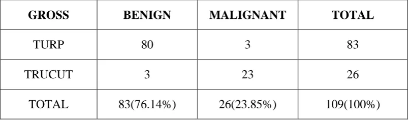
GROSS BENIGN MALIGNANT TOTAL
TURP 80 3 83
TRUCUT 3 23 26
TOTAL 83(76.14%) 26(23.85%) 109(100%)
PROSTATIC LESIONS:
Of the 109 total prostate specimens that were received in our Department, in the one year period from 2015 to 2016, there were a total of 83 cases that were benign, forming 76.14% and malignant cases were about 26 cases forming 23.85% of the total, vide chart no.2.
NATURE OF THE SPECIMEN:
45
CHART NO: 2 FREQUENCIES OF BENIGN AND MALIGNANT LESIONS
0 10 20 30 40 50 60 70 80
BENIGN MALIGNANT
46
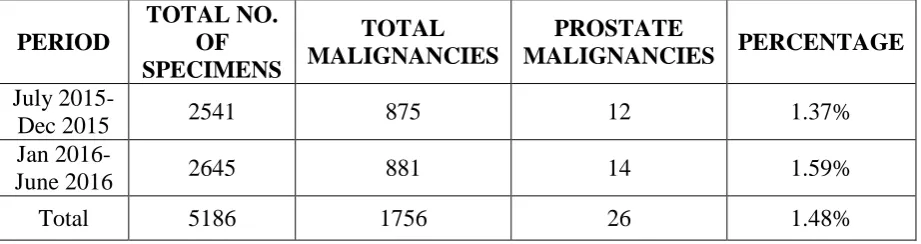
TABLE NO.:3 INCIDENCE OF PROSTATE CARCINOMA
PERIOD
TOTAL NO. OF SPECIMENS
TOTAL MALIGNANCIES
PROSTATE
MALIGNANCIES PERCENTAGE
July
2015-Dec 2015 2541 875 12 1.37%
Jan
2016-June 2016 2645 881 14 1.59%
Total 5186 1756 26 1.48%
47
CHART NO.:3 INCIDENCE OF PROSTATE CARCINOMA
0 200 400 600 800 1000
malignancie s prostate
48
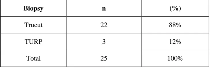
TABLE NO.:4 INCIDENCE OF PROSTATE CANCER IN GROSS SPECIMENS
Biopsy n (%)
Trucut 22 88%
TURP 3 12%
Total 25 100%
49
CHART NO.:4 INCIDENCE OF PROSTATE CANCER IN GROSS SPECIMEN
Trucut 88% Turp
12%
50
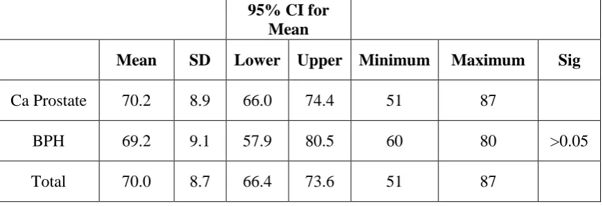
TABLE NO.:5 ASSOCIATION OF MEAN AGE WITH CLINICAL DIAGNOSIS
95% CI for Mean
Mean SD Lower Upper Minimum Maximum Sig
Ca Prostate 70.2 8.9 66.0 74.4 51 87
BPH 69.2 9.1 57.9 80.5 60 80 >0.05
Total 70.0 8.7 66.4 73.6 51 87
51
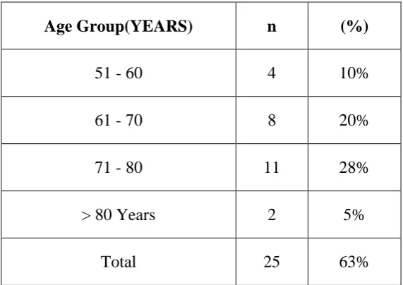
TABLE NO.: 6 ANALYSIS OF AGE AMONG THE PROSTATE MALIGNANCIES
Of the total 25 cases of prostate malignancies 28 cases were found in the age group of 71-80 years. The youngest individual was 51 years old and the oldest was 87 years old.
Age Group(YEARS) n (%)
51 - 60 4 10%
61 - 70 8 20%
71 - 80 11 28%
> 80 Years 2 5%
52
53
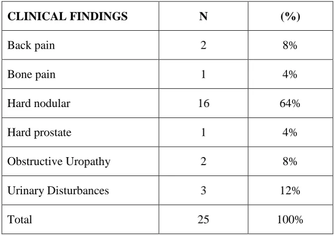
TABLE NO.7 COMPARING THE CLINICAL PRESENTATION
CLINICAL FINDINGS N (%)
Back pain 2 8%
Bone pain 1 4%
Hard nodular 16 64%
Hard prostate 1 4%
Obstructive Uropathy 2 8%
Urinary Disturbances 3 12%
Total 25 100%
54
CHART NO.:6 COMPARING THE CLINICAL PRESENTATION
Back pain Bone pain Hard nodular Hard prostate
Obstructive Uropathy
Urinary Disturbances
(%) 8% 4% 64% 4% 8% 12%
0% 10% 20% 30% 40% 50% 60%
55
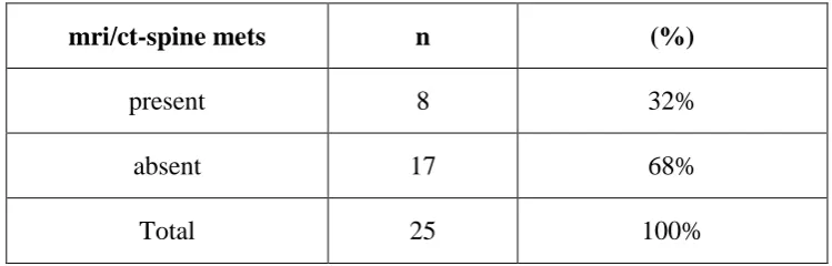
TABLE NO.:8 INCIDENCE OF METASTASIS WITH MRI FINDINGS
mri/ct-spine mets n (%)
present 8 32%
absent 17 68%
Total 25 100%
56
CHART NO.:7 INCIDENCE OF METASTASIS WITH MRI FINDINGS
Spine mets 32%
Nil 68%
57
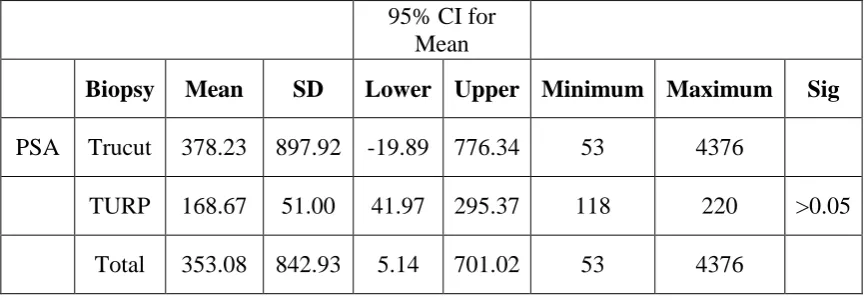
TABLE NO.:9 COMPARISON OF MEAN PSA WITH BIOPSY
While comparing the mean PSA values with the form of biopsy specimens sent, both the highest and lowest values were with that of trucut biopsy, which had negative values as lower confidence limits. Thus the p value is insignificant (>0.05) and nullifying the correlation.
95% CI for Mean
Biopsy Mean SD Lower Upper Minimum Maximum Sig PSA Trucut 378.23 897.92 -19.89 776.34 53 4376
58
CHART NO.:8 COMPARISON OF MEAN PSA WITH BIOPSY
59
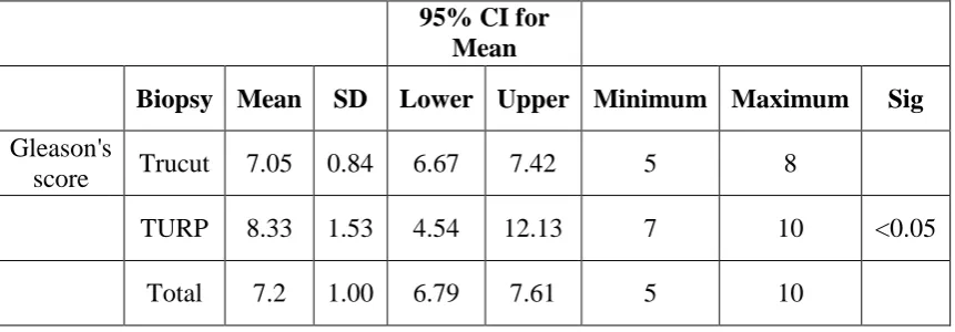
TABLE NO.:10 COMPARISON OF MEAN GLEASON’S SCORE WITH BIOPSY
95% CI for Mean
Biopsy Mean SD Lower Upper Minimum Maximum Sig Gleason's
score Trucut 7.05 0.84 6.67 7.42 5 8
TURP 8.33 1.53 4.54 12.13 7 10 <0.05 Total 7.2 1.00 6.79 7.61 5 10
60
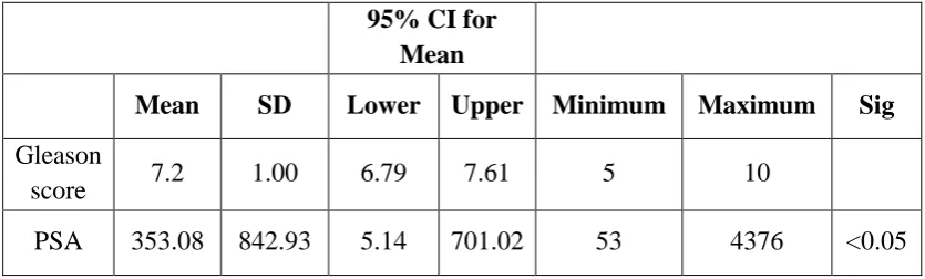
TABLE NO.: 11 ASSOCIATION OF MEAN OF GLEASON’S SCORE AND PSA
95% CI for Mean
Mean SD Lower Upper Minimum Maximum Sig
Gleason
score 7.2 1.00 6.79 7.61 5 10
PSA 353.08 842.93 5.14 701.02 53 4376 <0.05
61
CHART NO.: 10 ASSOCIATION OF MEAN OF GLEASON’S SCORE AND PSA
Gleascore PSA
Mean 7.2 353.08
0 50 100 150 200 250 300 350 400
62
CHART NO.:11 COMPARISON OF PSA VALUES AND GLEASON’S SCORE
It was found that the p value was significant, with <0.05,i.e, when the Serum PSA values and the Gleason’s score, were compared, with significant rise in PSA levels, the Gleason’s scoring also seemed to increase correspondingly in some of the patients. But it was also observed that there were also patients with lower PSA levels and slightly higher grade of Gleason’s scoring, which shows that there is a correlation with the PSA levels and the Gleason’s scoring.
-500 0 500 1000 1500 2000 2500 3000 3500 4000 4500 5000
0 2 4 6 8 10 12
PS
A
Gleason score Score
63
TABLE NO.:12 GRADING IHC EXPRESSION OF E-CADHERIN MARKER
ECADH n (%)
1+ 14 56%
2+ 4 16%
Neg 7 28%
Total 25 100%
64
CHART NO.:12 GRADING IHC EXPRESSION OF E-CADHERIN MARKER
1+ 56% 2+
16% Neg 28%
65
CHART NO.:13 CORRELATION OF E-CADHERIN AND GLEASON’S SCORE
The scatter diagram shows a linear plot of Gleason’s scoring with that of E-Cadherin expression with increasing scores mostly seen with 1+ intense expression.
0 0.5 1 1.5 2 2.5
0 2 4 6 8 10 12
ECA
D
H
Gleason score Score
66
CHART NO.:14 CORRELATIONS OF E-CADHERIN AND PSA VALUES
The scatter diagram shows that even with minimal elevation of PSA values, the expression of E-cadherin tends to become weaker in most of the cases and the case with maximum PSA value has negative E-Cadherin expression.
-0.5 0 0.5 1 1.5 2 2.5
0 1000 2000 3000 4000 5000
ECAD
H
PSA
67
TABLE NO.:13 GRADING IHC EXPRESSION OF VEGF MARKER
VEGF n (%)
1+ 3 12%
2+ 12 48%
3+ 8 32%
Neg 2 8%
Total 25 100%
68
CHART NO.:15 GRADING IHC EXPRESSION OF VEGF MARKER
1+ 12%
2+ 48% 3+
32%
Neg 8%
69
CHART NO.:16 CORRELATION OF VEGF AND GLEASON’S SCORE
There is almost a linear plot on comparing the expression of VEGF and Gleason’s score, with a strong expression as the score increases to the maximum of 10(5+5).
0 0.5 1 1.5 2 2.5 3 3.5
0 2 4 6 8 10 12
VE
GF
Gleason score Score
70
CHART NO.:17 CORRELATIONS OF VEGF AND SERUM PSA VALUES
The scatter diagram shows clustering of the expression intensity values with increasing PSA values and lower number of negative cases. There is also strong expression of VEGF with the maximum PSA value of 4376ng/ml
0 0.5 1 1.5 2 2.5 3 3.5
0 1000 2000 3000 4000 5000
VE
GF
PSA
71
TABLE NO.:14 COMPARISON OF MEAN OF GLEASON WITH VEGF
95% CI for Mean
VEGF Mean SD Lower Upper Minimum Maximum Sig Gleason
score 1+ 6.3 1.2 3.5 9.2 5 7
2+ 6.9 0.7 6.5 7.3 6 8
3+ 8.0 1.1 7.1 8.9 6 10
NIL 7.0 0.0 7.0 7.0 7 7 <0.05
Total 7.2 1.0 6.8 7.6 5 10
72
CHART NO.:18 COMPARISON OF MEAN OF GLEASON WITH VEGF
73
TABLE NO.:15 COMPARISON OF MEAN OF PSA WITH VEGF EXPRESSION
95% CI for Mean
VEGF Mean SD Lower Upper Minimum Maximum Sig
PSA 1+ 118.3 47.1 1.4 235.2 64 146 2+ 175.3 73.5 128.6 222.1 53 285 3+ 758.3 1466.2 -467.6 1984.1 53 4376
NIL 151.0 7.1 87.5 214.5 146 156 >0.05 Total 353.1 842.9 5.1 701.0 53 4376
74
TABLE NO.:16 CORRELATIONS OF VEGF AND E-CADHERIN EXPRESSION IN PROSTATE CANCER
Upon analysis of the expression of VEGF and E-Cadherin IHC markers, it was found that 28% of the cases had 3+ intense staining with VEGF and the same cases had negative staining with E-Cadherin. It has also been observed that there were no cases having negative staining with VEGF and high intense staining with E-cadherin, ruling out the benignity of the cases. But very few cases had weak 2+ staining of both the markers.
VEGF
ECADH 1+ 2+ 3+ Neg Total (%)
1+ 3 8 1 2 14 56%
2+ 0 2 2 0 4 16%
Neg 0 2 5 0 7 28%