ELECTRON MICROSCOPE ANALYSIS
Dissertation submitted to
THE TAMILNADU Dr. M.G.R. MEDICAL UNIVERSITY
In partial fulfillment for the Degree of MASTER OF DENTAL SURGERY
BRANCH IV
Conservative Dentistry and Endodontics, Ragas Dental College and Hospital, for his perseverance in motivating, guiding and supporting me throughout my study period.
My sincere thanks to Dr. R. Indira, M.D.S.,Professor and formerHOD,Department of Conservative Dentistry and Endodontics, Ragas DentalCollege and Hospital, who helped me with her guidance, support andconstant encouragement throughout my study period.
My sincere thanks to Dr. S. Ramachandran, M.D.S., Professor&former Principal, Department of Conservative Dentistry and Endodontics,Ragas Dental College and Hospital, who helped me with his advice andimmense support throughout my post graduate curriculum.
I extend my sincere thanks to Dr.P.Shankar, M.D.S., Professor, Ragas Dental College and Hospital, for his guidance, and constant encouragement during the completion of my study.
My sincere thanks toDr.Aravind, M.D.S., Senior lecturer, Department of Conservative Dentistry and Endodontics, Ragas Dental College and Hospital, who have helped me with his guidance, support and constant encouragement throughout my study period wherever and whenever needed.
I would like to solemnly thank Dr. Veni Ashok, M.D.S., Dr. Shankar Narayan,M.D.S., Dr.S.M. Venkatesan,M.D.S.,Readers, for all the help during my study period.
I would also like to thankDr.Sabari,M.D.S.,Dr.B.Venkatesh, M.D.S.,Senior lecturers for their friendly guidance and support.
I also wish to thank the management of Ragas Dental College and Hospital, Chennai for their help and support.
I sincerely thank,Dr.Sandhya,Dr.Linda, andDr.Bency for their constant support and encouragement throughout my study.
I remain ever grateful to all my batchmates, juniors and friends for their support.
1
2 CHX Chlorhexidine
3 EDTA Ethylenediamine-tetraacetic acid
4 MCP Mixture of castor detergent and papain enzyme
5 Er:YAG Erbium Yttrium Aluminium Garnet
6 Nd:YAG Neodymium-Doped Yttrium Aluminium
Garnet
7 Er:YSGG Erbium yttrium, scandium, gallium, garnet
8 SEM Scanning electron microscope
9 CSI Conventional syringe needle irrigation
10 LAI Laser activation irrigation
S. NO. INDEX PAGE.NO
1. INTRODUCTION 1
2. AIM AND OBJECTIVES 10
3. REVIEW OF LITERATURE 11
4. MATERIALS AND METHODS 33
5. RESULTS 42
6. DISCUSSION 51
7. SUMMARY 64
8. CONCLUSION 66
9. BIBLIOGRAPHY 67
Table 1 Distribution of smear scores (in %) in the apical, middle, and coronal thirds of the root canal in the Qmix no activation group (CSI)
Table 2 Distribution of smear scores (in %) in the apical, middle, and coronal thirds of the root canal in the Qmix LASER activation group
Table 3 Distribution of smear scores (in %) in the apical, middle, and coronal thirds of the root canal in the MCP no activation group (CSI)
Table 4 Distribution of smear scores (in %) in the apical, middle, and coronal thirds of the root canal in the MCP LASER activation group
Table 5
Mean smear layer score in Qmix before and after LASER activation
Table 6
Mean smear layer score in MCP before and after LASER activation
Table 7 Mean smear layer score in Qmix before activation and MCP before activation
Graph 1 Distribution of smear scores in the apical, middle, and coronal thirds of the root canal in the Qmix CSI group
Graph 2 Distribution of smear scores in the apical, middle, and coronal thirds of the root canal in the Qmix LASER activation group
Graph 3 Distribution of smear scores in the apical, middle, and coronal thirds of the root canal in the MCP CSI group
S.NO. TITLE FIGURE 1 Files and Protaper universal system
FIGURE 2 Saline and sodium hypochlorite
FIGURE 3 Endomotor (X smart plus)
FIGURE 4 Er:YAG laser equipment (Fotana laser)
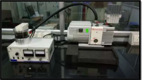
FIGURE 5 Scanning Electron Microscope– Gold sputter machine FIGURE 6 Scanning Electron Microscope analysis machine
FIGURE 7 QMIX 2 in 1 irrigating solution
FIGURE 8
MCP irrigating solution (Mixture of Castor detergent and Papain enzyme)
FIGURE 9 Preparation of MCP irrigating solution FIGURE 9a Castor oil
FIGURE 9b Sodium castorate
FIGURE 9c Filtration of MCP
FIGURE 9d MCP irrigating solution
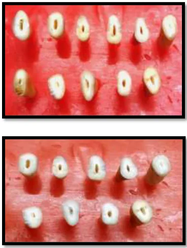
FIGURE10 Teeth samples
[image:11.595.92.516.176.727.2]FIGURE 14 Er:YAG laser activation
FIGURE 15 SEM - Conventional syringe irrigation group -QMIX
FIGURE 16 SEM- Conventional syringe irrigation group - MCP
FIGURE 17 SEM- Laser activation group- QMIX
[image:12.595.92.518.99.390.2]1
INTRODUCTION
Successful endodontic procedures depend on complete root-canal cleaning and shaping, three-dimensional hermetic root-canal system obturation, with no leakage of coronal restorations and prevention of reinfection.20 Earlier studies have demonstrated that chemo-mechanical instrumentation of a root canal create a bacterium-free environment and maintain disinfection.60,78 Many types of hand- and engine-driven rotary instruments and irrigation solutions have been developed for root-canal preparation.17 Mechanical instrumentation of the root canals produces a smear layer composed of organic and inorganic substances such as dentin particles, necrotic debris, and odontoblastic processes.54,88 Bodye et al (1963) first described the presence of smear layer on surface of cut enamel and McComb and Smith (1975) observed this layer on the walls of instrumented root canals.78
The smear layer is a structure composed of organic and inorganic parts, including fragments of odontoblastic processes, microorganisms and necrotic tissue.78,88,29 Electron microscopy shows the smear layer as an amorphous substance with an irregular surface attached to the root canal which covers the anatomical structures of the root canal. The smear layer thickness is not constant but ranges from 1 to 5 μm. The smear layer can be distinguished in
2
dentin and a deeper layer of debris condensed into the dentinal tubules called the smear plug.29,88
There are various controversies regarding the removal of smear layer. Studies done by Vojinovic et al 1973, and Michelich et al 1980, reported that the smear layer acts as a physical barrier to bacteria and bacterial byproducts. Conversely, Baker et al (1975) and Yamada et al (1983) observed that bacteria could remain in the smear layer and in the dentinal tubules despite instrumentation of the root canal and survive and multiply. Therefore some investigations had focused on the removal of the smear layer. Their findings reported the following;78,88
1. It has unpredictable thickness, volume and also contains bacteria and their byproducts. (Cergneux et al. 1987, McComb & Smith 1975, Brannstrom & Nyborg 1973). Bacteria such as Psedumonas aeruginosa, A.viscosus, Corynebacterium spp and S.sanguis digested the smear layer which could result in a gap between the obturating material and canal walls. (Bergenholtz 1977)
2. It may limit the optimum penetration of disinfecting agents. (Outhwaite et al. 1976, Goldberg & Abramovich 1977)
3
4. It is a loosely adherent structure and a potential avenue for leakage and bacterial contaminant passage between the root canal and dentinal walls. (Mader et al. 1984, Cameron 1987)
4 QMIX 2 IN 1 IRRIGATING SOLUTION:
Composition:45
(a) Ethylenediamine tetraacetic acid (a polyaminocarboxylic acid chelating agent)
(b) Chlorhexidine or orally acceptable addition salt (a bisbiguanide antimicrobial agent)
(c) N-cetyl-N, N,Ntrimethylammonium bromide (cetrimide -microbially active quaternary ammonium bromide.)
(d) Water.
5
The composition of this agent offers advantages over various other irrigating solutions. The traditionally used EDTA cannot be combined with chlorhexidine in high concentration, as this forms a precipitate and hence cannot remove the smear layer. Whereas in QMix, chlorhexidine is first mixed with cetrimide before EDTA is added. Chlorhexidine and cetrimide in water appears to form a micelle formulation that protects the combination to precipitate.44,72
MCP (MIXTURE OF CASTOR DETERGENT AND PAPAIN ENZYME):
Composition:91
a. 4% Papain enzyme powder
b. 20% Castor detergent (Ricinus communis)
a. Papain enzyme acts as a debris-removing agent. It acts only on the affected tissues, which lack the α 1-antitrypsine plasmatic antiprotease that inhibits proteolysis in healthy tissues.30 Bhardwaj (2002) observed that papain enzyme has the comparable antibacterial effect of calcium hydroxide when used in gel form as an intracanal medicament against E faecalis13.
6
Pecora JD et al, reported that this agent increases root dentin permeability similarly to 0.5% NaOCL and a 0.4% papaine gel.91 The Ricinus communis detergent acts by breaking sugar leakage of the cellular wall of pathogenic microorganisms, consequently the loss of cytoplasmic material leads to cell destruction.68
7
Different agitation techniques have been proposed to improve the efficacy of irrigation solutions, including hand agitation, sonic and ultrasonic devices and laser systems.41 Stojicic S, George R and De Moor RJG, had investigated the ability of some laser wavelengths to activate the commonly used irrigant solutions within the canal and observed that the laser-activated irrigation (LAI), has been more effective in removing debris and smear layer in root canals compared to traditional techniques (hand irrigation and passive ultrasonic irrigation).64
Laser-activated irrigation (LAI) has been introduced as a powerful method for root canal irrigation (Blanken & Verdaasdonk 2007, George & Walsh 2008, George et al. 2008). The laser radiation produces transient cavitations in the liquid through optical breakdown by strong absorption of the laser energy (Blanken & Verdaasdonk 2007). The use of lasers at different wavelengths has been proposed to supplement conventional endodontic cleaning procedures.64
8
Different lasers activation systems such as CO2, Nd:YAG, Er:YAG, Er,Cr:YSGG have been used for debris and smear layer removal from the canals. In addition different laser wavelengths have been used directly or as an adjunctive to disinfect canals. Laser light can penetrate areas of canals where irrigating and disinfecting solutions cannot reach, like secondary canals and deep dentinal tubules and also can eliminate microorganisms. Water or other irrigants are used during lasing to reduce thermal stress to the radicular dentine and periodontium. These pulsed lasers absorb in water and create pressure waves from explosions followed by implosions.80,33
Various studies on the efficacy of Er:YAG laser irradiation for cleaning root canal walls have already demonstrated that this type of laser is more effective in removing the smear layer than other laser types. The dentinal walls mostly show open tubules and are free of debris or a smear layer. A laser-activated irrigation treatment with an erbium laser (Er:YAG with a wavelength of 2,940 nm) has been presented as an activation method of irrigation solution.33,23,76 An Er:YAG laser has the highest absorption in water and a high affinity for hydroxyapatite, and it provides effective removal of the debris and smear layer from the complex root canal systems. The effect is based on explosive vapor bubbles with a secondary cavitation effect by the pulsed energy transferred to solutions.14,24
9
10
AMI AND OBJECTIVES
AIM :
To compare the smear layer removal ability of Qmix and MCP irrigating solutions using Er:YAG laser activation system- A Scanning Electron Microscope analysis.
OBJECTIVES:
1. To compare smear layer removal ability of Qmix and MCP using conventional syringe needle irrigation
11
REVIEW OF LITERATURE
Duarte, Marcos Antonio Hungaro et al (2001)25 evaluated the apical leakage in teeth obturated after they had been prepared using two different endodontic irrigants. Group 1: was instrumented using 0.8% papain gel as an endodontic irrigant. Group 2 used the 1% sodium hypochlorite solution as irrigant. Both experimental groups were instrumented manually using the step-back technique; the irrigation was performed after the use of each endodontic instrument. The samples were then obturated and immersed into a 2% methylene blue solution for 7 days at 37ºC. The results concluded that there was no statistical difference between the experimental groups (p>0.05).
Marcos Pozzetti Meneghin et al (2006)61 evaluated the cleaning of
12
Roy George et al (2008)33 examined the ability of laser tips when
Er:YAG and Er,Cr:YSGG lasers were used in root canals in which thick smear layers had been created intentionally to provide a challenge for the laser system. Smear layer was assessed from scanning electron microscopy images with an objective digital method. Lasing improved the action of ethylene diamine tetraacetic acid with cetavlon (EDTAC) in removing smear layer. Conical fibers performed better than plain fibers, but there was no difference in performance between the 2 laser systems when matched for all other parameters. The study concluded that Conical fiber tips performed better than plain fibers for removal of smear layer when matched for the same laser system and the same irrigant. Additional studies are needed to establish the usefulness of these modified fibers in other endodontic applications such as enlarging the canal or canal disinfection.
13
evaluators (kappa analysis) across the range of the Hulsmann scores. Image analysis might be useful for evaluating the degree of smear layer removal in endodontic research.
Letícia Helena Theodoro et al (2009)83 evaluated the effect of
erbium: yttrium–aluminum–garnet (Er:YAG) laser (2.94 μm) irradiation on
the removal of root surface smear layer of extracted human teeth. It compared
its efficacy with that of citric acid, ethylenediamine tetra-acetic acid (EDTA),
or a gel containing a mixture of tetracycline hydrochloride (HCl) and citric
acid, using scanning electron microscopy (SEM). Thirty human dentin
specimens were randomly divided into six groups: G1 (control group),
irrigated with 10 ml of physiologic saline solution; G2, conditioned with 24%
citric acid gel; G3, conditioned with 24% EDTA gel; G4, conditioned with a
50% citric acid and tetracycline gel; G5, irradiated with Er:YAG laser (47
mJ/10 Hz/5.8 J/cm2/pulse); G6, irradiated with Er:YAG laser (83 mJ/10
Hz/10.3 J/cm2/pulse). The study concluded that all treatment modalities were
effective in smear layer removal.
E. DiVito & O. Et al (2010)23 analyzed in vitro the debriding ability
of an Er:YAG laser system (2,940 nm) equipped with a newly designed radial
and stripped tip of 400 μm diameter by scanning electron microscopy (SEM).
14
four different final protocols were used. Group 1 was irrigated for 2 min with
saline water as a control group. Groups 2, 3 and 4 were irradiated with an
Er:YAG laser at 25 mJ and 15 Hz with a pulse duration of 50 !s and laser
spray off using the tip in the coronal opening of the wet root canal. Different
solutions and irradiation times were used: group 2 20 s, laser irradiation in
sterile distilled water, wet canal; group 3 20 s, laser irradiation in 17% EDTA,
wet canal; and group 4 40 s, laser irradiation in 17% EDTA, wet canal. The
study concluded that standardized instrumentation, followed by a final
Er:YAG laser irradiation in wet canals with EDTA irrigation resulted in more
cleaning of the root canal walls and a higher quantity of open tubules in
comparison with the traditional irrigation method.
15
standardized instrumentation, followed by a final Er:YAG laser irradiation in EDTA-wetted canals, showed more debriding and cleaning of root canal surfaces in comparison with Er:YAG laser irradiation in saline solution or saline solution alone.
16
EDTA in removing canal wall smear layers after the use of 5.25% NaOCl as the initial rinse.
S. Stojicic. Et al81 (2011)examined the efficacy of a novel root canal irrigant, QMiX, against Enterococcus faecalis and mixed plaque bacteria in planktonic phase and biofilms, and its ability to remove smear layer. Enterococcus faecalis and mixed plaque bacteria were exposed to QMiX, 2% chlorhexidine (CHX), MTAD and 1% sodium hypochlorite (NaOCl) for 5 s, 30 s and 3 min. Following exposure, samples were taken, serially diluted and grown aerobically and anaerobically on tryptic soy agar (TSA) plates or on blood agar plates for 24 and 72 h, respectively, to measure killing of bacteria. The amount of killed bacteria in biofilms was analysed by confocal laser scanning microscopy using viability staining. Dentin blocks were exposed to Qmix and 17% EDTA for 5 min. The effectiveness of smear layer removal by the solution was evaluated using scanning electron microscopy. The results concluded that QMiX and NaOCl were superior to CHX and MTAD under laboratory conditions in killing E. faecalis and plaque bacteria in planktonic and biofilm culture. Ability to remove smear layer by QMiX was comparable to EDTA.
Rebecca Guidotti et al (2012)39 evaluated the effectiveness of
Er:YAG laser fiber in removing the smear layer produced during root canal
walls instrumentation. Forty-eight single-rooted teeth were prepared with
17
Samples were randomly subdivided into groups and treated with: three
irradiations of 5 s each, with 300-μm Er:YAG endodontic fiber, 1 W and 2.5
% NaOCl solution (A Group); two laser irradiations with 17 % EDTA solution
and 2.5 % NaOCl solution (B Group); laser irradiation plus 17 % EDTA
solution and 2.5 % NaOCl (C Group); only in the final wash of 17 % EDTA
(control group D). Each sample was finally observed by scanning electron
microscope (SEM) at the coronal, medium, and apical thirds at ×500
magnification. The study concluded that The Er:YAG fiber double irradiation
with EDTA 17 % and NaOCl 2.5 % has been demonstrated to be effective in
removing smear layer, even in the apical third which is described as the
hardest area to clean during endodontic treatment.
18
Veeramachaneni Chandrasekhar. Et al (2013)19 evaluated the biocompatibility of a new root canal irrigant Q mix™ 2 in 1 in comparison to 0.9% sterile saline, 3% sodium hypochlorite (NaOCl), 2% chlorhexidine (CHX), and 17% ethylenediaminetetraacetic acid (EDTA). The results concluded that QMix™ 2 in 1 was shown to be less toxic to the rat
subcutaneous tissue than 3% NaOCl, 2% CHX, and 17% EDTA.
Vivek Hedge et al (2013)46evaluated the smear layer removal using hand activation and Laser with PIPS. Forty single rooted, extracted human teeth were used in the study. Standard endodontic access cavity preparation was performed and instrumentation was done upto F3 using rotary protapers. These teeth were divided into two groups for the final irrigant; group 1: 5.25% NaOCl and group II: 17% EDTA. Group 1 was activated mechanically and group II was activated using Er:YAG Laser with wavelength of 2940nm(PIPS). The study concluded that laser with PIPS showed better smear layer removal than hand activation group.
Birang Reza, Hasheminia Seyed Mohsen (2013)73 compared the effects of Erbium: Yttrium‑Aluminium‑Garnet (Er: YAG), and Neodimium:
Yttrium‑Aluminium‑Garnet (Nd: YAG) lasers on removing the smear layer
using scanning electron microscopy. In this experimental study, 55 human
single‑rooted teeth were examined. Instrumentation was done using the
19
#80 at the coronal area. The samples were divided into three groups: Samples irradiated by the Er: YAG laser (1 W, 10 Hz, 130.7 J/cm2) in Group 1 (n=25), the Nd: YAG laser (2 W, 15 Hz, 188.25 J/cm2) in Group 2 (n=25) and samples irrigated by 5.25% NaOCl as the control in Group 3 (n=5). Next, roots were bisected longitudinally and prepared for scanning electron microscopy. The study concluded that irradiation by the Er: YAG laser was more effective in smear layer removal than the Nd: YAG laser.
Arturo Javier Aranda-Garcia et al (2013)5 evaluated the efficacy of
QMix, SmearClear, and 17% EDTA for the debris and smear layer removal from the root canal and its effects on the push-out bond strength of an epoxy-based sealer by scanning electron microscopy (SEM). Forty extracted human
canines (n510) were assigned to the following final rinse protocols: G1-distilled water (control), G2–17% EDTA, G3-SmearClear, and G4-QMix.
The specimens were submitted to a SEM analysis to evaluate the presence of debris and smear layer, respectively, in the apical or cervical segments. The study concluded that the ability to remove the debris and smear layer by SmearClear and QMix was as effective as the 17% EDTA.
20
BioRaCe instruments up to BR7 (60/0.02). In group 1, canals were irrigated with a combination of 1% papain, 17% EDTA, Tween 80 and 2% CHX; in group 2, canals were irrigated with a combination of 0.1% papain, 17% EDTA, Tween 80 and 2% CHX. In group 3 (the negative control), the canal was irrigated with 2.5% NaOCl during instrumentation and at the end of preparation with 1 mL of 17% EDTA was used; in group 4 (positive control), normal saline was used for irrigation. The amount of the remaining smear layer was quantified according to Hulsmann method using scanning electron microscopy (SEM). The study concluded that combination of 1% papain, EDTA, 2% chlorhexidine and Tween 80 can effectively remove smear layer from canal walls.
21
microhardness, it was found that QMix, 17% EDTA + 2% CHX and 17% EDTA + 2.5% NaOCl cause the same reduction in the microhardness of root canal dentine in the coronal and middle regions.
Nawfal A.A. Zakarea et al (2014)90 evaluated in vitro, the efficacy of a newly prepared endodontic irrigant solution, against E. Faecalis. Sixty human extracted single rooted teeth samples were prepared by using protaper NiTi rotary system and concluded that Castor detergent 20% and papain enzyme 4% (MCP) has ability to completely eradicate Enterococcus Faecalis bacteria from the infected root canal in vitro in 5 min. It's antibacterial action is similar to the action of 2.5 % NaOCl.
22
levels (apical, middle, and cervical part of the canal). The results of this study concluded that MCP solution had the ability to remove the smear layer partially at the three levels of root canal without dentin erosion. However EDTA had the ability to remove the smear layer completely at the three levels of canal with obvious dentinal erosion.
M.C. Prado. et al (2014)71 compared the effectiveness of QMix on Smear layer (SL) removal using passive ultrasonic irrigation (PUI). For SL removal, QMix was used for 1 min, with or without passive ultrasonic irrigation. Distilled water was used as control (DW). The groups evaluated were: NaOCl +DW; CHX+DW; NaOCl + QMix; CHX+ QMix; NaOCl + QMix + PUI and CHX+ QMix + PUI. After irrigation protocols, the teeth were prepared and analyzed by Scanning Electron Microscopy. The results concluded that the use of QMix for 1 min is effective only when associated with passive ultrasonic irrigation.
23
remaining Smear layer was quantified according to Hulsmann’s method with
scanning electron microscopy (SEM). The study concluded that EDTA was more effective in removing Smear layer compared to Er: YAG and etidronate.
Sibel Kocak et al (2015)56 compared the efficacy of QMiX and ethylenediaminetetraacetic acid (EDTA) solutions with diode laser treatment in smear layer removal. Seventy-five extracted mandibular premolars were used. After root canals were, prepared the specimens were divided into five groups (n = 15): Group 1, no irrigation; Group 2, 17% EDTA; Group 3, QMiX solution; Group 4, 17% EDTA with diode laser; and Group 5, QMiX with diode laser. The roots were split longitudinally and prepared for scanning electron microscopic (SEM) investigation. The smear layer was evaluated under · 500, · 1000, and · 2000 magnifications. The study concluded that Diode laser treatment with solutions decreased the amount of smear layer, without significance.
24
the flexural strength after treatment with substances. The study concluded that CHX and GSE were more effective than NaOCl and QMix against E. faecalis. Furthermore, they did not harm dentine mechanical properties as observed for NaOCl and QMix.
Ankur Mahesh Banode et al (2015)12 compared the effectiveness of 17% ethylenediaminetetraacetic acid (EDTA) with that of 10% citric acid and newer irrigant QMix in the removal of smear layer from root canal wall
dentin. Twenty single‑rooted teeth were accessed and instrumented with
crown down technique up to protaper F3. Between each instrument used, the canals were irrigated with 1 ml of 5% sodium hypochlorite (NaOCl). After instrumentation, the irrigants used were 1% NaOCl, 17% EDTA, 10% citric acid, and newer irrigant QMix. The samples were prepared and observed by means of scanning electron microscopy. The study concluded that 17% EDTA, 10% citric acid, and QMixTM all chelating agent removes smear layer effectively from cervical and middle parts of canal as compared to apical third. In future, QMixTM may act as a promising chelating agent as well as antimicrobial irrigant
Sefika Nur Akyuz Ekim et al (2015)2 evaluated the efficiency of
25
irrigation activation technique; distilled water was used as an irrigant in Group 1. The other groups were treated with 2.5% NaOCl and 17% EDTA, respectively. Conventional syringe irrigation (CSI) was used in Group 2. Irrigation solutions were activated using passive ultrasonic irrigation (PUI, Group 3), EndoVac apical negative pressure (ANP, Group 4), diode laser (Group 5), Nd:YAG laser (Group 6), Er:YAG laser (Group 7), and Er:YAG laser using with photon-induced photoacoustic streaming (PIPSTM, Group 8). Teeth were split longitudinally and subjected to scanning electron microscope (SEM). The study concluded that all the irrigation activated/delivered techniques except diode laser have a positive effect on removing of smear layer.
Shilpi Gupta et al (2015)42 compared the smear layer removal efficacies of 5.25% sodium hypochlorite (NaOCl) and QMixTM (Dentsply Tulsa Dental Specialties, Tulsa, OK, USA) in the coronal, middle, and apical thirds of the root canal using a common irrigation protocol in deciduous teeth.
Forty extracted human single‑rooted deciduous teeth were prepared to 40 K
26
The results showed groups 1 and 2 had statistically significant differences in the coronal (P = 0.001) and middle thirds (P = 0.032); however, in the apical third the canal surfaces were cleaner in samples from group 2 (P = 0.046) as compared to group 1. The study concluded that QMixTM is effective as a final irrigation agent for the removal of smear layer in the coronal, middle, and apical thirds of the root canals in deciduous teeth.
E Kalyoncuoglu et al (2015)52 evaluated and compared in vitro the antifungal efficacy of QMix 2in1, 5.25% NaOCl, 2% CHX, and 17% EDTA as a final rinse against Candida albicans (C. albicans). Following root canal preparation, teeth were inoculated with C. albicans and incubated for 72 h. Teeth were irrigated with one of the following solutions as a final irrigant: (1) 5.25% NaOCl, (2) 2% CHX, (3) QMix 2in1, and (4) 17% EDTA. The results concluded that QMix 2in1 proved to be effective against C. albicans when used as a final rinse.
27
followed by 2% CTR; (4) MTAD; (5) QMix; and (6) control, 0.9% saline. Bacterial samples collected before instrumentation and after final irrigation were cultured and the colony-forming units (CFUs) were counted. The results concluded that the antimicrobial activity of QMix was comparable to that of EDTA/CHX and EDTA/CTR and more effective than that of EDTA/NaOCl against intracanal E. faecalis.
Alexander Pompermayer Jardine et al (2015)49 compared the effect
of QMix, BioPure MTAD, 17 % EDTA, and saline on the penetrability of a
resin-based sealer into dentinal tubules using a confocal laser scanning
microscope (CLSM) and to describe the cleaning of root canal walls by SEM.
Eighty distobuccal roots from upper molars were selected and randomly
divided into four groups (n=20) before root canal preparation according to the
solution used in the final rinse protocol (FRP): QG (QMix), MG (BioPure
MTAD), EG (17 % EDTA), and CG (control group: saline). Ten roots of each
group were prepared for SEM, and images (×2000) from the canal walls were
acquired. The remaining canals were filled with a single gutta-percha cone and
AH Plus with 0.1 % Rhodamine B. The specimens were horizontally sectioned
at 4 mm from the apex, and the slices were analyzed in CLSM (×10). Sealer
penetration was analyzed with Adobe Photoshop software. The results showed
that QG and EG presented similar amounts of sealer penetration (P>.05). MG
and CG presented the lowest penetrability values (P<.05). The best results for
28
QG and EG groups when compared with MG and CG (P<.05). The study
concluded that Seventeen percent EDTA and QMix promoted sealer
penetration superior to that achieved by BioPure MTAD and saline.
Dilara Arslan et al (2016)6 evaluated in vitro Smear Layer Removal Ability of QMix with Different Activation Techniques using the EndoActivator (EA) system (Dentsply Tulsa Dental Specialties), photon-initiated photoacoustic streaming (PIPS), and an Er:YAG laser with an endodontic fiber tip and concluded that the Er:YAG laser, PIPS, and EA techniques enhanced the smear removal capacity of the QMix solution when compared with needle irrigation. Er:YAG laser–activated QMix removed the smear layer more effectively than other techniques in the apical third of the root canal system, whereas PIPS had the same effect in the coronal third.
Birang Reza et al (2016)73 compared the effects of Erbium: TYttrium‑ Aluminium‑Garnet (Er:YAG),and Nodimium: Yttrium‑
Aluminium‑Garnet (Nd: YAG) lasers on removing the smear layer using
scanning electron microscopy. In this experimental study, 55 human single‑rooted teeth were examined. Instrumentation was done using the
step‑back technique with hand files up to file #40 at the apical area and file
29
samples irrigated by 5.25% NaOCl as the control in Group 3 (n=5). Next, roots were bisected longitudinally and prepared for scanning electron microscopy. The results concluded that irradiation by the Er: YAG laser was more effective in smear layer removal than the Nd: YAG laser.
Shaheen Venghat et al (2016)87 studied in vitro the effects of four endodontic irrigants and on a smear layer created by hand and rotary instrumentation in the middle and apical thirds of root canals. Cleaning and shaping up to size F5 using Protaper Universal System; the root canals were irrigated with 3 mL of 5.25% NaOCl, between each file size. Group 1 (G1) were irrigated with a final flush of QMix 2in1. The teeth in group 2 (G2) were irrigated with a final flush of 0.2%Chitosan, group 3 (G3) with Smear Clear and group 4 (G4) with Glyde. The teeth were split longitudinally and prepared for examination by scanning electron microscopy. The results of this study concluded irrigation with QMix 2in1, Smear Clear, 0.2%Chitosan, and Glyde and 6% did not remove all the smear layer from the root canal system. All these irrigants showed less effectiveness in removal of smear layer from apical 3rd.
30
1); ethylenediaminetetraacetic acid (EDTA) and sodium hypochlorite (NaOCl) (Group 2), and NaOCl + QMix (Group 3) or bonded with OptiBond adhesive after irrigation with saline (Group 4), EDTA and NaOCl (Group 5), and NaOCl + QMix (Group 6). All the samples were restored with composite. Ten samples per group were assessed for dye penetration. Fifteen samples were assessed under scanning electron microscope. The results concluded that EDTA + NaOCl or NaOCl + QMix irrigation of the pulp chamber was not deleterious to the bonding of any of the adhesives tested.
31
concluded that QMix 2 in 1 is most effective in removal of smear layer when compared to other tested irrigants.
Nidambur Vasudev Ballal et al (2016)11 evaluated the canal wall smear layer removal capacity and mineral content distribution of root canal dentine after irrigation with QMix, 7% maleic acid (MA) and 17% ethylenediaminetetraacetic acid(EDTA). Forty single rooted teeth were subjected to root canal instrumentation and divided into four groups: [1] 7% MA + 2.5% sodium hypochlorite (NaOCl), [2] 17% EDTA+ 2.5% NaOCl, [3] QMix + 2.5% NaOCl and [4] 0.9% saline (negative control). After irrigation, the teeth were examined by scanning electron microscopy (SEM) to determine the presence or absence of smear layer. For mineral content assessment, 40 root-halves were divided into four groups and treated with 7% MA, QMix, 17% EDTA and saline. Mineral content was evaluated using SEM-energy dispersive X-ray analysis. The study concluded that 7% MA had superior smear layer removal ability compared with QMix and 17% EDTA. Calcium level was decreased more with QMix while phosphorus level was decreased more with 7% MA and QMix respectively.
32
33
MATERIALS AND METHODS
ARMAMENTARIUM AND MATERIALS
1. ISO size # 15 to # 40 K-files stainless steel Endo handfiles (Kerr Dental)
2. Protaper Universal treatment (Rotary) (S1, S2,F1,F2,F3,F4) (Dentsply, India)
3. Sodium hypochlorite irrigation 5.25% and saline 4. Qmix 2 in 1 irrigating solution (Dentsply, Tulsa, USA) 5. MCP – Mixture of Castor detergent and Papain enzyme
Papain enzyme (Rashi Biotech, Pune)
MCP Preparation (Hubert Enviro Care Systems,
34 IRRIGANT SOLUTION:
1. QMIX 2 IN 2 IRRIGATING SOLUTION:
Qmix 2 in1 Irrigating Solution (Dentsply Tulsa Dental Specialties, Tulsa, OK) is a novel endodontic irrigant containing composed of a poly- aminocarboxylic acid chelating agent (EDTA), a bisbiguanide antimicrobial agent (chlorhexidine gluconate), a surfactant and deionized water. This is a readily available irrigating solution obtained from the manufacturer.
2. MCP (MIXTURE OF CASTOR DETERGENT AND PAPAIN ENZYME):
PREPARATION OF SODIUM CASTORATE
Requirements:
Castor oil
Sodium hydroxide
Distilled water
35 Procedure:
1. In 35 ml of distilled water, 1.75g of sodium hydroxide was added and mixed well to get base solution. Further 100 ml of castor oil was taken in a 250 ml beaker and kept in magnetic stirrer.
2. The base solution was slowly added into castor oil under magnetic stirring.
3. The stirring was done till the mixture began to thicken and when we could see trails of the mixture on the surface upon lifting the spoon/stirrer above the surface.
4. This stage is called ‘trace’ and few oils take longer than others to reach this stage.
5. The excess glycerin was separated by filtering using Whatmann No. 40 filter paper.
6. The sodium castorate obtained was stored in refrigerator till further usage.
PREPARATION OF MCP SOLUTION
Requirements:
Papain enzyme
Sodium castorate
Distilled water
36 Procedure:
1. In a 250 ml beaker 20 ml of sodium castorate and 4 gms of papin enzyme was added and the solution was made upto 100 ml using distilled water.
2. All the above reactions were carried out under magnetic stirring. 3. After 30 minutes, the solution was filtered using Whatmann No.40
filter paper to obtain MCP irrigant solution.
37
FLOWCHART ILLUSTRATING THE METHODOLOGY OF THE STUDY:
Selection of the study samples (32 teeth)
Cleaning and shaping done (Protaper rotary instrument upto size F4 and NaOCl irrigating solution)
Working length determination (using ISO #15 K files) Decoronation of the teeth (32 roots)
Teeth divided randomly in two experimental groups (32 roots)
QMIX 2 IN 1 IRRIGATING SOLUTION (16 roots)
MCP (MIXTURE OF CASTOR DETERGENT AND PAPAIN (16 roots)
1. QMIX without activation (n=8) 2. QMIX with LASER activation (n=8)
1. MCP without activation (n=8) 2. MCP with LASER activation (n=8)
SCANNING ELECTRON MICROSCOPY
38
SELECTION AND PREPARATION OF THE SAMPLES:
Thirty two single-rooted human mandibular premolars extracted for periodontal and orthodontic reasons from patients aged 16–19 years were obtained from the Department of Oral and Maxillofacial Surgery with the approval of the Ethical committee of Ragas Dental College and Hospital. Digital radiographs were taken to ensure the presence of a single root canal, no calcifications, and the absence of a complicated root canal anatomy. Soft tissue remnants and debris were cleaned mechanically and ultrasonically. The teeth were then stored in 10% formalin until use. The teeth were decoronated, and roots were standardized using a diamond disc operated at low speed to 12 mm in length. ISO size # 15 K-file (Kerr Dental) was inserted into the root canal until just visible at the apical foramen. The working length (WL) of each root canal was then established 1 mm short of the apical foramen. Each apex was sealed using sticky wax to simulate the clinical situation. Further the root canals were prepared using ProTaper rotary instruments (Dentsply, India) up to apical size (F4). The canals were irrigated with 2 mL 5.25% NaOCl between each file. At the end of instrumentation, 5mL 5.25%NaOCl for 1 minute and then 5mL saline for 1 minute were used.
After the instrumentation, the specimens were randomly divided into two groups of 16 roots each depending on the type of irrigating solution and activation process to be used.
39 Group I – QMIX 2 in 1 irrigating solution
a. QMIX without activation b. QMIX with activation
Group II – MCP (Mixture of castor detergent and Papain enzyme) a. MCP without activation
b. MCP with activation
LASER ACTIVATION PROTOCOL:
Er:YAG laser activation was performed with a wavelength of 2940 mm (Fidelis; Fotona, Ljubljana, Slovenia) and an R14 handpiece with a 300-mm endodontic fiber tip (Preciso, Fotona). The water and air were turned off. The protocol had been set to 1 W, 20 Hz, and 50 mJ as specified by the manufacturer. The laser was introduced with spiral movement from the apex to the coronal region. The laser was applied 5 times, and each application lasted 3s along the root canal system.
EVALUATION BY SCANNING ELECTRON MICROSCOPY:
40
and the optimum half was used for further analysis. This portion of the root was dehydrated in the ascending alcohol series for 24 hours (70%–100%). The specimens were left to dry overnight, mounted on copper stubs and coated with gold using the sputter machine (Sputter Coater SC7620). These were then examined and photographed using a scanning electron microscope (Model: EVO MA 15, Carl Zesis Pvt.Lts.UK). Scanning electron microscopic photomicrographs were taken at 2000x magnification at the coronal, middle, and apical thirds of the root canals using the SEM analysis software (Smart SEM User Interface).
CRITERIA FOR SMEAR LAYER EVALUATION:
The smear layer was scored according to the criteria given by Hulssman et al 1997.47
Score 1: indicates no smear layer, and all dentin tubules are open and
clean.
Score 2: indicates a small amount of smear layer, and some dentin
tubules are open.
Score 3: indicates a homogenous smear layer covering the root canal
wall, and only a few dentin tubules are open.
Score 4: indicates a complete root canal wall covered by a
homogenous smear layer, and no dentin tubules are open.
Score 5: indicates a heavy, non-homogenous smear layer covering the
41 STATISTICAL ANALYSIS
The following statistical procedures were carried out:- 1. Data compilation and presentation
2. Statistical analyses
I. Data compilation and presentation:
Data obtained were compiled systematically in Microsoft Excel spreadsheet. The dataset was subdivided and distributed meaningfully and presented as graphs and tables.
II. Statistical analyses:
Statistical analyses were performed using a personal computer in Statistical Package for Social Sciences software (SPSS version 22, USA). Normality distribution of the data was analyzed and specific statistical tests were used to find out the statistical significance of the obtained results. The p value was set for 0.05 and any value equal to or less than was considered to be significant.
1. Normality was tested using Shapiro Wilk test and the data was not normally distributed.
2. For the Inter group comparison (Qmix and MCP), Mann Whitney Test was done.
3. For Intra group comparison before and after Er:YAG Laser activation in each group Wilcoxson signed rank test was done.
ARMAMENTARIUM
FIGURE 1: FILES AND PROTAPER UNIVERSAL SYSTEM
[image:59.612.149.462.380.663.2]
FIGURE 3: ENDOMOTOR (X SMART PLUS)
FIGURE 5: SEM – SPUTTER COATER MACHINE
IRRIGATING SOLUTION USED IN THE STUDY
[image:62.612.220.388.458.665.2]FIGURE 7: QMIX 2 IN 1 IRRIGATING SOLUTION
FIGURE 9: PREPARATION OF MCP IRRIGATING SOLUTION
CASTOR OIL SODIUM CASTORATE
STEPWISE PROCEDURE FIGURE 10: TEETH SAMPLES
FIGURE 12: RADIOGRAPH BEFORE BIOMECHANICAL PREPARATION
[image:65.612.108.498.303.422.2]
FIGURE 13: RADIOGRAPH AFTER BIOMECHANICAL PREPARATION
[image:65.612.131.479.465.663.2]
SCANNING ELECTRON MICROSCOPY IMAGE
FIGURE 15: CONVENTIONAL SYRINGE IRRIGATION GROUP -QMIX CORONAL THIRD
MIDDLE THIRD
FIGURE 16: CONVENTIONAL SYRINGE IRRIGATION GROUP - MCP CORONAL THIRD
MIDDLE THIRD
FIGURE 17: LASER ACTIVATION GROUP- QMIX CORONAL THIRD
MIDDLE THIRD
FIGURE 18: LASER ACTIVATION GROUP- MCP CORONAL THIRD
MIDDLE THIRD
42
RESULTS
This study was designed to evaluate the smear layer removal ability of two irrigating solutions namely Qmix 2 in 1 solution and Mixture of castor detergent and papain enzyme (MCP) using conventional syringe irrigation(CSI) method and after LASER activation using Er:YAG laser system. The smear layer was viewed under Scanning Electron Microscope (SEM) and scored according to the criteria given by Hulssman et al 1997.
The experimental groups were divided into two groups: 1. Qmix 2 in 1 solution
2. Mixture of castor detergent and papain enzyme (MCP)
Each of the group contains a conventional syringe needle irrigation method (CSI) and Er:YAG Laser activation system respectively.
The results of the present study revealed that the distribution of smear layer scores in the Qmix group using conventional syringe irrigation(CSI) method were 3(25.0%), 4(62.5%) and 5(12.5%) in the apical thirds of the root canal. In the middle thirds the scores were 1(25.0%), 2(37.5%) and 3(37.5%). And in the coronal thirds the scores were 1(50%) and 2(50%).
(Table 1 and Figure 1)
43
and 2(75%). In the coronal thirds the scores were 1(75%) and 2 (25%). (Table 2 and Figure 2)
The distribution of smear layer scores in the MCP group using conventional syringe irrigation (CSI) method were 3(25%), 4(50%) and 5(25%) in the apical thirds of the root canal. In the middle thirds the scores were 1(12.5%), 2(25%), 3(37.5%) and 4(25%). In the coronal thirds the scores were 1(37.5%) and 2(62.5%). (Table 3 and Figure 3)
The distribution of smear layer scores in the MCP group using Er:YAG Laser activation method were 3(25%) and 4(75%) in the apical thirds of the root canal. In the middle thirds the scores were 1(12.5%), 2(50%) and 3(37.5%). In the coronal thirds the scores were 1(50%) and 2(50%). (Table 4 and Figure 4)
44
The mean smear layer score in the MCP group using conventional syringe irrigation (CSI) method were 4.00(0.756), 2.75(1.035) and 1.63(0.518) in the apical, middle and coronal third of the root canal respectively. The mean smear layer score in the MCP group using Er:YAG Laser activation method were 3.75(0.463), 2.25(0.707) and 1.50(0.535) in the apical, middle and coronal third of the root canal respectively. There was no significant difference at the apical and the coronal thirds between the groups (p value= 0.157 and 0.317). There was statistically significant difference at the middle thirds between the groups (p value =0.046). (Table 6)
The difference between the mean smear layer score in the Qmix group using conventional syringe irrigation (CSI) method and MCP group using conventional syringe irrigation(CSI) method in the middle and coronal third of the root canal was not statistically significant (p=,0.206,0.626). There was significant difference among the two groups at the apical thirds. (Table 7)
45
TABLES AND GRAPHS
Table 1: Distribution of smear scores (in %) in the apical, middle, and coronal thirds of the root canal in the Qmix no activation group (CSI)
Smear layer score
Apical third Middle third Coronal third
1 0 2(25.0) 4(50)
2 0 3(37.5) 4(50)
3 2(25.0) 3(37.5) 0
4 5(62.5) 0 0
5 1(12.5) 0 0
Graph 1: Distribution of smear scores in the apical, middle, and coronal thirds of the root canal in the Qmix no activation group (CSI)
0 2 4 6 8 10
Apical Middle Coronal
[image:75.595.103.495.225.421.2]46
Table 2: Distribution of smear scores (in %) in the apical, middle, and coronal thirds of the root canal in the Qmix LASER activation group
Smear layer score
Apical third Middle third Coronal third
1 0 2(25.0) 6(75.0)
2 5(62.5) 6(75.0) 2(25.0)
3 1(12.5) 0 0
4 2(25.0) 0 0
5 0 0 0
Graph 2: Distribution of smear scores in the apical, middle, and coronal thirds of the root canal in the Qmix LASER activation group
0 1 2 3 4 5 6 7 8 9
Apical Middle Coronal
47
Table 3: Distribution of smear scores (in %) in the apical, middle, and coronal thirds of the root canal in the MCP no activation group (CSI)
Smear layer score
Apical third Middle third Coronal third
1 0 1(12.5) 3(37.5)
2 0 2(25.0) 5(62.5)
3 2(25.0) 3(37.5) 0
4 4(50.0) 2(25.0) 0
5 2(25.0) 0 0
Graph 3: Distribution of smear scores in the apical, middle, and coronal thirds of the root canal in the MCP no activation group (CSI)
0 1 2 3 4 5 6 7 8 9
Apical Middle Coronal
48
Table 4: Distribution of smear scores (in %) in the apical, middle, and coronal thirds of the root canal in the MCP LASER activation group
Smear layer score
Apical third Middle third Coronal third
1 0 1(12.5) 4(50.0)
2 0 4(50.0) 4(50.0)
3 2(25.0) 3(37.5) 0
4 6(75.0) 0 0
5 0 0 0
Graph 4: Distribution of smear scores in the apical, middle, and coronal thirds of the root canal in the MCP LASER activation group
0 1 2 3 4 5 6 7 8 9 10
Apical Middle Coronal
49
Table 5: Mean smear layer score in Qmix before and after LASER activation#
Qmix before activation (Mean ± SD)
Qmix after LASER activation (Mean ± SD)
p value
Apical 3.88(0.641) 2.63(0.916) 0.015*
Middle 2.13(0.835) 1.75(0.463) 0.083
Coronal 1.50(0.535) 1.25(0.463) 0.157
#
Wilcoxson signed rank test, *Statistically significant
Table 6: Mean smear layer score in MCP before and after LASER activation#
MCP before activation (Mean ± SD)
MCP after LASER activation (Mean ± SD)
p value
Apical 4.00(0.756) 3.75(0.463) 0.157
Middle 2.75(1.035) 2.25(0.707) 0.046*
Coronal 1.63(0.518) 1.50(0.535) 0.317
#
[image:79.595.103.495.493.661.2]50
Table 7: Mean smear layer score in Qmix before activation and MCP before activation#
Qmix before activation (Mean ± SD)
MCP before activation (Mean ± SD)
p value
Apical 3.88(0.641) 4.00(0.756) 0.054*
Middle 2.13(0.835) 2.75(1.035) 0.206
Coronal 1.50(0.535) 1.63(0.518) 0.626
#
Mann Whitney Test, *Statistically significant
Table 8: Mean smear layer score in Qmix after LASER activation and MCP after LASER activation#
Qmix after activation (Mean ± SD)
MCP after activation (Mean ± SD)
p value
Apical 2.63(0.916) 3.75(0.463) 0.016*
Middle 1.75(0.463) 2.25(0.707) 0.114
Coronal 1.25(0.463) 1.50(0.535) 0.317
#
[image:80.595.104.494.496.665.2]51
DISCUSSION
The success of endodontic treatment depends on the eradication of microbes from the root-canal system and prevention of reinfection. The root canal is shaped with hand and rotary instruments under constant irrigation to remove the inflamed and necrotic tissue, microbes/biofilms, and other debris from the root-canal space. The mechanical instrumentation results in smear layer formation along the dentinal walls. Haapasalo et al. suggested that removal of the smear layer present in the dentinal tubules (smear plugs) allows both intracanal medicaments to penetrate the dentinal tubules for better disinfection and a better adherence and penetration of sealer into the dentinal tubules preventing apical/coronal microleakage.44,67
52
irrigation. Various devices have been tested in addition to traditional instrumentation and irrigation (i.e., different kinds of manual and rotary
instruments, ultrasonic tools, devices for irrigation), including lasers, to
improve preparation and disinfection. Among many types of lasers that are
used in endodontics, Mohammadi Z (2009), Stabholz A 2004, Gutknecht N
2004. Gouw-Soares S 2000, have demonstrated that Er:YAG laser, a
solid-state laser in which the active medium is erbium-doped yttrium aluminum
garnet (Er:Y3Al5O12), to be the most effective in removing smear layer and
debris.44
53
An ideal root canal irrigant solution should be nontoxic, with a broad antimicrobial spectrum and the ability to dissolve necrotic pulp tissue, inactivating endotoxins, and either prevent or remove the formation of a smear layer.53,89,26 Sodium hypochlorite (NaOCl) and chlorhexidine digluconate (CHX) are two common antibacterial agents used as root canal irrigants. Currently, sodium hypochlorite (NaOCl) (0.5–6.15%) and EDTA (15–17%) are the two most commonly used intracanal irrigants. Sodium hypochlorite can produce cytotoxicity and severe inflammatory reactions and Ethylenediamine-tetraacetic acid (EDTA) is effective only for removing the inorganic component of the smear layer. However, due to adverse outcomes, a combination of these irrigants is not advisable in their respective concentrations.77,88
QMIX 2 IN 1 IRRIGATING SOLUTION:
54
MCP (MIXTURE OF CASTOR DETERGENT AND PAPAIN ENZYME)
a. Papain is an endolytic plant cysteine protease enzyme which is isolated from papaya (Carica papaya L.) latex. It belongs to the papain superfamily, as a proteolytic enzyme. The proteolytic activity is towards proteins, short chain peptides, amino acid esters and amide links (Uhlig, 1998 and Tsuge et al., 1999). It preferentially cleaves peptide bonds involving basic amino acids, particularly arginine, lysine and residues following phenylalanine (Menard et al., 1990). It acts as a debris-removing agent, with no harmful effect on sound tissues because of the enzyme’s specificity, acting only on the tissues, which
lack the alpha 1- antitripsine plasmatic antiprotease that inhibits proteolysis in healthy tissues (Flindt, 1979). The action involves cleavage of polypeptide chains and/or hydrolysis of collagen crosslinkages and thus aids in the removal of smear layer (Beeley et al., 2000). It is also been reported as a potent enzyme in biochemical excavation procedures for dentin (Piva et al., 2008). Papain does not interfere in the bond strength of restorative materials to dentin (Lopes et al., 2007).4,59
b. Castor detergent (Ricinus communis):
55
Camargo CH, and Camargo SE, also reported its biocompatibility and antimicrobial activity on Gram-positive bacteria and yeasts. Derivatives of castor plant can be found as polyurethane polymers, detergents or gel.1
Er:YAG Laser systems (Activation System):
56
SCANNING ELECTRON MICROSCOPE AND SCORING SYSTEM: Consistent and reproducible evaluation techniques of the smear layer in root canals in scanning electron microscopy studies are needed when comparing various instruments and techniques. Comparing the effectiveness of smear layer removal methods typically involves scoring high magnification (1000x) photomicrographs from scanning electron microscopy (SEM). The images are coded and then scored by blinded evaluators by using qualitative or semi quantitative scales such as those described by Prati et al. and Hulsmann et al. with the latter the most commonly used.32
In the present study the Hulsmann et al (1997) criteria was used to score the smear layer status. The criteria were validated by the authors in their study wherein root cleanliness was evaluated after using different endodontic hand pieces and hand instruments. The smear layer definition used was given by using the American Association of Endodontist’s glossary “Contemporary Terminology For Endodontics”: as; “A surface film of debris retained on
dentin or other surfaces after instrumentation with either rotary instruments or endodontic files; consists of dentin particles, remnants of vital or necrotic pulp tissue. Bacterial components and retained irrigants.” The reproducibility
and intra-observer performance of the scoring system was assessed by reevaluation of 50 roots after 8 weeks by the same operator under the same conditions.47
57
gold standard of the ordinal scoring system with 3 observers of Hulsmann et al. The study demonstrated that there was good agreement between the digital analysis (SEM) method and the different evaluators (kappa analysis) across the range of the Hulsmann scores.32
58
While comparing the mean smear layer scores with respect to the sites in the root canal, there was highest mean score in the apical third followed by the middle third and coronal thirds among both the groups. There was significant difference at the apical third among both the groups. This might be attributed to irrigant activation techniques, which may be helpful in breaking the vapor lock effect and may have permitted fluid penetration into the dentinal tubule. A study done by Arslan D et al, 2016 comparing Qmix before and after Er:YAG activation gave similar results.6 Eliot C et al, 2013 had compared different Qmix with respect to the exposure time and also reported that there was more effective smear layer removal at the coronal and middle levels compared to the apical level.28 Venghat S 2016, also reported that smear layer removal was better removed in the coronal third followed by middle and apical third.87 Gupta S, 2016 in a study done in primary also reported similar results regarding the smear layer ability of Qmix.42 Nidambur Vasudev Ballal 2016, also reported that Qmix removed smear layer better in the coronal and middle thirds than apical thirds.11
In the present study the Laser activation system was Er:YAG for both the groups. There was significant difference in the Laser activation group among Qmix and MCP solutions. The ability of Er:YAG to remove smear layer has been evaluated by various studies. Rebecca Guidotti, 2012 demonstrated that Er:YAG fiber double irradiation with EDTA 17 % and
NaOCl 2.5 % has been demonstrated to be effective in removing smear
59
60
coincide with the results of David Uroz-Torres (2010) and Saito et al (2008) also found that the removal of smear layer was more complete in the cervical and middle thirds than in the apical third.85 Similar results were reported by
Sefika Nur Akyuz Ekim 2015, wherein Er:YAG activation results were significantly difference to the other groups. Ciucchi et al, also stated that there was definite decline in the efficiency of irrigating solution along the apical part of the canals. This was explained by the fact that dentin in the apical third is much more sclerosed and there are fewer dentinal tubules present.2
In the present study the distribution of smear layer scores in the MCP group using conventional syringe irrigation(CSI) method were 3(25%), 4(50%) and 5(25%) in the apical thirds of the root canal. In the middle thirds the scores were 1(12.5%), 2(25%), 3(37.5%) and 4(25%). In the coronal thirds the scores were 1(37.5%) and 2(62.5%). The distribution of smear layer scores in the MCP group using Er:YAG Laser activation method were 3(25%) and 4(75%) in the apical thirds of the root canal. In the middle thirds the scores were 1(12.5%), 2(50%) and 3(37.5%). In the coronal thirds the scores were 1(50%) and 2(50%). Regarding the mean smear layer scores, the MCP with conventional syringe methos had higher scores than the Er:Yag laser group. There was no significant difference among the two groups except at the middle third of the root canal.
61
apical third was significantly less debrided than the other two portions.91 In study done by Marcos Pozzetti Meneghin et al 2006, compared ricinus communis with NaOCl and showed no statistically significant difference (p>0.01) between the groups.61In another study done by Sampaio et al (2005) reported similar results such as castor-oil, detergents showed partial removal of the smear layer in compare with EDTA detergent.75 Duarte, Marcos Antonio Hungaro et al 2001, evaluated the efficiency of 0.8% papain gel and reported its efficiency similar to NaOCl.25 Further Vahid Zand et al 2013, also reported similar results to the present study stating papain enxyme partially removed smear in the root canal.92 Monika Chaves Medici 2006 also reported that Ricinus communis gels were not able to dissolve organic tissues and showed limited effectiveness to completely remove the smear layer from dentin walls.62
62
The Qmix group using Er:YAG method and MCP group using Er:YAG were compared in the present study. In the Qmix group the mean smear layer scores were lesser than the MCP group. The results revealed that the smear layer scores were higher in the apical third followed by middle and coronal third. Although there was no statistical significance among the groups at the middle third and coronal third of the root canal, there was significant difference among the two groups at the apical thirds.
Removal of the smear layer and debris in the apical third of the root canal remains challenging regardless of the solution used. In order to be effective in the instrumented root canal, the irrigant has to reach and be in contact with the entire root canal system for an adequate time. The canal shape, size, irrigant volume and pressure, irrigation needle design and size as well as depth of penetration are significant variables which play important role in irrigant dynamics. Three dimensional computational fluid dynamics modeling of root canal irrigation had demonstrated that the irrigant rinses a limited distance beyond the tip of the needle where irrigant velocity (pressure) is significant.79,66
63
root samples from older individuals. This indicates that the research in comparison of smear removal ability of irrigation protocol should consider tooth age and the use of appropriate tooth pairs.65
64
SUMMARY
In the present study two nirrigating solution namely, Qmix 2 in 1 irrigating solution and MCP were evaluated for their effectiveness of smear layer removing ability using conventional syringe method and Laser activation by Er:YAG laser systems. A total of 32 teeth were included in the study with 16 teeth in each group. Both the groups were divided into conventional syringe needle irrigation method and laser activation method respectively. Root canal instrumentation was performed with ProTaper nickel-titanium instruments, and canals were enlarged to an apical size of a 40/0.06 file to allow adequate penetration of solutions to the apical third and to improve the performance of irrigation activation. The smear layer removal ability was evaluated using the criteria given by Hulssman et al. Scanning electron microscopy was done for the samples in both the groups and according to the smear layer scoring criteria the score were given.
1. The present study revealed that the smear layer scores were higher in the Qmix CSI method than the Qmix Er:YAG system. There was no significant difference at coronal and middle third, while there was significant difference at the apical third.