0022-538X/05/$08.00⫹0 doi:10.1128/JVI.79.1.245–256.2005
Copyright © 2005, American Society for Microbiology. All Rights Reserved.
The Epstein-Barr Virus Replication Protein BBLF2/3 Provides an
Origin-Tethering Function through Interaction with the Zinc
Finger DNA Binding Protein ZBRK1 and the
KAP-1 Corepressor
Gangling Liao,
1Jian Huang,
2Elizabeth D. Fixman,
2† and S. Diane Hayward
1,2*
Sidney Kimmel Comprehensive Cancer Center1and Department of Pharmacology and Molecular Sciences,2
Johns Hopkins School of Medicine, Baltimore, Maryland
Received 8 June 2004/Accepted 12 August 2004
Herpesviruses encode a set of core proteins essential for lytic replication of their genomes. Three of these proteins form a tripartite helix-primase complex that, in the case of Epstein-Barr virus (EBV), consists of the helicase BBLF4, the primase BSLF1, and the linker protein BBLF2/3. BBLF2/3 and its homologs in the other herpesviruses remain relatively poorly characterized. To better understand the contribution to replication made by BBLF2/3, a yeast two-hybrid screen was performed with BBLF2/3 as the bait protein. This screen identified as interactors a number of cell replication-related proteins such as DNA polymerase beta and subunits of DNA polymerase delta along with the EBV-encoded DNase BGLF5. The screen also identified the DNA binding zinc finger protein ZBRK1 and the ZBRK1 corepressor KAP-1 as BBLF2/3 interactors. Inter-action between BBLF2/3 and ZBRK1 and KAP-1 was confirmed in coimmunoprecipitation assays. A binding site for ZBRK1 in the EBV oriLyt enhancer was identified by electrophoretic mobility shift assay. ZBRK1, KAP-1, and the ZBRK1 binding protein BRCA1 were shown by indirect immunofluorescence to be present in replication compartments in lytically induced D98-HR1 cells, and additionally, chromatin immunoprecipita-tion assays determined that these proteins associated with oriLyt DNA. Replicaimmunoprecipita-tion of an oriLyt plasmid and a variant oriLyt (⌬ZBRK1) plasmid was examined in lytically induced D98-HR1 cells. Exogenous ZBRK1, KAP-1, or BRCA1 increased the efficiency of oriLyt replication, while deletion of the ZBRK1 binding site impaired replication. These experiments identify ZBRK1 as another cell protein that, through BBLF2/3, provides a tethering point on oriLyt for the EBV replication complex. The data also suggest that BBLF2/3 may serve as a contact interface for cell proteins involved in replication of EBV oriLyt.
Herpesvirus lytic DNA replication initiates at defined ori-gins that combine sequences essential for replication with an-cillary elements that enhance replication efficiency. Epstein-Barr virus (EBV) contains two lytic origins, oriLyt, that are highly homologous and appear to have arisen through a dupli-cation event (29). The BamHI-H fragment oriLyt consists of an essential promoter region that drives the BHLF1 open reading frame and contains binding sites for the EBV Zta lytic regu-latory protein (42, 68), an adjacent region that contains ele-ments essential for replication but not transcription, and an enhancer region that affects replication efficiency (69). The central replication-specific region contains AT-rich repeats that, when deleted, result in reduced replication of an oriLyt-containing plasmid (65) and a homopyrimidine sequence that forms triplex DNA (59). Embedded in this region are binding sites for the cellular transcription factors SP1 and ZBP-89 (4, 26, 85). The enhancer region contains binding sites for the EBV Zta and Rta transactivator proteins (9, 25, 30, 44) that together control EBV lytic gene induction (10, 17, 61, 76, 83).
The bZIP family Zta transactivator not only regulates EBV lytic gene expression but also serves an essential role in the replication of oriLyt. In transient replication assays in cells transfected with expression plasmids for the six core replica-tion proteins, Zta is essential for replicareplica-tion, whereas the Rta and Mta lytic regulatory proteins influence replication effi-ciency but are nonessential (18, 66, 72). The BHLF1 promoter contains four Zta binding sites, and deletion of the two pro-moter-proximal sites severely impairs replication of an oriLyt plasmid. Substitution of the oriLyt Zta binding sites and pro-vision of other transcription factors such as VP16 or human papillomavirus E2 did not restore replication, indicating that Zta provides replication activities beyond those of a transcrip-tional activator (69). Reinforcing this point was the demon-stration that N-terminal regions of the Zta transactivation do-main that did not affect transcriptional activation of Zta target genes were essential for oriLyt replication (14, 66).
Zta makes a number of different contributions to oriLyt replication. Zta induces G1/S and G2/M cell cycle arrest, which
presumably creates an environment that favors replication of the EBV genomes at the expense of cellular DNA replication (19, 51). Cell cycle arrest has been linked to upregulation of the kinase inhibitors p21 and p27 and downregulation of c-Myc (8, 62), and effects on p53 levels may also contribute (53). CAAT/enhancer binding protein alpha (C/EBP␣) is a key player in this process, and Zta is unable to induce cell cycle
* Corresponding author. Mailing address: Sidney Kimmel Compre-hensive Cancer Center at Johns Hopkins, School of Medicine, Bunting-Blaustein Building CRB308, 1650 Orleans St., Baltimore, MD 21231. Phone: (410) 955-2548. Fax: (410) 502-6802. E-mail: [email protected].
† Present address: Meakins-Christie Laboratories, Department of Medicine, McGill University, Montreal, Canada.
245
on November 8, 2019 by guest
http://jvi.asm.org/
arrest in C/EBP␣null cells. Zta binds to C/EBP␣to coopera-tively activate C/EBP␣-responsive genes, upregulates expres-sion of C/EBP␣and p21 at the transcriptional level, and sta-bilizes C/EBP␣protein (81). Although cells are arrested in the cell cycle, selective changes occur that are normally associated with cell cycle progression. For example, retinoblastoma pro-tein is hyperphosphorylated, an event that is mediated by the cyclin A/E-CDK2 complex (38), and the CDK2 inhibitors pur-valanol A and roscovitine block EBV lytic replication (37).
A gene array analysis performed on telomerase-immortal-ized human keratinocytes infected with an adenovirus vector expressing the EBV BZLF1 protein identified a number of genes involved in cell cycle progression as being upregulated by Zta. These included E2F-1, cyclin E, and Cdc25A (52). Dis-ruption of the structure of promyelocytic leukemia (PML) oncogenic domains is a common feature of the lytic stage of herpesvirus replication (2, 16, 35, 50), and PML oncogenic domains are dispersed, although incompletely, in cells induced for the EBV lytic cycle (1, 5). Overexpression of Zta results in PML oncogenic domain disruption, but other viral proteins may contribute at physiological levels of Zta (5). Zta is sumoy-lated on amino acid 12, and competition for sumoylation of PML may be a factor in dispersion of PML from the PML oncogenic domain structures (1, 15, 40, 56).
Another way in which Zta supports oriLyt replication is by interacting with components of the core replication complex. The six core EBV replication genes are the polymerase BALF5, the polymerase processivity factor BMRF1, the single-stranded DNA binding protein BALF2, the helicase BBLF4, the primase BSLF1, and the primase-associated factor BBLF2/3. In cotransfection replication assays, these six pro-teins plus Zta are sufficient to replicate oriLyt (18). The func-tions of the core replication proteins are sufficiently well con-served between the different herpesviruses that the six core proteins of herpes simplex virus (HSV) or of Kaposi’s sarcoma-associated herpesvirus can replicate EBV oriLyt in the pres-ence of Zta (18, 80), and those of EBV can replicate cytomeg-alovirus oriLyt in the presence of cytomegcytomeg-alovirus UL84 (67). Zta has been shown to interact with BMRF1 through the bZIP domain of Zta (4, 86), with the BSLF1-BBLF2/3 primase sub-complex through amino acids 11 to 25 of the Zta activation domain (22, 66), and with BBLF4 through amino acids 24 to 86 of the Zta activation domain (41). These interactions presum-ably serve to stabilize the formation of an active replication complex on oriLyt, and in this capacity Zta is acting as an origin binding protein. However, there are numerous Zta bind-ing sites throughout the EBV genome, and therefore other mechanisms must exist to preferentially elicit replication com-plex formation at oriLyt.
One way in which to generate specificity is through combi-natorial interactions utilizing contacts between other oriLyt binding proteins and the replication complex. The transcrip-tion factors SP1 and ZBP-89 bind to the essential replicatranscrip-tion- replication-specific domain of oriLyt, and these proteins interact with the polymerase processivity factor BMRF1 and the DNA polymer-ase BALF5 (4, 85), thus providing a cell protein tether to the replication complex. We now describe another set of contacts between oriLyt and the replication complex, in this case me-diated by interactions between the zinc finger protein ZBRK1
and the its corepressor KAP-1 with the primase-associated factor BBLF2/3.
MATERIALS AND METHODS
Cell lines.Vero cells, 293T, HeLa cells, and EBV-positive D98-HR1 cells were grown in Dulbecco’s modified Eagle’s medium containing 10% fetal bovine
serum in a humidified 5% CO2incubator at 37°C. EBV-positive AGS/BX1 cells
were grown in F12 medium containing 10% fetal bovine serum.
Plasmids.BBLF2/3 cDNA was obtained from 293T cells transfected with a BBLF2/3 genomic expression plasmid (pRTS25) with reverse transcription-PCR
and the primers 5⬘-GTCAGGATCCATGATGGAAACACCCGCGGA and 5⬘
-GACTGGATCCGAATAAACTGAGAACAGTC. Gal4DBD-BBLF2/3 (pGL77) was constructed by ligating the BBLF2/3 cDNA into the BamHI site of the yeast expression vector pAS1-CYH2. The ZBRK1 and KAP-1 cDNAs (MGC ID 3270 and 3849) were purchased from the American Type Culture Collection. The ZBRK1 and KAP-1 sequences were amplified by PCR techniques and the
prim-ers 5⬘-GACTAGATCTATGATCCAGGCCCAGGAATC and 5⬘-GACTAGAT
CTCTATGGGTTTTCTGTAACAT (ZBRK1) and 5⬘-GACTAGATCTATGG
CGGCCTCCGCGGCGGCA and 5⬘-GACTAGATCTTCAGGGGCCATCAC
CAGGGC (KAP-1).
For eukaryotic expression, ZBRK1 and KAP-1 were cloned into SG5 [ZBRK1 (pGL115) and KAP-1 (pGL118A)] or the SG5-based vectors pJH253 [Flag-ZBRK1 (pGL116) and Flag-KAP-1 (pGL119A)], and HYC66 [HA-KAP-1
(pGL190)]. The glutathioneS-transferase-ZBRK1zn fusion plasmid (pGL136)
was constructed by PCR amplification of ZBRK1 codons 169 to 439 with the
primers 5⬘-AGTCAGATCTGAACGACTTCATACTGCAA and 5⬘-AGTCAG
ATCTGCAGGAGGATTTTCCAC and ligation into the BglII site of pGH413. The OriLyt BamHI/PstI fragment derived from pSL77 was cloned into the
pBluescript II SK(⫹) vector (Stratagene) to create pBluescript-Orilyt (pGL208).
pBluescript-Orilyt⌬ZBRK1 (pGL215) was constructed by a three-way ligation.
The ZBRK1 site on oriLyt of pGL208 was replaced by a HindIII site. PCR was
used to amplify an AflII/HindIII fragment with the primers 5⬘-GTACCTTAAG
GTGCGCCACCCTTCCTCCTT and 5⬘-AGTCAAGCTTAAAGGCAGCCAC
CACGCTGG, and a HindIII/BglII fragment with the primers 5⬘-AGTCAAGC
TTCCACTAAGCCCCCGTTGCTC and 5⬘-TGCAAGATCTGGCACAAATG
TAATTAAGAG. These fragments were ligated to form an AflII-HindIII-BglII fragment to replace the wild-type AflII-BglII fragment in pGL208.
The oriLyt-wtZBRK1-luciferase plasmid (pGL108) contained the equivalent wild-type oriLyt fragment amplified by PCR with pDH124 (43) as the template
and the primers 5⬘-GACTGGATCCGGCTCGCCTTCTTTTATCCTC and 5⬘
-AGTCACGCGTGGGTTAGTGATGAAACAGGC and ligated into the BglII
and MluI sites of pGL2 (CloneTech). The oriLyt-⌬ZBRK1-luciferase plasmid
(pGL133) was constructed by three-way ligation. A BamHI/HindIII fragment
PCR amplified with the primers 5⬘-GACTGGATCCGGCTCGCCTTCTTTTA
TCCTC and 5-AGTCAAGCTTAAAGGCAGCCACCACGCTGG and a
Hin-dIII/MluI fragment amplified the primers with 5⬘-AGTCAAGCTTCCACTAA
GCCCCCGTTGCTC and 5⬘-AGTCACGCGTGGGTTAGTGATGAAAC
AGGC were ligated to form a BamHI-HindIII-MluI fragment and then inserted into the BglII and MluI sites of pGL2 (CloneTech). The Flag-BRCA1 plasmid was a gift from David B. Young (Queensland Institute of Medical Research, Australia) The expression plasmids for BBLF2/3 (pRTS25), Zta (pRTS21), Rta (pRTS15), and Myc-BBLF2/3 (pDH318) have been described elsewhere (22).
Yeast two-hybrid screen.The yeast two-hybrid screen was performed with
Saccharomyces cerevisiae AH109 transformed with pAS1-CYH2-BBLF2/3 (pGL77) and a commercial EBV-infected B-cell Gal4ACT library (Clontech).
Transformants (5⫻106) were plated and screened on 150-mm-diameter plates
with medium containing 20g of 5-bromo-4-chloro-3-indolyl-␣-D
-galactopyrano-side (X-␣-Gal) and lacking adenine, histidine, leucine, and tryptophan. Blue
colonies were picked and transferred to SD medium lacking Ade, His, Leu, Trp,
and X-␣-Gal to verify the phenotype. A filter assay with
5-bromo-4-chloro-3-indolyl--D-galactopyranoside (X-Gal) was performed to check for
-galactosi-dase production by the putative positive clones. The library plasmid was rescued
via transformation ofEscherichia coli, and the inserts were sequenced.
Immunoprecipitation and Western blotting.293T or HeLa cells seeded at 106
per 10-cm-diameter culture dish were transfected with 20 g of expression
plasmid by the calcium phosphate method. Two days after transfection, the cells
were washed in phosphate-buffered saline (PBS, pH 7.4; 0.144 g of KH2PO4,
9.0 g of NaCl, and 0.795 g of Na2HPO4䡠7 H2O per liter) and lysed in 2.5 ml of
ice-cold lysis buffer (50 mM Tris-HCl, pH 7.8, 100 mM NaCl, 0.5 mM MgCl2, 1
mM EDTA, 0.2% Nonidet P-40, 1 mM dithiothreitol, 0.5 mM
phenylmethylsul-fonyl fluoride, 1g of pepstatin per ml and 5g of aprotinin per ml) at 4°C and
on November 8, 2019 by guest
http://jvi.asm.org/
homogenized. Lysates were precleared with Sepharose beads, and the superna-tant was mixed with rabbit anti-Flag, mouse anti-Myc or anti-hemagglutinin (HA) monoclonal antibody (Sigma), or rabbit anti-Rta antibodies (gift from J. M. Hardwick) at 4°C overnight and protein A- and G-Sepharose beads for 2 h at 4°C. The beads were washed six times with cold lysis buffer and resuspended
in 30l of 2⫻sodium dodecyl sulfate protein sample loading buffer (10 mM
Tris-HCl [pH 6.8], 10% glycerol, 2% sodium dodecyl sulfate, 5%
2-mercapto-ethanol, and 0.05% bromophenol). The sample (15l) was subjected to sodium
dodecyl sulfate-polyacrylamide gel electrophoresis on a 10% polyacrylamide gel. The gel was transferred to a membrane that was probed with antibody against Myc, Flag, or HA (Sigma, St Louis, Mo.), and the bands were visualized by chemiluminescence (Amersham Biosciences).
Electrophoretic mobility shift assays.Wild-type and mutant ZBRK1 oligonu-cleotide probes were synthesized as described (87). The OriLyt ZBRK1 probe
was formed by annealing the complementary oligonucleotides 5⬘-GATCGCCT
TTGGGTAGCATCACTTTGAGCC and 5⬘-GATCGGCTCAAAGTGATGCA
TCCCAAAGGC. The probes were labeled with Klenow enzyme and [32
P]dCTP.
In each reaction, 50,000 cpm of 32P-labeled probe was mixed with
GST-ZBRK1Znfusion protein purified by elution from glutathione-agarose beads with
50 mM Tris-HCl (pH 8.0) and 5 mM reduced glutathione. Poly(dI/dC) (100 ng) was added to each reaction. After 30 min of incubation at room temperature, the mixture was loaded onto a 5% polyacrylamide gel in running buffer containing 20 mM HEPES (pH 7.5) and 0.1 mM EDTA. The dried gel was subjected to autoradiography.
Immunofluorescence assays.Vero, D98-HR1, or AGS-BX1cells were seeded
at 8⫻104cells per well in two-well slide chambers. Cells were transfected with
a maximum of 3g of DNA by the calcium phosphate procedure. After
trans-fection, cells were incubated in Dulbecco’s modified Eagle’s medium plus 10%
fetal bovine serum for 16 h at 35°C in 3% CO2, followed by a medium change and
a further 24 h of incubation. Cells were washed in PBS, fixed with 1% parafor-maldehyde in PBS for 10 min at room temperature, and permeabilized for 20 min on ice in 0.2% Triton X-100 in PBS. Cells were incubated with primary antibody for 60 min at 37°C and with secondary antibody at 37°C for 30 min. Between each staining step, the cells were washed in PBS three times for 5 min each time. The antibodies used were anti-BMRF1 monoclonal antibody (1:200; ABI Advanced Biotechnologies, Inc., Columbia, Md.), rabbit Rta antibodies, rabbit anti-Myc antibodies (Santa Cruz Biotechnology, Santa Cruz, Calif.), and mouse anti-Myc monoclonal and rabbit anti-Flag antibodies (Sigma, St. Louis, Mo.). Fluorescein isothiocyanate-conjugated goat anti-mouse immunoglobulin G (1: 200), rhodamine-conjugated donkey anti-rabbit immunoglobulin (1:200) and indodicarbocyanine-conjugated donkey anti-mouse immunoglobulin G (Jackson Laboratories, West Grove, Pa.).
Chromatin immunoprecipitation assay.EBV-positive D98-HR1 cells seeded
at 106per 10-cm-diameter culture dish were cotransfected with 10g of Zta
(pRTS21) plus 10g of Flag-ZBRK1 (pGL116), Flag-KAP-1 (pGL119A), or
Flag-BRCA1 plasmid by the calcium phosphate method. Two days after trans-fection, the chromatin immunoprecipitation assay was performed with a
com-mercial protocol (Upstate, Lake Placid, N.Y.). For immunoprecipitation, 1g of
anti-BMRF1 monoclonal antibody (ABI Advanced Biotechnologies, Inc., Co-lumbia, Md.), rabbit Flag antibodies, or mouse Myc monoclonal anti-body (Sigma, St. Louis, Mo.) was mixed with 2.5 ml of sodium dodecyl sulfate lysis extract-chromatin immunoprecipitation dilution buffer overnight at 4°C with
rotation, followed by addition of 60l of salmon sperm DNA-protein A-agarose
slurry and incubation for 1 h at 4°C with rotation to collect the antibody complex. After five washes with the buffer provided by the kit, the protein A-antibody-histone-DNA complex was eluted in 0.5 ml of 1% sodium dodecyl sulfate and 0.1
M NaHCO3; 20l of 5 M NaCl was added to the combined eluates (0.5 ml),
which were heated at 65°C overnight to reverse the protein-DNA cross-links.
After treatment with 4g of proteinase K per ml in 10 mM EDTA, 40 mM
Tris-HCl (pH 6.5), and 200 mM NaCl for 1 h at 45°C, the DNA was recovered by phenol-chloroform extraction and ethanol precipitation, and resuspended in 10 mM TE buffer (pH 8.0).
EBV oriLyt primers LGH 5036 (5⬘-CAGGTGTGTCATTTTAGCCC-3⬘) and
LGH5040 (5⬘-TCCTGGTTCAACCCTATGGAG-3⬘), specific for a 185-bp
re-gion in the oriLyt enhancer, were used for PCR amplification. Two primers,
LGH5038 (5⬘-CTGACTTGTCACCTTTGCAC-3⬘) and LGH5041 (5⬘-GCTTA
GTGTGTCATGGTGAG-3⬘), specific for a 165-bp region in the oriLyt
pro-moter that is 522 bp away from the ZBRK1 site were used for comparison. The PCR products were analyzed on a 1.5% agarose gel and quantitated in the MultiImage Light Cabinet (Alpha-Innotech Corp.) with the accompanying Flu-orChem (version 1.02) software.
Transient replication assays.For D98-HR-1 assays, 2g of oriLyt plasmid
DNA (pGL208) was electroporated into D98-HR1 cells with or without 5g of
Zta and 5g of Rta plasmid DNA in 0.5 ml of Dulbecco’s modified Eagle’s
medium containing 10% fetal bovine serum with a Bio-Rad Gene Pulser II with
[image:3.585.44.283.80.193.2]a 0.4-cm cuvette and set at capacitance 950F and 180 V. Two days after
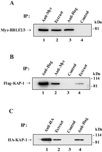
[image:3.585.318.522.277.580.2]FIG. 1. Interactions between BBLF2/3, ZBRK1, and KAP-1. West-ern blots of epitope-tagged proteins immunoprecipitated (IP) with the indicated antibodies from cells cotransfected with (A) Myc-BBLF2/3 and Flag-ZBRK1, (B) Flag-KAP-1 and Myc-BBLF2/3, and (C) HA-KAP-1 and ZBRK1. (A) Myc-BBLF2/3 interacts with Flag-ZBRK1. Lane 1, direct precipitation of Myc-BBLF2/3; lane 2, cell extract; lane 3, coprecipitation of BBLF2/3 with Flag-ZBRK1; lane 4, precipitation with control immunoglobulin G. (B) Flag-KAP-1 inter-acts with Myc-BBLF2/3. Lane 1, direct precipitation of Flag-KAP-1; lane 2, coprecipitation of Flag-KAP-1 with Myc-BBLF2/3; lane 3, pre-cipitation with control immunoglobulin G; lane 4, cell extract. (C) HA-KAP-1 interacts with Flag-ZBRK1. Lane 1, direct precipitation of HA-KAP-1; lane 2, cell extract; lane 3, precipitation with control immunoglobulin G; lane 4, coprecipitation of HA-KAP-1 with Flag-ZBRK1. The amount of extract used in the direct precipitations was 1/20th of that used in the coprecipitations.
TABLE 1. BBLF2/3-interacting proteins
Protein GenBank no.
Polymerase (DNA-directed) beta NM-002690 Polymerase (DNA-directed) delta4 BC-001334 Polymerase (DNA-directed) delta2 BC-000459 DNA-polymerase delta-interacting protein (p38) AF-179891 DEAD/H (Asp-Glu-Ala-Asp/His) box
polypeptide 5 (RNA helicase/DDX5/p68)
NM-004396
3⬘–5⬘TREX2 exonuclease AF-319570
BGLF5 (EBV DNase) NL-001345
Zinc finger protein ZBRK1 NM-021632
Transcriptional corepressor KAP-1/TIF1 BC-004978
on November 8, 2019 by guest
http://jvi.asm.org/
cotransfection, the DNA was prepared with the Wizard SV genomic DNA purification system (Promega). The DNA was cleaved with EcoRI and DpnI or EcoRI and MboI to distinguish between replicated plasmid DNA and input DNA that had not replicated in eukaryotic cells. After the digestion had gone to completion, the DNA was purified and concentrated by phenol-chloroform
ex-traction and ethanol precipitation in 20l of TE (pH 8.0). A 1-kb ApaLI
fragment from the oriLyt plasmid vector (pBluescript II SK[⫹]) was used as the
probe for Southern blot hybridization and autoradiography. Cotransfection-rep-lication assays were performed as described (18). OriLyt (pEF52) contains EBV sequences from BamHI-H (48,852) to BclI (53,770). OriLyt (cytomegalovirus enhancer) has the SmaI fragment of pEF52 replaced with the 860-bp
cytomeg-alovirus immediate-early enhancer. OriLyt (⌬KpnI-BclI) is deleted for the
es-sential replication domain and for the oriLyt enhancer.
Luciferase assays.HeLa cells were plated in six-well plates (Nunc) at 105
cells per well 1 day prior to transfection. Transfections were performed essentially as
described previously (36) with 1g of each plasmid DNA. Vector DNA was used
to keep the total amount of transfected DNA constant. The experiment was performed in triplicate.
RESULTS
BBLF2/3-interacting proteins. Herpesviruses encode a tri-partite helicase-primase complex which comprises a helicase, a primase, and a third associated protein that is necessary for the efficient functioning of the helicase and primase but whose role in viral DNA replication is not otherwise well characterized. In the case of EBV, the third component of the helicase-primase complex is BBLF2/3. To obtain additional information on the possible contribution of BBLF2/3 to EBV DNA replication, a yeast two-hybrid screen was performed to identify BBLF2/3-interacting proteins. The library used in the screen was a com-mercial library derived from an EBV-positive B-cell line. Nine of the most frequently identified positive interactors found in this screen are listed in Table 1. These include known
replica-FIG. 2. ZBRK1 binds to the oriLyt enhancer. (A) Diagram of the region of EBV BamHI-H containing oriLyt. The locations of the oriLyt (BHLF1) promoter and enhancer (69), the AT-rich region (65), and the binding sites for Zta (ZRE1 to ZRE7) (68), Rta (RRE1 and RRE2) (27), and SP1 and ZBP-89 (4, 26, 85) are indicated relative to a potential ZBRK1 binding site (ZBRK1). (B) Electrophoretic mobility shift assay in which GST-ZBRK1znor control GST protein was incubated with32P-labeled probes containing the potential ZBRK1 site in A (lanes 1 to 5), a consensus
ZBRK1 site (lanes 6 to 10), or a mutated ZBRK1 site (lanes 11 to 15). Unlabeled competitor oligonucleotides were added at 100-fold excess. (C) Cotransfection-replication assay showing the reduction in replication efficiency that occurs with substitution of the oriLyt enhancer region by the cytomegalovirus immediate-early enhancer. Southern blot of transfected Vero cell DNA digested with BamHI plus DpnI and probed with vector DNA to detect replicated oriLyt. Lane 1, oriLyt (cytomegalovirus enhancer). Lane 2, oriLyt(⌬KpnI-BclI). Lane 3, oriLyt(pEF52).
on November 8, 2019 by guest
http://jvi.asm.org/
[image:4.585.53.531.65.446.2]tion proteins such as DNA polymerase beta, two subunits of DNA polymerase delta, POLD2 and POLD4, plus the p38 DNA polymerase delta-interacting protein. There are also en-zymes with a DNA repair or replication association such as the 3⬘-5⬘exonuclease TREX2, the EBV-encoded DNase BGLF5, and the RNA helicase DDX5/p68 that is associated with rep-lication of hepatitis C virus genomes (23). The remaining two, the zinc finger transcription factor ZBRK1 and the ZBRK1-associated transcriptional corepressor KAP-1 (20), did not have an obvious connection to DNA replication. We therefore chose to investigate the relevance of the interaction of BBLF2/3 with these two proteins further.
BBLF2/3 interacts with ZBRK1 and KAP-1. Mammalian vectors that expressedepitope tagged BBLF2/3, ZBRK1, and KAP-1 were generated. Immunoprecipitation assays were per-formed on extracts of 293T cells or HeLa cells that had been cotransfected with different combinations of these vectors, and the precipitated proteins were visualized by Western blotting. In extracts from cells cotransfected with Myc-BBLF2/3 and Flag-ZBRK1 (Fig. 1A), Myc-BBLF2/3 was directly precipi-tated with anti-Myc antibody and was also precipiprecipi-tated with anti-Flag antibody indicating coprecipitation with ZBRK1. In cells cotransfected with Myc-BBLF2/3 and Flag-KAP-1 (Fig. 1B), Flag-Flag-KAP-1 was directly precipitated from the cell extract by anti-Flag antibody and was also precipitated with anti-Myc antibody, indicating coprecipitation with Myc-BBLF2/3. ZBRK1 is known to interact with KAP-1 (20), and this interaction was confirmed in cells cotransfected with HA-KAP-1 and Flag-ZBRK1 (Fig. 1C). HA-HA-KAP-1 was directly precipitated from the cell extract by anti-HA antibody and was also present in the anti-Flag precipitate, indicating coprecipi-tation with Flag-ZBRK1. There was no precipicoprecipi-tation of Myc-BBLF2/3, Flag-KAP-1, or HA-KAP-1 by control heterologous antibody (Fig. 1A, lane 4; Fig. 1B, lane 3; Fig. 1C, lane 3).
ZBRK1 binds to EBV oriLyt.ZBRK1 is a DNA binding zinc finger protein that recognizes the sequence GGGxxxCAG xxxTTT (58, 87). Examination of the sequence of EBV oriLyt revealed a motif at position 53581 in the oriLyt enhancer, GGGxxxCATxxxTTT, that differed in only one position from the consensus ZBRK1 binding site (Fig. 2A). Electrophoretic mobility shift assays were performed with bacterially expressed control GST protein and GST expressed as a fusion with the zinc finger DNA binding domain of ZBRK1 (GST-ZBRK1zn). 32P-labeled oligonucleotide probes that contained the
consen-sus ZBRK1 binding site (WT-ZBRK1), a site with six nucleo-tide changes in the core positions (Mut-ZBRK1) or the se-quence of the predicted ZBRK1 site from oriLyt (oriLyt-ZBRK1) were synthesized. In an electrophoretic mobility shift assay analysis (Fig. 2B), ZBRK1 bound to the oriLyt-ZBRK1 probe (Fig. 2B, lane 2) comparably to its binding to the WT-ZBRK1 probe (Fig. 2B, lane 7) and did not bind to the Mut-ZBRK1 probe (Fig. 2B, lane 12). Binding to the oriLyt-ZBRK1 probe was competed away by unlabeled, competitor oriLyt and WT-ZBRK1 oligonucleotides (Fig. 2B, lanes 3 and 4), as was binding to the WT-ZBRK1 probe (Fig. 2B, lanes 8 and 9). The Mut-ZBRK1 competitor oligonucleotide did not affect ZBRK1 binding to either the oriLyt-ZBRK1 or WT-ZBRK1 probe (Fig. 2B, lanes 5 and 10). Control GST protein did not bind to any of the probes (Fig. 2B, lanes 1, 6, and 11). Thus, the oriLyt sequence represents a functional ZBRK1 binding site.
The oriLyt enhancer sequences are not essential for repli-cation, as replacement with the cytomegalovirus immediate-early enhancer allows detectable oriLyt replication (29). How-ever, compared to the replication observed with a wild-type oriLyt plasmid, the cytomegalovirus immediate-early enhancer is an inefficient substitute (Fig. 2C, lane 1 versus lane 3). OriLyt (⌬KpnI-BclI) is deleted for the essential
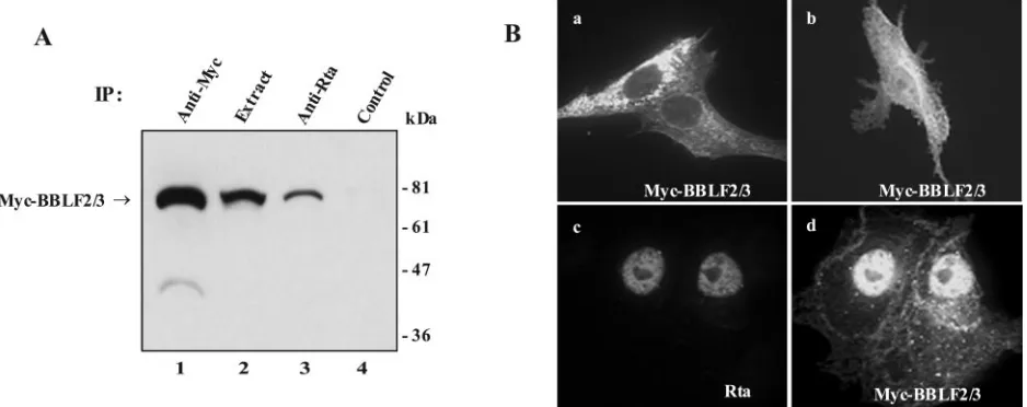
relation-spe-FIG. 3. BBLF2/3 interacts with Rta. (A) Western blot analysis of Myc-BBLF2/3 proteins immunoprecipitated from extracts of 293T cells transfected with expression vectors for Myc-BBLF2/3 and Rta. Lane 1, direct precipitation of Myc-BBLF2/3; lane 2, cell extract; lane 3, coprecipitation of Myc-BBLF2/3 with anti-Rta antibody; lane 4, precipitation with heterologous antibody. The amount of extract used in the direct precipitates was 1/20th of that used in the coprecipitates. (B) Indirect immunofluorescence assay showing the intracellular localization of Myc-BBLF2/3 when transfected into Vero cells alone (a and b) or in the presence of cotransfected Rta (d). Myc-BBLF2/3 was detected with anti-Myc antibody and fluorescein isothiocyanate-conjugated secondary antibody. Rta (c) was detected with anti-Rta polyclonal antiserum and rhodamine-conjugated secondary antibody. Nuclear staining for Myc-BBLF2/3 is increased in the presence of Rta (panel d versus panels a and b).
on November 8, 2019 by guest
http://jvi.asm.org/
[image:5.585.59.528.71.257.2]cific domain and is replication incompetent (Fig. 2C, lane 2). Thus, the ZBRK1 binding site would not be expected to be essential for replication but could contribute to replication efficiency.
BBLF2/3 also interacts with Rta. In transient replication assays in transfected cells, Rta is not essential for replication of an oriLyt containing plasmid but replication by Zta mutated at codons 12 and 13 can be partially rescued by Rta, suggesting that Rta may provide an ancillary replication function (66). The ZBRK1 binding site in oriLyt lies adjacent to two binding sites for the EBV Rta transactivator. The close proximity of these sites, along with the fact that the Zta 12/13 mutation impairs the interaction of Zta with the BBLF2/3-BSLF1 pri-mase subcomplex (22), raised the possibility that BBLF2/3 might make contacts with Rta. Interaction between BBLF2/3 and Rta was detected experimentally with two approaches. Western blot analysis of extracts from 293T cells cotransfected with Myc-BBLF2/3 and Rta showed that Myc-BBLF2/3 was directly precipitated by anti-Myc antibody (Fig. 3A, lane 1) and was also present in the Rta precipitate (lane 3). Myc-BBLF2/3 was not precipitated by control heterologous antibody (lane 4). Intracellular relocalization can also serve as an indication of protein-protein interaction. Complete nuclear localization of the tripartite helicase-primase complex naturally occurs in the presence of all three members of the complex. When expressed in transfected Vero cells in the absence of the other two mem-bers of the complex and visualized by indirect immunofluores-cence, Myc-BBLF2/3 exhibits either a cytoplasmic localization or mixed cytoplasmic plus nuclear localization (Fig. 3B, panels a and b). However, in the presence of the nuclear Rta protein (Fig. 3B, panel c), Myc-BBLF2/3 assumed a very different and predominantly nuclear localization (Fig. 3B, panel d). These results suggest that BBLF2/3 interacts either directly or indi-rectly with Rta.
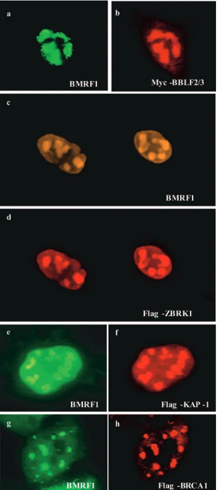
BBLF2/3-interacting proteins are present in replication compartments in induced D98-HR1 cells.BBLF2/3 functions in EBV DNA replication, and the interaction of BBLF2/3 with ZBRK1 and KAP-1 is of interest primarily in the context of the potential contribution of these proteins to oriLyt replication. To determine whether the intracellular localization of ZBRK1 and KAP-1 was compatible with a replication-associated activ-ity, the localization of epitope-tagged ZBRK1 and KAP-1 was examined in D98-HR1 and AGS-BX1 cells that were induced for lytic cycle replication by transfection with Zta. Immunoflu-orescence assays were performed on the transfected cells, and staining with anti-BMRF1 antibody was used to detect endog-enous BMRF1, which served as a marker for viral replication compartments (Fig. 4). In these assays, Myc-BBLF2/3 showed the expected colocalization with BMRF1 in replication com-partments (Fig. 4a and b). Transfected Flag-ZBRK1 (Fig. 4c and d) and transfected Flag-KAP-1(Fig. 4e and f) were also detected in BMRF1 staining replication compartments.
ZBRK1 also interacts with BRCA1 (87), a protein involved in cellular DNA damage responses, double-strand break re-pair, and the bypass of stalled DNA replication forks (60). Flag-BRCA1 was therefore included in these assays. In lytically induced D98-HR1 cells, transfected Flag-BRCA1 was ob-served to colocalize with BMRF1 in replication compartments (Fig. 4g and h). The presence of ZBRK1 and the ZBRK1-interacting proteins KAP-1 and BRCA1 in replication com-partments strengthens the case for the interaction between BBLF2/3 and ZBRK1 having biological relevance.
[image:6.585.55.274.69.559.2]ZBRK1, KAP-1, and BRCA1 are associated with oriLyt DNA. To assess whether ZBRK1 and the ZBRK1 binding
FIG. 4. BBLF2/3, ZBRK1, KAP-1, and BRCA1 are present in viral replication compartments. Immunofluorescence assays performed on D98-HR1 or AGS-Bx1 (c and d) cells transfected with Zta to induce the EBV lytic cycle plus epitope-tagged BBLF2/3 (a and b), ZBRK1 (c and d), KAP-1(e and f) or BRCA1 (g and h). Cells were stained for BMRF1 as a marker for viral replication compartments with BMRF1 antibody and indodicarbocyanine-conjugated secondary body. Epitope-tagged proteins were detected with Flag or anti-Myc antibodies and rhodamine-conjugated secondary antibody.
on November 8, 2019 by guest
http://jvi.asm.org/
proteins KAP-1 and BRCA1 were associated with oriLyt in EBV-infected cells, chromatin immunoprecipitation assays were performed (Fig. 5). D98-HR1 cells were electroporated with Zta to induce EBV lytic replication plus either Flag-ZBRK1 (Fig. 5A), Flag-KAP-1 (Fig. 5B), or Flag-BRCA1 (Fig. 5C). Extracts of the electroporated cells were immunoprecipi-tated with the indicated antibodies, and the presence of asso-ciated oriLyt DNA was assessed with PCR primers that am-plified a 185-bp region (53454 to 53642) from the oriLyt enhancer or a 165-bp fragment (52886 to 53051) from the oriLyt promoter region. Immunoprecipitation of endogenously expressed BMRF1 served as a positive control (Fig. 5A, 5B, and 5C) and immunoprecipitation with anti-Myc antibody served as a negative control (Fig. 5A, 5B, and 5C).
The PCRs detected oriLyt enhancer DNA (182 bp) associ-ated with the anti-Flag immunoprecipitates from the cells transfected with Flag-ZBRK1 (Fig. 5A, lane 2), Flag-KAP-1 (Fig. 5B, lane 2), and Flag-BRCA1 (Fig. 5C, lane 2). BMRF1 binding to the oriLyt promoter DNA fragment was also de-tected (165 bp; Fig. 5A, 5B, and 5C, lanes 1). The anti-Flag immunoprecipitate detected no binding of ZBRK1 or BRCA1 to the oriLyt promoter probe (165 bp; Fig. 5A and 5C, lanes 2). However, KAP-1 binding was observed (165 bp; Fig. 5B, lane 2), suggesting that the KAP-1 corepressor is also associated with other oriLyt binding proteins. The chromatin immuno-precipitation assays showed that ZBRK1 and the ZBRK1-interacting proteins KAP-1 and BRCA1 are associated with EBV oriLyt in induced D98-HR1 cells.
ZBRK1 contributes to oriLyt replication efficiency.To eval-uate whether ZBRK1 had any effect on oriLyt replication, the replication of an oriLyt plasmid was examined in D98-HR1 cells electroporated with ZBRK1 or the ZBRK1-interacting protein KAP-1 or BRCA1 plus Zta and Rta to induce the EBV lytic cycle. In this assay, replicated DNA was identified by exploiting the ability of the restriction enzyme DpnI to dis-criminate between input oriLyt DNA that carries the bacteri-ally imposed methylation pattern and oriLyt DNA that has been replicated in the cell and no longer carries the bacterial methylation markers. Input DNA is cleaved, and DpnI-resis-tant DNA represents the replicated DNA. Aliquots of total cell DNA were subjected to digestion with EcoRI and DpnI. This results in linearization of the replicated 8.8-kb oriLyt plasmid. The DNA was then subjected to Southern blotting with a 1-kb ApaLI fragment of the pBluescript II SK plasmid vector as the probe. The use of a vector probe allowed analysis of the rep-lication of the input oriLyt plasmid without the readout being complicated by replication of the endogenous EBV genomes. Aliquots from the same samples were also digested with EcoRI plus MboI. MboI has the same four-base recognition sequence as DpnI but in contrast to DpnI does not cleave the input methylated DNA. Thus, an identical 8.8-kb linear oriLyt
plas-FIG. 5. ZBRK1, KAP-1, and BRCA1 are associated with oriLyt DNA. Chromatin immunoprecipitation assays performed on extracts of D98-HR1 cells induced for the lytic cycle by electroporation of Zta and also receiving (A) Flag-tagged ZBRK1, (B) Flag-KAP-1, or (C) Flag-BRCA1. Anti-BMRF1 antibody was used to precipitate en-dogenous BMRF1 (lane 1), and anti-Flag antibody was used to pre-cipitate the Flag-tagged ZBRK1, KAP-1, and BRCA1 proteins (lane
2). Precipitates generated with anti-Myc antibody served as negative controls (lane 3). Input DNA in the cell lysate (lane 4) and DNA associated with the immunoprecipitates was amplified by PCR with primers that generated a 185-bp fragment of the oriLyt enhancer or primers that generated a 165-bp fragment of the oriLyt promoter. The PCR products were displayed on an agarose gel and stained with ethidium bromide. Lane 5, DNA size markers.
on November 8, 2019 by guest
http://jvi.asm.org/
[image:7.585.53.272.63.665.2]mid is generated by this cleavage. The EcoRI/MboI fragment provides a measure of the amount of input oriLyt plasmid present in the cell extracts and serves to ensure that the dif-ferent samples were transfected and extracted with equal effi-ciency.
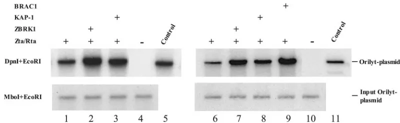
Addition of ZBRK1 increased the efficiency of oriLyt repli-cation, as shown in two independent replication assays (Fig. 6, lane 2 versus lane 1 and lane 7 versus lane 6). Addition of KAP-1 also increased oriLyt replication, although the increase was less than that seen with ZBRK1 (Fig. 6, lane 3 versus lane 1 and lane 8 versus lane 6). Exogenous BRCA1 also had a positive effect on oriLyt replication efficiency (Fig. 6, lane 9 versus lane 6). In the absence of lytic cycle induction by Zta and Rta, input oriLyt plasmid was detected by Mbo/EcoRI cleavage but no DpnI-resistant replicated DNA was observed (Fig. 6, lane 4 and lane 10).
To address whether there was a direct contribution medi-ated by binding of ZBRK1 to oriLyt, a pair of oriLyt plasmids in which the only difference was a deletion of 17 bp across the ZBRK1 binding site were constructed. These plasmids con-tained EBV BamHI-H sequences from the BamHI site at 48850 to the PstI site at 54712. A comparison of the replication of these two oriLyt plasmids in electroporated D98-HR1 cells induced for lytic replication with Zta and Rta revealed that deletion of the ZBRK1 site reduced replication efficiency. As illustrated in two independent assays, oriLyt-(⌬ZBRK1) rep-licated less efficiently than wild-type OriLyt in the absence of added exogenous ZBRK1 (Fig. 7A, lane 3 versus lane 1 and lane 9 versus lane 6) and in the presence of added exogenous ZBRK1 (Fig. 7A, lane 4 versus lane 2 and lane 10 versus lane 7). The oriLyt-(⌬ZBRK1) plasmid still showed some increase in replication efficiency in the presence of added ZBRK1 (Fig. 7A, lane 4 versus lane 3 and lane 10 versus lane 9), suggesting that ZBRK1 also had some effect on replication beyond that mediated by the oriLyt ZBRK1 binding site. In contrast, the mild increase in replication efficiency seen with the addition of KAP-1 (Fig. 7A, lane 8 versus lane 6) was not observed with OriLyt-(⌬ZBRK1) (Fig. 7A, lane 11 versus lane 9), implying that interaction of KAP-1 with ZBRK1 at the oriLyt ZBRK1 site was the dominant source of the KAP-1 stimulation. The
increase observed in the replication of oriLyt with the addition of ZBRK1 and the decreased replication efficiency of oriLyt(⌬ZBRK1) were replication-specific changes. Examina-tion of the expression of luciferase driven by the BHLF1 pro-moter in an oriLyt-luciferase plasmid revealed that the low basal activity of the BHLF1 promoter was not significantly affected by the addition of ZBRK1 or by the deletion of the ZBRK1 site from the oriLyt enhancer region (Fig. 7B).
DISCUSSION
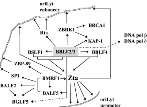
Herpesviruses encode a tripartite helicase-primase complex that has recently received attention as the target of a new class of thiazolylphenyl-containing inhibitors of herpes simplex virus replication (6, 11, 12, 34). The three EBV proteins that com-prise this complex, BBLF4, BSLF1, and BBLF2/3, have been shown to associate in immunofluorescence assays in trans-fected cells (22), and coimmunoprecipitation of baculovirus-expressed proteins provided evidence that each of the three proteins interacts directly with the other two (82). The BBLF4, BSLF1, and BBLF2/3 proteins have also been shown to make contacts with other viral proteins in the replication complex. The tripartite complex interacts with the DNA polymerase BALF5 (21), the primase subcomplex BSLF1-BBLF2/3 inter-acts with Zta, and the helicase BBLF4 also interinter-acts with a separate region of the Zta activation domain (22, 41) (Fig. 8). The interaction between BBLF4 and Zta is necessary to recruit Zta into replication compartments, and a mutant Zta that has lost BBLF4 interaction is excluded from these compartments and is replication defective (41).
[image:8.585.90.490.67.190.2]These interactions also occur in HSV, where the HSV pri-mase-associated protein UL8 has been shown to interact with the HSV UL9 origin binding protein (54) and the HSV DNA polymerase (49) as well as the UL27 single-stranded-DNA-binding protein (28). In HSV-infected cells, the early-stage prereplicative foci contain the HSV helicase-primase proteins (UL5, UL8, and UL52) plus the HSV-encoded single-strand-ed-DNA-binding protein UL29 and the UL9 origin binding protein, and these foci can also be demonstrated in cells trans-fected with expression plasmids for UL29, UL5, UL8, and
FIG. 6. ZBRK1, KAP-1, and BRCA1 increase oriLyt replication efficiency. Southern blots of DNA extracted from two independent experi-ments (lanes 1 to 5 and lanes 6 to 11) in which D98-HR1 cells were electroporated with Zta and Rta to induce lytic replication gene expression plus ZBRK1, KAP-1, or BRCA1. Replicated oriLyt plasmid DNA was identified by digestion with DpnI and EcoRI (upper panels) and input DNA was identified by digestion with MboI and EcoRI (lower panels). DNA was visualized with32P-labeled oriLyt vector DNA as a probe. Cells were
electroporated with: lane 1, oriLyt, Zta, and Rta; lane 2, oriLyt, Zta, Rta, and ZBRK1; lane 3, oriLyt, Zta, Rta, and KAP-1; and lane 4, oriLyt and SG5 vector DNA. Lane 5 contains oriLyt plasmid DNA. Cells were electroporated with the following: lane 6, oriLyt, Zta, and Rta; lane 7, oriLyt, Zta, Rta, and ZBRK1; lane 8, oriLyt, Zta, Rta, and KAP-1; lane 9, oriLyt, Zta, Rta, and BRCA1; lane 10, oriLyt and SG5 vector DNA. Lane 11 contains oriLyt plasmid DNA.
on November 8, 2019 by guest
http://jvi.asm.org/
UL52 (46, 48, 88). The primase is necessary for the recruitment of HSV polymerase to these sites (7), and the interaction between the EBV polymerase and the helicase-primase com-plex suggests that a similar situation may occur during lytic EBV replication.
The primase-associated factor is known to influence the intracellular localization of the helicase-primase complex, to stimulate primase activity (3, 73, 77), and, as described above, to be involved in interactions with other virally encoded rep-lication proteins. In transfected cells, the prereprep-lication foci established by the HSV single-stranded-DNA-binding protein and the helicase-primase proteins contain the cellular recom-bination and repair proteins RPA, RAD51, and NSB1,which are components of the homologous recombination pathway (79). The suggestion was therefore made that these four pro-teins are involved in the recruitment of cellular propro-teins that participate in the HSV DNA replication process. The results of our yeast two-hybrid screen suggest that the EBV primase-associated factor BBLF2/3 may form an interaction platform for a variety of cellular protein partners with replication-re-lated functions.
The interactions observed inSaccharomyces cerevisiaewith cellular DNA polymerase beta and two subunits of DNA poly-merase delta plus a polypoly-merase delta-interacting protein (47)
[image:9.585.44.284.490.666.2]FIG. 7. Deletion of the oriLyt ZBRK1 binding site impairs oriLyt replication but not transcriptional activity. (A) Southern blots showing two independent replication assays (lanes 1 to 5 and lanes 6 to 12) comparing the replication efficiency of the Wt-OriLyt plasmid and OriLyt-⌬ZBRK1. The assays were performed as described for Fig. 6. Cells were electroporated with (lanes 1 and 3) oriLyt, Zta, and Rta and (lanes 2 and 4) oriLyt, Zta, Rta, and ZBRK1. Lane 5 contains oriLyt plasmid DNA. Cells were electroporated with the following: lanes 6 and 9, oriLyt, Zta, and Rta; lanes 7 and 10, oriLyt, Zta, Rta, and ZBRK1; lanes 8 and 11, oriLyt, Zta, Rta, and KAP-1. Lane 12 contains oriLyt plasmid DNA. (B) Luciferase reporter assay performed in HeLa cells transfected with OriLyt-luciferase or OriLyt (⌬ZBRK1)-luciferase reporters (1.0 g), together with ZBRK1 or vector (1.0g) as indicated. Results are the averages of three experiments, with the standard deviation shown.
FIG. 8. Summary of known interactions plus interactions identified in this work between the EBV core replication proteins and between these proteins, the Zta and Rta transactivators, and cellular proteins. The complexity of the protein-protein interactions and the multiple points of contact with oriLyt DNA are illustrated. Interactions identi-fied only inS. cerevisiaeare indicated by dotted lines.
on November 8, 2019 by guest
http://jvi.asm.org/
are speculative, since they were not followed up experimen-tally. However, the interactions are likely to be relevant. DNA polymerase beta is a single-subunit DNA repair protein that functions in base excision repair (31), while DNA polymerase delta is a four-subunit holoenzyme that participates in both leading- and lagging-strand synthesis at the replication fork and interacts with proliferating cell nuclear antigen (32). The TREX2 3⬘to 5⬘exonuclease associates with DNA polymerase delta and is important for the fidelity of polymerase delta (74, 75). The literature on the RNA helicase p68 focuses on a role in dissociation of double-stranded RNA structures and gene expression, but p68 is also capable of unwinding RNA-DNA hybrids (63). The EBV-encoded DNase BGLF5 has previously been shown to interact with the viral DNA polymerase BALF5, the polymerase processivity factor BMRF1, and the single-stranded-DNA-binding protein BALF2 (13, 45, 84) (Fig. 8).
The interactions between BBLF2/3 and ZBRK1 and KAP-1 were validated in coimmunoprecipitation assays, and both cell proteins were detected in EBV replication compartments and associated with oriLyt in chromatin immunoprecipitation anal-yses. ZBRK1 is an eight-zinc finger DNA-binding protein that contains an N-terminal Kruppel-associated box (KRAB) do-main. KRAB domains mediate repression through interaction with the corepressor KAP-1 (70, 71). ZBRK1 and KAP-1 themselves have binding partners of potential relevance to oriLyt replication. ZBRK1 has a C-terminal BRCA1 binding domain (87). BRCA1 was isolated as a susceptibility gene for familial breast cancer and functions in multiple ways to main-tain the integrity of the genome (33, 78). The 3,418-amino-acid BRCA1 protein has a C-terminal motif called a BRCT domain that is present in many DNA repair proteins and an N-terminal ring domain (60). BRCA1 binds to the Rad50-Mre11-Nbs1 complex and can also be isolated in a complex with BRCA2 and Rad51 (55, 90).
BRCA1 stimulates DNA double-strand break repair by ho-mologous recombination and may also have some effect on repair by nonhomologous end joining (55, 89). Homologous recombination is utilized during DNA replication to bypass stalled replication forks (24). We detected BRCA1 in EBV replication compartments present in lytically induced cells and also found BRCA1 to be associated with oriLyt by chromatin immunoprecipitation assay and to increase oriLyt replication efficiency. In addition to its association with a macromolecular histone deacetylase-containing complex (71), KAP-1 binds to heterochromatin protein 1 (39, 64). In Drosophila
melano-gaster, heterochromatin protein 1 was found to bind to the
origin recognition complex (57).
The EBV core replication proteins are linked through a series of contacts with each other and with the viral origin binding protein Zta, which in turn binds to multiple sites within oriLyt. (Fig. 8). However, it seems that additional contacts between the replication complex and oriLyt are important, either to generate structural specificity or to stabilize the com-plex. Contacts between the BMRF1 and BALF5 proteins and the DNA binding transcription factors SP1 and ZBP-89 have been reported previously (4, 26, 85). We have now identified additional tethering interactions between BBLF2/3 and Rta and between BBLF2/3 and ZBRK1. The interaction with ZBRK1 increased oriLyt replication efficiency but did not ap-pear to be obligatory for replication of an oriLyt plasmid. This
is consistent with the location of the ZBRK1 site within the oriLyt enhancer, which is important for replication efficiency but whose sequences are partially substitutable. BBLF2/3 and its HSV homolog UL8 have a recognized role in stimulating helicase-primase activity and in providing contacts with other virally encoded components of the replication complex. The interaction data provided here suggest an expanded contribu-tion of BBLF2/3 to lytic EBV DNA replicacontribu-tion through origin tethering and through recruitment of cellular replication-re-lated proteins.
ACKNOWLEDGMENTS
We thank David Young for BRCA1 plasmid DNA, Lindsey Hutt-Fletcher for AGS-BX1 cells, and Feng Chang for manuscript prepa-ration.
This work was supported by Public Health Service grant R01 CA30356.
REFERENCES
1.Adamson, A. L., and S. Kenney.2001. Epstein-Barr Virus immediate-early protein BZLF1 is SUMO-1 modified and disrupts promyelocytic leukemia
bodies. J. Virol.75:2388–2399.
2.Ahn, J. H., E. J. Brignole, and G. S. Hayward.1998. Disruption of PML subnuclear domains by the acidic IE1 protein of human cytomegalovirus is mediated through interaction with PML and may modulate a RING
finger-dependent cryptic transactivator function of PML. Mol. Cell. Biol.18:4899–
4913.
3.Barnard, E. C., G. Brown, and N. D. Stow.1997. Deletion mutants of the herpes simplex virus type 1 UL8 protein: effect on DNA synthesis and ability to interact with and influence the intracellular localization of the UL5 and
UL52 proteins. Virology237:97–106.
4.Baumann, M., R. Feederle, E. Kremmer, and W. Hammerschmidt.1999. Cellular transcription factors recruit viral replication proteins to activate the
Epstein-Barr virus origin of lytic DNA replication, oriLyt. EMBO J. 18:
6095–6105.
5.Bell, P., P. M. Lieberman, and G. G. Maul.2000. Lytic but not latent replication of Epstein-Barr virus is associated with PML and induces
sequen-tial release of nuclear domain 10 proteins. J. Virol.74:11800–11810.
6.Betz, U. A., R. Fischer, G. Kleymann, M. Hendrix, and H. Rubsamen-Waigmann.2002. Potent in vivo antiviral activity of the herpes simplex virus primase-helicase inhibitor BAY 57–1293. Antimicrob. Agents Chemother.
46:1766–1772.
7.Carrington-Lawrence, S. D., and S. K. Weller.2003. Recruitment of poly-merase to herpes simplex virus type 1 replication foci in cells expressing
mutant primase (UL52) proteins. J. Virol.77:4237–4247.
8.Cayrol, C., and E. K. Flemington.1996. The Epstein-Barr virus bZIP tran-scription factor Zta causes G0/G1 cell cycle arrest through induction of
cyclin-dependent kinase inhibitors. EMBO J.15:2748–2759.
9.Chevallier-Greco, A., H. Gruffat, A. Manet, A. Calender, and A. Sergeant.
1989. The Epstein-Barr virus (EBV) DR enhancer contains two functionally different domains: domain A is constitutive and cell specific, domain B is
transactivated by the EBV early protein R. J. Virol.63:615–623.
10.Countryman, J., and G. Miller. 1985. Activation of expression of latent Epstein-Barr herpesvirus after gene transfer with a small cloned subfragment
of heterogeneous viral DNA. Proc. Natl. Acad. Sci. USA82:4085–4089.
11.Crumpacker, C. S., and P. A. Schaffer.2002. New anti-HSV therapeutics
target the helicase-primase complex. Nat. Med.8:327–328.
12.Crute, J. J., C. A. Grygon, K. D. Hargrave, B. Simoneau, A. M. Faucher, G. Bolger, P. Kibler, M. Liuzzi, and M. G. Cordingley.2002. Herpes simplex virus helicase-primase inhibitors are active in animal models of human
dis-ease. Nat. Med.8:386–391.
13.Daibata, M., and T. Sairenji.1993. Epstein-Barr virus (EBV) replication and expressions of EA-D (BMRF1 gene product), virus-specific
deoxyribonucle-ase, and DNA polymerase in EBV-activated Akata cells. Virology196:900–
904.
14.Deng, Z., C. J. Chen, D. Zerby, H. J. Delecluse, and P. M. Lieberman.2001. Identification of acidic and aromatic residues in the Zta activation domain
essential for Epstein-Barr virus reactivation. J. Virol.75:10334–10347.
15.Everett, R. D., P. Freemont, H. Saitoh, M. Dasso, A. Orr, M. Kathoria, and J. Parkinson.1998. The disruption of ND10 during herpes simplex virus infection correlates with the Vmw110- and proteasome-dependent loss of
several PML isoforms. J. Virol.72:6581–6591.
16.Everett, R. D., and G. G. Maul.1994. HSV-1 IE protein Vmw110 causes
redistribution of PML. EMBO J.13:5062–5069.
17.Feederle, R., M. Kost, M. Baumann, A. Janz, E. Drouet, W. Hammer-schmidt, and H. J. Delecluse.2000. The Epstein-Barr virus lytic program is
on November 8, 2019 by guest
http://jvi.asm.org/
controlled by the co-operative functions of two transactivators. EMBO J.
19:3080–3089.
18.Fixman, E. D., G. S. Hayward, and S. D. Hayward.1995. Replication of
Epstein-Barr virusoriLyt: lack of a dedicated virally encoded origin-binding
protein and dependence on Zta in cotransfection assays. J. Virol.69:2998–
3006.
19.Flemington, E. K.2001. Herpesvirus lytic replication and the cell cycle:
arresting new developments. J. Virol.75:4475–4481.
20.Friedman, J. R., W. J. Fredericks, D. E. Jensen, D. W. Speicher, W.-P. Huang, E. G. Neilson, and F. J. Rauscher III.1996. KAP-1, a novel core-pressor for the highly conserved KRAB repression domain. Genes Dev.
10:2067–2078.
21.Fujii, K., N. Yokoyama, T. Kiyono, K. Kuzushima, M. Homma, Y. Nish-iyama, M. Fujita, and T. Tsurumi.2000. The Epstein-Barr virus pol catalytic subunit physically interacts with the BBLF4-BSLF1-BBLF2/3 complex. J.
Vi-rol.74:2550–2557.
22.Gao, Z., A. Krithivas, J. E. Finan, O. J. Semmes, S. Zhou, Y. Wang, and S. D. Hayward.1998. The EBV lytic transactivator Zta interacts with the
helicase-primase replication complex. J. Virol.72:8559–8567.
23.Goh, P. Y., Y. J. Tan, S. P. Lim, Y. H. Tan, S. G. Lim, F. Fuller-Pace, and W. Hong.2004. Cellular RNA helicase p68 relocalization and interaction with the hepatitis C virus (HCV) NS5B protein and the potential role of p68 in
HCV RNA replication. J. Virol.78:5288–5298.
24.Goodman, M. F.2000. Coping with replication ‘train wrecks’ in Escherichia
coli using Pol V, Pol II and RecA proteins. Trends Biochem. Sci.25:189–195.
25.Gruffat, H., E. Manet, A. Rigolet, and A. Sergeant.1990. The enhancer factor R of Epstein-Barr virus (EBV) is a sequence-specific DNA binding protein.
Nucleic Acids Res.18:6835–6843.
26.Gruffat, H., O. Renner, D. Pich, and W. Hammerschmidt.1995. Cellular proteins bind to the downstream component of the lytic origin of DNA
replication of Epstein-Barr virus. J. Virol.69:1878–1886.
27.Gruffat, H., and A. Sergeant.1994. Characterization of the DNA-binding site repertoire for the Epstein-Barr virus transcription factor R. Nucleic Acids
Res.22:1172–1178.
28.Hamatake, R. K., M. Bifano, W. W. Hurlburt, and D. J. Tenney.1997. A functional interaction of ICP8, the herpes simplex virus single-stranded DNA-binding protein, and the helicase-primase complex that is dependent
on the presence of the UL8 subunit. J. Gen. Virol.78:857–865.
29.Hammerschmidt, W., and B. Sugden.1988. Identification and characteriza-tion of oriLyt, a lytic origin of DNA replicacharacteriza-tion of Epstein-Barr virus. Cell
55:427–433.
30.Hardwick, J. M., P. M. Lieberman, and S. D. Hayward.1988. A new Epstein-Barr virus transactivator, R, induces expression of a cytoplasmic early
anti-gen. J. Virol.62:2274–2284.
31.Hubscher, U., G. Maga, and S. Spadari.2002. Eukaryotic DNA polymerases.
Annu. Rev. Biochem.71:133–163.
32.Hubscher, U., and Y. S. Seo.2001. Replication of the lagging strand: a
concert of at least 23 polypeptides. Mol. Cell12:149–157.
33.Jhanwar-Uniyal, M.2003. BRCA1 in cancer, cell cycle and genomic stability.
Front. Biosci.8:s1107–1117.
34.Kleymann, G., R. Fischer, U. A. Betz, M. Hendrix, W. Bender, U. Schneider, G. Handke, P. Eckenberg, G. Hewlett, V. Pevzner, J. Baumeister, O. Weber, K. Henninger, J. Keldenich, A. Jensen, J. Kolb, U. Bach, A. Popp, J. Maben, I. Frappa, D. Haebich, O. Lockhoff, and H. Rubsamen-Waigmann.2002. New helicase-primase inhibitors as drug candidates for the treatment of
herpes simplex disease. Nat. Med.8:392–398.
35.Korioth, F., G. G. Maul, B. Plachter, T. Stamminger, and J. Frey.1996. The nuclear domain 10 (ND10) is disrupted by the human cytomegalovirus gene
product IE1. Exp. Cell Res.229:155–158.
36.Krithivas, A., D. B. Young, G. Liao, D. Greene, and S. D. Hayward.2000. Human herpesvirus 8 LANA interacts with proteins of the mSin3 corepres-sor complex and negatively regulates Epstein-Barr virus gene expression in
dually infected PEL cells. J. Virol.74:9637–9645.
37.Kudoh, A., T. Daikoku, Y. Sugaya, H. Isomura, M. Fujita, T. Kiyono, Y. Nishiyama, and T. Tsurumi.2004. Inhibition of S-phase cyclin-dependent kinase activity blocks expression of Epstein-Barr virus immediate-early and
early genes, preventing viral lytic replication. J. Virol.78:104–115.
38.Kudoh, A., M. Fujita, T. Kiyono, K. Kuzushima, Y. Sugaya, S. Izuta, Y. Nishiyama, and T. Tsurumi.2003. Reactivation of lytic replication from B cells latently infected with Epstein-Barr virus occurs with high S-phase cy-clin-dependent kinase activity while inhibiting cellular DNA replication.
J. Virol.77:851–861.
39.Lechner, M. S., G. E. Begg, D. W. Speicher, and F. J. Rauscher, 3rd.2000. Molecular determinants for targeting heterochromatin protein 1-mediated gene silencing: direct chromoshadow domain-KAP-1 corepressor interaction
is essential. Mol. Cell. Biol.20:6449–6465.
40.Lee, H. R., D. J. Kim, J. M. Lee, C. Y. Choi, B. Y. Ahn, G. S. Hayward, and J. H. Ahn.2004. Ability of the human cytomegalovirus IE1 protein to mod-ulate sumoylation of PML correlates with its functional activities in tran-scriptional regulation and infectivity in cultured fibroblast cells. J. Virol.
78:6527–6542.
41.Liao, G., F. Y. Wu, and S. D. Hayward.2001. Interaction with the
Epstein-Barr virus helicase targets Zta to DNA replication compartments. J. Virol.
75:8792–8802.
42.Lieberman, P. M., and A. J. Berk.1990. In vitro transcriptional activation, dimerization, and DNA-binding specificity of the Epstein-Barr virus Zta
protein. J. Virol.64:2560–2568.
43.Lieberman, P. M., J. M. Hardwick, and S. D. Hayward.1989. Responsive-ness of the Epstein-Barr virus NotI repeat promoter to the Z transactivator is mediated in a cell-type-specific manner by two independent signal regions.
J. Virol.63:3040–3050.
44.Lieberman, P. M., J. M. Hardwick, J. Sample, G. S. Hayward, and S. D. Hayward.1990. The Zta transactivator involved in induction of lytic cycle gene expression in Epstein-Barr virus-infected lymphocytes binds to both AP-1 and ZRE sites in target promoter and enhancer regions. J. Virol.
64:1143–1155.
45.Lin, S. F., T. Y. Hsu, M. Y. Liu, L. S. Lin, H. L. Yang, J. Y. Chen, and C. S. Yang.1995. Characterization of Epstein-Barr virus DNase and its interaction
with the major DNA binding protein. Virology208:712–722.
46.Liptak, L. M., S. L. Uprichard, and D. M. Knipe.1996. Functional order of assembly of herpes simplex virus DNA replication proteins into
prereplica-tive site structures. J. Virol.70:1759–1767.
47.Liu, L., E. M. Rodriguez-Belmonte, N. Mazloum, B. Xie, and M. Y. Lee.
2003. Identification of a novel protein, PDIP38, that interacts with the p50 subunit of DNA polymerase delta and proliferating cell nuclear antigen.
J. Biol. Chem.278:10041–10047.
48.Lukonis, C. J., and S. K. Weller.1996. Characterization of nuclear structures in cells infected with herpes simplex virus type 1 in the absence of viral DNA
replication. J. Virol.70:1751–1758.
49.Marsden, H. S., G. W. McLean, E. C. Barnard, G. J. Francis, K. MacEach-ran, M. Murphy, G. McVey, A. Cross, A. P. Abbotts, and N. D. Stow.1997. The catalytic subunit of the DNA polymerase of herpes simplex virus type 1 interacts specifically with the C terminus of the UL8 component of the viral
helicase-primase complex. J. Virol.71:6390–6397.
50.Maul, G. G., D. Negorev, P. Bell, and A. M. Ishov.2000. Review: properties and assembly mechanisms of ND10, PML bodies, or PODs. J. Struct. Biol.
129:278–287.
51.Mauser, A., E. Holley-Guthrie, D. Simpson, W. Kaufmann, and S. Kenney.
2002. The Epstein-Barr virus immediate-early protein BZLF1 induces both
a G2and a mitotic block. J. Virol.76:10030–10037.
52.Mauser, A., E. Holley-Guthrie, A. Zanation, W. Yarborough, W. Kaufmann, A. Klingelhutz, W. T. Seaman, and S. Kenney.2002. The Epstein-Barr virus immediate-early protein BZLF1 induces expression of E2F-1 and other proteins involved in cell cycle progression in primary keratinocytes and
gastric carcinoma cells. J. Virol.76:12543–12552.
53.Mauser, A., S. Saito, E. Appella, C. W. Anderson, W. T. Seaman, and S. Kenney.2002. The Epstein-Barr virus immediate-early protein BZLF1
reg-ulates p53 function through multiple mechanisms. J. Virol.76:12503–12512.
54.McLean, G. W., A. P. Abbotts, M. E. Parry, H. S. Marsden, and N. D. Stow.
1994. The herpes simplex virus type 1 origin-binding protein interacts
spe-cifically with the viral UL8 protein. J. Gen. Virol.75:2699–2706.
55.Moynahan, M. E., J. W. Chiu, B. H. Koller, and M. Jasin.1999. BRCA1
controls homology-directed DNA repair. Mol. Cell4:511–518.
56.Muller, S., and A. Dejean.1999. Viral immediate-early proteins abrogate the modification by SUMO-1 of PML and Sp100 proteins, correlating with
nuclear body disruption. J. Virol.73:5137–5143.
57.Pak, D. T., M. Pflumm, I. Chesnokov, D. W. Huang, R. Kellum, J. Marr, P. Romanowski, and M. R. Botchan.1997. Association of the origin recognition
complex with heterochromatin and HP1 in higher eukaryotes. Cell91:311–
323.
58.Peng, H., L. Zheng, W. H. Lee, J. J. Rux, and F. J. Rauscher, 3rd.2002. A common DNA-binding site for SZF1 and the BRCA1-associated zinc finger
protein, ZBRK1. Cancer Res.62:3773–3781.
59.Portes-Sentis, S., A. Sergeant, and H. Gruffat.1997. A particular DNA structure is required for the function of a cis-acting component of the
Epstein-Barr virus OriLyt origin of replication. Nucleic Acids Res.25:1347–
1354.
60.Powell, S. N., and L. A. Kachnic.2003. Roles of BRCA1 and BRCA2 in homologous recombination, DNA replication fidelity and the cellular
re-sponse to ionizing radiation. Oncogene22:5784–5791.
61.Ragoczy, T., L. Heston, and G. Miller.1998. The Epstein-Barr virus Rta protein activates lytic cycle genes and can disrupt latency in B lymphocytes.
J. Virol.72:7978–7984.
62.Rodriguez, A., E. J. Jung, Q. Yin, C. Cayrol, and E. K. Flemington.2001. Role of c-myc regulation in Zta-mediated induction of the cyclin-dependent
kinase inhibitors p21 and p27 and cell growth arrest. Virology284:159–169.
63.Rossler, O. G., A. Straka, and H. Stahl.2001. Rearrangement of structured RNA via branch migration structures catalysed by the highly related
DEAD-box proteins p68 and p72. Nucleic Acids Res.29:2088–2096.
64.Ryan, R. F., D. C. Schultz, K. Ayyanathan, P. B. Singh, J. R. Friedman, W. J. Fredericks, and F. J. Rauscher 3rd.1999. KAP-1 corepressor protein inter-acts and colocalizes with heterochromatic and euchromatic HP1 proteins: a potential role for Kruppel-associated box-zinc finger proteins in
heterochro-matin-mediated gene silencing. Mol. Cell. Biol.19:4366–4378.
on November 8, 2019 by guest
http://jvi.asm.org/
65.Ryon, J. J., E. D. Fixman, C. Houchens, J. Zong, P. M. Lieberman, Y.-N. Chang, G. S. Hayward, and S. D. Hayward.1993. The lytic origin of herpes-virus papio is highly homologous to Epstein-Barr herpes-virus ori-Lyt: evolutionary conservation of transcriptional activation and replication signals. J. Virol.
67:4006–4016.
66.Sarisky, R. T., Z. Gao, P. M. Lieberman, E. D. Fixman, G. S. Hayward, and S. D. Hayward.1996. A replication function associated with the activation
domain of the Epstein-Barr virus Zta transactivator. J. Virol.70:8340–8347.
67.Sarisky, R. T., and G. S. Hayward.1996. Evidence that the UL84 gene product of human cytomegalovirus is essential for promoting oriLyt-depen-dent DNA replication and formation of replication compartments in
cotrans-fection assays. J. Virol.70:7398–7413.
68.Schepers, A., D. Pich, and W. Hammerschmidt.1993. A transcription factor with homology to the AP-1 family links RNA transcription and DNA
repli-cation in the lytic cycle of Epstein-Barr virus. EMBO J.12:3921–3929.
69.Schepers, A., D. Pich, J. Mankertz, and W. Hammerschmidt.1993.cis-acting elements in the lytic origin of DNA replication of Epstein-Barr virus. J.
Vi-rol.67:4237–4245.
70.Schultz, D. C., K. Ayyanathan, D. Negorev, G. G. Maul, and F. J. Rauscher,
3rd.2002. SETDB1: a novel KAP-1-associated histone H3, lysine 9-specific
methyltransferase that contributes to HP1-mediated silencing of
euchro-matic genes by KRAB zinc-finger proteins. Genes Dev.16:919–932.
71.Schultz, D. C., J. R. Friedman, and F. J. Rauscher, 3rd.2001. Targeting histone deacetylase complexes via KRAB-zinc finger proteins: the PHD and bromodomains of KAP-1 form a cooperative unit that recruits a novel
isoform of the Mi-2alpha subunit of NuRD. Genes Dev.15:428–443.
72.Semmes, O. J., L. Chen, R. T. Sarisky, Z. Gao, L. Zhong, and S. D. Hayward.
1998. Mta has properties of an RNA export protein and increases
cytoplas-mic accumulation of EBV replication gene mRNA. J. Virol.72:9526–9534.
73.Sherman, G., J. Gottlieb, and M. D. Challberg.1992. The UL8 subunit of the herpes simplex virus helicase-primase complex is required for efficient
primer utilization. J. Virol.66:4884–4892.
74.Shevelev, I. V., and U. Hubscher.2002. The 3⬘5⬘exonucleases. Nat. Rev.
Mol. Cell. Biol.3:364–376.
75.Shevelev, I. V., K. Ramadan, and U. Hubscher.2002. The TREX2 3⬘–⬎5⬘
exonuclease physically interacts with DNA polymerase delta and increases its
accuracy. Sci. World J.2:275–281.
76.Takada, K., N. Shimizu, S. Sakuma, and Y. Ono.1986.trans-Activation of the latent Epstein-Barr virus (EBV) genome after transfection of the EBV
DNA fragment. J. Virol.57:1016–1022.
77.Tenney, D. J., W. W. Hurlburt, P. A. Micheletti, M. Bifano, and R. K. Hamatake.1994. The UL8 component of the herpes simplex virus helicase-primase complex stimulates primer synthesis by a subassembly of the UL5
and UL52 components. J. Biol. Chem.269:5030–5035.
78.Venkitaraman, A. R.2002. Cancer susceptibility and the functions of BRCA1
and BRCA2. Cell108:171–182.
79.Wilkinson, D. E., and S. K. Weller.2004. Recruitment of cellular
recombi-nation and repair proteins to sites of herpes simplex virus type 1 DNA replication is dependent on the composition of viral proteins within prerep-licative sites and correlates with the induction of the DNA damage response.
J. Virol.78:4783–4796.
80.Wu, F. Y., J. H. Ahn, D. J. Alcendor, W. J. Jang, J. Xiao, S. D. Hayward, and G. S. Hayward.2001. Origin-independent assembly of Kaposi’s Sarcoma-Associated Herpesvirus DNA replication compartments in transient cotrans-fection assays and association with the ORF-K8 protein and cellular PML.
J. Virol.75:1487–1506.
81.Wu, F. Y., H. Chen, S. E. Wang, C. M. ApRhys, G. Liao, M. Fujimuro, C. J. Farrell, J. Huang, S. D. Hayward, and G. S. Hayward. 2003. CCAAT/ enhancer binding protein alpha interacts with ZTA and mediates ZTA-induced p21(CIP-1) accumulation and G(1) cell cycle arrest during the
Epstein-Barr virus lytic cycle. J. Virol.77:1481–1500.
82.Yokoyama, N., K. Fujii, M. Hirata, K. Tamai, T. Kiyono, K. Kuzushima, Y. Nishiyama, M. Fujita, and T. Tsurumi.1999. Assembly of the Epstein-Barr virus BBLF4, BSLF1 and BBLF2/3 proteins and their interactive properties.
J. Gen. Virol.80:2879–2887.
83.Zalani, S., E. Holley-Guthrie, and S. Kenney.1996. Epstein-Barr viral la-tency is disrupted by the immediate-early BRLF1 protein through a
cell-specific mechanism. Proc. Natl. Acad. Sci. USA93:9194–9199.
84.Zeng, Y., J. Middeldorp, J. J. Madjar, and T. Ooka.1997. A major DNA binding protein encoded by BALF2 open reading frame of Epstein-Barr virus (EBV) forms a complex with other EBV DNA-binding proteins:
DNAase, EA-D, and DNA polymerase. Virology239:285–295.
85.Zhang, Q., E. Holley-Guthrie, J. Q. Ge, D. Dorsky, and S. Kenney.1997. The Epstein-Barr virus (EBV) DNA polymerase accessory protein, BMRF1, activates the essential downstream component of the EBV oriLyt. Virology
230:22–34.
86.Zhang, Q., Y. Hong, D. Dorsky, E. Holley-Guthrie, S. Zalani, N. A. Elshiekh, A. Kiehl, T. Le, and S. Kenney.1996. Functional and physical interactions between the Epstein-Barr virus (EBV) proteins BZLF1 and BMRF1: effects
on EBV transcription and lytic replication. J. Virol.70:5131–5142.
87.Zheng, L., H. Pan, S. Li, A. Flesken-Nikitin, P. L. Chen, T. G. Boyer, and W. H. Lee.2000. Sequence-specific transcriptional corepressor function for
BRCA1 through a novel zinc finger protein, ZBRK1. Mol. Cell6:757–768.
88.Zhong, L., and G. S. Hayward.1997. Assembly of complete, functionally active herpes simplex virus DNA replication compartments and recruitment of associated viral and cellular proteins in transient cotransfection assays.
J. Virol.71:3146–3160.
89.Zhong, Q., C. F. Chen, P. L. Chen, and W. H. Lee.2002. BRCA1 facilitates microhomology-mediated end joining of DNA double strand breaks. J. Biol.
Chem.277:28641–28647.
90.Zhong, Q., C. F. Chen, S. Li, Y. Chen, C. C. Wang, J. Xiao, P. L. Chen, Z. D. Sharp, and W. H. Lee.1999. Association of BRCA1 with the
hRad50-hMre11-p95 complex and the DNA damage response. Science285:747–750.