METALLOPROTEINASE INHIBITORS ON THE
MICROTENSILE BOND STRENGTH OF SELF ETCH
ADHESIVE – AN IN VITRO STUDY
Dissertation submitted to
THE TAMILNADU Dr. M.G.R. MEDICAL UNIVERSITY In partial fulfilment for the Degree of
MASTER OF DENTAL SURGERY
BRANCH IV
This is to certify that this dissertation titled “Effectiveness of different matrix metalloproteinase inhibitors on the microtensile bond strength of self etch
adhesive – An In Vitro Study” is a bonafide record of work done by Dr.SHALINI. V under my guidance and to my satisfaction during her postgraduate
study period, 2015 – 2018. This dissertation is submitted to THE TAMILNADU Dr. M.G.R. MEDICAL UNIVERSITY, in partial fulfilment for the award of the degree of Master of Dental Surgery in Conservative Dentistry and Endodontics, Branch IV. It has not been submitted (partially or fully) for the award of any other degree or diploma.
________________________ ________________________ Dr. Minu Koshy, MDS Dr. Subha Anirudhan, MDS
Guide, Professor, Co-Guide, Professor
Department of Conservative Dentistry and Department of Conservative Dentistry and
Endodontics, Endodontics,
Sri Ramakrishna Dental College Sri Ramakrishna Dental College and Hospital, Coimbatore. and Hospital, Coimbatore.
_______________________ Dr. V. Prabhakar, MDS,
Principal, Professor and HOD,
Department of Conservative Dentistry and Endodontics, Sri Ramakrishna Dental College and Hospital,
Coimbatore
Date :
NAME OF THE CANDIDATE Dr. Shalini V
TITLE OF DISSERTATION Effectiveness of Different Matrix
Metalloproteinase Inhibitors on the Bond Strength of Self Etch Adhesive – an in Vitro Study
PLACE OF STUDY Sri Ramakrishna Dental College and Hospital
DURATION OF THE COURSE 2015-2018
NAME OF GUIDE Dr. Minu Koshy
HEAD OF THE DEPARTMENT Dr.V.Prabhakar
I hereby declare that no part of the dissertation will be utilized for gaining financial assistance for research or other promotions without obtaining prior permission from the Principal, Sri Ramakrishna Dental College and Hospital. In addition, I declare that no part of this work will be published either in print or in electronics without permission from the guide who has been actively involved in the dissertation. The author solely has the rights for publishing the work with prior permission from the Principal, Sri Ramakrishna Dental College and Hospital, Coimbatore.
Urkund Analysis Result
Analysed Document: ALL CHAPTERS.pdf (D34305621) Submitted: 1/3/2018 12:15:00 PM
Submitted By: shaliniviswanathan3@gmail.com Significance: 1 %
Sources included in the report:
md pdf.pdf (D34132051)
Instances where selected sources appear:
3
CERTIFICATE II
This is to certify that this dissertation work titled “EFFECTIVENESS OF DIFFERENT MATRIX METALLOPROTEINASE INHIBITORS ON THE BOND STRENGTH OF SELF ETCH ADHESIVE – AN IN VITRO STUDY” of the candidate Dr.Shalini V with registration number 241517302 for the award of Masters of Dental Surgery in the branch of Conservative Dentistry and Endodontics. I personally verified the urkund.com website for the purpose of plagiarism check. I found that the uploaded thesis file contains from introduction to conclusion pages and result shows 1 percentage of plagiarism in the dissertation.
This thesis is the result of work done with immense support from many people and it is with immense pleasure that I express my heartfelt gratitude to all of them.
I devote my heartfelt thanks to Dr. V. Prabhakar,MDS, Principal & Head of Department, whose discipline and skills that run deep under his authoritative yet natural care during my post graduate period which enabled me to successfully conclude my thesis.
I would like to thank and acknowledge Dr.Minu Koshy, MDS,Professor, my Guide who has always been a source of support and encouragement at any moment, in and out of the department. I am grateful to Dr. Subha Anirudhan, MDS, Professor, for her innovative ideas, constructive suggestions, valuable criticism and constant encouragement.
I take this opportunity to express my sincere gratitude to Dr. Ambalavanan, MDS, Reader, Dr. Sriman Narayanan, MDS, Reader, Dr.Gayathri V, MDS, Senior Lecturer and Dr.Remya Verghese, MDS, Senior Lecturer for their valuable guidance that enabled me to comprehend this dissertation and supported me at every juncture throughout my postgraduate curriculum.
lending a helping hand with the sectioning of tooth samples .
I am thankful to Dr. Sadhana.K, for her guidance in the statistical works of this study.
I am thankful to my teachers Dr.Sudhakar M.D.S and Dr.Prabhu M.D.S, my seniors, my colleagues and my juniors, who have been together as friends and of great support throughout my period of study here. I am thankful to all other department staff members, my fellow colleagues in other departments, all UG staff members and non-clinical staffs of my department for their great support and encouragement.
I express my dearest gratitude to my mother, my husband and my son, and the special people in my life who contributed in various ways towards my study and this dissertation.
Last but not the least, I am greatly indebted to God the Almighty, for blessing me with all the good things in my life and guiding me throughout.
DR. SHALINI .V
TITLE
PAGE NO.
1
Introduction
1
2
Aim and Objective
4
3
Review of Literature
5
4
Materials and Methods
18
5
Results
32
6
Discussion
43
7
Summary and Conclusion
51
INTRODUCTION
The success of restorations hangs on the adhesion stability between composite
resin and tooth structure. To maintain adhesion stability, the presence of a hybrid
layer is essential so that it forms a resistant structure against bacterial invasion.
Currently, simplified systems such as self-etching primer/adhesive systems have
demonstrated good clinical performance to dentin. This approach is less
technique-sensitive and reduces the time required for the bonding procedure clinically.
Although advances in adhesive dentistry have brought about improvements in
bonding systems and techniques, bond failures at the tooth-restoration interface still
remain a challenge in the dental field 1.
Proper adhesion depends on the proper infiltration of resin monomer or
partial/ complete degradation of dental substrate to allow formation of hybrid layer.
Hybrid layer has a mixture of organic matrix and hydroxyapatite crystal residue of
dentin with resin monomers and solvent adhesives 2. This layer is subject to
hydrolytic or enzymatic degradation. It was observed that hydrolytic degradation of
exposed collagen fibres occurs without the presence of bacterial colonization.
Research on dentin non collagenous proteins has demonstrated that dentin collagen
fibrils contain inactive proforms of proteolytic enzymes called the Matrix
metalloproteinases. Areas not infiltrated by adhesives, expose collagen fibrils that are
slowly degraded by MMP’s. The recent literature revealed that the matrix
metalloproteinase (MMPs) are abundant in the dentin layer after the etching process
Matrix metalloproteinases get activated in an acidic environment , either due
to acid etching or acidic monomers and resin monomers , that leads to increased
digestion of collagen within the hybrid layer , thus decreasing the bond strength 4.
Different strategies have aimed to improve the bond durability by applying
enzyme inhibitors as a pretreatment before resin infiltration or by admixing enzyme
inhibitors to primers. The common MMP inhibitors used are chlorhexidine,
tetracycline, ammonium compounds, green tea polyphenol epigallo-catechin 3 gallate
and chelating agents like ethylene diamine acetic acid etc.
Grape seed extract , a naturally occurring cross linking agent ,is composed of
proanthocyanidin. It has been used to induce exogenous cross links of collagen and
has the property to inhibit MMP’s 5.
Green tea extract has been described as a natural inhibitor of MMPs. It is
made from Camellia sinensis and is composed of polyphenols named catechins, such
as epicatechin (EC), epigallocatechin (EGC), epicatechin gallate (ECG) and
epigallocatechin gallate (EGCG).It also has antioxidant property 6.
Fluoride (F) has produced a favourable balance in tooth demineralisation and
remineralization and its role in preventing dental caries has been well documented.
More recently, it was found that 1.23% Sodium fluoride (NaF) gel significantly
reduced the demineralised organic matrix degradation. In addition to its established
effect on the demineralisation and remineralization cycle, Fluoride could also inhibit
However, there are only few studies that have evaluated the efficacy of these
MMP inhibitors to prevent dentin bond strength loss over time 6,8,9. Thus, the aim of
the present study was to evaluate the microtensile bond strength (microTBS) of a
self-etching adhesive (Clearfil SE Bond), with pretreatment of different MMP inhibitors
(Grape seed extract , green tea extract and Clearfil SE Protect ) over 7 days , 30 days
AIM AND OBJECTIVE
To evaluate the effectiveness of different Matrix metalloproteinase inhibitors
(Green tea extract , Grape seed extract and Sodium Fluoride containing adhesive ) on
the microtensile bond strength over 90 days using self etch adhesives – an in vitro
REVIEW OF LITERATURE
Ebrahimi-Chaharom et al (2017)10, evaluated the effect of different
concentrations of specific inhibitor of MMPs (galardin) on the shear bond strength of
self-adhesive resin cements to dentin. The samples were divided into 3 groups (with
no galardin, galardin at a high concentration and galardin at a low concentration),
resin blocks were bonded to dentin surface ,stored in water for 24 hrs and shear bond
strength values were determined.Pretreatment with galardin increased the shear bond
strength of self-adhesive resin cements to dentin and this increase had a direct
relationship with the concentration of galardin in the solution.
Gajjela et al (2017)11 , studied the influence of 2 % chlorhexidine (CHX),
6.5% grape seed extract (GSE) and 1 % riboflavin/ chitosan modification on
microtensile bond strength (µTBS) of composite resin to dentin after polymerase
chain reaction (PCR) thermocycling. Composite build ups were constructed over the
specimens and PCR thermocycling (5000 cycles) was performed and tested under
universal testing machine. It was concluded that pretreatment with CHX and GSE
leads to a significant increase in µTBS of composite resin to dentin.
Gerhardt et al (2016)9 , evaluated the effect of dentin pretreatment with
matrix metalloproteinase inhibitors ( 2% green tea extract,2 % chlorhexidine ,2%
epigallocatechin 3-gallate(EGCG ) on long term microtensile bond strength of self
etch adhesive to dentin. Microtensile bond strength testing were carried out
immediately and after 6 months of storage in water.It was concluded that bond
strength increased in dentin pretreatment with EGCG , green tea groups and in non
Correa B et al (2016)12, evaluated the effect of matrix metalloproteinase
(MMP) inhibitors - 2% (CHX) and sodium fluoride (NaF) (5000 ppm) - on
microtensile bond strength (μTBS) of composite resin to Er:YAG laser-irradiated
dentin after chemical degradation of the bond interface using mechanical testing
machine. Fracture pattern was analyzed using SEM.It was found that 2% CHX and
NaF 5000 ppm presented similar μTBS of composite resin to laser-irradiated dentin
before and after chemical degradation.
Carlvaho et al (2016) 6 , evaluated the effect of pretreatment of two matrix
metalloproteinase inhibitors (2 % green tea extract and 2 % chlorhexidine) on the
bonding durability of etch and rinse adhesive to caries affected dentin over 24 hours
and 6 months storage in water.Dentin beams were prepared and subjected to
microtensile bond strength testing.It was concluded that the application of 2% green
tea extract was able to increase bond durability of the etch-and-rinse system to dentin.
Moreira da Silva et al (2015)13, evaluated the degree of conversion ,water
sorption, solubility and bond strength of experimental etch and rinse adhesive
containing Galardin, Batimastat , GM-1489 and cholrhexidine diacetate. Degree of
conversion was measured using FT-IR spectroscopy, water sorption and solubility
calculated based on ISO4049 and tensile bond strength tested using universal testing
machine over 24 hrs,6 months and 12 months storage. They concluded that
experimental adhesive with GM- 1489 inhibitor and chlorhexidine diacetate had better
physiochemical properties and good bond stability over time.
Kim park et al (2015) 14, tested the ability of green tea catechins ( methanol
extract of green tea) and commercially available epigallocatechin 3-gallate (EGCG) to
gelatine zymography. It was concluded that GTX and EGCG completely inhibited
the MMP-9 acitivity and EGCG significantly inhibited the neutrophil release.
Zheng et al (2014)8 , analyzed the effect of different matrix metalloproteinase
(MMP) inhibitors (2% chlorhexidine digluconate [CHX], 0.05% green tea extract, 1
mM ferrous sulfate, or 0.2 mM galardin) on the microtensile bond strength
(microTBS) of an etch-and-rinse (Optibond FL) and a selfetching adhesive (Clearfill
SE bond) immediately and after 9 months of aging. It was concluded that MMP
inhibitors prevented the decrease in microTBS upon aging of the etch-and-rinse but
not of the self-etching adhesive.
Rupam kaur et al (2014)15, determined the effect of pretreatment with grape
seed extract on the bond strength of a self-etch adhesive (All Bond SE ) with dentin.
Occulsal cavities were prepared and restored with hybrid composite.Dentin beams
were prepared and microtensile bond strength were tested using universal testing
machine. It was concluded that pretreatment with grape seed extract leads to
significant increase in the bond strength of composite with dentin.
Srinivasalu et al(2013)5,evaluated the shear bond strength of composite resin
to deep dentin, bonded using a self-etch adhesive, after treatment with two collagen
cross-linkers (10% sodium ascorbate and 6.5% proanthocyanidin) at varying time
intervals. Human incisors were sectioned longitudinally , pretreatment were done for
5 and 10 minutes and shear bond strength were tested using universal testing machine.
It was concluded that dentin surface pre-treatment with both 10% sodium ascorbate
and 6.5% proanthocyanidin resulted in significant improvement in bond strength of
Yi Liu et al (2013)16, evaluated the effects of Proanthocyanidin PAand the
photoinitiator type on the degree of conversion and polymerisation rate of a model
dental adhesive.Three types of photoinitiators were introduced into the Bis-
GMA/HEMA comonomer mixture and for each resin mixture , adhesive containing
0%, 2.5%, 5% and 10% PA with respect to weight of resin were produced. When
light cured , the rate of polymerisation and degree of conversion were analysed using
ATR- FTIR Spectroscopy. It was observed that PA hampered the polymerisation of
all adhesives regardless of the photoinitiators used and also found that photoinitiation
formulations could maintain a satisfactory degree of conversion while a significant
amount of PA is incorporated.
Xijin Du et al (2012)17, studied the antibacterial potential and physiochemical
properties of a dental adhesive incorporated with epigallocatechin-3-gallate(EGCG) in
different concentrations over time. Growth of streptococcus mutans were determined
by direct contact test. Microtensile bond strength and degree of conversion were
evaluated immediately and after 6 months. It was concluded that 200μg/ml EGCG
incorporated adhesive showed better therapeutic value while sustaining the bond
durability of resin- dentin
Epasinghae et al (2012) 18 , studied the effect of varying concentration of
proanthocyanidin incorporation into the dental adhesive resin on resin dentin bond
strength. Experimental hydrophilic adhesives were prepared by combining 50%
weight comonomer with 50% weight ethanol along with 1%, 2 % ,3%
proanthocyanidin concentration respectively with control group free of it. Bonding
procedures were carried out and subjected to tensile forces. They concluded that
effect on bond strength, whereas 3% proanthocyanidin significantly lowered bond
strength.
Fang et al (2012)19, investigated whether transient pretreatment by PA based
preconditioner can improve the resin dentin bonds of various etch and rinse adhesives.
PA based preconditoners were prepared by adding powdered grape seed extract , to
various solvents at different concentrations. Bond strength , failure mode and degree
of conversion were determined via microtensile testing, field emission scanning
electron microscopyand Fourier transform infrared spectroscopy.PA preconditioning
improved the bond strength of tested water / ethanol based adhesive better than the
water/ acetone based system.So it was observed that PA preconditioning improved
resin dentin bonds without compromise on the curing behaviour on the tested
adhesives.
Kato et al (2012)20 , evaluated the impact of protease inhibitors on dentin
matrix degradation by colleganase. Specimens were treated for 1 min with gels
containing epigallocatechin 3-gallate (EGCG), chlorhexidine (CHX, 0.012%), FeSO4
(1 mM), NaF (1.23%), or no active compound (P, placebo) and matrix loss was
analysed using profilometry.It was concluded that treatment with gels containing
EGCG, CHX, or FeSO4 led to significantly lower dentin matrix loss when compared
with the other treatments.
Dourado Loguercia et al (2011)21 , evaluated the effect of the method of
application on the immediate and 3 year resin dentin bond strength for 3 one step self
etch adhesives ( Aper Prompt L- Pop, Clearfil S3 bond and Xeno III. Adhesives were
applied under Inactive application (adhesive layer after application on tooth were left
Dentin sticks were tested for bond strength immediately and after 3 years of water
storage. Nanoleakage was detected using silver nitrate deposition. They observed that
active application improved the immediate bonding strength and showed decreased
degradation rate in active application.
Liu et al(2011)22, analysed the effect of transient proanthocyanidins
preconditioning on the cross-linking and mechanical properties of demineralized
dentin by adding powdered proanthocyanidins-rich grape seed extract to various
solvents at different concentrations. Demineralized dentin specimens were
preconditioned for 20,30, 60 or120 s,followed by the evaluation of their crosslinking
degree, mechanical properties and micromorphology using Ninhydrin assay,
Universal testing machine and field emission scanning electron microscope
(FESEM). It was found that the cross-linking degree of the demineralized dentin
collagen exhibited concentration- and time- dependent increase after preconditioning
treatment, irrespective of the preconditioner and the solvent. 15% proanthocyanidins
preconditioning resulted in thehighestmean ultimate tensile strength and FESEM
confirmed that the demineralized dentin collagen was in a homogeneous and regular
arrangement after preconditioning and maintained expanding.
Castellan et al (2011)23 ,studied the long term stability of dentin matrix
following preptreatment using natural collagen cross linking agent rich in
Proanthocyanidin(PA) from grape seed , cocoa seeds , cranberry, cinnamon and acai
berry. Apparent elastic modulus were analysed over 3 , 6 and 12 months using three
point bend flexural testing. Grape seed extract and cocoa seed extract were able to
De Munck et al(2011)24, evaluated the bonding effectiveness using five
different adhesives to different dentin locations after 1 week, 3, 6 and 12 months of
water storage. The beams were divided into ‘center’ and ‘periphery’ dentin
specimens and were subjected to microtensile bond strength testing. It was determined
that only for the mild self-etch adhesives, µTBS to ‘periphery’ dentin was higher than
for the ‘center’ specimens , whereas for etch and rinse adhesives it was insensitive to
regional variability.
Mutluay et al (2011)25, compared the MMP-inhibitory activity of a series of
alcohols (i.e. methanol, ethanol, propanols, butanols, pentanols, hexanols, the ethanol
ester of methacrylic acid, heptanols and octanol). It was measured against soluble
rhMMP-9 and insoluble matrix-bound endogenous MMPs of dentin in completely
demineralized dentin. The results indicated that alcohols with 4 methylene groups
inhibit MMPs more effectively than methanol or ethanol.
Green et al (2010)26, compared the morphological differences of hybrid layers
created by Bis GMA/HEMA model adhesives with and without the addition of grape
seed extract PA under the conditions of enzymatic collagen degradation. Following
collagenase treatment with 0.1% collagenase solution for 0,1,and 6 days , the
specimens were analysed with SEM/TEM. It was concluded that the presence of
grape seed extract PA in dental adhesives may inhibit the biodegradation of
unprotected collagen fibrils within the hybrid layer.
De Munck et al (2010)27 , evaluated the endogenous enzymatic degradation
for several simplified self-etch adhesives in which primers were modified by adding
two MMP inhibitors: chlorhexidine, a non specific MMP inhibitor and SB-3CT, a
etch-and-rinse adhesives, the involvement of endogenous MMP-2 and MMP-9 in the
bond-degradation process is minimal for mild self-etch adhesives.
Castellan et al (2010)28 , studied the properties of dentin matrix treated with 2
proanthocyanidin rich cross-linking agents (Grape seed extract and Cocoa seed
extract) and their effect on dentin bonded interface. Results showed that both cross
linkers increased elastic modulus of dimeneralised dentin as exposure time increased
and resin dentin bonds also significantly increased following treatment with grape
seed extract regardless of the application time or adhesive used.
Breschi et al (2010)29 , determined the use of a specific MMP inhibitor
(Galardin) for preservation of hybrid layer and its mechanical properties after aging.
Microtensile bond strength and interfacial nanoleakage were analysed. MMP
inhibition were analysed zymographically. Galardin showed inhibition of MMP-2 and
MMP-9. It was also observed that the use of galardin had no effect on immediate
bond strength , while it significantly decreased bond strength after 1 year.
Tezvergil-Mutluay et al (2010)60 , evaluated the effect of different storage
media on changes in matrix stiffness, loss of dry weight or solubilization of collagen
from demineralized dentin beams incubated in vitro for up to 60 days.Dentin beams
were prepared and stored in either media containing both zinc and calcium designated
as complete medium (CM), calcium-free medium, zinc-free medium, a doubled-zinc
medium or water. Quantitation of hydroxyproline (HOP) was taken as an index of
solubilization of collagen by MMPs and measured. It was found that both the storage
medium and the storage time showed significant effects on E, mass loss and HOP
release. The incubation in CM resulted in relatively rapid and significant decrease in
Al Ammar et al (2009)30, investigated the effect of three different
cross-linking agents (5% Glutaraldehyde [GD], 6.5% Grape seed extract [GSE], and 0.5%
Genipin [GE]) on the tensile bond strength (TBS) of resin-dentin bonds. Dentin
surfaces were etched using phosphoric acid and then teeth were pretreated with
collagen cross linkers before adhesive application and restored. After 24 hours,
specimens were tested using universal testing machine. This study concluded that
Chemical modification to the dentin matrix promoted by GD and GSE, but not GE,
resulted in increased bond strength.
Qian xie et al (2008)31, evaluated the effect of 6.5% grape seed extract and
1000 ppm sodium fluoride on the remineralisation of artificially induced root caries
lesions. Fragments were pH cycled through treatment solutions , acidic buffer and
neautral buffer for 8 days at 6 cycles per day. Microhardness and mineral
precipitation were tested using microhardness tester, polarized light microscopy and
confocal laser scanning microscopy. It was concluded that grape seed extract
positively affects the remineralisation/ demineralisation processes of artificial root
caries.
Walter et al (2008) 32, evaluated the effects of dentin collagen modifications
induced by various cross-linkers (5% glutaraldehyde (GA), 0.5% proanthocyanidin
(PA), 0.625% genipin (GE))on the stability of collagen matrix and the inhibition of
root caries. Cross-linker-treated demineralized human root dentin were digested with
bacterial collagenase, centrifuged, and the supernatants were subjected to amino acid
analysis to determine collagen content. The residues were analyzed by SDS-PAGE
and hydroxyproline analysis. Bovine root surfaces were conditioned with phosphoric
acid, treated with the cross-linkers, incubated with Streptococcus mutans and
with confocal microscopy. It was observed that naturally occurring cross-linkers,
especially PA, could be used to modify root dentin collagen to efficiently stabilize
collagen and to increase its resistance against caries.
Carilho et al (2007) 33, analysed the dentin bond preservation using 2%
chlorhexidine pretreatment . Microtensile bond strengths and failure mode distribution
under SEM were analyzed immediately after specimen preparation and 6 months
later.It was observed that significantly better preservation of bond strength was
observed after 6 months and suggested that chlorhexidine might be useful for the
preservation of dentin bond strength.
Bedran Russo et al (2007)34, evaluated the effect of two collagen crosslinking
agents (glutaraldehyde, GD and grape seed extract, GSE) on the modulus of elasticity
of demineralized dentin. Demineralised dentin specimens were immersed in 2.5%
GD; 5% GD, 25% GD; 0.65% GSE; 6.5% GSE and tested at baseline, 10 min, 30
min, 1 h, 2 h, 4 h for modulus of elasticity by three point bend method. It was
concluded that the use of these collagen crosslinkers to increase the stiffness of
demineralized dentin, was both concentration and time dependent.
Shinohara et al (2006) 1, evaluated the influence of a fluoride-containing
adhesive (Clearfill SE Protect bond) on microtensile bond strength to dentin, as well
as analyzed the dentin-adhesive interface after acid-base challenge. Microtensile bond
strength was tested using Universal testing machine.For interfacial analysis, adhesive
system was applied on dentin surface and a low-viscosity resin was placed between
two dentin disks. Then, the specimens were subjected to acid-base challenge,
sectioned, and polished to be observed by SEM. It was observed that a thick acid-base
fluoride-releasing adhesive was used and the presence of fluoride in an adhesive contributed
significantly to prevent secondary caries and did not interfere with dentin-adhesive
bond strength.
Bedran Russo et al (2006)35, evaluated the effect of three collagen
crosslinking agents[5% glutaraldehyde (GD), 0.5% proanthocyanidin PBS solution
(PA), and 0.625% genipin PBS solution (GE)] on the ultimate tensile strength (UTS)
of undemineralized and demineralized dentin. Specimens were prepared and kept in
their respective solutions for either 4 or 40 hour. It was concluded that the application
of two naturally occurring crosslinkers (Proanthocyanidin and Geipin), to dentin
collagen significantly improved the ultimate tensile bond strength.
Nishitani et al (2006)36, compared the ability of all in one adhesives (Clearfil
Tri-S Bond, G-Bond, Adper Prompt L-Pop) or a self etching primer (Clearfil SE Bond
primer ) to activate gelatinolytic and collagenolytic activities in powdered mineralized
dentin made from human tooth. Powered dentin was mixed with adhesive or primer
for varying times and then the reaction was stopped by extracting the adhesives using
acetone. Gelatinolytic activity were recorded using relative fluorescent units (RFU)
per mg dry weight. It was concluded that self-etch adhesives may activate latent
MMP and increase the activity to near-maximum levels and contribute to the
degradation of resin–dentin bonds over time.
Rochaa et al (2006)37, evaluated the effects of dentin surface treatments on
the tensile bond strength (TBS) of the self-etching primer Clearfil SE Bond (CSE) and
the one-step self-etching One-Up Bond F (OUB). The exposed flat dentin surfaces
were prepared with diamond bur at high-speed, carbide bur at low-speed or wet
water for 24 hours, specimens were sectioned and tensile bond strength were tested. It
was observed that the groups treated with CSE exhibited the highest bond strength
for all surface treatments. Dentin surfaces prepared with carbide bur at low speed
reduced tensile bond strength in the CSE group; however, OUB was not affected by
surface treatments.
Mazzoni et al (2006)38, studied the proteolytic activities of mineralised dentin,
acid etched dentin and etch and rinse adhesive treated acid etched dentine. Powdered
dentin prepared from human teeth were treated with 17% EDTA, 10% phosphoric
acid or with five simplified etch and rinse adhesives that were applied to 10%
phosphoric acid etched dentine. Gelatinolytic activity were assayed using fluorescein
labelled gelatine. It was concluded that simplified etch and rinse adhesives can
reactivate endogenous enzymatic activites in dentin that are previously inactivated by
phosphoric acid etching.
Imazato et al(2003)39, investigated the antibacterial activity of an adhesive
resin incorporating the antibacterial monomer 12-methacryloyloxydodecylpyridinium
bromide (MDPB) as well as its bonding characteristics in terms of bond strength into
dentin and curing ability. 2.5% MDPB was incorporated into the bonding agent and
its inhibitory effect against the growth of Streptococcus mutans on its surface was
determined. Bond strength to human dentin and degree of conversion of the
experimental adhesive were evaluated by conventional tensile bond strength test and
Fourier transformation infrared spectroscopy.The results indicated that an adhesive
resin with antibacterial activity after curing could be produced by incorporation of
Demule M et al (2000)49, investigated the effects of different biologically
active components from natural products, including green tea polyphenols (GTP),
resveratrol, genistein and organosulfur compounds from garlic, on matrix
metalloproteinase (MMP)-2, MMP-9 and MMP-12 activities. The inhibition of the
enzymes were measured by Fluorescence assays using gelatin or elastin substrates.
The inhibition were further confirmed using gelatin zymography. The results
indicated that catechins from green tea inhibit MMP activities and proMMP-2
MATERIALS
1. CLEARFIL SE BOND (KURARAY AMERICA, INC)
2. GRAPE SEED EXTRACT ( ZENITH PHARMACEUTICALS)
3. GREEN TEA EXTRACT ( ZENITH PHARMACEUTICALS)
4. CLEARFIL SE PROTECT BOND (KURARAY AMERICA, INC)
5. MICROHYBRID COMPOSITE (VOCO POLOFILL)
6. EXTRACTED HUMAN MOALR TEETH
7. DISTILLED WATER
ARMAMENTARIUM
1. DIAMOND DISC
2. DIAMOND ABRASIVE BUR
3. TWEEZER
4. APPLICATOR TIPS
5. ANODIZED ALUMINIUM COMPOSITE INSTRUMENT
6. POLYWAVE LED CURING LIGHT (IVOCLAR VIVADENT, USA)
7. HARD TISSUE MICROTOME ( LEICA SP 1600)
8. UNIVERSAL TESTING MACHINE (INSTRON)
MATERIALS
[image:31.595.322.483.124.308.2] [image:31.595.112.278.125.312.2]
Fig 1 - CLEARFIL SE BOND Fig 2- GREEN TEA EXTRACT
[image:31.595.122.282.381.584.2] [image:31.595.327.513.397.583.2]
ARMANENTARIUM
Fig 5 - INSTRUMENTS USED Fig 6 - LIGHT CURE UNIT
[image:32.595.170.463.387.582.2]
Fig 7 - HARD TISSUE MICROTOME
Fig 8 - UNIVERSAL TESTING MACHINE
[image:33.595.252.380.410.620.2]
METHODOLOGY
1. Preparation of natural human tooth samples
- Selection and preservation of tooth samples
- Grouping of samples
2. Preparation of dentin surface
3. Application of MMP inhibitor / Bonding agent
4. Composite build up
5. Preparation for microtensile bond strength evaluation
6. Statistical evaluation
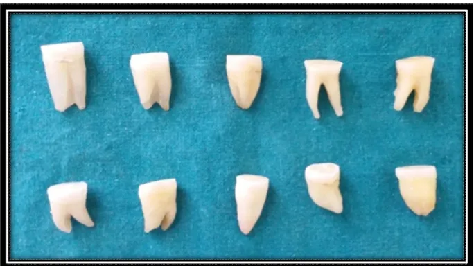
PREPARATION OF NATURAL HUMAN TOOTH SAMPLES:
SELECTION AND PRESERVATION OF TOOTH SAMPLES
Sound molar teeth (figure 10) were obtained and used for current study. Teeth
with caries , attrition and fracture were excluded. The selected teeth were then
disinfected and stored in physiologic saline for no longer than 4 weeks after
extraction.
GROUPING
The selected teeth were randomly divided into 4 groups. (10 in each group)
according to the material used.
GROUP 1 – Clearfil SE bond
GROUP 2 – Green tea extract
GROUP 3 – Grape seed extract
PREPARATION OF DENTIN SURFACE:
The occlusal enamel of the teeth was removed. A flat mid coronal dentin
surface (figure 12) were prepared by means of a water cooled slow speed diamond
saw(Figure 11). It was then made as an even surface using diamond abrasive bur.
Dentin surface was then checked for the absence of pulp chamber exposure.
APPLICATION
GROUP 1 :
Clearfil SE bond, a 2 step self etch adhesive was used (figure1). Equal drops
of primer and adhesive were added equally and mixed together. It was applied over
the dentin surface using microbrush and light cured for 20 seconds as per
manufacturer’s instructions.(figure- 14,15)
GROUP 2 :
0.5% Green tea extract were prepared by mixing 50 g of green tea extract
powder (ZENITH PHARMACEUTICALS) in 100 ml of distilled water (Figure -2).
The solution was rubbed over the dentin surface for 60 seconds using microbrush and
allowed to dry for one minute .Over the pretreated surface ,a layer of clearfil SE
bond were applied and cured for 20 seconds.(figure – 13,14,15)
GROUP 3 :
6.5% grape seed extract was prepared by mixing 650 mg of grape seed extract
powder (ZENITH PHARMACEUTICALS) in 100 ml of distilled water(Figure 3).
allowed to dry for one minute. Over the pretreated surface, a layer of clearfill SE bond
was applied and cured for 20 seconds.(figure -13,14,15)
GROUP 4 :
Clearfil SE Protect bond , a 2 step self ecth adhesive was used (Figure 4).
Primer and adhesive of equal proportion were mixed and was applied over the dentin
surface using a microbrush. It was then cured for 20 seconds.(Figure – 14,15)
COMPOSITE BUILD UP :
Over the bonded dentin surface, 3 increments (2 mm each) of composite
(Voco Polofil) build up were done using a aluminium coated instrument (Figure 16).
Each increment was cured for 20 seconds. Final cure of the build up was done for 40
seconds according to manufacturer’s instructions.
PREPARATION FOR MICROTENSILE BOND STRENGTH EVALUATION:
Specimen preparation :
The roots of the teeth were sectioned off and the coronal portions were
mounted on acrylic resin blocks (Figure 17). These acrylic blocks were mounted on a
hard tissue microtome (Leica SP 1600 Saw Microtome) and sectioned using water jet
as coolant (Figure 18). The teeth were sectioned longitudinally across the bonded
interface to obtain dentin sticks with a cross sectional area of approximately 1.0 ±
0.1mm2. Three or four resin dentin sticks were acquired from each tooth sample
(Figure 19). 10 sticks per group were stored in separate containers with water as the
Each bonded stick was then attached to a custom made jig with cyanoacrylate
resin (Figure 20 ) for microtensile bond strength testing and subjected to a tensile
force in a Universal Testing machine (Instron,Norwood,USA) at a cross head speed of
0.5mm/min (Figure 21). The load at which the failure occurred was recorded by
specialised software attached to the universal testing machine. The test was done on
the 7th day ,30th day and 90th day.
Thus values were obtained and tabulated. Then were subjected to statistical
evaluation using SPSS software version 21.
STATISTICAL EVALUATION:
The SPSS software version 21 was used for statistical analysis of the
microtensile bond strength values obtained for each sample in the study groups.
The values tabulated were tested for significance using one-way ANOVA and
FIGURE 10 – Sound molar teeth
[image:38.595.187.448.440.669.2]FIGURE 12- Flat mid coronal dentin surface prepared
Figure 13- Pretreatnent for Group Figure 14- Application of Bonding
2& Group 3 For 60 seconds agents in all four groups
[image:39.595.111.506.442.605.2]
Figure 15 – Curing done for 40 seconds
[image:40.595.210.431.123.351.2]
[image:41.595.170.465.410.624.2]
Figure 17- coronal portion embedded in acrylic resin block
Figure 19 – Dentin Resin Beams
Figure 20 – Dentin beams on custom made jigs
[image:42.595.217.415.397.660.2]RESULTS
The microtensile bond strength values of 10 samples for each group were
obtained and calculated in MegaPascals(MPa). Values were then subjected to
statistical analysis. The mean and standard deviation values obtained were subjected
to One way ANOVA test and Post Hoc test.
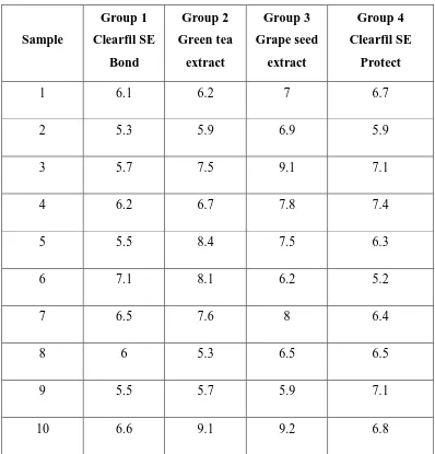
[image:45.595.101.499.334.749.2]7 th Day
Table – 1 Bond strength values in Mega Pascals
Sample Group 1 Clearfil SE Bond Group 2 Green tea extract Group 3 Grape seed extract Group 4 Clearfil SE Protect
1 6.1 6.2 7 6.7
2 5.3 5.9 6.9 5.9
3 5.7 7.5 9.1 7.1
4 6.2 6.7 7.8 7.4
5 5.5 8.4 7.5 6.3
6 7.1 8.1 6.2 5.2
7 6.5 7.6 8 6.4
8 6 5.3 6.5 6.5
9 5.5 5.7 5.9 7.1
Table – 2 Mean and standard deviation values
N Minimum Maximum Mean
Std.
Deviation
Group 1 10 5.300 7.100 6.050 .577
Group 2 10 5.300 9.100 7.050 1.271
Group 3 10 5.900 9.200 7.410 1.133
Group 4 10 5.200 7.400 6.540 .644
Table – 3 Oneway ANOVA test
Sum of Squares
Df Mean
Square
F Sig.
Between Groups
10.591 3 3.530 3.860 .017
Within Groups 32.923 36 .915
Total 43.514 39
Graph - 1 Bardiagram representing mean values of 4 groups on 7th day
M
P
[image:46.595.124.492.564.761.2]Post Hoc Tests
[image:47.595.106.546.188.621.2]Multiple Comparisons
Table – 4 Tukey HSD
(I) GROUP (J) GROUP Mean Difference (I-J) Std. Error Sig.
95% Confidence Interval
Lower
Bound
Upper
Bound
1
2 -1.000 .427 .108 -2.151 .151
3 -1.360* .427 .015 -2.511 -.208 4 -.490 .427 .664 -1.641 .661
2
1 1.000 .427 .108 -.151 2.151
3 -.360 .427 .834 -1.511 .791
4 .510 .427 .635 -.641 1.661
3
1 1.360* .427 .015 .2081 2.511 2 .360 .427 .834 -.791 1.511
4 .870 .427 .195 -.281 2.021
4
1 .490 .427 .664 -.661 1.641
2 -.510 .427 .635 -1.661 .641
3 -.870 .427 .195 -2.021 .281
The mean difference is significant at the 0.05 level.
When comparing the Group 4 (Clearfil SE Protect) with other groups, there
was no statistical difference between the mean microtensile bond strength values.
When comparing the Group 3 (Grape seed extract) with Group 1 (Clearfil SE ), there
30th day
Table – 5 - Bond strength values in Mega Pascals
Sample Group 1 Group 2 Group 3 Group 4
1 8.2 12.1 15.1 11.2
2 7.9 10.1 13.4 9.3
3 10.5 15.3 18.5 14.4
4 9.1 12.1 11.1 16.3
5 7.9 16.2 16.2 12.1
6 11.3 13.4 13.5 11.2
7 10 9.2 17.9 13.1
8 8.5 8.9 13.2 17.1
9 7.9 12 14.1 15.2
10 9.3 14.3 18.0 11.9
Table – 6 Mean and standard deviation values
N Minimum Maximum Mean
Std.
Deviation
Group 1 10 7.900 11.300 9.060 1.207
Group 2 10 8.900 16.200 12.360 2.484
Group 3 10 11.100 18.900 15.190 2.604
[image:48.595.107.525.493.701.2]Table – 7 One way ANOVA test
Sum of Squares
Df Mean
Square
F Sig.
Between
Groups
195.407 3 65.136 12.601 .000
Within Groups 186.093 36 5.169
Total 381.500 39
Graph – 2 Bar diagram representing mean values of 4 groups on 30th day
M
P
Multiple Comparisons
Table – 8 Tukey HSD
(I) Group (J) Group Mean Difference (I-J) Std. Error Sig.
95% Confidence Interval
Lower Bound
Upper Bound
1
2 -3.300* 1.016 .013 -6.038 -.561 3 -6.130* 1.016 .000 -8.868 -3.391 4 -4.1200* 1.016 .001 -6.858 -1.381
2
1 3.300* 1.016 .013 .561 6.038 3 -2.830 1.016 .041 -5.568 -.091 4 -.820 1.016 .851 -3.558 1.918
3
1 6.130* 1.016 .000 3.391 8.868 2 2.830* 1.016 .041 .091 5.568 4 2.0100 1.016 .216 -.728 4.748
4
1 4.120* 1.016 .001 1.381 6.858 2 .820 1.016 .851 -1.918 3.558
3 -2.010 1.016 .216 -4.748 .728
* The mean difference is significant at the 0.05 level.
On 30th day, Mean microtensile bond strength values were higher in Grape seed
extract (15.19) when comparing with Clearfil SE (9.06), Green Tea extract (12.36)
and Clearfill SE Protect (13.18)
When comparing Clearfil SE group with other three groups , there was a significant
difference.
When comparing Clearfil SE Protect group with Green tea extract and Grape seed
90 th day
Table – 9 Bond strength values in Mega Pascals
Sample Group 1 Group 2 Group 3 Group 4
1 7.1 11.2 14.2 11.3
2 6.9 10.2 12.8 9
3 9.1 14.5 16.3 13.4
4 8.5 11.8 11 13.3
5 7.5 15.1 15.8 11.8
6 10.5 13.1 13.2 10.6
7 9 8.9 16.4 12.5
8 7.9 8.7 10.1 16.4
9 7.6 11.6 11.1 15.3
10 8.6 12.5 16.4 11.2
Table – 10 Bond strength values in Mega Pascals
N Minimum
Maximu
m
Mean
Std.
Deviation
Group 1 10 6.900 10.500 8.270 1.094
Group 2 10 8.700 15.100 11.760 2.142
Group 3 10 10.100 16.400 13.730 2.450
[image:51.595.101.520.479.649.2]Table – 11 One way ANOVA Test
Sum of Squares
Df Mean
Square
F Sig.
Between Groups 164.194 3 54.731 13.123 .000
Within Groups 150.142 36 4.171
Total 314.336 39
Graph – 3 Bar diagram representing mean values of 4 groups on 90th day
M
P
Multiple comparison
[image:53.595.107.533.146.699.2]Post Hoc analysis
Table- 12 Tukey HSD
(I) group (J) group Mean Difference (I-J) Std.
Error Sig.
95% Confidence Interval
Lower
Bound
Upper
Bound
1
2 -3.490* .913 .003 -5.949 -1.030
3 -5.460* .913 .000 -7.919 -3.000
4 -4.210* .913 .000 -6.669 -1.750
2
1 3.490* .913 .003 1.030 5.949
3 -1.970 .913 .155 -4.429 .489
4 -.720 .913 .859 -3.179 1.739
3
1 5.460* .913 .000 3.000 7.919
2 1.970 .913 .155 -.489 4.429
4 1.250 .913 .527 -1.209 3.709
4
1 4.210* .913 .000 1.750 6.669
2 .720 .913 .859 -1.739 3.179
3 -1.250 .913 .527 -3.709 1.209
On 90th day , the mean microtensile bond strength values were higher in Grape
seed extract group(13.73) comparing to Clearfil SE group(8.27), Green tea extract
group(11.76) and Clearfil SE Protect group(12.48)
When comparing the Clearfil SE group with Green tea extract, Grape seed
extract and Clearfil SE Protect group , there was a significant difference. When
comparing the Clearfil SE Protect group with Green tea extract and Grape seed
Graph – 4 Bond strength values in 3 different time periods.
0 2 4 6 8 10 12 14 16
group 1 group 2 group 3 group 4
7 th day
30 th day
90 th day
From the above line diagram comparing the 7th day, 30th day and 90th day
mean microtensile bond strength values of all four groups, there was a significant
increase in bond strength after 30 and 90 days comparing to 7 days. Among the
groups, Grape seed extract had shown increase in bond strength followed by Clearfil
DISCUSSION
Adhesive restorations are routinely used to replace the lost dental tissue.
Current adhesive systems bond to dentin through a micromechanical mechanism
based on the formation of a hybrid layer. The hybrid layer, a collagen-resin interface,
is the most vulnerable portion of the bonded interfaces where stress tends to
concentrate and most failures take place40. While bonding to enamel substrate has
been shown to be reliable over-time, bonding to dentin substrate is a great challenge41.
Dentin represents the bulk of the tooth and a reliable long-term bond is essential for
the success of adhesive restorations. It has been speculated that a decreasing
concentration gradient of resin monomer diffusion within the acid-etched dentin, and
a subsequent resin elution from hydrolytically unstable polymeric hydrogels within
the hybrid layers leave the collagen fibrils unprotected and vulnerable to degradation
by endogenous metalloproteinases (MMPs) 20.
MMPs are a group of 23 mammalian enzymes capable of degrading all
extracellular matrix components. Human dentin contains collagenase (MMP-8),
gelatinases MMP-2 and -9, and enamelysin MMP-2042. MMPs were shown to be
expressed during tooth development and to be necessary for normal dentin formation .
After dentin mineralization, they remain trapped in the calcified matrix either under
active or proenzyme forms, which may explain their persistent presence within the
dentin of adult teeth 43. It was found that their exposure and activation during the
caries process would allow these enzymes to promote matrix degradation and caries
progression. Dentin collagenolytic and gelatinolytic activities can be suppressed by
protease inhibitors indicating that MMP inhibition could be beneficial in the
Two main methods to increase the dentin/resin interface properties have to be
considered: the continuing improvement/development of new adhesive systems and
the establishment of tissue engineering/biomimetics approaches to improve the
intrinsic properties of the substrate. Intrinsic collagen cross-links provide the tensile
properties of collagen molecules. The use of extrinsic collagen cross-linking agents
can induce additional formation of inter and intramolecular cross-links44. Selective
cross-linking agents have been demonstrated to increase the ultimate tensile strength
and elastic modulus of demineralized dentin45. This can be achieved by the use of
various collagen cross-linkers, both synthetic and natural, on the dentin substrate prior
to the bonding procedure. Naturally occurring collagen cross-linkers such as sodium
ascorbate, epigallectonin and proanthocyanidin have been reported to increase the
collagen cross-linking in sound and caries-affected dentin46. Several synthetic
collagen cross-linkers (Glutaraldehyde, Carbodiimide, formaldehyde and epoxy
resins) have been investigated to improve on the mechanical properties of dentin and
the resin-dentin interface. The major drawback with these are they are toxic to the
tissues.
The two main strategies that are currently in use for adhesive bonding to
enamel and dentin are the total-etch technique and the self-etch technique. Although
etch-and-rinse technique is still considered as the most effective approach to achieve
efficient and stable bonding to enamel and dentin, the multiple application steps, the
critical rinsing step and the frustratingly high incidence of post-operative sensitivity
led to the evolution of more simplified, less technique-sensitive and user-friendly
self-etch adhesives47 .
The importance of fluoride (F) in preventing dental caries by favorably
its ability to inhibit matrix metalloproteinases (MMPs), has been investigated
recently and found to be effective 7. Thus a fluoride containing self etch
adhesive was studied on the bond strength against different naturally occuring MMP
inhibitors for a period of 7 days, 30 days and 90 days, if effective this could minimize
the clinical step of pretreating with mmp inhibitors as they are incorporated in the
adhesive.
Proanthocyanidins (PA) are naturally occurring bioflavonoids found in high
concentrations in grape seed, pine bark, cranberries, lemon tree bark and hazel nut
tree leaves. PA from grape seed extract has been shown to effectively cross-link
collagen in few vitro studies28,46. Their effect on the bond strength of resin composite
bonded using a self-etch adhesive to deep dentin has proved to be effective. It
interacts with proteins to induce cross-links by four different mechanisms: covalent
interaction, ionic interaction, hydrogen bonding interaction, or hydrophobic
interactions 48.
Green tea, has been described as a natural inhibitor of MMPs. It is made from
Camellia sinensis and is composed of polyphenols named catechins, such as
epicatechin (EC), epigallocatechin (EGC), epicatechin gallate (ECG) and
epigallocatechin gallate (EGCG)49. EGCG ,a major polyphenol of green tea has
known to contain potential health ingredients including antioxidant , antimicrobial,
antidiabetic, anti inflammatory and cancer preventing properties. Significant
antimicrobial activity in vitro has been demonstrated against a variety of gram
positive , gram negative and fungal pathogens. It was also found to have distinct
A two-step self-etching primer/adhesive system, Clearfil Protect Bond-
composed of an antibacterial primer containing MDPB
12-methacryloyloxydodecylpyridinium bromide) and a fluoride-releasing adhesive
- has shown the potential in artificial secondary caries inhibition around
restorations51. NaF, the most common F compound in oral hygiene products,has
the ability to completely inhibit the activity of MMPs in clinically relevant
concentrations The possible mechanism by which NaF inhibits the MMPs is not
known clearly 7.
Bond strength is referred to the force per unit area that is required to debond
the adhesive or adherent interface. To measure the ultimate tensile strength and
modulus of elasticity of dentin . Schreiner et al (1994) 52 introduced microtensile
bond strength testing to dentistry. According to Pashley et al (1999) 53, the
microtensile bond strength test presented several advantages in comparison to the
macro and shear testing as it permitted a greater number of adhesive failures and
the measurement of regional bond strengths. Of all the in- vitro tests, microtensile
bond strength testing is deemed better despite its technical limitations, as the
bonded interface of smaller cross sectional area of specimens has a better stress
distribution during loading 54. Considering these factors microtensile bond
strength testing methodology was selected for the current study. The dimension of
the specimen also plays an important role in the determination of the bond
strength. In this study , stick shaped dentin beam specimens were produced and it
has a more favourable stress distribution as investigated by Phrukkanom et al
(1998)58 regarding specimen geometry. The specimen’s dimensions were
In this study , microtensile bond strength values on 7 th day showed a
significant difference between grape seed extract- 0.015 Mpa (group 3 ) and
Clearfil SE bond (group 1 ) . There was no statistical difference between the
group 4 (Clearfil SE Protect) and other groups. On 30th day the mean bond
strength were greater in grape seed extract followed by Clearfill SE Protect and
Green Tea extract. Group 4 (clearfill SE Protect) showed a statistical similar
difference in comparison with Grape seed extract and Green tea extract group. On
the 90th day testing, Group 3 (Grape seed extract) mean bond strength values were
still higher. But the test Group 4 had no statistical difference with group 2 and
group 3 . On 90th day aging the control group seemed to have lower bond strength
values.
A study done by Kaur et al (2014)15 showed that microtensile bond strength
of grape seed extract group were higher and gave a significant difference
comparing to control group without grape seed extract pretreatment using self etch
adhesives. Bond strength of 1 hr treated Grape seed extract 6.5 % showed bond
strength values that were statistically higher than glutaraldehyde treated and
control group27. In a study by Castellan et al(2010) 28, comparing the effect of
Grape seed extract and Cocca seed extract for 10 minute pretreatment , only
Grape seed extract pretreatment showed higher bond strength values than control
group. On the contrary in a study done by Epasinghae et al (2012) 18, there was
significant decrease in bond strength when 3% Grape seed extract was
incorporated into an experimental adhesive . But there was no significant change
in groups with no treatment or 1 % or 2% proanthocyanidin incorporation.
Pretreatment for 1 hour or 10 minutes is not clinically possible so in this study
strength was evaluated. Shortening the pretreatment time did influence the bond
strength. It had a higher bond strength in comparison with other groups.
A study by Carvalho et al (2016)6 found that 2% Green tea extract increased
microtensile bond strength values using etch and rinse system after 6 months of
storage in water.The bond remained stable, without showing any determental or
beneficial effects. 2% green tea extract showed an increase in bond strength after
24 hour and 6 month water storage using self etch adhesive system 9. This increase
may be due to the hydrophobic interaction and antioxidant property of EGCG that
is present as 42.3 % in Green tea extract17 .
Zheng et al (2014)8 in a study determined the effect of different inhibitor
solution effects using etch and rinse and self etch technique. In their study the
bond strength of Clearfil SE immediately and after 9 months was found to be
statistically similar. Clearfil SE contains 10-MDP which is completely
hydrophobic and relatively resistant to hydrolysis monomer that promotes strong
ionic bond with calcium. So in addition in micromechanical retention ,clearfil se
also provides chemical adhesion with calcium, thus explaining why there was no
significant decrease in bond strength in all the groups after 9 months. Andre et al
(2015)56 compared a etch and rinse adhesive with 4 self etch adhesive and found
that adhesive interface formed by self etching primer 10- MDP functional
monomer, showed the highest bond stability among the adhesive systems after 12
months of storage.
Theoretically higher levels of MMP-2 and MMP-9 activity were demonstrated
for etch-and-rinse compared with self-etching adhesives.This might therefore
is affected to a higher extent than that of the Clearfil SE Bond(Self etch).
Moreover , it is likely that with longer aging times, the effect of enzymatic
degradation might become more apparent for Clearfil SE Bond as well57.
In this study, Group 4 (Clearfil SE Protect) seemed to maintain a stable bond
strength over 3 months of aging .The possible mechanism by which NaF inhibits
the MMPs is not known clearly. Considering that MMPs are Zn2+- and Ca2+
dependent enzymes, and F is highly electronegative, these excess F could make
these cations unavailable to participate in the catalytic process and inhibiting the
MMP activity to an extent7 . However group 4 (Clearfil SE Protect ) bond
strength seemed to be less compared to Grape seed extract but similar to that of
Green tea extract. So Group 4 can a provide stable bond strength over long time
in comparison with Clearfil SE and Green tea extract pretreatment. In a study by
Shinora et al (2006)1, Clearfil SE Protect gave increased bond strength values
compared to Clearfill SE .
According to De Munck, et al (2012)58, the simple water storage of specimens
has a clear bond degrading effect. However, the literature makes no definitive
conclusion in regard to the minimal period of water storage that promotes
degradation in the hybrid layer and a consequent decrease in bond strength. By
applying an adhesive system under simulated intrapulpal pressure and storing
specimens for 2 years in artificial saliva Mobark et al (2011)59 observed that the
bond strength to caries-affected dentin was similar between 2%
chlorhexidine-treated and non-chlorhexidine-treated dentin. But, at a concentration of 5%, chlorhexidine was
able to prevent loss in bond strength after 2 years. MMPs require zinc and calcium
Tezvergil-Mutluay, et al(2010)60 reported that using water as a storage medium
underestimates the hydrolytic activity of endogenous dentin MMPs, because it
promoted the loss of calcium and zinc ions from dentin matrices, rather than
restoring them. The use of better solutions that simulate oral fluids and longer
storage periods should be researched in future studies. So the use of water as a
SUMMARY AND CONCLUSION
The current study evaluated the microtensile bond strength of two naturally
occurring Matrix metalloproteinase inhibitors (Grape seed extract and Green Tea
extract ) and a two step self etch adhesive containing a synthetic MMP inhibitor
Sodium Fluoride (Clearfil SE Protect) against a Self etch adhesive (Clearfil SE ).
Four groups of tooth samples were evaluated Group 1- Clearfil SE, Group 2-0.5%
Green Tea Extract, Group 3 – 6.5% Grape seed extract , Group 4 – Clearfil SE
Protect . Methods were strictly based on manufacturer’s instructions and
standardisation. A flat ground section was prepared and pretreated according to
groups and bonding procedure was carried out and composite restorations were
done. The samples were then sectioned to obtain resin dentin sticks and were
mounted in a custom made jig and tested for microtensile bond strength with
samples n =10 per group on 7th day, 30th day and 90th day using Universal Testing
Machine.
The results of the study showed that on 7th day analysis Grape seed extract had
the highest bond strength followed by Green tea extract , Clearfil SE Protect and
Clearfil SE Bond. On 30th day Grape seed extract group had a significant
difference with Green tea extract group and Clearfil SE group but had no
significant difference with Clearfil SE Protect group. On 90th day Grape seed
extract showed higher bond strength followed by Clearfil SE Protect , Green Tea
Within limitations, it is concluded from this study that Grape seed extract on
pretreatment for 60 seconds can provide a better bond strength but it needs a
additional step before bonding procedure and also requires more research to either
make it readily available or to incorporate into an adhesive . Many previous
studies done on grape seed extract had a prolonged pretreatment time which was
not feasible clinically. Clearfil SE Protect ,which had a good bond strength and
durability than Clearfil SE and Green tea extract can be used clinically without
any additional procedure and is readily available with antibacterial properties.
However more research are required to incorporate natural MMP inhibitors to the