J
OURNAL OFV
IROLOGY, Sept. 2006, p. 9288–9299
Vol. 80, No. 18
0022-538X/06/$08.00
⫹
0
doi:10.1128/JVI.02138-05
Copyright © 2006, American Society for Microbiology. All Rights Reserved.
Cross-Interaction between JC Virus Agnoprotein and
Human Immunodeficiency Virus Type 1 (HIV-1) Tat
Modulates Transcription of the HIV-1 Long
Terminal Repeat in Glial Cells
Dorota Kaniowska,
1Rafal Kaminski,
1Shohreh Amini,
1Sujatha Radhakrishnan,
1Jay Rappaport,
1Edward Johnson,
2Kamel Khalili,
1* Luis Del Valle,
1and Armine Darbinyan
1Department of Neuroscience, Center for Neurovirology, Temple University School of Medicine, 1900 North 12th St., 015-96,
Room 203, Philadelphia, Pennsylvania 19122,
1and Department of Microbiology and Molecular Cell Biology,
Eastern Virginia Medical School, 700 W. Olney Road, P.O. Box 1980, Norfolk, Virginia 23501
2Received 12 October 2005/Accepted 13 June 2006
The human polyomavirus JC virus (JCV) is the causative agent of the fatal demyelinating disease
progres-sive multifocal leukoencephalopathy (PML), which is commonly seen in AIDS patients. The bicistronic viral
RNA, which is transcribed at the late phase of infection, is responsible for expressing the viral capsid proteins
and a small regulatory protein, agnoprotein. Immunohistochemical analysis of brain tissue from subjects with
AIDS/PML revealed colocalization of the human immunodeficiency virus type 1 (HIV-1) transactivator, Tat,
and JCV agnoprotein in nucleus and cytoplasm of “bizarre” astrocytes. In accord with this observation, we
detected the copresence of agnoprotein and Tat in human astrocytes upon infection with JCV and HIV-1 or in
astrocytic cells expressing these proteins after transfection. Interestingly, results from infection of human
astrocytes with HIV-1 and JCV showed a decrease in the level of HIV-1 replication in cells that are coinfected
with JCV. Conversely, a slight increase in the level of JCV replication was observed in the presence of HIV-1.
The copresence of JCV and HIV-1 in astrocytes prompted us to investigate the possible cross-interaction of
agnoprotein with Tat and its impact on HIV-1 gene transcription. Our results demonstrate that agnoprotein
through its N-terminal domain associates with Tat and the interaction causes the suppression of Tat-mediated
enhancement of HIV-1 promoter activity in these cells. Results from RNA and protein binding assays showed
that agnoprotein can inhibit the association of Tat with its target RNA sequence, TAR, and with cyclin T1.
Furthermore, agnoprotein is able to interfere with cross-interaction of Tat with the p65 subunit of NF-
B and
Sp1, whose functions are critical for Tat activation of the long terminal repeat. These observations unravel a
new pathway for the molecular interaction of these two viruses in biologically relevant cells in the brains of
AIDS/PML patients.
Human immunodeficiency virus type 1 (HIV-1) invasion of
the central nervous system (CNS) induces a variety of clinical
abnormalities, including dementia, ataxia, and memory loss
(51). Histologically, brain tissue samples from patients with
HIV encephalopathy exhibit astrogliosis, cerebral vasculitis,
neuronal loss, myelin pallor, formation of multinucleated giant
cells, and apoptosis (50). Progressive multifocal
leukoenceph-alopathy (PML) represents one of the most common
compli-cations of HIV-1 infection (5). PML is a subacute
demyelinat-ing disease that results from the cytolytic destruction of
oligodendrocytes, the myelin-producing cells of the CNS, by
the human neurotropic polyomavirus, JC virus (JCV) (68).
PML is most often encountered in patients with
immunocom-promised conditions that alter T-cell-mediated immunity (68).
At present, one of the most frequent underlying diseases in
PML patients is AIDS. In fact, PML is now considered as an
AIDS-defining illness (6), and there is a disproportionate
in-cidence of PML in HIV-1-infected individuals, as it affects 4 to
8% of AIDS patients (5). The significantly higher incidence of
PML in AIDS patients than in other immunosuppressed
indi-viduals has suggested that the presence of HIV-1 in the brain
may directly or indirectly contribute to the pathogenesis of
PML. Evidence for a direct role of HIV-1 in JCV activation
comes from several studies showing up-regulation of the JCV
late promoter by the HIV-1-encoded regulatory protein, Tat
(12–14, 59).
Activation of the JCV late gene leads to the production of
viral capsid proteins which eventually form virions and lyse the
infected cells. In addition, the late region of JCV encodes a
small, highly basic protein known as agnoprotein (35). Several
studies have shown that agnoprotein, which is conserved
among the members of the polyomavirus family, has a critical
role in the regulation of viral gene expression and replication
(35, 46, 52–56). Furthermore, agnoprotein has the ability to
modulate certain important host cell functions, including cell
cycle progression and DNA repair (20, 21). In addition to
oligodendrocytes, JCV replicates in astrocytes in cell culture,
and expression of its proteins in astrocytes of PML patients,
with or without AIDS, has been attributed to an abnormal
feature of these cells, so-called “bizarre” astrocytes. With
re-spect to HIV-1, unlike its productive replication in microglia
and resident macrophages in brain, astrocytes provide a poor
* Corresponding author. Mailing address: Department of
Neuro-science, Center for Neurovirology, Temple University School of
Med-icine, 1900 North 12th St., 015-96, Room 203, Philadelphia, PA 19122.
Phone: (215) 204-0678. Fax: (215) 204-0679. E-mail: kamel.khalili
@temple.edu.
9288
on November 8, 2019 by guest
http://jvi.asm.org/
host for viral replication (9, 39). Regardless, limited expression
of the HIV-1 regulatory and envelope proteins has been
de-tected in astrocytes of patients with CNS disorders (1, 57, 60,
65, 66). Thus, it is evident that in the brains of AIDS patients
with PML, astroglial cells can serve as a unique site where both
viruses may coexist during the course of the disease. With this
notion and in light of earlier observations demonstrating
cross-communication between JCV and HIV-1 through HIV-1 Tat
protein, we designed experiments to investigate whether
agno-protein of JCV has the capacity to physically and functionally
interact with HIV-1 Tat.
MATERIALS AND METHODS
Cell culture.Primary human fetal astrocytes were prepared according to a modified procedure based on the methods of Cole and de Vellis (16) and Yong
and Antel (79). Astrocytes were plated (2⫻105in 60-mm dishes) and
main-tained in regular growth medium (Dulbecco’s modified Eagle medium–F-12 supplemented with 15% fetal bovine serum). U-87MG (ATTC HTB14), a hu-man glioblastoma cell line, was grown in Dulbecco’s modified Eagle’s medium supplemented with 10% heat-inactivated fetal bovine serum. NIH 3T3 cells expressing JCV agnoprotein (agnopositive) and control agnonegative NIH 3T3 cell lines (20) and the HeLa cell line, containing an integrated HIV-1 long terminal repeat (LTR) with a chloramphenicol acetyltransferase gene have been described previously (73).
Transfection.Primary astrocytes were transfected using FuGENE 6 transfec-tion reagent (Roche, Inc., Indianapolis, IN). U-87MG cells and HL3T1 cells were transfected using the calcium phosphate precipitation method (30).
Plasmids. pCMV-Tat; pGST-Tat expression vectors for deletion mutants 1–86, 1–72, 20–72, and 50–72; and reporter constructs based on pGL3-basic vector (Promega Corp., Madison, WI) were used. The plasmid containing
full-length (⫺450 to⫹80) HIV LTR and its mutant variant with no TAR sequence
(⫹3 to⫹80) were previously described (58). The HIVNL4-3GFPVpr construct
was kindly provided by B. E. Sawaya and V. Planelles. The cyan fluorescent protein (CFP)-Tat construct has been described previously (19). pTR(AAV)-agnoprotein was generated by PCR amplification of the agnogene using forward
(5⬘-TATGCGGCCGCTAATACGACTCACTATAGG-3⬘) and reverse (5⬘-TAG
AATAGGGCCCTCTAGATGCATGCTCGA-3⬘) primers followed by
enzy-matic digestion of the PCR product by NotI endonuclease and subcloning of the resulting fragment into NotI-digested pTR-UF5 plasmid. pCMV-agnoprotein
and its deletion mutants, pGEX1T-agnoprotein and its deletion mutants, and
pYFP-agnoprotein were previously described (20).
Antibodies.Rabbit polyclonal antibody against JCV agnoprotein was previ-ously described (24). Anti-HIV-1 Tat (R705) was obtained from the AIDS Research and Reference Reagent Program, Division of AIDS, NIAID, NIH. Anti-cyclin T1 (T-18), anti-Cdk9 (C-20), anti-p65 (F-6), and anti-Sp1 (PEP 2) antibodies were purchased from Santa Cruz Biotechnology. Anti-lamin A was
from Cell signaling. Anti-␣-tubulin, clone B512, was obtained from
Sigma-Al-drich. Goat anti-rabbit immunoglobulin G (IgG)-phycoerythrin (PE)-conjugated secondary antibody was from Imgenex (San Diego, CA).
Coinfection.For immunostaining, primary cultures of human fetal astrocytes in the log phase of growth were infected with the JR-FL strain of HIV-1 as
follows. In all, 50 ng of p24-containing virus stock was added to every 1⫻106
cells. Cells were incubated with virus stock in a small volume of serum-free medium for 2 h at 37°C. The cells were then washed twice with phosphate-buffered saline (PBS), and fresh medium was added (Dulbecco’s modified Ea-gle’s medium–F-12 supplemented with 15% fetal bovine serum). After 1 day, the same cells were infected with the Turbo (Mad1/SVEdelta) strain of JCV at a multiplicity of infection of 1. Cells were incubated with virus stock in serum-free medium for 2 h at 37°C. The cells were then washed twice with PBS, and fresh medium was added. For flow cytometric analysis, human primary culture of
astrocytes was transduced with HIV-1(NL4-3)green fluorescent protein
(GFP)-Vpr (72). After 24 h, cells were infected with Mad-1/SVEdelta JCV at a multi-plicity of infection of 1.0 (67). In parallel, control uninfected cells or cells infected with JCV or HIV-1 alone were prepared at day 7.
Preparation of cellular protein extracts.For preparation of whole-cell extract, cells were lysed for 30 min on ice in LB1 buffer (50 mM HEPES, pH 7.5, 150 mM
NaCl, 1.5 mM MgCl2, 1 mM EGTA, 10% glycerol, 1% Triton X-100) containing
1g/ml leupeptin, 1g/ml aprotinin, 1 mM phenylmethylsulfonyl fluoride, and
0.2 mM Na-orthovanadate. Cell debris was pelleted by centrifugation at 14,000 rpm for 15 min at 4°C. The supernatant was assayed for protein content by
Bradford analysis (Bio-Rad) and was either used immediately or stored at
⫺80°C. Nuclear and cytoplasmic fractions were prepared using NE-PER nuclear
and cytoplasmic extraction reagents (Pierce Biotechnology, Rockford, IL). Prior to extract preparation, cells were counted and an amount of extract equiva-lent to the same number of cells was loaded in each lane for Western blot
analysis. For luciferase assays, experiments were performed with 5⫻106cells
in 60-mm dishes. Cells were harvested 36 h posttransfection, and protein extracts were used to examine the level of luciferase activity with the Promega assay kit (Madison, WI).
In vitro translation.Agnoprotein and Tat were synthesized in vitro and
ra-diolabeled with [35
S]methionine using the TNT T7 Quick Coupled transcription/ translation system (Promega).
Expression and purification of recombinant GST fusion proteins.One
hun-dred milliliters of overnight cultures ofEscherichia coli(DH5␣), transformed
with pGST-Tat or pGEX1T-agnoprotein and their respective deletion mutant
plasmids, was diluted in fresh Luria-Bertani medium broth supplemented with
ampicillin. Cultures were induced with 0.4 M isopropyl--D
-thiogalactopyrano-side (IPTG) at an optical density at 600 nm of 0.5 and were incubated for an additional 2 h at 37°C. Cells were collected by centrifugation and resuspended in 10 ml of lysis buffer containing 20 mM Tris (pH 8.0), 100 mM NaCl, 1 mM EDTA, and 0.5% Nonidet P-40 supplemented with 1 mM phenylmethylsulfonyl fluoride, 2 mM lysozyme, and 0.6 mM leupeptin. After sonication, lysates were
cleared by centrifugation at 12,000⫻gand incubated with 300 ml of
glutathione-Sepharose beads overnight at 4°C. GlutathioneS-transferase (GST) fusion
pro-teins were purified by three cycles of washing and centrifugation with 10 ml of lysis buffer and analyzed by sodium dodecyl sulfate-polyacrylamide gel electro-phoresis (SDS-PAGE) followed by Coomassie blue staining.
In vitro protein-protein interactions (GST pull-down assay) and Western blotting.GST pull-down assays were performed as we have previously described
(20, 21). For in vitro binding assays, either 8l of35S-labeled in vitro-translated
agnoprotein or Tat or 250 g of whole-cell protein lysate prepared from
U-87MG or NIH 3T3 cells with or without agnoprotein was used. These were
incubated with 15g of GST or fusion proteins GST-Tat 1–86, GST-Tat 1–72,
GST-Tat 20–72, and GST-Tat 50–72 or GST-agnoprotein 1–71, agnoprotein 1–54, agnoprotein 1–36, agnoprotein 18–71, agnoprotein 37–71, and agnoprotein
18–54. These proteins were coupled to glutathione Sepharose beads in 300l of
HNTG buffer (2 0 mM HEPES, pH 7.5, 150 mM NaCl, 0.1% Triton X-100, 10%
glycerol) containing 1g/ml leupeptin, 1g/ml aprotinin, 1 mM
phenylmethyl-sulfonyl fluoride, and 0.2 mM Na-orthovanadate for 2 h at 4°C. After incubation, the beads were pelleted and washed five times with 1 ml of HNTG buffer. The bound proteins were eluted with Laemmli sample buffer, heated to 95°C for 10 min, and separated by SDS-PAGE. Agnoprotein and Tat were detected by either fluorography or immunoblot analysis with antiagnoprotein or anti-Tat antibod-ies, respectively. For detection of Cdk9, p65, and Sp1 in GST pull-down assays, Western blots with appropriate antibodies were used. For Western blots with
total cell protein, 50g of protein was run on SDS-PAGE, transferred to a
nitrocellulose membrane, and immunoblotted with antibody. Bound antibody was detected using the ECL enhanced chemiluminescence detection kit (Amer-sham, Arlington Heights, IL) according to the manufacturer’s recommendations.
Coimmunoprecipitation.HL3T1 cells were transfected with plasmids express-ing Tat or agnoprotein alone or in combination. Total protein extract was
prepared and 250g was incubated with an anti-cyclin T1 antibody in 500l of
HNTG buffer overnight at 4°C. Immunocomplexes were precipitated by the addition of protein A-Sepharose beads, washed four times with rocking at 4°C in 1 ml of HNTG buffer, and resolved by SDS-PAGE followed by Western blotting using an anti-Tat antibody. Radiograms were analyzed by the Quantity One program (Molecular Imager FX; Bio-Rad) to determine the intensity of bands.
Flow cytometric analysis.Cells were harvested, rinsed with PBS, and fixed in suspension in 1% methanol-free formaldehyde in PBS on ice for 20 min. Cells
were then resuspended in 73% ethanol for at least 16 to 20 h at⫺20°C. After
being washed twice with PBS, cells were gently resuspended in 0.2% Triton X-100 with 1% (wt/vol) bovine serum albumin in PBS for 30 min. Following low-speed centrifugation, cells were incubated with antiagnoprotein antibody in 1% bovine serum albumin in PBS overnight at 4°C. Cells were then washed twice
and resuspended in 100l of goat anti-rabbit IgG-PE-conjugated secondary
antibody (Imgenex, San Diego, CA) for 30 min at room temperature in the dark. As a negative control, samples were left without antiagnoprotein antibody but PE-conjugated secondary antibody alone was applied. After washing cells with PBS, cellular fluorescence was measured using FACScan flow cytometry for detection of GFP (for HIV-1-positive cells) and/or PE (for JCV-positive cells). Flow cytometry was performed with a Coulter EPICS FACScan flow cytometer.
Electrophoretic mobility shift assay.The TAR RNA sequence of the LTR (5⬘ -UGGGUCUCUCUGGUUAGACCAGAUCUGAGCCUGGGAGCUCUCU
V
OL. 80, 2006
JC VIRUS–HIV-1 Tat CROSS-INTERACTION
9289
on November 8, 2019 by guest
http://jvi.asm.org/
GGCUAACUAGGAACCCACUGCUUAAGCCUCA-3⬘) was end labeled with
[␥-32P]ATP using T4 polynucleotide kinase (Boehringer Mannheim, Indianapolis,
IN). A 0.3M concentration of bacterially produced and eluted GST,
GST-agno-protein, or in vitro-synthesized Tat was incubated with 60,000 cpm of labeled
probe in a final volume of 20l binding buffer containing 12 mM HEPES (pH
7.9), 4 mM Tris-HCl (pH 7.5), 60 mM KCl, 5 mM MgCl2, 0.8 mM dithiothreitol,
0.5g of poly[dI-dC] as nonspecific competitor, 10% glycerol, and 10g/ml
DNase-free RNase for 1 h at 4°C. The binding mixture was resolved by
electro-phoresis in a 6% native polyacrylamide–0.5⫻Tris-borate-EDTA (TBE) gel and
analyzed by autoradiography. The integrity and equal loading of proteins used in these assays were verified by SDS-PAGE.
Detection of fluorescent proteins.U-87MG cells (1⫻105) were transfected
with 5g of pCFP-Tat or pYFP-agnoprotein, alone or in combination, and then
seeded in poly-L-lysine-coated glass chamber slides. Cells were fixed in 4%
paraformaldehyde in 1⫻PBS after 16 h of incubation. Cells were washed in PBS,
and proteins were visualized for cyan blue or yellow fluorescence.
Immunocytochemistry. Primary human fetal astrocytes were infected with HIV-1 and JCV alone or in combination. After 7 days, the cells were seeded on
poly-L-lysine-coated glass chamber slides at low density and were fixed in 10%
buffered formalin after 24 h. Fixed cells were blocked with 5% bovine serum
albumin in PBS for 2 h and incubated with antiagnoprotein rabbit polyclonal primary antibodies for 1 h. Cells were then washed three times with PBS–0.01% Tween 20 at 10-min intervals and incubated with fluorescein isothiocyanate (FITC)-conjugated antirabbit secondary antibody for 45 min. Cells were then blocked with 5% bovine serum albumin in PBS for 2 h and incubated with anti-Tat rabbit polyclonal primary antibody for 1 h, after which the cells were washed three times with PBS–0.01% Tween 20 at 10-min intervals and incubated with an antirabbit rhodamine-conjugated secondary antibody for 45 min. Finally, the slides were washed three times with PBS, mounted, and visualized by fluo-rescence microscopy.
Immunohistochemistry.Tissue samples were obtained from the archives of the Manhattan Brain Bank at Mt. Sinai School of Medicine. These had been previously fixed in 10% buffered formalin, embedded in paraffin, and sectioned
at a thickness of 4m. After deparaffination and antigen retrieval with citrated
buffer heated to 97°C, a primary anti-Tat antibody was applied overnight at room temperature (courtesy of Avindra Nath, Department of Neurology, Johns Hop-kins School of Medicine, Baltimore, MD). After being rinsed thoroughly with PBS, samples were incubated with a Texas red-tagged secondary antibody for 1 h. Then rabbit polyclonal antiagnoprotein antibody (24) was incubated overnight, and, finally, after being rinsed thoroughly with PBS, a fluorescein-tagged
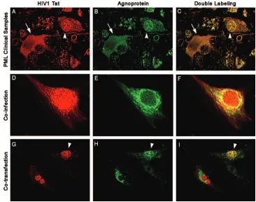
sec-FIG. 1. Colocalization of HIV-1 Tat and JCV agnoprotein in astrocytes. (A to C) Immunofluorescence for the HIV-1 transactivator protein
Tat demonstrates robust expression in the cytoplasm and weaker expression in the nuclear compartment of bizarre astrocytes within demyelinated
plaques in a case of AIDS-related PML (A; rhodamine). The tissue obtained from the archives of the Manhattan Brain Bank at Mt. Sinai School
of Medicine, which had been previously fixed in 10% buffered formalin and embedded in paraffin, was sectioned at a thickness of 4
m. After
deparaffination and antigen retrieval with citrated buffer heated to 97°C, a primary anti-Tat antibody was applied overnight at room temperature
(courtesy of Avindra Nath, Department of Neurology, Johns Hopkins School of Medicine, Baltimore, MD). After being rinsed thoroughly with
PBS, samples were incubated with a Texas red-tagged secondary antibody for 1 h. Then rabbit polyclonal antiagnoprotein antibody (24) was
incubated overnight, and finally, after a thorough rinsing with PBS, a fluorescein-tagged secondary antibody was incubated and sections were
visualized in an inverted, fluorescent Nikon microscope with deconvolution software (SlideBook 4.0.1.34; Intelligent Imaging, Denver, CO). The
JCV late regulatory product, agnoprotein, is detected in the cytoplasm of the same astrocytes (B; fluorescein). Superimposition of both
fluorochromes shows colocalization of both proteins in the cytoplasm (arrow) of the majority of cells and in the nuclei (arrowhead) of few bizarre
astrocytes (C; double labeling). (D to F) Primary astrocytes were coinfected with HIV-1 and JCV, and by immunofluorescence we detected Tat
in both nuclei and cytoplasm of infected cells (D; rhodamine), and agnoprotein in the cytoplasm (E; fluorescein). Deconvolution demonstrates the
colocalization of both proteins in the cytoplasmic compartment (F). (G to I) U-87MG cells cotransfected, as previously described (20), with
plasmids expressing Tat (CFP-Tat) and agnoprotein (YFP-agnoprotein) show Tat localization mainly in the nuclei, with some cells showing nuclear
and cytoplasmic labeling (G), and agnoprotein in the cytoplasm (H), where it colocalizes with Tat (I). Original magnification in all panels,
⫻
1,000.
YFP-agnoprotein has been previously described (20). CFP-Tat was created by removing the Tat gene from cytomegalovirus (CMV)-Tat with BglII
and EcoRI and cloning it into the BamHI and EcoRI sites of pECFP-C1.
9290
KANIOWSKA ET AL.
J. V
IROL.
on November 8, 2019 by guest
http://jvi.asm.org/
[image:3.585.107.473.68.357.2]ondary antibody was incubated and sections were examined. All fluorescent images were captured using an inverted, fluorescent Nikon microscope with deconvolution software (SlideBook 4.0.1.34; Intelligent Imaging, Denver, CO).
RESULTS
Expression and subcellular localization of agnoprotein and
Tat.
JCV agnoprotein is mostly a perinuclear protein with a
limited nuclear appearance both in cell culture and in clinical
samples (20, 24, 46, 47). In cell culture, HIV-1 Tat is mainly
found in the nuclei and nucleoli with some accumulation in the
cytoplasm. Of note, in earlier studies cytoplasmic Tat has been
demonstrated in reactive astrocytes of AIDS patients with
PML (23, 24). Here, we examined the expression and
subcel-lular localization of agnoprotein and Tat in clinical samples
from AIDS patient brain with PML. Samples from a total of 12
autopsy cases of HIV-related PML were obtained from
ar-chives of the Manhattan Brain Bank at Mount Sinai School of
Medicine in New York, NY. The clinical signs and symptoms
of the patients depended on the location of the demyelinated
lesions and included headaches, motor deficits, pareses,
par-esthesias, and cortical blindness in one case with occipital
le-sions. The brain tissue samples were analyzed by double
im-munostaining using antiagnoprotein and anti-Tat antibodies.
Results from immunohistochemical experiments revealed that
in bizarre astrocytes the majority of Tat and agnoprotein is
accumulated in the cytoplasm (Fig. 1A and B), although there
was a limited number of astrocytes exhibiting both cytoplasmic
presence and nuclear presence of these proteins (Fig. 1A).
Superimposition of fluorochromes in both cases demonstrates
clear colocalization of Tat and agnoprotein in cytoplasm
and/or nuclei of bizarre astrocytes (Fig. 1C). Next, we
exam-ined the subcellular localization of these two proteins in
pri-mary culture of human fetal astrocytes after infection with JCV
and HIV-1 by immunofluorescence and were able to detect Tat
in both nuclei and cytoplasm and agnoprotein mainly in the
cytoplasm of infected cells (Fig. 1D and E). Evaluation of these
cells by deconvolution microscopy revealed the colocalization
of both proteins in the cytoplasmic compartment (Fig. 1F). In
a third approach, we assessed the subcellular colocalization of
Tat and agnoprotein in human astrocytic cells after
transfec-tion with plasmids expressing these proteins. In this
experi-ment, U-87MG cells were transfected with yellow fluorescent
protein (YFP)-agnoprotein and CFP-Tat encoding plasmids
either alone or in combination. Once expressed alone,
YFP-agnoprotein was mostly accumulated in the cytoplasmic
com-partment of cells, whereas CFP-Tat was predominantly
local-ized in the nuclei (Fig. 1G and H). Interestingly, in cells
transfected with both agnoprotein- and Tat-producing
plas-mids, some levels of agnoprotein were detected in the nuclei.
Conversely, Tat protein was also detected in the cytoplasm,
suggesting that coproduction of agnoprotein and Tat in cells
may alter their preferred subcellular compartmentalization
(Fig. 1I).
[image:4.585.71.502.73.343.2]To further verify the subcellular localization of Tat and
agnoprotein in astrocytes, U-87MG cells were transfected with
YFP-agnoprotein and CFP-Tat expression plasmids and then
cytoplasmic and nuclear proteins were prepared and analyzed
by Western blotting. As shown in Fig. 2, agnoprotein was
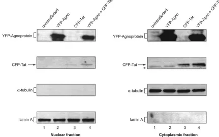
FIG. 2. Detection of Tat and agnoprotein in cytoplasmic and nuclear fractions. Nuclear and cytoplasmic proteins were prepared from equal
numbers of U-87MG cells cotransfected with YFP-agnoprotein and CFP-Tat-expressing plasmids as indicated above each lane. Protein extracts
prepared from an equal number of cells were analyzed by a series of Western blots using antiagnoprotein antibody (24), anti-HIV-1 Tat (R705
from the AIDS Research and Reference Reagent Program, Division of AIDS, NIAID, NIH), antitubulin antibody, and anti-lamin A antibody.
V
OL. 80, 2006
JC VIRUS–HIV-1 Tat CROSS-INTERACTION
9291
on November 8, 2019 by guest
http://jvi.asm.org/
detected in both nuclear and cytoplasmic fractions of cells
transfected with YFP-agnoprotein expression plasmid either
alone or together with CFP-Tat expression plasmid. We
ob-served a noticeably higher level of agnoprotein in the
cytoplas-mic fraction compared to the nuclear fractions. In parallel, a
similar experiment was performed for the detection of Tat
protein. Again, Tat was detected in the cytoplasm and nuclear
fraction of astrocytes transfected with CFP-Tat. The level of
Tat was slightly higher in the cytoplasm in the presence of
agnoprotein. The purity of the nuclear fractions was assessed
by Western blotting for the presence of
␣
-tubulin. As seen,
␣
-tubulin was detected in the cytoplasm, but not in nuclear
fractions (Fig. 2). Furthermore, results from Western blotting
showed the presence of nuclear marker (lamin A) only in the
nuclear but not in the cytoplasmic fractions.
Coinfection of human primary astrocytes with JCV and
HIV-1.
To assess the importance and frequency of JCV
coin-fection with HIV-1 on the expression and replication of HIV-1,
human primary culture of astrocytes was transduced with
HIV-1
(NL4-3)GFP-Vpr (72). After 24 h, cells were infected with
Mad-1/SVEdelta JCV at a multiplicity of infection of 1.0 (67).
In parallel, control uninfected cells or cells infected with JCV
or HIV-1 alone were prepared at day 7. Cells were harvested
and, after fixation in formaldehyde and ethanol and treatment
with 0.2% Triton X-100, were incubated with antiagnoprotein
antibody, followed by incubation with goat anti-rabbit
IgG-PE-conjugated secondary antibody. In a negative control, samples
were left without antiagnoprotein antibody but PE-conjugated
secondary antibody was applied. After washing cells with PBS,
cellular fluorescence was measured using FACScan flow
cy-tometry for detection of GFP (for HIV-1-positive cells) and/or
PE (for JCV-positive cells). Results from this experiment
re-vealed nearly 81% of the cells with expression of agnoprotein,
pointing to the efficient infection of the cells with JCV. In the
presence of HIV-1, the number of JCV-positive cells was
in-creased, exceeding 94.7%, which corroborates the previous
observation demonstrating stimulation of JCV gene
transcrip-tion by HIV-1 Tat (11, 37, 59). In vitro infectranscrip-tion of the primary
culture of astrocytes with HIV-1 was poor, as only 3.15% of the
cells were GFP positive, indicative of HIV-1 replication in
FIG. 3. Effect of JCV agnoprotein on Tat-mediated activation of the HIV-1 LTR. Primary human fetal astrocytes were prepared according to
a modified procedure based on the methods of Cole and de Vellis (16) and Yong and Antel (79). Astrocytes were plated (2
⫻
10
5in 60-mm plate)
and maintained in regular growth medium (Dulbecco’s modified Eagle’s medium–F-12 supplemented with 15% fetal bovine serum). Cells were
then transfected using FuGENE 6 transfection reagent with 1
g of luciferase-based reporter constructs containing full-length (
⫺
450 to
⫹
80)
HIV-LTR (A) or a deletion mutant of the LTR lacking the TAR sequence (
⫹
3 to
⫹
80) (B). Transfections were carried out in the absence or
presence of plasmids (1
g) expressing either Tat or agnoprotein under control of the cytomegalovirus (CMV) promoter (58). The concentration
of DNA in each transfection mixture remained constant by adding pCDNA3. Forty hours after transfection, cells were harvested and luciferase
enzymatic activity was measured. The average values of multiple experiments are presented as
n
-fold effects with a baseline remaining as an
arbitrary unit of 1. (C) Western blot analysis of protein extract from the transfected cells (as indicated in panel A) for detection of Tat and
agnoprotein.
9292
KANIOWSKA ET AL.
J. V
IROL.
on November 8, 2019 by guest
http://jvi.asm.org/
these cells. This value was decreased to 1.5% in the presence of
JCV infection, suggesting that coinfection of the
HIV-1-in-fected cells with JCV can decrease the level of HIV-1 gene
expression and replication in these cells.
Effect of JCV agnoprotein on Tat-mediated activation of the
HIV-1 LTR.
In light of our results showing colocalization of
agnoprotein and Tat, in the next series of experiments we
examined the effect of agnoprotein on Tat activation of HIV-1
promoter. Toward this end, primary culture of human fetal
astrocytes was transfected with an HIV-1 LTR reporter
plas-mid either alone or together with plasplas-mids expressing
agno-protein and HIV-1 Tat. As anticipated, expression of Tat
sig-nificantly enhanced the level of full-length LTR promoter
activity in the transfected cells (Fig. 3A). In accord with the
previous results (8, 17, 31, 62–64, 75), expression of Tat in
astrocytes also led to a modest activation of the LTR with no
TAR region (Fig. 3B). Interestingly, coexpression of
agnopro-tein in the cells caused a noticeable decrease in the level of Tat
activation of the TAR-containing full-length HIV-1 LTR but
showed no effect on the modest activation of
⌬
TAR LTR by
Tat (Fig. 3A and B). The presence of Tat and agnoprotein in
the transfected cells was monitored by Western blot analysis
(Fig. 3C).
[image:6.585.116.482.68.439.2]Identification of the region within Tat which is important
for its interaction with agnoprotein.
These observations
pro-vided a rationale to investigate the physical interaction of JCV
agnoprotein with HIV-1 Tat. In vitro-synthesized full-length
agnoprotein was prepared and incubated with GST-Tat and
GST, and the ability of agnoprotein to interact with Tat was
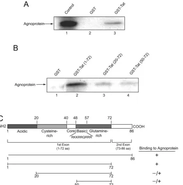
tested by pull-down assay. Figure 4A illustrates a typical gel
analysis showing detection of agnoprotein in fractions bound
to GST-Tat, but not GST alone. To further verify the
specific-ity of this interaction, by determining the region within Tat that
associates with agnoprotein, total protein extracts from NIH
FIG. 4. Association of agnoprotein with HIV-1 Tat and identification of the agnoprotein binding region within Tat. (A) In vitro-synthesized
35S-labeled full-length agnoprotein was incubated with either GST or GST-Tat 1–86 (58) immobilized on glutathione-Sepharose beads. Beads were
washed extensively, and protein complexes were resolved by SDS-PAGE and analyzed by autoradiography. (B) For the mapping assay, total protein
extract from NIH 3T3 cells expressing agnoprotein (20) was incubated with either GST, GST-Tat (1–72), or the deletion mutants of Tat fused to
GST as indicated in the figure, which were immobilized on glutathione-Sepharose beads. After washing bead-protein complexes, the bound
proteins were analyzed by SDS-PAGE followed by Western blot analysis using antibody against JCV agnoprotein (24). (C) Schematic
represen-tation of Tat protein depicting the various domains of Tat and the arginine-rich domain spanning residues 48 to 57. The ability of Tat and its
deletion mutants to interact with agnoprotein is highlighted on the right.
V
OL. 80, 2006
JC VIRUS–HIV-1 Tat CROSS-INTERACTION
9293
on November 8, 2019 by guest
http://jvi.asm.org/
3T3 cells expressing agnoprotein (20) were prepared and
mixed with GST, GST-Tat 1–86, and the various deletion
mu-tants of Tat in fusion with GST as indicated in Fig. 4B. From
the intensity of the band corresponding to agnoprotein, it was
evident that the region within the C terminus of Tat spanning
amino acids 72 to 86 can be deleted with no negative impact on
Tat interaction with agnoprotein. The N terminus of Tat
be-tween residues 1 and 20 is important for its binding to
agno-protein as the removal of this region noticeably decreased
Tat-agnoprotein association. Further deletion of Tat that
re-moves the residues between 20 and 50 further decreased Tat
association with agnoprotein. Thus, the region of Tat that
spans residues 1 to 50 is critical for interaction of Tat and
agnoprotein. The integrity of the bacterially produced
GST-Tat and its mutants was examined by gel electrophoresis (data
not shown). Similar to that in NIH 3T3, the interaction of Tat
and agnoprotein was observed in human astrocytic cells (data
not shown). Figure 4C schematizes the structural organization
of Tat, the mutants that were used in this experiment, and the
levels of Tat binding to agnoprotein.
Identification of the Tat binding region within agnoprotein
and functional interaction of agnoprotein and Tat.
In a
recip-rocal experiment, in vitro-synthesized Tat protein was
incu-bated with GST or GST-agnoprotein and the level of Tat
binding was examined by gel electrophoresis. A band
corre-sponding to Tat was detected in the elution fraction obtained
from GST-agnoprotein, but not GST alone (Fig. 5A).
As illustrated in Fig. 5B, the 71-amino-acid agnoprotein has
a peculiar feature in which its N- and C-terminal domains are
hydrophilic while the central portion of the protein is relatively
hydrophobic. The predicated secondary structure suggests the
presence of several potential helix-turn-helix domains, and the
central domain may adopt an
␣
-helical configuration. To
eval-uate the importance of the various regions of agnoprotein in its
interaction with Tat, in vitro-synthesized Tat protein was
mixed with deletion mutants of agnoprotein in fusion with
GST, and its binding was examined by gel electrophoresis.
Results from the binding assay revealed that a region between
amino acid residues 18 and 54 is the primary domain for
binding to Tat protein (Fig. 5B). From the comparison of the
intensity of the bands obtained from GST-agnoproteins 18–71
and 18–54, it seems that the C-terminal domain of agnoprotein
spanning amino acids 54 to 71 can have a negative effect on the
binding of the region of agnoprotein between residues 18 and
54 with Tat protein.
To correlate the results from binding studies with the
func-tional ability of agnoprotein to suppress Tat activation of the
LTR, we selected two mutants of agnoprotein—one which
exhibits strong binding ability to Tat and the other with no
binding activity to Tat—for use in the transfection assay. As
seen in Fig. 5C, agnoprotein mutant 37–71, which had no
binding activity to Tat, showed no drastic inhibitory effect upon
the LTR, whereas agnoprotein mutant 18–54 with strong
bind-ing to Tat severely hampered the level of Tat activation of the
LTR in astrocytes. This observation suggests that the physical
interaction of Tat and agnoprotein is an important event in the
functional interaction of these proteins upon HIV-1 gene
tran-scription.
Effect of agnoprotein on the interaction of Tat with TAR
RNA, cyclin T1, and cdk9.
In a TAR-dependent manner, Tat
exerts its activity by interacting with the TAR RNA sequence
of the LTR, where it can recruit several cellular factors such as
cyclin T1 and cdk9 that potentiate RNA polymerase II activity
(3, 34, 48, 49). As a first step to assess the impact of the
FIG. 5. Physical and functional interaction of agnoprotein and Tat.
(A) In vitro-synthesized
35S-labeled full-length Tat was incubated with
either GST or GST-agnoprotein (full-length; amino acids 1 to 71) (55)
immobilized on glutathione-Sepharose beads. Beads were washed
ex-tensively, and the protein complexes associated with GST or
GST-agnoprotein were resolved by SDS-PAGE and analyzed by
autoradiog-raphy. (B) For mapping, in vitro-synthesized
35S-labeled full-length
Tat was incubated with either GST, GST-agnoprotein (1–71), or the
deletion mutants of agnoprotein (as indicated) fused to GST and
immobilized on glutathione-Sepharose beads. The bound proteins
were analyzed by SDS-PAGE followed by autoradiography. (B)
Struc-tural organization of agnoprotein illustrating the various domains of
agnoprotein. The ability of agnoprotein and its deletion mutants to
interact with Tat is depicted on the right as follows:
⫹⫹⫹
, strong
interaction;
⫹⫹
, reduced interaction;
⫹
, weak interaction; and
⫺
, no
interaction. (C) Effect of agnoprotein mutants on transcriptional
ac-tivity of Tat. Primary human fetal astrocytes (2
⫻
10
5in 60-mm plate)
were transfected using FuGENE 6 transfection reagent with 1
g of
full-length (
⫺
450 to
⫹
80) HIV-LTR fused to the luciferase gene in the
absence or presence of plasmids encoding Tat (1
g) and full-length
agnoprotein (1–71) (1
g) and deletion mutants which demonstrated
strong binding activity (18–54) or no binding ability (37–71) to Tat. The
concentration of DNA in each transfection mixture remained constant
by adding pCDNA3. Forty hours after transfection, cells were
har-vested and luciferase enzymatic activity was measured. Average values
of multiple experiments are presented as
n
-fold effects.
9294
KANIOWSKA ET AL.
J. V
IROL.
on November 8, 2019 by guest
http://jvi.asm.org/
formation of a Tat-agnoprotein complex on the interaction of
Tat with TAR RNA, we performed an RNA band-shift assay
using synthetic TAR RNA as a probe (22, 36, 41, 74).
32P-labeled TAR RNA was incubated with Tat in the absence and
presence of GST and GST-agnoprotein. As shown in Fig. 6A,
results from gel analysis ruled out the binding of GST and
GST-agnoprotein to TAR RNA, yet demonstrated the ability
of GST-agnoprotein to block formation of the Tat-TAR
com-plex which was detected upon the addition of Tat protein to
the TAR RNA probe. Thus, these observations indicate that
the association of Tat and agnoprotein can have a negative
impact on the interaction of Tat and TAR. As stated above, in
addition to TAR, Tat interacts with cyclin T1 to stimulate
transcription of the LTR via a TAR-dependent pathway. As
such, in the next experiment we determined whether the
in-teraction of Tat with cyclin T1 is affected by agnoprotein of
JCV. Protein extracts from cells with or without agnoprotein
expression were utilized in immunoprecipitation followed by
FIG. 6. Effect of agnoprotein on the interaction of Tat with TAR and cyclin T1. (A) Electrophoretic mobility shift assay. Approximately 60,000
cpm of synthetic
32P-labeled TAR RNA (5
⬘
UGGGUCUCUCUGGUUAGACCAGAUCUGAGCCUGGGAGCUCUCUGGCUAACUAGGG
AACCCACUGCUUAAGCCUCA-3
⬘
) was incubated for 1 h on ice with 0.3
M eluted GST, GST-agnoprotein, or in vitro-synthesized Tat in 20
l binding buffer containing 12 mM HEPES (pH 7.9), 4 mM Tris-HCl (pH 7.5), 60 mM KCl, 5 mM MgCl
2, 0.8 mM dithiothreitol, 0.5
g of
poly(dI-dC) as nonspecific competitor, 10% glycerol, and 10
g/ml DNase-free RNase. The binding mixture was resolved on a 6% polyacrylamide–
0.5
⫻
TBE gel and analyzed by autoradiography. Integrity and equal loading of proteins used in the assay were verified by SDS-PAGE.
(B) Agnoprotein negatively affects binding of Tat to cyclin T1. HL3T1 cells (HeLa cells with stably integrated HIV-1 LTR in the genome) were
transfected with plasmids expressing Tat or agnoprotein alone or in combination. Total protein extract was prepared, and 250
g was incubated
with an anti-cyclin T1 antibody. Immunocomplexes were immunoprecipitated (IP) with the addition of protein A-Sepharose beads, resolved by
SDS-PAGE, and analyzed by Western blotting using an anti-Tat antibody. The presence of Tat was verified by Western blotting (lanes 1 to 4).
Radiograms were analyzed by the Quantity One program (Molecular Imager FX; Bio-Rad), and binding activity was determined by analyzing the
intensity of bands (adjusted volume of counts per mm
2). A total of 12.5% of Tat was bound to cyclin T1 in the absence of agnoprotein, and only
3% of Tat was found in the complex with cyclin T1 in the presence of agnoprotein. (C) Expression of agnoprotein in the transfected cells was tested
by Western blot analysis. (D) Presence of agnoprotein affects binding of Tat to cdk9. Total protein extracts from U-87MG cells transfected with
pCDNA3 or pCMV-agnoprotein were incubated with either GST or GST-Tat 1–86 immobilized on glutathione-Sepharose beads. After washing,
protein complexes were resolved by SDS-PAGE and analyzed by Western blotting using anti-cdk9 antibody. GST proteins were used at a 1
M
concentration in pull-down assays.
V
OL. 80, 2006
JC VIRUS–HIV-1 Tat CROSS-INTERACTION
9295
on November 8, 2019 by guest
http://jvi.asm.org/
Western blot analysis to assess binding of cyclin T1 with
en-dogenously expressed Tat. Results shown in Fig. 6B illustrate
reduced binding of Tat and cyclin T1 in cells that express
agnoprotein, suggesting that the interaction of Tat and
agno-protein may have a negative impact on Tat-cyclin T1 complex
formation. Expression of agnoprotein in the transfected cells
was assessed by direct Western blot assay (Fig. 6C). Similarly,
our results from the GST pull-down assay showed that
agno-protein had a moderate effect on the interaction of Tat and
cdk9, a partner of cyclin T1 which is responsible for the
phos-phorylation of the polymerase II carboxyl terminus (Fig. 6D).
Effect of agnoprotein on the interaction of Tat with the p65
subunit of NF-
B and Sp1.
In the next series of experiments,
we focused our attention on the effect of agnoprotein on the
interaction of Tat with upstream DNA binding transcription
factors whose involvement in Tat activation of the LTR had
been previously demonstrated. First, we tested Tat interaction
with the p65 subunit of NF-
B as cross-interaction between
Tat and upstream transcription activators, particularly the p65
subunit of NF-
B (2, 28, 33, 42, 70, 71, 80), plays an important
role in Tat activation of the LTR in susceptible cells, including
astrocytic cells (61, 75). In this respect, protein extracts from
cells expressing agnoprotein and the control cells with no
agno-protein were incubated with GST or GST-Tat fusion agno-protein.
The complexes were analyzed by Western blotting using
anti-p65 antibody. As shown in Fig. 7A, under similar conditions,
the intensity of the band corresponding to p65 from cells
ex-pressing agnoprotein is less than that seen in the control cells
(compare lanes 2 and 4), indicating that the interaction of
agnoprotein and Tat may also interfere with the ability of Tat
to associate with p65. This event may not be attributed to the
levels of p65 in the control and agnoprotein-expressing cells as
tested by Western blot analysis of p65 in these cells (Fig. 7B).
Next, we investigated the interaction of Tat and Sp1 in the
presence of agnoprotein. Of note, earlier studies demonstrated
that physical interaction of Tat and Sp1 may have an impact on
the level of LTR promoter activity (15, 32, 77). Our results, as
seen in Fig. 7C, show a modest negative effect of agnoprotein
on the cross-communication between Tat and Sp1. Altogether,
these observations demonstrate that the physical contact
be-tween agnoprotein and Tat can interfere with some of the
critical events involved in the interaction of Tat with its viral
(TAR) and cellular (p65 and cyclin T1/cdk9) partners and
hence decrease the level of LTR transcription in astrocytic
cells.
DISCUSSION
Here we provide evidence for the potential cross-interaction
of JCV and HIV-1 through the regulatory proteins Tat and
agnoprotein and the impact of this interaction on expression of
the HIV-1 genome. Results from infection studies showed that
human fetal astrocytes can be coinfected with JCV and HIV-1
and the presence of JCV in cells alters HIV-1 gene expression.
At the same time, JCV gene expression was slightly increased
in HIV-1-replicating cells. We demonstrate that the physical
interaction of Tat and agnoprotein partially affects their
sub-cellular localization and has a negative impact on the
interac-tion of Tat with its target RNA, TAR, and the critical upstream
transcription factors such as the p65 subunit of NF-
B. Earlier
results from several laboratories have ascribed an important
role for Tat in recruiting pTEFb, the positive elongation factor
b composed of cdk9 and cyclin T complex, in close proximity to
the transcription start site via its association with TAR (3, 27,
38, 69). Our results show that agnoprotein can interfere with
the interaction of Tat with cyclin T and cdk9. Thus, it is likely
that the interaction of agnoprotein with Tat negatively affects
its cross-association with TAR through the cyclin T-cdk9
com-plex. More recently, it has been demonstrated that Tat activity
upon the HIV-1 LTR can be inhibited by HEXIM1 (26). The
inactive form of pTEFb, which is associated with inhibited
cdk9 kinase activity, forms a large complex consisting of
HEXIM1 and 7SK small nuclear RNA (40, 45, 76, 78). The
interaction of agnoprotein with Tat that interferes with the
Tat-cyclin T complex may result in the presence of free cyclin
T1, which, in turn, can form a complex with HEXIM1. Thus, it
is plausible to envision cooperativity between agnoprotein and
HEXIM1-7SK snRNA in disruption of active
pTEFb-Tat-TAR complex. On the other hand, the interaction of
agnopro-tein and Tat may alter the subcellular distribution of Tat,
leading to its retention in the cytoplasm. Under these
circum-stances, Tat may not exert its nuclear function such as
tran-scriptional activation of several cellular genes, including
cyto-kines such as tumor necrosis factor alpha, whose downstream
transcription factor, NF-
B, is critical for LTR activity.
Alto-gether, these observations suggest that agnoprotein may utilize
FIG. 7. Effect of agnoprotein on the interaction of Tat with p65
and Sp1. (A) Total protein extracts (250
g) from U-87MG cells
transfected with pCDNA3 or pCMV-agnoprotein were incubated with
either GST or GST-Tat 1–86 immobilized on glutathione-Sepharose
beads. After washing, protein complexes were resolved by SDS-PAGE
and analyzed by Western blotting using anti-p65 antibody. GST
pro-teins were used in a 1
M concentration in pull-down assays. (B)
Ex-pression of p65 and agnoprotein was verified by direct Western
blot-ting. (C) Total protein extracts (250
g) from U-87MG cells
transfected with pCDNA3 or pCMV-agnoprotein were incubated with
either GST or GST-Tat 1–86 immobilized on glutathione-Sepharose
beads. After washing, protein complexes were resolved by SDS-PAGE
and analyzed by Western blotting using anti-Sp1 antibody. GST
pro-teins were used in a 1
M concentration in pull-down assays.
9296
KANIOWSKA ET AL.
J. V
IROL.
on November 8, 2019 by guest
http://jvi.asm.org/
both direct and indirect pathways to interfere with
transcrip-tional activation of the LTR by Tat in astrocytes upon its
association with Tat. It is known that HIV-1 is poorly
repli-cated in astrocytes in cell culture (9, 39, 43, 44). There have
been several speculations and experimental data that may
ex-plain, at least in part, the inability of astrocytes to fully support
HIV-1 gene expression and replication. While the exact events
leading to nonproductive replication of HIV-1 in astrocytes
remain to be elucidated, HIV-1 interaction with other
patho-gens in astrocytes may contribute to the level of HIV-1 gene
expression and replication in these cells. In earlier
observa-tions, it was demonstrated that the JCV early gene product, T
antigen, had the capacity to stimulate transcription of the
HIV-1 LTR (29). On the other hand, results from transcription
studies revealed the ability of Tat to stimulate transcription of
the JCV genome in glial cells (59). Tat activation of the JCV
late promoter led to the notion that the molecular dialogue
between HIV-1 and JCV may contribute to a higher incidence
of PML in people with AIDS than in any other
immunosup-pressed individuals (59). Activation of the late gene of JCV by
Tat can allow, in addition to expression of the viral capsid
proteins, expression of JCV agnoprotein, whose function is
important for JCV replication. Our results on the effect of
agnoprotein on Tat activation of the LTR suggest that a
deli-cate balance in the level of JCV and HIV-1 gene expression
can control the level of JCV and HIV-1 gene transcription in
astrocytes. On the other hand, our preliminary data show that
cross-interaction of Tat with agnoprotein has less negative
effect on JCV gene expression and its activation by Tat. Thus,
it is likely that while agnoprotein may also interfere with Tat
interaction with the TAR-like sequence within JCV (13, 14,
37), the secondary pathway by which Tat can stimulate the JCV
genome (4, 7, 10, 18, 25) may remain operative in the presence
of agnoprotein. Our results further identified a region within
the N terminus of agnoprotein that has a helix-loop-helix
struc-ture as a potential Tat binding site. The ability of this small
domain of agnoprotein to decrease Tat function may provide a
new biological tool for the development of molecules that
interfere with the activation of HIV-1 LTR by Tat.
ACKNOWLEDGMENTS
We wish to thank past and present members of the Department of
Neuroscience and Center for Neurovirology for their continued
sup-port, insightful discussions, and sharing of reagents and ideas,
partic-ularly Martyn White for his insightful comments and critical reading of
the manuscript. We would like to thank S. Morgello, Director of the
NIH-funded Manhattan Brain Bank at Mt. Sinai School of Medicine,
for providing the brain tissue samples from patients with PML. We
also thank C. Schriver for editorial assistance.
This work was made possible by grants awarded by NIH to K.K.
REFERENCES
1.Anderson, C. E., G. S. Tomlinson, B. Pauly, F. W. Brannan, A. Chiswick, R. Brack-Werner, P. Simmonds, and J. E. Bell. 2003. Relationship of Nef-positive and GFAP-reactive astrocytes to drug use in early and late HIV
infection. Neuropathol. Appl. Neurobiol.29:378–388.
2.Barboric, M., R. M. Nissen, S. Kanazawa, N. Jabrane-Ferrat, and B. M. Peterlin.2001. NF-kappa B binds P-TEFb to stimulate transcriptional
elon-gation by RNA polymerase II. Mol. Cell8:327–337.
3.Beiniasz, P. D., T. A. Grdina, H. P. Bogerd, and B. R. Cullen.1999. Recruit-ment of cyclin T1/P-TEFb to an HIV type 1 long terminal repeat promoter proximal RNA target is both necessary and sufficient for full activation of
transcription. Proc. Natl. Acad. Sci. USA96:7791–7796.
4.Berger, J. R.2003. Progressive multifocal leukoencephalopathy in acquired immunodeficiency syndrome: explaining the high incidence and
dispropor-tionate frequency of the illness relative to other immunosuppressive
condi-tions. J. Neurovirol.9(Suppl. 1):38–41.
5.Berger, J. R., and M. Concha.1995. Progressive multifocal leukoencepha-lopathy: the evolution of a disease once considered rare. J. Neurovirol.
1:5–18.
6.Berger, J. R., B. Kaszovitz, M. J. Post, and G. Dickinson.1987. Progressive multifocal leukoencephalopathy associated with human immunodeficiency virus infection. A review of the literature with a report of sixteen cases. Ann.
Intern. Med.107:78–87.
7.Berger, J. R., A. Chauhan, D. Galey, and A. Nath.2001. Epidemiological evidence and molecular basis of interactions between HIV and JC virus.
J. Neurovirol.7:329–338.
8.Berkhout, B., A. Gatignol, A. B. Rabson, and K. T. Jeang. 1990. TAR-independent activation of the HIV-1 LTR: evidence that tat requires specific
regions of the promoter. Cell62:757–767.
9.Brack-Werner, R.1999. Astrocytes: HIV cellular reservoirs and important
participants in neuropathogenesis. AIDS13:1–22.
10.Chen, N. N., C.-F. Chang, G. L. Gallia, D. A. Kerr, E. M. Johnson, C. P. Krachmarov, S. M. Barr, R. J. Frisque, B. Bollag, and K. Khalili.1995. Cooperative action of cellular proteins YB-1 and Pur alpha with the tumor antigen of the human JC polyomavirus determines their interaction with the
viral lytic control element. Proc. Natl. Acad. Sci. USA92:1087–1091.
11.Chepenik, L. G., A. P. Tretiakova, C. P. Krachmarov, E. M. Johnson, and K. Khalili.1998. The single-stranded DNA binding protein, Pur␣, binds HIV-1
TAR RNA and activates HIV-1 transcription. Gene210:37–44.
12.Chowdhury, M., J. P. Taylor, H. Tada, J. Rappaport, F. Wong-Staal, S. Amini, and K. Khalili.1990. Regulation of the human neurotropic virus
promoter by JCV-T-antigen and HIV-1 tat protein. Oncogene5:1737–1742.
13.Chowdhury, M., J. P. Taylor, C.-F. Chang, J. Rappaport, and K. Khalili.
1992. Evidence that a sequence similar to TAR is important for induction of the JC virus late promoter by human immunodeficiency virus type 1 Tat.
J. Virol.66:7355–7361.
14.Chowdhury, M., M. Kundu, and K. Khalili.1993. GA/GC-rich sequence
confers Tat responsiveness to human neurotropic virus promoter, JCVL, in
cells derived from CNS. Oncogene8:887–892.
15.Chun, R. F., O. J. Semmes, C. Neuveut, and K.-T. Jeang.1998. Modulation of Sp1 phosphorylation by human immunodeficiency virus type 1 Tat. J.
Vi-rol.72:2615–2629.
16.Cole, R., and J. de Vellis.1997. Astrocyte and oligodendrocyte cultures, p.
117–130.InS. Federoff and A. Richardson (ed.), Protocols for neural cell
culture, 2nd ed. Humana Press, Totowa, NJ.
17.Dandekar, D. H., K. N. Ganesh, and D. Mitra.2004. HIV-1 Tat directly binds to NF kappaB enhancer sequence: role in viral and cellular gene
expression. Nucleic Acids Res.32:1270–1278.
18.Daniel, D. C., Y. Kinoshita, M. A. Khan, L. Del Valle, K. Khalili, J. Rappaport, and E. M. Johnson.2004. Internalization of exogenous human immuno-deficiency virus-1 protein, Tat, by KG-1 oligodendroglioma cells followed by stimulation of DNA replication initiated at the JC virus origin. DNA Cell
Biol.23:858–867.
19.Darbinian-Sarkissian, N., A. Darbinyan, J. Otte, S. Radhakrishnan, B. E. Sawaya, A. Arzumanyan, G. Chipitsyna, Y. Popov, J. Rappaport, S. Amini, and K. Khalili.2006. p27SJ, a novel protein in St. John’s Wort that
sup-presses expression of HIV-1 genome. Gene Ther.13:288–295.
20.Darbinyan, A., N. Darbinian, M. Safak, S. Radhakrishnan, A. Giordano, and K. Khalili.2002. Evidence for dysregulation of cell cycle by human
polyoma-virus, JCV, late auxiliary protein. Oncogene21:5574–5581.
21.Darbinyan, A., K. M. Siddiqui, D. Slonina, N. Darbinian, S. Amini, M. K. White, and K. Khalili.2004. Role of JC virus agnoprotein in DNA repair.
J. Virol.78:8593–8600.
22.Darfeuille, F., A. Arzumanov, S. Gryaznov, M. J. Gait, C. Di Primo, and J. J. Toulme.2002. Loop-loop interaction of HIV-1 TAR RNA with N3⬘-P5⬘ deoxyphosphoramidate aptamers inhibits in vitro Tat-mediated
transcrip-tion. Proc. Natl. Acad. Sci. USA99:9709–9714.
23.Del Valle, L., S. Croul, S. Morgello, S. Amini, J. Rappaport, and K. Khalili.
2000. Detection of HIV-1 Tat and JCV capsid protein, VP1, in AIDS brain
with progressive multifocal leukoencephalopathy. J. Neurovirol.6:221–228.
24.Del Valle, L., J. Gordon, S. Enam, S. Delbue, S. Croul, S. Abraham, S. Radhakrishnan, M. Assimakopoulou, C. D. Katsetos, and K. Khalili.2002. Expression of human neurotropic polyomavirus JCV late gene product
agno-protein in human medulloblastoma. J. Natl. Cancer Inst.94:267–273.
25.Enam, S., T. M. Sweet, S. Amini, K. Khalili, and L. Del Valle.2004. Evidence for involvement of transforming growth factor beta 1 signaling pathway in activation of JC virus in human immunodeficiency virus 1-associated
pro-gressive multifocal leukoencephalopathy. Arch. Pathol. Lab. Med.128:282–
291.
26.Fraldi, A., F. Varrone, G. Napolitano, A. A. Michels, B. Majello, O. Bensaude, and L. Lania. 2005. Inhibition of Tat activity by the HEXIM1 protein.
Retrovirology2:42.
27.Garber, M. E., T. P. Mayall, E. M. Suess, J. Meisenhelder, N. E. Thompson, and K. A. Jones.2000. CDK9 autophosphorylation regulates high-affinity binding of the human immunodeficiency virus type 1 Tat-P-TEFb complex to
TAR RNA. Mol. Cell. Biol.20:6958–6969.
V
OL. 80, 2006
JC VIRUS–HIV-1 Tat CROSS-INTERACTION
9297
on November 8, 2019 by guest
http://jvi.asm.org/
28.Garcia-Martinez, L. F., G. Mavankal, J. M. Neveu, W. S. Lane, D. Ivanov, and R. B. Gaynor.1997. Purification of a Tat-associated kinase reveals a
TFIIH complex that modulates HIV-1 transcription. EMBO J.16:2836–
2850.
29.Gendelman, H. E., W. Phelps, L. Feigenbaum, J. M. Ostrove, A. Adachi, P. M. Howley, G. Khoury, H. S. Ginsberg, and M. A. Martin.1986. Trans-activation of the human immunodeficiency virus long terminal repeat
se-quence by DNA viruses. Proc. Natl. Acad. Sci. USA83:9759–9763.
30.Graham, F. L., and A. J. van der Eb.1973. A new technique for the assay of
infectivity of human adenovirus 5 DNA. Virology52:456–467.
31.Harrich, D., J. Garcia, R. Mitsuyasu, and R. Gaynor.1990. TAR-indepen-dent activation of the human immunodeficiency virus in phorbol ester
stim-ulated T lymphocytes. EMBO J.9:4417–4423.
32.Jeang, K.-T., R. Chun, N. H. Lin, A. Gatignol, C. G. Glabe, and H. Fan.1993. In vitro and in vivo binding of human immunodeficiency virus type 1 Tat
protein and Sp1 transcription factor. J. Virol.67:6224–6233.
33.Jones, K. A., and B. M. Peterlin.1994. Control of RNA initiation and
elongation at the HIV-1 promoter. Annu. Rev. Biochem.63:717–743.
34.Kashanchi, F., G. Piras, M. F. Radonovich, J. F. Duvall, A. Fattaey, C. M. Chiang, R. G. Roeder, and J. N. Brady.1994. Direct interaction of human
TFIID with the HIV-1 transactivator tat. Nature367:295–299.
35.Khalili, K., M. K. White, H. Sawa, K. Nagashima, and M. Safak.2004. The agnoprotein of polyomaviruses: a multifunctional auxiliary protein. J. Cell.
Physiol.207:1–7.
36.Kolb, G., S. Reigadas, C. Boiziau, A. van Aerschot, A. Arzumanov, M. J. Gait, P. Herdewijn, and J. J. Toulme.2005. Hexitol nucleic acid-containing
aptamers are efficient ligands of HIV-1 TAR RNA. Biochemistry44:2926–
2933.
37.Krachmarov, C. P., L. G. Chepenik, S. Barr-Vagell, K. Khalili, and E. M. Johnson.1996. Activation of the JC virus Tat-responsive transcriptional control element by association of the Tat protein of human immunodefi-ciency virus 1 with cellular protein Pur alpha. Proc. Natl. Acad. Sci. USA
93:14112–14117.
38.Kwak, Y. T., D. Ivanov, J. Guo, E. Nee, and R. B. Gaynor.1999. Role of the human and murine cyclin T proteins in regulating HIV-1 tat activation. J.
Mol. Biol.288:57–69.
39.McCarthy, M., J. He., and C. Wood.1998. HIV-1 strain-associated variability
in infection of primary neuroglia. J. Neurovirol.4:80–89.
40.Michels, A. A., V. T. Nguyen, A. Fraldi, V. Labas, M. Edwards, F. Bonnet, I. Lania, and O. Bensaude. 2003. MAQ1 and 7SK RNA interaction with CDK9/cyclin T complexes in a transcription-dependent manner. Mol. Cell.
Biol.23:4859–4869.
41.Murchie, A. I., B. Davis, C. Isel, M. Afshar, M. J. Drysdale, J. Bower, A. J. Potter, I. D. Starkey, T. M. Swarbrick, S. Mizra, C. D. Prescott, P. Vaglio, F. Aboul-ela, and J. Karn.2004. Structure-based drug design targeting an inactive RNA conformation: exploiting the flexibility of HIV-1 TAR RNA.
J. Mol. Biol.336:625–638.
42.Nabel, G., and D. Baltimore.1987. An inducible transcription factor
acti-vates expression of human immunodeficiency virus in T cells. Nature326:
711–713. (Erratum,344:178, 1990.)
43.Neumann, M., B. K. Felber, A. Kleinschmidt, B. Froese, V. Erfle, G. N. Pavlakis, and R. Brack-Werner.1995. Restriction of human immunodefi-ciency virus type 1 production in a human astrocytoma cell line is associated
with a cellular block in Rev function. J. Virol.69:2159–2167.
44.Neumann, M., E. Afonina, F. Ceccherini-Silberstein, S. Schlicht, V. Erfle, G. N. Pavlakis, and R. Brack-Werner.2001. Nucleocytoplasmic transport in human astrocytes: decreased nuclear uptake of the HIV Rev shuttle protein.
J. Cell Sci.114:1717–1729.
45.Nguyen, V. T., T. Kiss, A. A. Michels, and O. Bensaude.2001. 7SK small nuclear RNA binds to and inhibits the activity of cdk9/cyclin T complexes.
Nature414:322–325.
46.Okada, Y., S. Endo, H. Takahashi, H. Sawa, T. Umemura, and K. Nagashima.
2001. Distribution and function of JCV agnoprotein. J. Neurovirol.7:302–
306.
47.Okada, Y., H. Sawa, S. Endo, Y. Orba, T. Umemura, H. Nishihara, A. C. Stan, S. Tanaka, H. Takahashi, and K. Nagashima.2002. Expression of JCV Agnoprotein in progressive leukoencephalopathy brain. Acta Neuropathol.
(Berlin)104:130–136.
48.Okamoto, H., C. T. Sheline, J. L. Corden, K. A. Jones, and B. M. Peterlin.
1996. Trans-activation by human immunodeficiency virus Tat protein re-quires the C-terminal domain of RNA polymerase II. Proc. Natl. Acad. Sci.
USA93:11575–11579.
49.Parada, C. A., and R. G. Roeder. 1996. Enhanced processivity of RNA polymerase II triggered by Tat-induced phosphorylation of its
carboxy-ter-minal domain. Nature384:375–378.
50.Petito, C. K., E. S. Cho, W. Lemann, B. A. Navia, and R. W. Price.1986. Neuropathology of acquired immunodeficiency syndrome (AIDS): an
au-topsy review. J. Neuropathol. Exp. Neurol.45:635–646.
51.Price, R. W., B. J. Brew, and M. Rosenblum.1990. The AIDS dementia complex and HIV-I brain infection: a pathogenetic model of virus-immune
interaction, p. 269–290.InB. H. Waksman (ed.), Immunologic mechanisms
in neurologic and psychiatric disease. Raven Press, New York, N.Y.
52.Radhakrishnan, S., J. Gordon, L. Del Valle, J. Cui, and K. Khalili.2004. Intracellular approach for blocking JC virus gene expression by using RNA
interference during viral infection. J. Virol.78:7264–7269.
53.Safak, M., and K. Khalili.2001. Physical and functional interaction between viral and cellular proteins modulate JCV gene transcription. J. Neurovirol.
7:288–292.
54.Safak, M., and K. Khalili.2003. An overview: human polyomavirus, JC virus,
and its associated disorders. J. Neurovirol.9(Suppl. 1):3–9.
55.Safak, M., R. Barrucco, A. Darbinyan, Y. Okada, K. Nagashima, and K. Khalili.2001. Interaction of JC virus Agno protein with T antigen modulates transcription and replication of the viral genome in glial cells. J. Virol.
75:1476–1486.
56.Safak, M., B. Sadowska, R. Barrucco, and K. Khalili.2002. Functional interaction between JC virus late regulatory agnoprotein and cellular Y-box
binding transcription factor, YB-1. J. Virol.76:3828–3838.
57.Saito, Y., L. R. Sharer, L. G. Epstein, J. Michaels, M. Mintz, M. Louder, K. Golding, T. A. Cvetkovich, and B. M. Blumberg.1994. Overexpression of nef as a marker for restricted HIV-1 infection of astrocytes in postmortem
pediatric central nervous tissue. Neurology44:474–481.
58.Sawaya, B. E., K. Khalili, J. Gordon, R. Taube, and S. Amini.2000. Coop-erative interaction between HIV-1 regulatory proteins Tat and Vpr
modu-lates transcription of the viral genome. J. Biol. Chem.275:35209–35214.
59.Tada, H., J. Rappaport, M. Lashgari, S. Amini, F. Wong-Stahl, and K. Khalili. 1990. Trans-activation of the JC virus late promoter by the tat protein of type 1 human immunodeficiency virus in glial cells. Proc. Natl.
Acad. Sci. USA87:3479–3483.
60.Takahashi, K., S. L. Wesselingh, D. E. Griffin, J. C. McArthur, R. T. Johnson, and J. D. Glass.1996. Localization of HIV-1 in human brain using polymer-ase chain reaction/in situ hybridization and immunocytochemistry. Ann.
Neurol.39:705–711.
61.Taylor, J. P., and K. Khalili.1994. Activation of HIV-1 transcription by Tat in cells derived from the CNS: evidence for the participation of NF-kappaB.
Adv. Neuroimmunol.4:291–303.
62.Taylor, J. P., R. Pomerantz, O. Bagasra, M. Chowdhury, J. Rappaport, K. Khalili, and S. Amini.1992. TAR-independent transactivation by Tat in cells derived from the CNS: a novel mechanism of HIV-1 gene regulation. EMBO
J.11:3395–3403.
63.Taylor, J. P., M. Kundu, and K. Khalili.1993. TAR-independent activation of HIV-1 requires the activation domain but not the RNA-binding domain of
Tat. Virology195:780–785.
64.Taylor, J. P., R. J. Pomerantz, G. Raj, F. Kashanchi, J. N. Brady, S. Amini, and K. Khalili.1994. Central nervous system-derived cells express a B-binding activity that enhances human immunodeficiency virus type 1 tran-scription in vitro and facilitates TAR-independent transactivation by Tat.
J. Virol.68:3971–3981.
65.Tornatore, C., R. Chandra, J. R. Berger, and E. O. Major.1994. HIV-1 infection of subcortical astrocytes in the pediatric central nervous system.
Neurology44:481–487.
66.Trillo-Pazos, G., A. Diamanturos, L. Rislove, T. Menza, W. Chao, P. Belem, S. Sadiq, S. Morgello, L. Sharer, and D. J. Volsky.2003. Detection of HIV-1 DNA in microglia/macrophages, astrocytes, and neurons isolated from brain tissue with HIV-1 encephalitis by laser capture microdissection. Brain
Pathol.13:144–154.
67.Vacante, D. A., R. Traub, and E. O. Major.1989. Extension of JC virus host range to monkey cells by insertion of a simian virus 40 enhancer into the JC
virus regulatory region. Virology170:353–361.
68.Walker, D. L.1985. Progressive multifocal leukoencephalopathy, p. 503–524.
InP. J. Vinken, G. W. Bruyn, H. L. Klawans, and J. C. Kloetsier (ed.),
Handbook of clinical neurology, vol. 47. Elsevier, New York, N.Y. 69.Wei, P., M. E. Garber, S. M. Fang, W. H. Fischer, and K. A. Jones.1998. A
novel CDK9-associated C-type cyclin interacts directly with HIV-1 Tat and
mediates its high-affinity, loop-specific binding to TAR RNA. Cell92:451–
462.
70.West, M. J., A. D. Lowe, and J. Karn.2001. Activation of human
immuno-deficiency virus transcription in T cells revisited: NF-B p65 stimulates
transcriptional elongation. J. Virol.75:8524–8537.
71.Westendorp, M. O., V. A. Shatrov, K. Schulze-Osthoff, R. Frank, M. Kraft, M. Los, P. H. Krammer, W. Dro¨ge, and V. Lehmann.1995. HIV-1 Tat potentiates TNF-induced NF-kappaB activation and cytotoxicity by altering
the cellular redox state. EMBO J.14:546–554.
72.White, S. M., M. Renda, N.-Y. Nam, E. Klimatcheva, Y. Zhu, J. Fisk, M. Halterman, B. J. Rimel, H. Federoff, S. Pandya, J. D. Rosenblatt, and V. Planelles.1999. Lentivirus vectors using human and simian
immunodefi-ciency virus elements. J. Virol.73:2832–2840.
73.Wright, C. M., B. K. Felber, H. Paskalis, and G. N. Pavlakis.1986. Expres-sion and characterization of the transactivator of HTLV-III/LAV virus.
Science234:988–992.
74.Yamamoto, R., M. Katahira, S. Nishikawa, T. Baba, K. Taira, and P. K. Kumar.2000. A novel RNA motif that binds efficiently and specifically to the Tat protein of HIV and inhibits the transactivation by Tat of transcription in
vitro and in vivo. Genes Cells5:371–388.
75.Yang, L., G. F. Morris, J. M. Lockyer, M. Lu, Z. Wang, and C. B. Morris.
9298
KANIOWSKA ET AL.
J. V
IROL.
on November 8, 2019 by guest
http://jvi.asm.org/
1997. Distinct transcriptional pathways of TAR-dependent and TAR-inde-pendent human immunodeficiency virus type-1 transactivation by Tat.
Vi-rology235:48–64.
76.Yang, Z., Q. Zhu, K. Luo, and Q. Zhou.2001. The 7SK small nuclear RNA
inhibits the cdk9/cyclin T1 kinase to control transcription. Nature414:317–
322.
77.Yedavalli, V. S. R. K., M. Benkirane, and K. T. Jeang. 2003. Tat and trans-activation-responsive (TAR) RNA-independent induction of HIV-1 long terminal repeat by human and murine cyclin T1 requires Sp1. J. Biol.
Chem.278:6404–6410.
78.Yik, J. H., R. Chen, R. Nishimura, J. L. Jennings, A. J. Link, and Q. Zhou.
2003. Inhibition of P-TEFb (CDK9/Cyclin T) kinase and RNA polymerase II transcription by the coordinated actions of HEXIM1 and 7SK snRNA. Mol.
Cell12:971–982.
79.Yong, V. W., and J. P. Antel.1997. Culture of neural cells from human brain
biopsies, p. 81–96.InS. Federoff and A. Anderson (ed.), Protocols for neural
cell culture. Humana Press, Totowa, N.J.
80.Zhu, Y., T. Pe’ery, J. Peng, Y. Ramanathan, N. Marshall, T. Marshall, B. Amendt, M. B. Mathews, and D. H. Price.1997. Transcription elongation factor P-TEFb is required for HIV-1 Tat transactivation in vitro. Genes Dev.
11:2622–2632.
V
OL. 80, 2006
JC VIRUS–HIV-1 Tat CROSS-INTERACTION
9299
on November 8, 2019 by guest
http://jvi.asm.org/