ASSESSMENT OF OCCLUSAL FISSURE
MORPHOLOGY IN DECIDUOUS MOLAR TEETH
Dissertation Submitted to
THE TAMILNADU Dr. M.G.R. MEDICAL UNIVERSITY
In Partial Fulfillment for the Degree of
MASTER OF DENTAL SURGERY
BRANCH VIII
from the bottom of the heart with genuineness, evading the rationalising
ability of the brain.’
Keeping the above mentioned statement in mind, I would like to
holeheartedly, thank few people whose presence at various points of time in
my life, have ade me what I am today.
I would like to thank Almighty, for having blessed me with conducive
environment, throughout my life. My greatest boon, for having born to my
parents, getting trained from eminent, skillful, highly knowledgeable teachers,
who were highly gracious enough to impart me with their valuable possession
of knowledge and skill.
I would like to take this opportunity to thank my Guru, Guide and
Head of the Department, Professor Dr. M. Jayanthi, M.D.S., for her constant
support and intellectual inputs through out my post-graduation period. It was
the initial belief that she bestowed upon me when we started the study, which
kept me motivated and urged me to complete this dissertation, within the
stipulated time frame. Her constant reassurance, and valuable guidance
regarding the nuances of the art paediatric dentistry, helped me to learn the
art of child management and successfully execute the same with impeccable
had taken to ensure, that I completely understand. I have been inspired by her
depth of knowledge, clarity of surgical plan, immense surgical skill in
executing the same, prioritizing perfection both in planning and action.
Madam, these three years of my postgraduation period under your mentorship
and guidance, will be the most enchanting, enriching and enlightening period
of my life. I owe you a lot Madam.
I would like to extend my gratitude to Professor Dr Nilaya Reddy,
M.D.S., who has been a great trainer, philosopher to me during the three
years of my fruitful postgraduate study period. Her constant, persisting quest
for surgical and academic innovation and excellence , has activated an entire
area of dormant grey matter in my brain .I have been awestruck , by her
confidence, speed and precision of surgical work. I would like to personally
thank her, for guiding me, and patiently instructing me while remaining
besides me throughout three years of clinical procedures .I have been inspired
by her unconventional, unorthodox style of thinking and execution. Apart from
the, academic knowledge that Madam has imparted upon me, I have learnt
from her, the way of dealing with problems or difficulties by facing them head
on and management of adverse situations in appropriate manner. Madam, I
will always remain indebted to you Madam.
I would like to convey my heartfelt gratitude to our beloved Principal,
Professor Dr. N.S. Azhagarasan, M.D.S., for believing in us and allowing us
caring nature were a great support at every step and moment during these
three years, and this will be remembered all my life.
I would like to take the opportunity to whole-heartedly thank Reader
Dr Sakthivel for his invaluable and prompt guidance, enlighting discussions,
and constant support throughout my postgraduate life. I also thank him for
teaching me the abstract features of esthetics, precise nature of rotary system
and perfection in root canal treatment, be it in the field esthetics dentistry.
I would like to thank, Reader Dr Poornima, for sharing with me her
deep knowledge and teaching me the essence of medical science in terms of
assessing and handling a patient. I thank her for the enthusiasm, constant
support that she had extended throughout my postgraduate course, especially
lending a helping hand when ever I struggled. She has always showed belief in
me and reassured my abilities in dealing with tough, new challenges in my
life.
I don’t have words to express my heartfelt thanks to Dr Arun .E Senior
Lecturers, for always encouraging me and providing a conducive learning and
knowledge sharing experience in the department. I would genuinely thank sir
for teaching me the pathway for child management and also the tricks to excel
I would like to thank, Dr. Radhika Krishnan, Anaesthesiologist for
sharing with me her deep knowledge and teaching me the essence of medical
science in terms of general anaesthesia and handling patient .I thank her for
the enthusiasm, constant support that she has extended throughout my
postgraduate course.
It would be a crime on my part, if I ignore Dr. Uma Devi for being a
constant source of knowledge, wisdom, encouragement. I would like to
specifically thank Dr. Kavitha wilson for answering all my doubts both subject
and otherwise, during the entire duration my dissertation. Madams, I owe you
a lot.
I would like to genuinely thank my seniors Dr. Madhan, Dr. Porselvi,
Dr. Lakshmi and Dr. Rama devi for teaching me various aspects of oral
surgery, from their perspective.
I would like to thank my batch mate precisely unborn sister
Dr. Bhuvanesswari for providing a healthy and competitive learning
atmosphere through out my postgraduate learning period.
I would also like to acknowledge and thank my juniors,
Dr. Devichandrika , Dr.Keerthi, Dr.Gayathri and Dr.Akila
I would also like to mention the constant selfless help offered to me by
be her student.
I would like to acknowledge the constant support rendered to me by
non-teaching staffs- Sister Eshawari, Sister Veni, Brother Venugopal and
others, at our department, during the three year post graduate period.
I would be failing in my duty, if don’t acknowledge the constant and
timely support of Mr Thavamani and Ms. Sudha, in compiling and printing
my dissertation work.
I would like to extend my gratitude to all those who directly or
indirectly, helped me in completing this dissertation to the best of my ability,
within time, without compromising the quality of dissertation.
As I have reserved the best for the last, I am nowhere without my
parents Mr K.Karunakaran and Mrs Anithakaruanakaran and my late
grandfathers and grandmother Mr. Kattiyakaran, Mr. Muthunayagam and
Mrs. Lilly and my grandmother Mrs. Vellaiammal Words cannot express
the magnitude of things that, I owe them. I dedicate this dissertation to my
parents and grandfathers and grandmothers. The stage that I have reached
of numerous failures in my life. They have always given me the best, often
beyond their abilities. I considered myself fortunate to have been born to them.
‘ Jai Hind ! ’
To study the complex anatomy of the pit and fissure system of human
primary first and second molar teeth under stereomicroscope.
Background:
The pit and fissure patterns on the occlusal surface of the human teeth
represent vulnerable sites for initiation of dental caries due to their
morphological complexity. However the decision making for sealants is based
on the personnel, tooth and surface at risk. Hence it is important to understand
the pit and fissure patterns in the application of appropriate preventive
measures.
Materials and methodology:
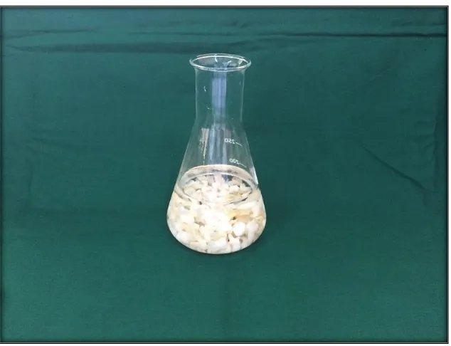
100 Maxillary and mandibular first and second primary molars were
collected and stored in neutral 10% formalin, cleaned with slurry of pumice
and water. The teeth were sectioned longitudinally (buccolingually), thickness
ranging from 40µm to100µm with the help of carborundum disc. The ground
sections of the teeth were fixed on the glass slide and examined under
stereomicroscope with 10 X magnification for the fissure pattern . The results
Results:
The U-TYPE (56%) of fissure pattern was more prevalent in both
the maxillary and the mandibular molar teeth followed by V –TYPE (37%) of
fissure pattern.
Conclusion:
The U and V types of fissure patterns were predominantly seen in the
primary molars compared to the other fissure patterns.
KEY WORDS:
PITS AND FISSURES, U- TYPE AND V – TYPE OF FISSURE
PATTERN IN PRIMARY MOLARS, OCCLUSAL SURFACE OF
S.NO. TITLE PAGE NO.
1. INTRODUCTION 1
2. AIMS AND OBJECTIVES 5
3. REVIEW OF LITERATURE 6
4. MATERIALS AND METHODS 36
5. RESULTS 40
6. DISCUSSION 44
7. CONCLUSION 50
8. SUMMARY 51
9. BIBLIOGRAPHY 53
LIST OF TABLES
TABLE
NO. TITLE
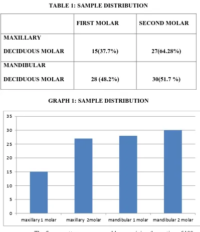
1. SAMPLE DISTRIBUTION
2.
OCCLUSAL FISSURE MORPHOLOGY OF DECIDUOUS MOLAR TEETH
LIST OF GRAPHS
GRAPH
NO. TITLE
FIGURE
NO. TITLE
1. 100 PRIMARY MOLAR TEETH
2. ARMAMENTARIUM
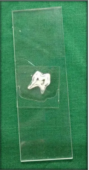
3. SECTIONS MOUNTED ON SLIDES
4. STEREOMICROSCOPE
5. MAXILLARY MOLAR TEETH – V SHAPE FISSURE PATTERN
6. MANDIBULAR MOLAR TEETH – U SHAPE FISSURE PATTERN
LIST OF ANNEXURES
S.NO TITLE
I
INSTITUTIONAL REVIEW BOARD APPROVALIntroduction
1
INTRODUCTION
Caries process involves a large number of interrelating factors
affecting the tooth with changes in the environment. Literature shows that pits
and fissures are areas which are more susceptible to carious attack compared
to the smooth surfaces.
Kraus Jordan and Abrams 1969 defined “A fissure is a cleft or crevice
in a tooth surface thought to result from the imperfect fusion of the enamel of
adjoining cusp or lobes” and “A pit is a sharp pointed depression usually
located at the junction of 2 or more intersecting developmental grooves.1
The researchers have all investigated the patterns of the occlusal
surface of the teeth and explained the fissure pattern through their drawing as
the invagination extending from the occlusal surface to the enamel and
sometimes into the dentin which are quite common and these teeth also have
areas at the base of pits and fissure where there is little enamel covering the
dentin. The presence of deep invagination of the enamel is thought by many to
be an important predisposing factor because decay often starts in pits and
fissure. More over many clinical studies have demonstrated the susceptibility
of these areas to caries and tooth type in the dentition has its own specific
2
the same specific anatomical configuration in identical tooth types
(Brekhus 1931, McCall 1934, Prime 1937, Brucker 1944, Paynter and
Grainger 1962).2-6.
The pit and fissures in both primary and permanent dentition are areas
which are highly liable to decay and act as a reservoir for the initiation and
progression of the disease. The anatomy of pit and fissures of the teeth have
been a subject of research as the recent trends focus more on prevention.
In the 1970s research had focused on prevention of occlusal caries and
as a result the number and intensity of caries involving pits and fissure and
smooth surface have decreased. 80% of all carious lesions in young permanent
teeth involve a fissure surface which makes up 13% of total tooth surface.7
Prevention of pit and fissure caries has progressed from early treatment
modalities like, mechanical fissure eradication and chemical treatment using
silver nitrate to the development of more innovative and progressive materials
and methods, such as micromechanical bonding of artificial resins to enamel
substrate using acid etching techniques. The introduction of materials designed
to seal pits and fissures so as to eliminate them as stagnation sites for
microbial fermentation is a promising adjunct to existing measures. A
Introduction
3
judged „AT RISK FOR CARIES „and not necessarily directed to all teeth with
deep pits and fissures.8
The fissure patterns in the primary teeth was described by Mortimer
(1970) as U and V type.9 The permanent teeth fissure patterns were described
by Nagano and Gustafson found that the prevalence of V- type was
34 %, IK-type 26 %, I- type 19 %, U- type 14 % and other as 7 %. Nagona
also observed the relation between the localization of the primary carious
lesion and form and depth of the fissure and he revealed that caries starts from
the bottom in V type, it starts halfway down in the U- type, and from the top in
the I-type and IK- type. A deep, narrow fissure may resist carious progression
by hindering the impaction or even diffusion of considerable amounts of
substrate which seems to be less liable to carious attack than one providing
some space for plaque and debris to accumulate. Steepness of walls and ample
space for retention above the entrance to the fissure appear to be the most
important feature, with the depth of the fissure proper, being of secondary
significance.10,11 It is therefore mandatory to know about the pits and fissure
patterns to preserve the teeth.
The morphology of fissures and their relationship to enamel caries in
permanent teeth have been investigated by many authors under ordinary light,
4
deciduous tooth fissures pattern and its relationship to caries is scarce. Hence
this study was aimed to investigate the pit and fissure pattern in the deciduous
5
AIM AND OBJECTIVES
6
REVIEW OF LITERATURE
Profitable investigations in relation to caries of pits and fissures
were made when scientific observations on teeth first started. John Hunter
wrote his book “Practical Treatise of the Disease of Teeth” in 1778 in which
he mentioned that fissures are cracks or the hollow path of grinding surfaces
of molars filled with black substance.13
Fox, writing in 1803, described fissures as irregularities of the
grinding surface of the molars which leads into a cavity in the center of the
tooth. The chief predisposition to decay is defective formation in either
enamel or bony parts of teeth.14
W. Robertson (1835) in his book “A Practical Treatise on the
Human Teeth” described the presence of small openings on the occlusal
surface which leads to large cavities. He also drew attention to the shape and
form which increases the liability of the tooth to decay. He tried to describe
the etiology of caries in relation to the form of teeth. His observation on
occlusal surfaces and on pits and fissures were remarkable in that he
mentioned that the size and depth of fissures differ greatly and that “we rarely
meet two molar teeth exactly alike” and also in his “Practical treatise of the
diseases of the teeth “called attention to the fact that it is the shape and form
of the tooth which increases the liability of the tooth to decay and also
Review of Literature
7
attack on teeth was made at such points where food in retained. He reported
that surface is often intersected with numerous smaller projections or ridges
running transversely and in various directions and corresponding with an equal
number of depressions, which constitute so many smaller cavities or deep pits,
occasionally a fissure extends across the ridge of the mastication surface, and
forms a cavity in the side of the teeth, in other cases the masticating surface
presents three or four prominence around a deep pit in the centre. These
indentations are in size, depth and number infinitely variable, so much so that
we rarely meet with two molar teeth exactly alike, with the exception of the
corresponding teeth of the same jaw, in which we always find a great
similarity of structure He is considered the first writer to tell us that all decay
is the result of chemical action and he believed that pits and fissures often
found on teeth were the principal cause of their destruction.15
Goddard, (1843), spoke of fissures as original openings in enamel
and that carious process is very rapid in them.14
Kelly, (1843), described decay as a process commencing in the
body of the dentine of the tooth directly beneath the enamel, he even called it
internal decay. He also attributes cause of tooth decay to the presence of deep
fissures on bicuspids and molars.14
8
Baron Jones (1853) described decay as a process which affects teeth
on opposite sides. This he attributes to the same morphology and the same
environmental factors and considers decay occurs in pairs.14
James Truman (1870) quoted whether the filling of teeth be
regarded as a subject already barren of interest, or a filed so thoroughly
explored that no new result can be obtained, or still a theme open to further
explanations, it cannot be denied that much more needs to be said to change
bad practices if not bad theories. It is certainly astonishing that the progress in
some directions had not kept pace with the great advances made ion others.
This Category may be placed the knowledge and treatment of those
depressions in teeth technically termed fissures .and advised treatment of
depressions and that they should be given promptest attention and if there is a
slight doubt the teeth should be given the benefit of the doubt and fissures
must be filled.16
Magitot (1870) described fissures as congenital imperfections,
where from vary infinitely and consist most commonly of vices of
conformations of the enamel with large, dark, coloured, irregular grooves on
the masticating face of molars and bicuspids. They have been considered
“intrafollicular disturbances of their dentification”.17
J.H. McQuillan, (1871) stressed that a fissure could extend from the
bottom of the sulcus on the grinding surface of the tooth through the enamel
Review of Literature
9
that these fissures could be due to the result of defective formation and that
enamel prisms have failed to coalesce at that point leading to the formation of
the fissure and subsequently it‟s filling with food material. He considered the
presence of fissures as an abnormal condition which must be treated.18
In 1871 Robert Arthur published a book entitled “Treatment and
Prevention of Decay of the Teeth” in which he claimed that the source of the
agent causing decay, and contaminants at a point which form the lodgement
and retention of particles in the mouth. He points to a rule by saying that
wherever there is a defect in the continuity of the enamel of tooth decay is sure
to occur , suggested that decay would not occur if the defective places could
be obliterated by being filled with some substance capable of resisting the
action of decomposing agents.19
Salter (1875) thought that defects between cusps of molars and
premolars are common. Also that the fissures are deep and at their bottom
exits only a “confused” developed enamel that is cracked and porous affording
a most incomplete protection of the dentine from external influences. He also
considered these depressions could happen on any unused tooth portion
leading to the same results. He claimed that imperfectly formed enamel is
more or less faulty in structure and this leads to decay. 20 This view was later
10
Andrews (1889) reviewed the literature of pits and fissures. He stated
that pits and fissures are minute depressions extending from existing natural
depression which separate the cusps of the molars .such sites he considered to
be a predisposing cause of decay. Andrews mentioned pits and fissures are
constantly present in the bicuspids as in molars differing with Black who
found that pits were very often absent in bicuspids. He believed that fissures
were found due to inherited tendencies although he was not sure of this
opinion and claimed they could be caused by accidents subsequent to birth.
Andrews also described an area of imperfectly developed dentine under a deep
fissure and believed this to be the real cause of the formation of the fissure
itself. He called such areas soft dentine, formed of imperfect global
structures.14
Johnson (1898) considered fissures as structural imperfections by
which the developing islands of calcification, beginning in the tips of the
cusps, failed to properly unite on approaching each other, leaving a crack for
the ingress of foreign matter.21
G.V Black (1897)in his book “dental anatomy “defined pits as sharp
pointed depressions in the enamel. Pits occur mostly where several
developmental grooves join as in the occlusal surface of molars and at the
ending of the buccal grooves on the buccal surfaces of the molars. He defined
Review of Literature
11
enamel of different lobes. Fissures occurs along the lines of the developmental
grooves “Blacks extensive works in operative dental procedures are well
known and they always included pits and fissures as sites of decay.22
Bodecker (1927) was also an advocate of the same theory of
prophylactic odontotomy. A classification was made by bodecker into
complete and incomplete fissures. Complete fissures were considered to be
those which extend up to the dentine and incomplete fissures those which
extend between the enamel and dentine and have a certain amount of enamel
between them. Consequently with Hyatt and bodecker leading a vanguard of
those interested in the prevention of caries, a new field was created in
preventive dental procedures.23
Thaddeus P.Hyatt (1930) reported a review of literature on pits and
fissures in the year 1853 Alfred Barron jones published a book entitled
“observations on the diseases and loss of teeth” the corresponding teeth on
opposite sides of the jaw are identical in structure. In the year 1871 JH Mc
Quillan reported enamel prisms having failed to coalesce at that point and thus
a condition is presented favourable to the retention of fluids and semi- solids,
which undergo decomposition, would speedily destroy the thin septum of
enamel covering the dentine. Tomes says from the natural depressions which
separate the cusps of molars, minute but deep fissures may extended through
12
as they recede from the surface of the tooth. Dr. J. Leon Williams took the
position that “the predisposing causes of dental caries in the tooth are 1) shape
or form 2) density or structure 3) reaction or vitality.”24
Thaddeus P.Hyatt (1931) reported the new aspect of prophylactic
odontotomy and suggested that 1) the acidity of the saliva is nerve sufficiently
strong to cause decay. 2) Decay does not start from within the tooth and work
outward. 3) the undisturbed retention of food debris creates a condition
favourable to caries development 4) For caries to progress in dentin, there
must be an opening to the surface 5) there is a distinct difference between the
progress of decay in enamel defects.6) The structure of the tooth governs the
rate of progress of decay but does not affect the liability to decay. And also
reported that the crown form of the tooth develops from certain definite
primary lobes which unite in a variety of combination to from the different
teeth. The differentiation in terminology between coalescing surfaces and
developmental lines is used for the following reasons : 1 A line has length
without breadth 2 A surface is a plane which has both length and breadth .
There for coalescing surfaces. Having both length and breadth cannot be
classed ad lines. Thus, we have the developmental lines as the external
indication of the fusion of the primary lobes. These external evidences of
union, or what are called developmental lines, vary in number in the different
Review of Literature
13
Often, we find that some one of these variable factors has failed to perform its
normal function, and along the developmental line there is an imperfect
coalescence, leaving what is technically called pits and fissure.25
Walter and Bossert (1933) conducted a study to check the
relationship between the shape of the occlusal surfaces of molars and the
prevalence of decay in the year 1933.Measurements made with
Stantonsurveyer of upper right first permanent molars of 7 to 25 years group
people to access the relation between cuspal angulation and the caries liability
at pits and fissures . The highest portion, or upper extremity of mesio – lingual
cusp to the mesio –buccal cusp. Distance and was measured as X, Y, Z, M of
100 teeth 38 – had caries in the central pit and 62 – were non carious tooth.
The study result shows that steeper the sides of the cusp, the greater the
likelihood of caries in the pit.26
Brucker (1944) reviewed the relation between the caries and fissures
and presented evidence not in favour of the prophylactic odontotomy that is
against concept by hyatt and bodecker.27
Grainger R.M et al (1959) conducted a study to check the difference
in the morphology and size of the teeth of a caries – susceptible and a caries –
resistant strain of rats . The basic morphologic pattern of the teeth in human
beings and in animals is apparently determined by a genetic mechanism this
14
morphology and size of molar teeth in two groups of rats, one caries –
susceptible and the other caries- resistant, which had been selectively bred on
the basis of caries susceptibility in the laboratories. The upper right and upper
left first molars were dissected from each head. The upper right molars were
cemented to a stiff wire so that photographs could be made of the occlusal and
lingual surfaces of each tooth. Standard enlargements were made from
negatives on a non – shrink photographic paper. The magnification factor was
30 times the original tooth diameter. The left maxillary first molar was also
cemented to a stiff wire, this time so that the lingual surfaces were positioned
at right angles to the long axis of the wire. The photographs were used to
measure both the depth and the mesio- occlusal fissure. The only correlation
that approached significantly was the difference in the diameter of the crown
and at the cervix, and the depth of the section fissure. The angle and the depth
of the fissure appeared generally low that factor were believed to be separately
associated with caries susceptibility in the animals.28
Grainger R.M et al (1959) conducted a study on a comparison
between the width of the fissures of the lower molars of caries-resistant and
caries-susceptible albino rats the importance of heredity in the development of
dental caries has been demonstrated by Hunt and Hoppert. The lower molar
teeth of a rat are characterised by deep, narrow, transverse fissures in which
Review of Literature
15
in such a depression facilitates the carious process. Difference in the width of
fissures might therefore contribute to difference in resistance to caries. The
object of this investigation was to determine whether the fissures of the
susceptible rats differ significantly in width from the fissures of resistants.
Resistant animals used in this excrement were produced from mattings of
nineteenth generation resistant adults from Hunt‟s and Hoppert‟s experiments.
Both groups of rats (susceptible and resistant) contained 24 females and 16
males at the begging of the investigation. Some fissures in the susceptible
group were damaged by caries and secondary fracturing following the carious
process. Since these fissures could not be accurately measured, they were not
included in the study. In the resistant group the fissures of all the original 24
females and 16 males were measured. Factors which might conceivably,
affect, or be correlated with, the widths of the fissures in the lower molar are:
degree of resistance to dental caries, sex and the side (right or left) on which
the tooth is located.29
Gillings and Buonocore (1961) conducted a study to check the pit and
fissure pattern in the human molars and bicuspids and confirmed the presence
of pit and fissure in human tooth as normal. A total of 40 – caries free upper
and lower first and second molar and 12 – upper and lower bicuspids were
assessed. Mesiodistal fissure was taken as a standard region for checking the
16
reconstruction‟. Molars were made into 24 – sections per tooth and bicuspids
16- sections per tooth. Totally 1,312 sections were assessed. True enamel
thickness, probe enamel thickness, ideal enamel thickness was checked. They
confirmed the presence of deep invagination on the teeth surface sometimes
dentin has been also exposed.30
Klaus G. Konig (1963) conducted a study to check the co relation
between the dental morphology in relation to caries resistance with special
reference to fissures as susceptible areas 12 human premolars. 140 non –
decalcified serial sections were studied to see the relationship between
morphology and early caries in the occlusal fissures. In all teeth, the initial
lesions over the whole length of a fissure were small in shallow parts and
comparatively well progressed in narrow parts with steep walls. In 24 highly
susceptible OM and 24 relatively caries – resistance spd rats, the relationship
between the frequency distribution of sulcal lesion in first and second lower
molars and measurable morphologic characteristics in these teeth was
investigated. In MO rats, the susceptible fissure was the shallowest one with
the widest opening angle, a finding which corroborates the result from human
teeth. Fissure almost as narrow and deep as in the susceptible MO rats –
fissures, therefore which might be supposed to become decayed environment
conditions favouring caries – are rather resistant to decay. On the other hand,
Review of Literature
17
step between occlusal level of the first and second molars. These steep favours
the food entrapment. This study confirms the narrowness and the steepness of
fissure walls apparently favours the onset of caries with favourable food
entrapment surface.12
Birgit angmar, D. Carlstrom and J.E Glas (1963) conducted a study
to see the ultrastructure of dental enamel, the mineralization pattern of normal
permanent enamel of ten human incisors was studied with quantitative contact
microradiography. And the technique was also described in detail. The general
trend in the mineralization pattern consisted in a smooth decrease in the
amount of mineral salts from the surface of the enamel towards the dentin
enamel junction. This decrease varied considerably in individual samples, the
extreme values being 0.7 and 4.4% mineral salts by weight. A simultaneous
investigation with polarized light microscopy revealed no co relation between
then birefringence and the mineral content. Permanent enamel was found to be
non imbibable with immersion liquids other than water.31
Fusayama and kurosu (1964) conducted a study to access the
accuracy of clinical by complaining with the finding of ground section in
extracted 31- posterior tooth. Examination was done with naked eye and
photograph were taken for diagnosis of the fissure, groves and pits. The same
teeth were examined by longitudinal sectioning and tooth section replica was
18
was recorded by clinical examination of 4,887 teeth. The differential clinical
diagnosis between fissures and grooves has been verified by histologic
methods. A well – defined dark line establishes a fissure. A broader shadow
indicates a groove. The same criteria serve to establish fissures and grooves
which persist after attrition. The incidence of pits and fissures in
developmental groove has been calculated. They concluded that well
demarcated, defend dark lines as fissure, a board shallow indicates a groove
,no isolated pit on the buccal surface where as in lower molar on isolated pits
were found on the lingual surface.32
Louis W.Ripa (1966)conducted a study to examine the histology of
the early carious lesion in primary teeth with special reference to a prism less
outer layer of primary enamel. Longitudinal polished ground sections,
approximately 100µ thick, were prepared through 20 white spots lesions from
the proximal enamel surface of primary teeth, embedded in Wards bioplastic
and cut on a Gillinga – hamco thin section machine, lesion of permanent teeth
were prepared similarly for comparison. And reported that the direction of the
striae of Retzius appears to represent a major histologic difference between
proximal white spot lesions of primary and permanent enamel. In the primary
teeth the straie are more or less parallel to the surface as said by pickerills. The
significant of this study was the existence of negatively birefringent, highly
Review of Literature
19
teeth examined strongly suggests it is a product of the terminals stage of
amelogenesis and not acquired postoperatively.33
L.W. Rippa , A.J. Gwinnett and M.G. Buonocore(1966) conducted
a study to examine the prismless outer layer deciduous and permanent enamel.
The outer layer of enamel was made on longitudinal, undecalcified sections
prepared from twenty eight erupted deciduous teeth, eighty - eight erupted and
forty unerupted permanent teeth were examined by means of polarized light
and phase contrast microscopy and microradiography. A layer of apparently
prismless enamel was found on all the deciduous teeth and on 70 per cent of
the permanent teeth. This layer, which averaged approximately 30µ in
thickness, frequently showed surface parallel laminations and the crystallines
with in the layers. For the twenty – eight deciduous teeth, the crystalline
orientation of the prismsless differed from that of the underlying enamel.34
A.J. Gwinnett (1967) conducted a study to view the ultra-structure of
the prismless enamel of permanent human teeth of fifteen permanent erupted
human pre- molars was investigated by polarized light and electron micro
radiography and X- ray diffraction analysis. The crystallites in the prismless
enamel were arranged parallel to each other. The increase in the negative
birefringence of this enamel compared to that underlying it was attributed
partly to its parallel crystallite arrangement contrasting with the underlying
20
microradiography. Unlike its counterpart in the deciduous teeth only single
arch.35
K.V. Mortimer (1970) conducted a study to check the relationship of
deciduous enamel structure to dental diseases. 150 freshly extracted deciduous
teeth from age group 15 months to 14 years and 40 exfoliated teeth were
examined. The teeth were sliced with the section thickness between 25 µm -
100µm with microradiography. The images showed the thickness of the
enamel of the deciduous teeth was much thinner than that found in the
permanent teeth. Fissures caries predominate in the young teeth .older age
group there was a predominance of interstitial lesions even though occlusal
lesion were seen .Fissure seen in the deciduous molars teeth were shallower
than those found in the molars of the permanent teeth.9
A. J Gwinnett and MG Buonocore (1972) conducted a study in a
scanning electron microscope study of pit and fissure surfaces conditioned for
adhesive sealing. The occlusal surfaces of twenty – five caries free, erupted,
permanent human molariform teeth were checked. A modified 50%
phosphoric acid solution was used to condition occlusal surface prior to the
application of adhesive sealant. The extent to which this agent affects the
occlusal sites of permanent molar and pre molar teeth was investigated using
Review of Literature
21
Catherine L.Taylor and A John Gwinnett (1973) conducted a study
to check the penetration of sealant into pits and fissure. The study sample was
divided into four groups and three resin sealants were placed, the extended of
the sealant filled the pits and fissures were checked and no change was found
with the various cleaning methods. The fissures were also classified as wide
V type, a narrow V type and a bottle neck configuration .This study confirmed
that in tooth possible transition of a wide V to a narrow V to a bottle neck
configuration. The wide V was the most common, where as the bottle was the
least common.37
K.A. Galil and A.J. Gwinnett (1975) conducted a study by three
dimensional replicas of pits and fissures in human teeth: scanning electron
microscopy study. Eighty unerupted molars and premolars were surgically
removed and stored in 10 percent neutral formalin vinyl resin replica was
taken .The replicas were observed under the light microscope of replicas
mounted for examination in the scanning electron microscope . The premolars
were showing curved fissure with number of pits arising from it. Upper and
lower molars the outline of the fissures was extremely variable. The most
common were pointed, clubbed a rose headed.38
K.A. Galil and A.J. Gwinnett (1975) conducted the study to check
the histology of fissures in human 193 unerupted teeth. The teeth were
22
subjected to either light or scanning electron microscope. The ameloblast were
often directly in contact with enamel wall. Despite the narrow, constrictive
confines of the pits and fissure sites, these cellular elements exist in a
presumably viable state until or shortly after eruption. Their ultimately fate is
unknown.39
Marianne juhl (1983) conducted a study in three dimensional replicas
of pits and fissures morphology in human teeth where the replicas of pits and
fissures from surgically removed unerupted human third molars and
premolars, and from erupted premolars extracted for orthodontic reasons, were
examined in the scanning electron microscope. The results confirm that the
morphology on the occlusal surfaces of third molars is extremely variable with
numerous pits of considerable length positioned at various angles relative to
the fissures. In contrast, the morphology of unerupted and erupted premolars is
simples with fewer and shorter pits positioned perpendicular to the fissure. It
is pointed out that fine details of the morphology are lost when reconstruction
of the occlusal morphology are made from serial ground section. However,
such reconstructions seems to be less reliable in third molars than in
premolars.40
Marianne juhl (1983)conducted a study to localize the carious lesion
in occlusal pits and fissures of human premolars. The position of carious
Review of Literature
23
sections in polarized light using air and various aqueous media for imbibition.
Most frequently the carious lesion was localized in the lower part of fissures
61% .Multiple foci both in the upper and lower part were found in 36% of the
fissure. Only 13% had carious lesions positioned above the entrance. No
relationship was found between fissure and morphology and site of the carious
lesion. However this study has confirmed that fissured areas are highly
susceptible to caries in premolars.41
Shellis R P (1984) conducted a invitro study to determine the mean
lesion depth in deciduous and permanent teeth by producing caries – like
lesion in 21 human deciduous teeth and 18 permanent teeth by exposing the
tooth to acidified hydroxymethyl – cellulose gel with gel with ph 4.5 for
21 days. The teeth after incubation was sectioned using A-S Hallswarth
technique. The sections was examined through scanning electron microscope
SEM. The negative of SEM photograph were projected at a constant
magnification in an enlarged paper. The lesion depth was w more in deciduous
teeth then the permanent teeth which was in accordance with the study by
feartheson and mellberg and tyler et al. this study also hypothesised that there
is difference in lesion formation between deciduous and permanent teeth.42
Kim Ekstrand V. Qvist A. Thylstrup (1987)conducted a study in
light microscope study of the effect of probing in occlusal surfaces. The aim
24
defects in occlusal fissures. The study was carried out in 10 young male adults
each of whom was due to have on pair of newly erupted third molars to be
extracted. The teeth were sectioned a total 196 ground section were examined
in a stereomicroscope .Results indicated that classical use of sharp explorers
may produce irreversible traumatic defects in demineralised areas in occlusal
fissures conditions for isolated lesion progression.43
J.C. CARVALHO, K.R. EKSTRAND, and A. THYLSTRUP (1989)
conducted a study to check the occlusal surfaces of partly and fully erupted
first right permanent molars were examined with respect to the occurrence and
distribution of plaque and dental caries in a group of 57 six- to eight-year-old
children. The children were classified into four groups ranging from one tooth
partially erupted to full occlusion. Occlusal plaque was recorded at two levels
of examination: (1) visible plaque and (2) detailed mapping by means of a
plaque detector system. Dental caries was recorded after professional cleaning.
The recording of plaque was repeated after 48hr without oral hygiene. The
findings showed a significant reduction in the easily detectable plaque in fully
erupted teeth, compared with the three groups representing partly erupted
teeth. The detailed mapping of plaque showed a clear pattern of preferential
locations related to the macro morphology of the occlusal surfaces, and
revealed reduction in the frequency of thick plaque accumulation in the fully
Review of Literature
25
teeth, and arrested lesions were mainly observed in the same group. This
indicated that erupting teeth are more likely to develop dental caries, due to
favourable conditions for plaque accumulation. Functional usage of teeth in
addition to improved access for tooth brushing promoted arrestment of lesions
initiated during eruption.44
John Brownhill et al (1990) investigated the treatment selection for
fissures grooves of permanent molar teeth .Six permanent third molar teeth,
with morphology similar to the first molar were chosen. These teeth were then
evaluated by paediatric dentist and other dentists, for diagnosis and treatment
strategies. It was found out that the paediatric dentist were more conservative
in their approach, they used sealants. But all the other dentists were more
radical, and when the fissure was opened up they found caries, which later was
restored using amalgam /composite. They stated that if the fissures are non –
carious, then they would be sealed and resealed ad needed. The authors felt
that usage of radical and too conservative management techniques was
unpopular.45
Ekstrand KR, Garlsen O, thylstrup (1991) conducted study to
check the morphometric analysis of occlusal groove in mandibular third molar
Based on serially cut 200-microns-thick sections from 21 human mandibular
third molars, a quantitative characterization was made of the morphology in
26
interlobular groove and structure angle. The interlobular groove depth varied
between 0.13 and 0.55, taken in relation to the maximum crown height. The
structure angle varied between 2 degrees and 170 degrees. If the structure
angle was less than or equal to 25 degrees, the interlobular groove was
classified as a fissure; if the angle was greater than 25 degrees, the interlobular
groove was classified as a groove. Only in 18% of the sections did the
interlobular groove manifest itself as a fissure. The study demonstrates that it
was possible unambiguously to describe the two-dimensional profile of
interlobular grooves by groove depth and structure angle.46
Lussi. a (1991) conducted a diagnostic study The purpose of this in
vitro study was to test the accuracy and the reproducibility of diagnostic and
treatment decisions of fissure caries with and without explorer. 34 dentists
were asked to diagnose 61 teeth and decide upon possible treatment. The teeth
were then histologically prepared and diagnosed. The agreement between
histological and clinical diagnoses was assessed. The results showed no
difference in diagnostic accuracy between explorer and visual technique only.
Sensitivity (62%) and specificity (84%) showed that the dentists were more
likely not to treat decayed teeth than to restore sound teeth. The percentage
'correctly diagnosed teeth' was approximately 42%. As there was an inherent
possibility of a correct diagnosis by chance, this value had to be corrected to
Review of Literature
27
decisions, however, was 73%. The reproducibility test gave kappa values of
0.47 for diagnostic and 0.44 for treatment decisions. It was concluded that the
use of an explorer does not improve the validity of the diagnosis of fissure
caries when compared to that of a visual inspection alone.47
Jean R jasmin et al (1991) conducted a scanning electron microscopic
study of the fitting surfaces of fissure sealants. After sealing 10 young
permanent teeth with helioseal, they were immersed in 30 % nitric acid for 6
hrs to be dissolved and to obtain the sealant. The base of the sealant served as
a replica of the base of the fissure. They were the seen under SEM. They
found that even though the sealant penetrated deep into the fissure, they did
not consistently reach the bottom of the fissures. All specimens exhibited
bubbles and gaps of different sizes and shapes in the depth of the fissures. All
cases showed tag formation at the slopes and upper part of the fissures, but tag
formation was absent at the base of the fissure. Their emphasized the difficulty
involved in removing fissure contents using pumice and acid etching.48
Masato Futatsuki (1995) in a clinical and a SEM study evaluated the
early loss of pits and fissure sealant. They stated that the presence of prismless
enamel might significantly influence sealant retention due to its limited
porosity and penetrability following acid conditioning. They found that after
28
suggested that a mechanical preparation of the fissure would produce a fresh
enamel surface porosity free from debris and prismless enamel.49
Ekstrand et al (1995) conducted the study to check the relationship
between the external and histological features of progressive stages of caries.
The material comprised 140 extracted maxillary third molars. The central
fossa area was examined with a stereomicroscope (SM) (x16) and
macroscopically (M) under standardized conditions after cleaning and
air-drying. Signs of caries were classified using a detailed scoring system
involving 12 (SM) and 8 (M) classification criteria, ranging from 'sound' to
'cavitation with dentine involvement'. Six radiographic scores were used in the
classification. Sections 250 microns in thickness were cut in buccolingual
direction through the central fossa, and the fossa section with the most
extensive stereomicroscopic changes was selected for histologic examination
(x16). The histologic enamel and dentine changes were classified
independently using 9 and 7 scores, respectively. The correlation between SM
and the histologic enamel changes (HE scores) in terms of progressive
demineralization and destruction were highly correlated (rs = 0.90). Dentinal
changes were also highly correlated with enamel changes (rs = 0.85). The
histologic classifications in conjunction with the macroscopical observations
made it possible to demonstrate a clear relationship between the external
Review of Literature
29
The data did not support routine usage of radiographic examination for
occlusal caries diagnosis.50
Ferreira zandona AG et al (1998) conducted study to check the
demineralization in pit and fissure it has been demonstrated that when excited
by laser light carious enamel appears dark compared to luminescent sound
enamel. The aim of this study was to compare the sensitivity and specificity of
visual exams (V), laser fluorescence (LF) and dye-enhanced LF (DELF) for
detecting demineralization in occlusal pits and fissures. The actual presence of
lesions was determined by subsequent confocal laser microscopy (CM), which
was compared to histology (H). Independent clinical examiners visually
graded three sites on occlusal surfaces of extracted, human premolars as sound
or carious and also rated the colour of each graded site as: 0 = same as
surrounding enamel; 1 = white; 2 = light brown, or 3 = brown/dark brown. An
argon laser was used to illuminate the teeth for LF and DELF; the images were
captured with a CCD camera and then analyzed. DELF images were captured
after the teeth had been exposed to 0.075% sodium fluorescein. Sections were
then cut from each specimen and analyzed by CM and H for the presence or
absence of caries. Results showed that DELF (0.72) was significantly more
sensitive (p<0.05) than LF (0.49) and V (0.03) for detecting caries, but there
were no significant differences among the methods in specificity (V 1.00; LF
30
V (VC, sensitivity 0.47; specificity 0.70), V exams were not different from
LF. The area under the ROC curve, using H as the gold standard and CM as
the test, was 0.78. Results indicated that DELF was the best diagnostic tool
and that VC and LF were equally effective as diagnostic methods, when
colour of fissures was included as an indication of demineralization in the
visual exam.51
Mass E et al ( 1999) studying the effect of sealant on the presence of
S.mutans in situ found that sealing caused a significant reduction in S.mutans
levels on the treated occlusal surfaces , which lasted ,in most cases, up to six
months . They suggested that sealants enable a prolonged reduction of S.
mutans presence in situ, indicating an additional prevention effect, by
eliminating some of the cariogenic bacterial reservoirs from the oral cavity.52
Irinoda et al (2000) investigated the effect of viscosity on the
penetration of sealants into etched enamel by morphological observation of the
resin infiltrated enamel at the enamel sealant interface sixty unerupted lower
first premolars extracted from patients for orthodontic reasons were
thoroughly cleaned A. “WINDOW” on the occlusal of 15 of the premolars,
including both mesial and distal pits, was developed by painting nail varnish
around the border of the occlusal surfaces. Etching was then done with 35%
phosphoric acid for 60 sec to all 60 premolars. Five window teeth were
Review of Literature
31
superficial and subsurface enamel. Five were prepared for SEM analysis to
observe the change of the superficial etched enamel surface. Another five
window teeth were embedded in epoxy resin and sectioned parallel to the long
axis of the tooth through the fissures in order to observe the subsurface depth
of the etch SEM analysis. The other 45 teeth were divided into three groups of
15 teeth each. Fissures of each group of teeth were sealed using prism – shield
and concise white sealant or teethmate a sealants. They were then sectioned
and demineralized before being examined by a scanning electron microscope.
Photographs of secondary electron image (SEI) were done to graduate the
resin – infiltrated enamel and resin tags for these sealants. After SEM
observation the 15 sample of each applied sealant were polished to a high
gloss again and placed in a silver nitrate solution for 24 hours before being
examination under the SEM equipped with back – scatter electron detector.
Results showed that fissured enamel of unerupted human lower first premolars
became porous after etching with 35 % phosphoric acid. The low viscosity
sealant teethmate penetrated fully and formed a resin – infiltrated layer in
enamel beyond the etched depth, when compared to the high viscosity
sealants. They also stated that the resin infiltrated enamel and resin tags might
be able to offer adequate protection in the event of sealant loss.53
Hassal et al (2001) states the fissure system be opened up
32
determine whether caries is wide spread. All suspicious fissures are involved
in the preparation. He considers the success rate for sealant restoration are
comparable to those of amalgam restoration but with an advantage that they
are less invasive and hence sound tooth is not removed . He also advocates the
usage of glass ionomers to compensate for the problem faced with resin based
sealant, such as marginal shrinkage and recurrent caries.54
El-Housseiny A, Jamjoum H(2002) This study aimed at disclosing a
correlation between length of experience by the dentist and accuracy of caries
diagnosis, by traditional clinical technique, that included visual and explorer
examination, and evaluating the accuracy of the DIAGNOdent laser device.
Histological sections confirmed clinical data obtained. Results showed that a
reverse relationship between length of experience and accuracy of caries
diagnosis i.e. younger dentist could achieve more accurate diagnosis. Laser
diagnosis was superior to clinical diagnosis, whether by vision or explorer. It
is concluded that DIAGNOdent laser is a promising instrument that can
represent a valuable contribution to the dental practice.55
Fabiano bassalobre Valera (2005) Conducted a study to
morphometric analysis of the occlusal surface; the influence on the prevalence
of carious lesion. The aim of the study was to test the two null hypothesis
1) There is no morphological difference between molars and premolars in
Review of Literature
33
to the presence of pits and fissures and the prevalence of carious lesion.
Twenty two human teeth were used in the study which were sectioned and
evaluated for the presence of pits and fissures and the prevalence of carious
lesions. The results showed that there were no difference between molars and
premolars regarding to the presence of pits and fissures and in general a
prevalence of 22.5 % of pits and fissures. There was carious lesion in 92 % of
the pits and fissures and in 34 % of the grooves and fossae areas.56
James B.Selecmanet al (2007) conducted a invitro study the effect of
preparation technique, fissure morphology, and material characteristics on the
in vitro margin permeability and penetrability of pits and fissure sealants.
A total of 100 extracted permanent molars were randomly assigned to 10
groups that combined the materials and preparation technique. Following the
placement of the specimen were subjected for dye immersion , sectioned for
microscopic examination and results showed that fissure morphology was not
a significant factor regarding micro leakage, significant impact was there with
sealant penetration with u type displaying high values. They also twenty –
nine percent of the specimen showed U type of fissure with V,Y1, and
Y2 fissure respectively.57
Grewal N, chopra R (2008) his study was designed to examine the
effect of fissure morphology on penetration and adaptation of fissure sealants
34
extracted molars and premolars were divided into two groups on the basis of
their eruption time. The two groups were further divided into five subgroups
on the basis of fissure morphology. An scanning electron microscopic analysis
of penetration and adaptation of sealant was done. V- and U-shaped fissures
were found to have the maximum penetration. Penetration was very poor for
I- and IK-types of fissures. No significant difference in penetration was found
in relation to eruption time. Adaptation of sealant was not affected by any of
the factors. Concluded that Even the well-applied sealant does not necessarily
provide complete obturation of pits and fissures, thus necessitating periodical
clinical observation to determine the success or potential failure of the sealant
treatment.58
Asma – al- jobair (2013) conducted a study to check the sealant
penetration and adaptation in contaminated fissures through scanning electron
microscope. Totally 56 teeth were taken in the study randomly divided into
eight groups and the treatments were assigned accordingly .The sealants used
were glass ionomer fissure sealant and resin – based fissure sealant. Then the
conditioning like dry – conditioning, water contamination, saliva
contamination or saliva contamination and air – drying. Penetration depth and
the fissure types were evaluated. The micromorphological types of fissure
Review of Literature
35
U and V type as wide and shallow fissure and Y1 and Y2 were together
combined as narrow and deep fissure.59
Richa Khanna et al (2015) conducted a study to check the
morphology of pits and fissures reviewed through scanning electron
microscope. Human permanent posterior teeth with intact occlusal surface
were collected stored in 10% neutral formalin. Teeth were embedded in self
cure acrylic resin blocks, cleaned with pumice slurry with bristle brush in slow
speed hand piece. Teeth were divided into two groups 1) 10 teeth – for
checking occlusal aspect of the fissure 2) 20 – teeth for examination of vertical
aspect (depth) of the fissure. Samples were again divided into 10 teeth –no
sealant application and 10 – teeth with sealant application .Both the subgroup
teeth were slit longitudinally through the fissures with water cooled diamond
blade creating sections of 1 to 1.5 mm thickness and dimensions no more than
2mm by 2mm length. Specimens were cleaned examined under scanning
electron microscope and observation were recorded. Group 1 revealed the
complex anatomy of the fissure system from the occlusal aspect with its
numerous ramification. Group 2 revealed longitudinal depths of the fissure
systems both with and without sealant reported the average distance of the
base of the fissure from dentino- enamel junction was found to be 0.54 mm.60
Materials & Methods
36
MATERIALS AND METHODS
This study was conducted in Ragas Dental College and Hospital,
Department of Paedodontics and Preventive Dentistry, in collaboration with
the Department of Oral Pathology to assess the occlusal morphology of pits
and fissure in molar primary teeth.
Armamentarium:
100 extracted or normally exfoliated primary first and second upper and
lower molar teeth
Saline
Disposable mask and gloves
Cotton
Pumice slurry and polishing cups/ brushes
Mouth mirror, Probe and Tweezer
Carborundum disc and mantle
Glass slide
Cover slip
37 INCLUSION CRITERIA:
Extracted / exfoliated primary molar teeth with intact crown structure
with or without root surface.
EXCLUSION CRITERIA:
Teeth with caries, fracture, crack and malformed teeth were excluded.
100 primary molars (maxillary and mandibular) were selected after
thorough examination. The teeth were cleaned with a slurry of pumice,
rubber polishing cups and then with water, preserved in neutral 10%
formalin, until the time it was sectioned and examined under
stereomicroscope.
The teeth were categorized as maxillary or mandibular according to
Materials & Methods
38 Sample distribution table:-
100 (n) Primary Molars
Maxillary Molars Mandibular Molars
39 SPECIMEN PREPARATION:-
The tooth was first sectioned longitudinally in a buccolingual direction
with the water cooled carborundum disc. Then the serial sections were
grounded and polished resulting in a final thickness of 40µm to 100µm. The
prepared sections were mounted to the glass slide and cover slips were placed.
The examination of the specimen and photomicrograph was carried out using
stereomicroscope with 10 X magnification.
STATISTICAL ANALYSIS:-
The type of fissure patterns were recorded, tabulated and analyzed
Figures
Figure 3: Sections mounted on slides
Figure 4: Stereomicroscope
Figures
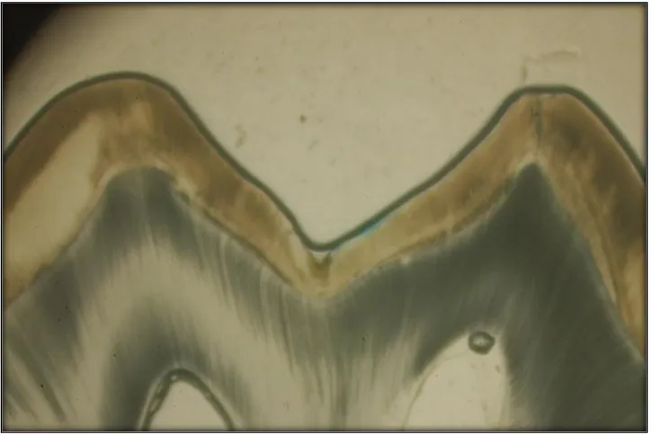
[image:63.595.115.480.463.708.2]Figure 5: Maxillary Molar Teeth – V Shape Fissure Pattern
Figure 7: Linear Depression
40
RESULTS
Table 1 shows the sample distribution that included 15 first primary
maxillary molar and 27 second primary maxillary molar, 28 first primary
mandibular molar and 30 second mandibular molar teeth. In this 56 (56 %)
sections showed U- type of fissure pattern 37 (37%) sections showed
V –type and 7 (7 %) sections showed other type of linear depression.
Table 2 shows fissure morphology where , U- type of fissure pattern
was more prevalent in the mandibular molars whereas the U and V – type
patterns were equidistributed in the maxillary molar sections. There was no
significant difference in the distribution of type of fissure pattern between first
and second molars (P ˃ 0.05)*** or between maxillary and mandibular molar
teeth (P ˃ 0.05)***