METHOD-A PROSPECTIVE STUDY
Dissertation Submitted to
THE TAMILNADU Dr. M.G.R. MEDICAL UNIVERSITY
In partial fulfillment for the Degree of MASTER OF DENTAL SURGERY
BRANCH III
ORAL AND MAXILLOFACIAL SURGERY
the foundation of my life and all my work.
I wish to thank my mother Mrs.Mekala for the sacrifices she made and for giving me a great foundation in my life and for being the most wonderful
mother . I thank my uncles Mr.Malarvannan, Mr.Swaminathan, Mr.Suriya Devan, Mr.Rajendran for being the pillars of my life and showering me love and encouragement.I sincerely thank Mr.Ezhavazhgan, Mr.Pannerselvam, Mr.Jothimani for believing in me & supporting me unconditionally.I thank my grandparents Mr.Azhagu sivanandan and Mrs.Mayavathi for praying and supporting me in all my works.
With deep satisfaction and immense pleasure, I present this work
undertaken as a Post Graduate student specializing in Oral and Maxillofacial
Surgery at Ragas Dental College and Hospital. I would like to acknowledge
my working on this dissertation which has been wonderful and enriching
learning experience.
I convey my heartfelt gratitude and my sincere thanks to my Head of
the department and my guide Professor Dr M Veerabahu, Department of Oral and Maxillofacial Surgery, Ragas Dental College and Hospital, Chennai
for his exceptional guidance, tremendous encouragement, well timed
consideration you have shown towards me. Your presence in the department
creates an aura and motivates me to reach heights. I would cherish these
memories throughout my life.
I would like to extend my heartfelt gratitude to Professor Dr N S Azhagarasan, Principal, Ragas Dental College and Hospital for allowing us to use the scientific literature and research facilitates of the college and for
providing the platform to meet such wonderful academicians and people.
I owe enormous gratitude to my guide Professor Dr Malini Jayaraj for her invaluable guidance and support throughout my course. She has always
been a source of provoking new thoughts in me. Her loving and caring nature
lightened the burden of many hardships. I shall forever remain thankful to her
for her valuable guidance and input throughout the making of this
dissertation. It was an enriching experience to have spent three years of my
life under her guidance.
I wish to convey my heartfelt thanks to Professor Dr B Vikraman, a great teacher who has always been a source of inspiration. His way of looking
I would also thank my Professor Dr J A Nathan for everlasting inspiration, constant encouragement, constructive criticism and valuable
suggestion conferred upon me throughout my postgraduate period.
I am greatly indebted to Dr Radhika Krishnan Anaesthesiologist, for imparting and sharing her vast experience in the field of medicine. I thank her
for her valuable suggestion and constant encouragement through my
postgraduate course.
I am grateful and sincerely thankful to Dr D. Shankar, Dr Sathya Bama, Dr Saneemand Dr Satish Readers, for their vehement personal interest, wish and never-ending willingness to render generous help to me
throughout my dissertation and post graduate with valuable advice.
I thank Dr Seema Alice Mathew, Dr James Bhagat, Dr Naren Kumar, Dr K.M. Harish Senior lecturers for their guidance, scholarly suggestion and whole-hearted support throughout my postgraduate course.
I thank Dr Venkatesh, Dr Raghu Department of surgical oncology Cancer Institute Adayar, Chennai, , for their vehement, valuable guidance and
SharnuSiviah , Dr Nambi Nayagi and Dr Nirmal for their support, constructive criticism at every step and selfless co - operation during my
dissertation. I wish them a successful career ahead.
I would like to thank my dear friends Dr Prasanth eager, Dr Sam prasanth,Dr.Nivetha,Mashshariff,ArunBalaji,Elakkuvan,Sindhupriya, Arulraj,Amudhan, Sathiyaseelan for inspiring and encouraging me during my postgraduate period. My heartfelt thanks to my dear friends, without them
my time wouldn’t have been enjoyable.
I offer my sincere thanks to my senior’s, Dr Narayana murthy, Dr Nirmal tony, Dr Sriraman, Dr Vivek, My Juniors Dr Manoj kumar, Dr Stephen, Dr Ajith, Dr Arun, Dr Deepan, Dr Kishok, Dr Diana, Dr Alka, Dr Veera Raghavan, Dr Aravindh sai for their encouragement and support during the course.
I sincerely thank Mr Thavamani, for helping in editing and printing of
my thesis. I would also thank Sis.Deepa, Sis.Laila,Sis. Leema, Sis.Mala for
The aim of this study is to evaluate the possibility of avoiding a
trans-alveolar method of extraction for mutilated tooth using physics forceps.
MATERIALS AND METHODS
In this prospective clinical study, a total of 30 patients seeking
transalveolar method of extraction were taken as study group. Out of 30
patients, complete success of extraction with physics forceps (Atraumatic
extraction) was selected as one group and failure to extract tooth with physics
forceps (Traumatic extraction) was selected as another group. Clinical
outcomes in form of operative time taken, gingival laceration, intra-operative
patient comfort, postoperative pain and healing were recorded and compared.
RESULTS
Statistically significant reduction in the operating time, marginal bone
loss, soft tissue loss, post-operative evaluvation of parameters including pain
,healing status and other complications were lesser in physics forceps
( Atraumatic extraction) when compared to transalveolar method of extraction
In our study, we would like to conclude that we could avoid
transalveolar extraction in 87% of mutilated teeth. Hence from the findings of
our study, we advocate that the use of physics forceps would reduce the rate
of transalveolar extraction in mutilated teeth. Further it provides a smoother,
uneventful post-operative healing in patients who require extraction of
mutilated teeth. We would like to highlight, inspite of the higher cost of
instrument and a steep learning curve, it would be commonly employed in
future to perform atraumatic extraction of mutilated tooth. In majority of cases
employment of transalveolar method can be avoided by proper usage of
physics forceps in mutilated tooth.
S.No TITLE PAGE NO
1. INTRODUCTION 1
2. AIMS AND OBJECTIVE 7
4. REVIEW OF LITERATURE 8
5. MATERIALS AND METHODS 19
6. RESULTS 32
7 TABLES AND GRAPHS 35
7 DISCUSSION 55
8 SUMMARY AND CONCLUSION 63
9 BIBLIOGRAPHY 66
[image:12.595.126.472.122.628.2]TABLE
NO
TITLE
1. DISTRIBUTION OF STUDY SUBJECTS ACCORDING TO GENDER
2. DISTRIBUTION OF STUDY SUBJECTS ACCORDING TO AGE
3.
DISTRIBUTION OF STUDY SUBJECTS ACCORDING TO THE
INDICATION FOR EXTRACTION
4.
DISTRIBUTION OF STUDY SUBJECTS ACCORDING TO THE
METHOD OF EXTRACTION
5.
DISTRIBUTION OF STUDY SUBJECTS ACCORDING TO THE
GINGIVAL CONDITION AT THE TIME OF EXTRACTION
6.
DISTRIBUTION OF STUDY SUBJECTS ACCORDING TO THE
TIME TAKEN TO COMPLETE THE PROCEDURE
7.
DISTRIBUTION OF STUDY SUBJECTS ACCORDING TO THE
STATUS OF ALVEOLAR BONE (SOCKET INTACT) AT THE
TIME OF EXTRACTION
8.
DISTRIBUTION OF STUDY SUBJECTS ACCORDING TO THE
STATUS OF POST-OPERATIVE COMPLICATIONS REVIEWED
FORCEPS(VAS SCALE) REVIEWED ON DAY 3, DAY5,DAY 7
FROM THE DAY OF EXTRACTION
10.
DISTRIBUTION OF STUDY SUBJECTS ACCORDING TO THE
STATUS OF POST-OPERATIVE PAIN RECORDED FOR
PATIENTS WHO UNDERWENT EXTRACTION WITH
TRANS-ALVEOLAR METHOD(VAS SCALE) REVIEWED ON DAY 3,
DAY5,DAY 7 FROM THE DAY OF EXTRACTION
11
DISTRIBUTION OF STUDY SUBJECTS ACCORDING TO THE
HEALING CONDITION RECORDED FOR PATIENTS WHO
UNDERWENT EXTRACTION USING PHYSICS FORCEPS
(LANDRY ET AL) REVIEWED ON DAY 3, DAY5,DAY 7 FROM
THE DAY OF EXTRACTION
12.
DISTRIBUTION OF STUDY SUBJECTS ACCORDING TO THE
HEALING CONDITION RECORDED FOR PATIENTS WHO
UNDERWENT EXTRACTION WITH TRANSALVEOLAR
METHOD (LANDRY ET AL) REVIEWED ON DAY 3, DAY5,DAY
1
INTRODUCTION
An extraction of tooth is considered as a very traumatic procedure by
any layman due to the horrifying experiences associated with the tooth
extraction in the past. Even in this current era; despite modern methods of
anaesthesia, removal of tooth is the most dreaded procedure in majority of
patients.
The history of dental extraction forceps goes back to the time of
Aristotle (384 to 322 BC) wherein Aristotle described the mechanics of
extraction forceps. This was documented 100 years before by Archimedes
who studied and discussed the principle of the lever. A tool called a pelican
(as it resembled a large-beaked bird ) was developed in the 14th century, and
was put in use to extract teeth untill the 18th century. The modern tooth
extraction forceps became popular in the early 20th century. Until the early
1900s, travelling dentists performed extractions at town fairs, where musicians
were instructed to play loud enough to cover the cries of pain of those unlocky
patients who had their teeth extracted.
An ideal tooth extraction is defined as the painless removal of the
whole tooth, or tooth-root with minimal trauma to the investing tissues, so that
the wound heals uneventfully and no postoperative prosthetic problem is
2
The three mechanical principles of extraction are: - 1.Expansion of the
bony socket to permit the removal of its contained tooth. This is achieved by
using the tooth as the dilating instrument, and is the most important factor in
„forceps extraction‟. In order to ensure access, it is recommended that
sufficient tooth structure be clinically available to be firmly grasped by the
forceps blades. The root pattern of the tooth must be such that it permits
dilatation of the socket sufficiently to facilitate the complete dislocation of the
tooth from its socket. The socket can be dilated only if the alveolar bone is
sufficiently elastic to permit such expansion. If the root pattern or consistency
of the investing bone is such that the dilation of the socket is impracticable,
then the trans-alveolar method of extraction, with or without the division of
the roots of multi-rooted teeth, must be employed 2. The use of a lever and
fulcrum to force a tooth or root out of the socket along the path of least
resistance 3.The insertion of a wedge or wedges between the tooth-root and
the bony socket wall, thus causing the tooth to rise in its socket.
Various possible complication in tooth extraction includes failure to
secure anaesthesia, failure to remove the tooth with either forceps or elevators;
fracture of crown or roots of tooth being extracted; fracture of alveolar bone,
maxillary tuberosity, adjacent or opposing tooth;fracture of mandible;
displacement of a root into the soft tissues, into the maxillary antrum;
excessive haemorrhage during tooth removal or on completion of the
3
its branches, lingual nerve, tongue and floor of mouth; postoperative pain may
be due to damage to hard and soft tissues, dry socket; postoperative swelling
due to oedema, haematoma formation, infection, trismus, creation of
oro-antral communication.
Delayed wound healing is one of the most common problem that can
occur after tooth extraction .It may be attributed to traumatic extraction which
causes great damage to the gingival tissue.. Gingival pain, swelling,
inflammation are the common problems that the patients usually encounter the
day after extraction. Clinically, traumatic damage to the dentoalveolar housing
during extraction can result in significant ridge deformities upon healing
Dar Es Salaam41 in his study found out that the frequency of post
extraction complications was low , and was mainly due to, infected sockets ,
followed by bleeding sockets and retained roots . In addition to the above
mentioned complications, he mentioned eight complications suffered by
patients, which includes necrotising fasciitis , herpes zoster, ludwig's angina,
infections of the submandibular , parapharyngeal , masticator and
submasseteric spaces ,and reaction to local anaesthesia (2ml of 2 per cent
lignocaine hydrochloride.
Transalveolar extraction method is the surgical technique which is
employed for recovering roots that were fractured during routine extraction of
teeth which could not be extracted by the routine closed methods for a
4
muco-periosteal flap, cutting of the bone obstructing the removal of the tooth
and if required sectioning of the roots and then removal. Postoperative
complications commonly include haemorrhage ,pain ,swelling.
Hence “Atraumatic dental extraction“ have gained prominence and
may ultimately become the standard technique for tooth removal which
preserves bone, gingival architecture and aids in preservation of the socket for
future implant or immediate dental implant placement.
One of the tool and technique that have been proposed for minimally
invasive tooth removal was by using “ PHYSICS FORCEPS”. Various other
advanced technique include powertome, proximators, periotomes and benex
extractor
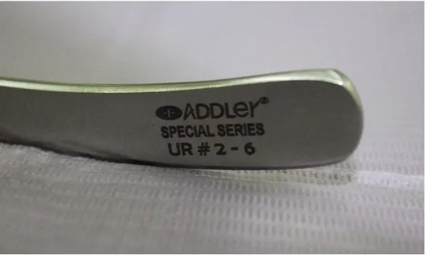
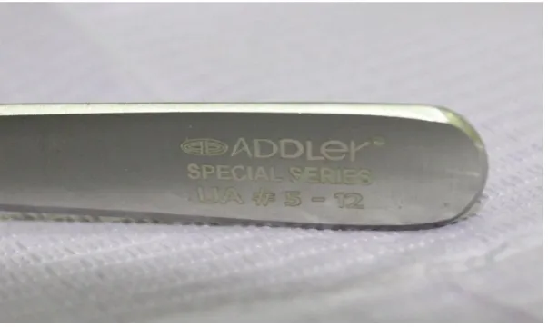
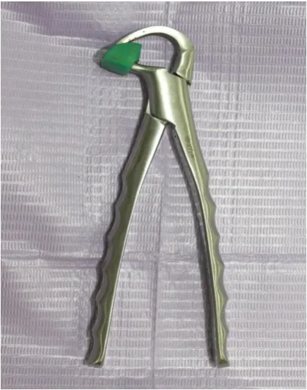
The Physics Forceps (ADDLEYTM) (Fig 1-8) is an atraumatic
extraction system that provides a simple, predictable and unconventional
method of extracting teeth regardless of a practitioner‟s extraction experience,
or the condition of the tooth, while providing a positive patient experience.
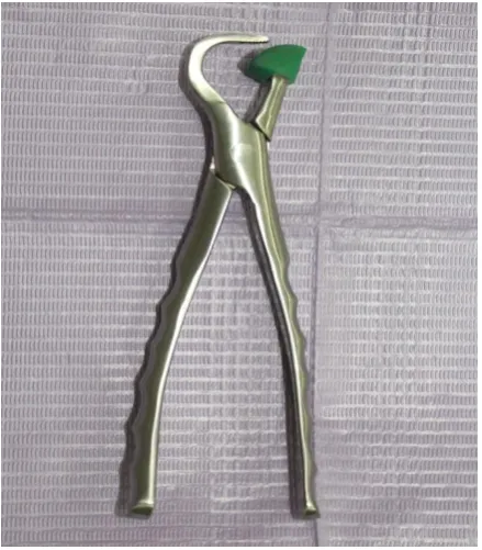
The Physics Forceps operate as an elevator, rather than forceps, using
first-class lever mechanics. One handle is connected to a "bumper" (Fig 9),
which acts as the fulcrum, that is placed deep in the vestibule. The other
handle is connected to the "beak," which is positioned most often on the
lingual or palatal root of the tooth into the gingival sulcus. No aspect of the
instrument grasps the crown and there is no advanced elevation needed. Once
5
only wrist movement in a slow, steady and controlled manner — no
squeezing.
The Physics Forceps are effective in most cases, including grossly
decayed or broken down teeth, endodontically treated teeth, fractured or
fragile teeth, curved or long rooted molars and cuspids, or any tooth that
would historically be challenging with conventional instrumentation. The
Physics Forceps is ideal for an implantologist who aims to preserve the
surrounding bone and tissue in preparation for placement of dental implants.
The Physics Forceps applies a constant and steady load on the tooth,
allowing "creep" to build, releasing hyaluronic acid, resulting in the
breakdown of the periodontal ligaments. This forceps works in shorter time
period than conventional methods that employ intermittent, rocking or brute
strength types of forces.
The principles of biomechanics are the basis for the development of
the physics forceps. This instrument was developed by Golden in 2004 and
has been modified with the help of several doctors. Implementation of a
first-class lever, creep, and the type of force provides the mechanical advantages
necessary to make this dental extraction devices more efficient.
The extraction of a tooth using Physics Forceps is similar to the
removal of a nail from wood using a hammer verses a pair of pliers
6
hammer‟s claw fit under the head of a nail. The hammer‟s head acts as a
fulcrum. A rotational force applied to the hammer handle magnifies the force
by the length of the handle, and the nail is elevated from the wood. Unlike a
nail in wood with parallel sides and friction along its full length, a tooth is
tapered. After being elevated a few millimeters, the periodontal ligament
fibres are broken and the tooth may then be easily removed without additional
rotational force.
The aim of this study is to evaluate the possibility of avoiding a
trans-alveolar method for extraction of mutilated tooth using physics forceps.
7
AIMS AND OBJECTIVES
The aim of this study is to evaluate the possibility of avoiding a
8
REVIEW OF LITERATURE
Dr.Soumen Mandal, Dr. Sunil Kr. Gupta, Dr. Ankur Mittal, Dr. Ritesh Garg (2015)13 evaluated efficacy of physics forcep in non-surgical
mandibular multirooted tooth extractions.They compared outcome variables
(laceration, cortical plate fracture, post-operative pain and complication) in
patients being treated for their multirooted tooth extraction with the physics
forcep and the conventional forcep and organised a double blind, randomized
controlled trial in which p value came to be statistically significant in support
following parameter for physics forcep. (p value Laceration of gingival tissue 0.032, cortical plate fracture 0.001, post-operative pain 0.035 and average time
taken for extraction was 2.33 with a standard deviation of ±1.588) .This study
concluded that physics forceps can be used as a helpful aid in atraumatic
extraction of mandibular tooth, in addition to decreasing patient’s
post-operative discomfort it also maintain the socket integrity by preventing any
damage to the soft tissue and hard tissue architecture and thus making
placement of future prosthesis easier.
Timothy Kosinski,DDS,MAGD (2012)25 in his study stated that the patient who underwent extraction with physics forceps were satisfied with
9
Harsh S Patel,Anil M Managutti,Shailesh Menat,Arvind Agarwal,Dishan Shah,Jigar Patel (2016)20 in their study compared the efficacy of physics forceps with conventional forceps in terms of operating
time, prevention of marginal bone loss & soft tissue loss, postoperative pain
and postoperative complications following bilateral premolar extractions for
orthodontic purpose. They found mild swelling on 1st postoperative day in
2.38% (1 case) of extraction done by physics forceps and 4.76% (2 cases)
extractions done by physics forceps. Tooth fracture (irrespective of crown and
root fracture) was noted in 6% of total extractions which was equal in both
physics forceps and conventional forceps groups (3% each). Buccal cortical
fracture was noted in 4.76% of total extractions done by conventional forceps
and no cortical fracture in extractions done by conventional forceps. This
study suggested that physics forceps was more efficient in reducing operating
time and prevention of marginal bone loss & soft tissue loss when compared
to conventional forceps in orthodontically indicated premolar extractions.
Khaled K. Abou-ayasha, Sherif S. Ayyad, Riham M. Eldibany (2016)24 designed a study to clinically and radiologically evaluate immediate implant placement into fresh extraction sockets of maxillary anterior teeth
following extraction by physics forceps. This study was conducted on ten
adult patients of both sexes with age ranged between 20-40 years, having
10
rehabilitation. Patients were divided into two groups; each group contained
5 patients (a study group and a control group), in the study group, teeth
extraction was performed using the maxillary anterior physics forceps while in
the control group extraction was performed using the maxillary anterior
conventional forceps. Clinical evaluations were done in the immediate
post-operative period, after one week, after 1, 3 and 6 months. Radiographic
evaluations were done after 1, 3 and 6 months. Clinical results revealed that
there were statistically significant (p=0.016) results obtained in each group
separately throughout the evaluation period. This study concluded that
immediate implant placement following tooth extraction using Physics
forceps showed superior results in the immediate post-operative phase alone.
N.J. Perkins, H.M. Perez, C.E. Misch, R. Golden. St. Helen’s (2010)39 in their study stated that as with any new technique there is a learning curve for operators in making the transition from conventional forceps to
Physics Forceps, especially the effective use of wrist movement. Physics
Forceps represent an important addition to the armamentarium for atraumatic
11
Soumen Mandal, Shriparna Biswas Mandal,Vaibav Kamal,Avanindra Kumar,Kamna Gorka,Avanish Kumar (2015)13 in their study evaluated and determined the efficacy of the physics forceps in
nonsurgical mandibular single-rooted tooth extraction.The comparision
between the mean time taken for the extraction of a single-rooted tooth with a
physics forceps and a conventional forceps, which was 1.868 minutes with
physics forceps with a standard deviation of 1.503 minutes whereas with a
conventional forceps the mean time was 2.584 minutes with a standard
deviation of 1.831 minutes.23 out of 25 subjects reported no laceration with
the use of physics forceps whereas 8 out of 25 subjects reported laceration
with the use of conventional extraction forceps. This study concluded that
the extraction using the physics forceps were predictable in time commitment,
faster ,less traumatic physically and pshychologically to the patient
Sathish Madathanapalli, Sanidhya Surana,Deepak Thakur, Pooja Ramnani, Sheetal Kapse (2016)29 conducted a study to compare the efficacy of physics forceps vs conventional forceps for the extraction of the right and
left maxillary 1 st molars. They compared the physics forceps with the
conventional forceps for the removal of maxillary 1 st molars in 30 patients
under the following parameters time taken, post operative pain on the
3rd,5th,7th day, incidence of crown/root/buccal plate fracture during extraction.
12
pain on the 3 rd postoperative day (p=0.031).On comparing all parameters
,they found out that the utility of this physics forceps is better in comparison to
the conventional forceps.
Marco Cicciù, Ennio Bramanti, Fabrizio Signorino, Alessandra Cicciù and Francesco Sortino (2013)11 in their article analysed all the applied movements when extracting healthy upper and lower jaw premolars
for orthodontic purposes. The results of the study demonstrated how the
strength, needed for performing the first upper premolar extraction is 25 times
higher than the lower jaw. This confirmed that tooth extraction of multi-root
teeth will result in a very difficult surgical operation. The rocking movement
recorded high values of +199 Nmm while the twisting recorded maximum
values of +8,69 Nmm. This data showed how the strength needed for double
rooted teeth extraction is not proportional to the strength needed for single
ones.This study concluded complications of tooth extraction are connected to
13
Egon Muska, DDS, Clemens Walter, DMD, Alan Knight, BDS, Pankaj Taneja, BDS, Yogesh Bulsara,Michael Hahn, BDS, Mayur Desai, and Thomas Dietrich, MD, DMD, MPH (2013)35 study conducted to evaluate the applicability and limitations of a novel atraumatic extraction
system. The present study demonstrated a high success rate for the atraumatic
extraction of severely decayed teeth not suitable for forceps extraction with
the use of a novel vertical extraction system. Ninety-eight out of 111 teeth
were successfully extracted with the device, indicating an overall success rate
of 83%. The success rate was higher in single-rooted teeth (89%), whereas
fewer than one-half (43%) of multirooted teeth were successfully extracted.
The results of this proof of principle study suggest that:1) the Benex extractor
system may be successfully used for atraumatic tooth extraction 2) the system
has a higher success rate with single-rooted teeth compared with multirooted
teeth.
Harry Dym, DDS, Adam Weiss, DDS(2012)14 their article reviewed and highlighted exodontia tips as well as new techniques to make simple and
complex exodontia more predictable and efficient with improved patient
outcomes. This article summarized that dentist requires some practice before
achieving complete mastery in the use of the physics forceps because the
14
extractions; however, once comfortable, the clinician will marvel at the ease
and the little force required to remove even difficult teeth.
Rudys Rodolfo De Jesus Tavarez, Washigton Luís Machado dos Reis, Adrycila Teixeira Rocha, Leily Macedo Firoozmand, Matheus Coêlho Bandéca, Mateus Rodrigues Tonetto, Adriana Santos Malheiros (2013) 52 presented a clinical case where the extraction was performed using atraumatic extractor with implant placement and immediate provisionalization
in a maxillary lateral incisor.The clinical case described in this paper
demonstrates a sequence of clinical atraumatic extraction, and then the
Immediate installation provisionalization. It was concluded that when
carefully indicated and planned, this technique can provide an immediate
result promising with maintaining the tooth gingival contour.
Mohamed H. El-Kenawy • Wael Mohamed Said Ahmed (2015)28
conducted a study to evaluate the efficacy of physics forceps versus
conventional forceps in simple dental extraction . The incidence of crown,
root, and buccal bone plate fractures decreased while using physics forceps
when compared to the conventional forceps. In physics forceps group(100
cases): crown fracture occurred in three cases (3 %), buccal bone fracture
15
while in conventional forceps group(100 cases): crown fracture occurred in 10
cases (10 %), buccal bone fracture occurred in seven cases (7 %), and root
fracture occurred in 27 roots(16.6 %).This study suggested that the physics
forceps and its associated beak and bumper technique is clinically valuable in
atraumatic tooth removal and in preserving the buccal bone plate, which is
mainly critical for implant dentistry.
Sonune Avinash M., Borle Rajiv M., JadhavAnendd A. (2017)47
conducted a study to evaluate the efficacy between the conventional
extraction forceps and physics forceps in orthodontic extraction of maxillary
premolars. In this study, mean time taken for extraction by conventional
forceps(19.42 sec) was found to be significantly lesser as compared to
physics forceps (33.64 sec) and there was buccal cortical plate fractures in 12
patients while using the conventional extraction forceps and no buccal plate
fracture with use of physics forceps group. This study suggested that physics
forceps have certain advantages over conventional method of extraction in
multi rooted, decayed tooth but there are no such obvious results found to
indicate consideration of physics forceps over conventional forceps for
16
Yong-Hoon Choi*, Ji-Hyun Bae (2011)10 in their study stated atraumatic safe extraction (ASE) can be regarded as a reproducible,
predictable method of extraction for Intentional Replantation. A Physics
Forceps was used for extraction and the success rate of ASE was assessed.
Ninety-six premolars and molars were treated by Intentional Replantation. The
complete success rate (no crown and root fracture) was 93% (n = 89); the
limited success rates because of partial root tip fracture and partial osteotomy
were 2% (n = 2) and 5% (n = 5), respectively. The clinical and overall success
rates of ASE were 95% and 100%, respectively; no failure was observed. This
study concluded that the ASE method should be considered as reliable
extraction method for safe and successful Intentional Replantation; it is
expected to contribute greatly to save natural teeth. However, long-term
observation of its prognosis is required.
Samyuktha Hariharan, Vinod Narayanan, Chen Loong Soh (2014)19 compared outcome variables (operative complications, inflammatory complications, and operating time) in patients being treated by orthodontic
extraction of upper premolars with the Physics forceps or the universal
extraction forceps. They organised a single blind, split mouth clinical trial to
compare the outcomes of the 2 groups (n=54 premolars). The Physics forceps
group had lower mean (SD) visual analogue scores (VAS) for pain (0.59
17
(p=0.03). There were no other significant differences between the 2 groups in
any other variable studied. The authors reported that the mean operating time
was less using Physics forceps (120.5seconds) than with the universal
extraction forceps (188.6seconds). The incidence of fractures of the bony plate
fractures was 22% in the Physics forceps group compared with 25% in the
conventional forceps group and found that the mean operative time using the
Physics forceps for extraction of upper premolars was 21.4 (27.3) seconds and
43.5 (49.5) seconds for the universal extraction forceps
Charles A. Babbush, DDS, MScD (2007)6 in their article reviewed the mechanisms involved in traditionally extracting teeth, and discusses a
technological breakthrough that enables non molar intact teeth to be extracted
mechanically and atraumatically. This article summarized “The Easy X-Trac
System” represents a revolution in exodontia. Instead of pulling and twisting
the tooth root to be extracted, the surgeon secures a screw within the root, then
uses a simple mechanical device to lift the screw and root atraumatically.
Carl E.Misch, DDS MDS; Helena M.Perez, DDS (2008)33 in their article reviewed the biomechanics of a periotome, an elevator, and extraction
forceps. Those methods will be compared to the biomechanical principles used
18
summarized the mechanical advantages available to extract the teeth were
primarily applied to hold the crown of the tooth, rather than aiding in its
extraction. An extraction device (physics forceps) has been developed to apply
a biomechanical rationale to the extraction process of a tooth using a class 1
19
MATERIALS AND METHODS
This study was conducted at Department of Oral and Maxillofacial
Surgery, Ragas Dental college and Hospital, Uthandi. A total of 30 patients
who required tooth extraction were included in this clinical study. A clearance
for performing the study was obtained from the institutional ethical
committee, and each patient was given a brief description of the intended
procedure with follow period and was required to sign an informed consent
sheet.
The recorded Data included age, sex, chief complaint, medical and
dental history, clinical findings, radiographic findings, duration of procedure,
The following parameters was evaluvated, ie PAIN-(VAS) ,GINGIVAL
INDEX- (Landry et al) on 3rd,5th,7th post-operative day.
INCLUSION CRITERIA
Ellis class III,IV,V,VI,VII,VIII,IX
Grossly decayed tooth
Fracture of teeth below alveolar crest
20
EXCLUSION CRITERIA
• Ellis class I fracture • Ellis class II fracture
• Severely malpositioned teeth
• Tooth with Grade II, Grade III mobility • Teeth with true pocket
• Teeth with clinical attachment loss • Medically compromised patients • Patients not willing for extraction
ARMAMENTARIUM USED
• 2% lignocaine with1:80,0000 adrenalin • Molt no 9 periosteal elevator
• William’s probe
• Maxillary physics forceps • Mandibular physics forceps • Gauze
• Suture material ( if needed )
21
SURGICAL TECHNIQUE DURING EMPLOYMENT OF PHYSICS
FORCEPS
• Tooth to be extracted is anaesthetised with Local Anaesthesia
(2% Lignocaine with 1:80,000 adrenalin )
• Lingual and buccal gingival attachment is separated from the tooth
using mucoperiosteal elevator (Molt No.9)
• Forceps handles are opened widely, beak is set into the depth of the
lingual or palatal sulcus on solid root surface. Secure purchase point on
solid root surface is critical to successfully rolling out the tooth.
• The bumper (which is covered by plastic or rubber to avoid trauma to
the buccal soft tissues) is set perpendicular to the tooth at about the
level of the mucogingival junction.
• Position was held securely without squeezing the handles.
• A steady and slow rotational force is applied in the direction of the
bumper without squeezing the handles or moving the arm.
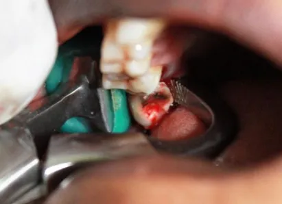
• As the periodontal ligament disengages, the tooth literally “pop”
(Fig 12,15).
• After that releasing pop, the tooth is easily delivered with a
22
• All the patients were prescribed following medications: Cap.
Amoxicillin 500mg (TID)-3days,Tab.Metrogyl 400mg
(TID)-3days,Tab. Diclofenec Sodium 50mg (BD)-3 days, Tab. Ranitidine
150mg (BD)-3days
EVALUATION CRITERIA
Tooth delivered using
PHYSICS FORCEPS OPEN METHOD
Trauma to the gingiva (mucosal tear)
YES NO
Fracture of alveolar bone (socket intact)
YES NO
Time taken for procedure ----
23
HEALING INDEX -LANDRY ET AL
Very poor
-tissue colour: greater than or equal to 50% of gingiva red
-response to palpation: bleeding
-granulation tissue: present
- incision margin: not epithelialised, with loss of epithelium
beyond incision margin
- suppuration present
Poor
-tissue colour: greater than or equal to 50% of gingiva red
-response to palpation: bleeding
-granulation tissue: present
-incision margin: not epithelialised with connective tissue
exposed
Good
-tissue colour: greater than or equal to and less than 50% of
gingiva red
-response to palpation: no bleeding
24
-incision margin: no connective tissue exposed
Very good
-tissue colour: less than 25%of gingiva red
-response to palpation: no bleeding
-granulation tissue: none
-incision margin: no connective tissue exposed
Excellent
-tissue colour: all tissues pink
-response to palpation: no bleeding
-granulation tissue: none
-incision margin: no connective tissue exposed
DAY-3
POST-OP Healing index (Landry et al )
Very Poor
Poor
Good
Very Good
25
POST-OP PAIN (VAS)
DAY-5
POST-OP Healing index (Landry et al )
Very Poor
Poor
Good
Very Good
26
POST-OP PAIN (VAS)
DAY -7
POST-OP Healing index (Landry et al )
Very Poor
Poor
Good
Very Good
27
28
CASE HISTORY
OP NO
DATE
NAME
AGE/SEX
OCCUPATION
WEIGHT
RELIGION
PHONE NO
ADDRESS
CHIEF COMPLAINT
29
PAST MEDICAL HISTORY
PAST DENTAL HISTORY
PERSONAL HISTORY
EXTRA-ORAL EXAMINATION
FACE -
LIPS -
TEMPOROMANDIBULAR JOINT –
30
INTRA-ORAL EXAMINATION
INVESTIGATIONS
DIAGNOSIS
TREATMENT PLAN
31
PROCEDURE
STARTING TIME –
FINISHING TIME –
TIME TAKEN FOR PROCEDURE -
TOOTH DELIVERED USING
PHYSICS FORCEPS
OPEN METHOD
TRAUMA TO THE GINGIVA (MUCOSAL TEAR )
YES
NO
INSTRUMENTATION
[image:51.595.149.450.529.708.2]Fig 1.Right maxillary posterior physics forceps
Fig 3.Left maxillary posterior physics forceps
Fig 5.Maxillary anterior physics forceps
Fig 7.Mandibular universal physics forceps
Fig 9. Bumper –which act as a fulcrum
CASE I
Fig 11.Pre-operative IOPA
Fig 12.Intra-operative application of physics forceps
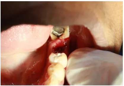
[image:56.595.194.400.351.500.2]CASE II
Fig 14.Pre-operative IOPA
[image:57.595.193.403.542.687.2]Fig 15. Intra-operative application of physics forceps
32
RESULTS
The present prospective, clinical study was conducted to evaluvate the
possibility of avoiding a trans-alveolar method for extraction of mutilated
tooth using physics forceps. In our study, 30 patients who required extraction
of mutilated tooth was taken into the study design. All those 30 patients
reported to the outpatient Department, Department of oral and maxillofacial
surgery, Ragas Dental College, Uthandi, Chennai for tooth extraction.
All 30 patients were examined and assessed with radiographs
(Fig 11,14) .Out of 30 patients, (Table,Graph 1) 26 were male and 4 were
female in ratio of 6.5:1 . The mean (SD) age who underwent extraction using
physics forceps was 37.92 years(Table 13), range 21-65, of which 9 were
female and 17 male whereas who underwent transalveolar extraction was
28.25 years(Table 13) ,range from 21-65 of which 2 were female and 2 male.
Out of 30 patients,26 patients underwent extraction using physics
forceps and 4 patients underwent transalveolar method of extraction (Table 4).
All 4 patients with transalveolar method (Table,Graph 3) had grossly decayed
with root canal treated few years back.
On day of extraction; operating time, patient comfort, gingival trauma
were assessed for made for both the group of patients who underwent
33
Postoperative healing was recorded with HEALING INDEX-LANDRY ET
AL and postoperative pain was recorded on VAS SCALE .Both were
recorded on 3rd ,5th,7th post-op day.
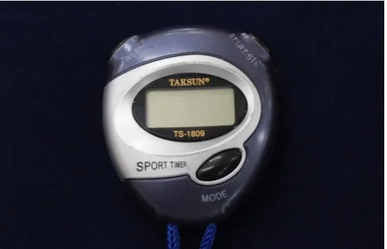
EXTRACTION TIME: Operating time was recorded using a stop
watch (Fig 10) from the beginning of extraction after the local anaesthetic had
been injected to the completion of the extraction. Time taken to extract a tooth
can be considered as a time period from engaging a tooth by forceps to
completely removing it out of socket. Out of 26 subjects who underwent
extraction using physiscs forceps, (Table,Graph 6) 18 reported extraction
below 5mins;8 reported between 5-10 mins; and none reported more than 10
min. Out of 4 subjects who underwent transalveolar extraction reported
extraction time more than 10 min. The operating time was shorter when the
patient underwent extraction using Physics forceps than for the transalveolar
extraction and there was statistically significant difference in the operating
time between two groups(p<0.001)
PATIENT COMFORT: All 26 patients had good comfort during
extraction with physics forceps while 4 patients had mild discomfort with
transalveolar extraction, there was statistically significant difference in the
patient comfort between two groups (p<0.001)(Table 13)
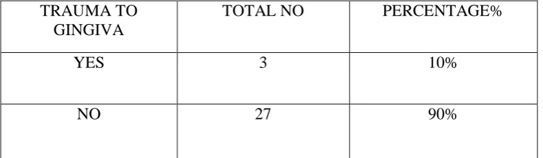
TRAUMA TO GINGIVA: Trauma to gingiva was noted in 3 patient
34
(Table,Graph 5), there was statistically significant difference in the gingival
trauma between two groups (p<0.014)(Table 13)
PAIN SCORE: Postoperative pain measured on VAS scale was
recorded on 3rd, 5th,7th post-op day. Out of 26 patients (Table 9), only 7
reported with pain on 3rd day with physics forceps. On follow up on 5th day,
only 1 reported with pain and on 7th day none reported with pain. Mean VAS
score within group (physics forceps) was 0.001(Table 15). Out of 4 patients
who underwent transalveolar extraction, (Table 10) 3 reported with pain on
3rd day. On follow up on 5th day 2 reported with pain and on 7th day none
reported with pain. Mean VAS score within group (transalveolar extraction)
was 0.082(Table 15).Mean VAS Score (Table 15) on 3rd post-op day with
physics forceps and transalveolar extraction were 0.002 and on 5th day 0.039
.There was statistically significant reduction in the pain score in physics
forceps group postoperatively.
HEALING SCORE: All 26 patients who underwent extraction using
physics forceps had good healing postoperatively and hence mean healing
score were p<0.001.While all 4 patients who underwent transalveolar
extraction had poor healing on 3rd day post-op, and 3 had poor healing on 5th
day post-op and on 7th day all 4 had good gingival healing. Hence mean
healing score with transalveolar extraction were p<0. 039.There was
statistically significant difference between two groups in the gingival healing
35
TABLE1: DISTRIBUTION OF STUDY SUBJECTS ACCORDING TO GENDER
TABLE2: DISTRIBUTION OF STUDY SUBJECTS ACCORDING TO AGE
AGE TOTAL NO PERCENTAGE%
18-28
10 33.3%
29-38
9 30%
39-49
7 23.3%
50 ABOVE
4 13.3%
GENDER TOTAL NO PERCENTAGE %
MALE 19 63.4%
36
TABLE 3: DISTRIBUTION OF STUDY SUBJECTS ACCORDING TO THE INDICATION FOR EXTRACTION
INDICATION FOR EXTRACTION
TOTAL NO PERCENTAGE%
GROSSLY DECAYED 20 66.7%
FRACTURED TOOTH 3 10%
ROOT CANAL TREATED TOOTH
4 13.3%
FRACTURE OF TOOTH BELOW ALVEOLAR
CREST
3 10%
TABLE 4: DISTRIBUTION OF STUDY SUBJECTS ACCORDING TO THE METHOD OF EXTRACTION
TOOTH DELIVERED TOTAL NO PERCENTAGE%
PHYSICS FORCEPS 26 86.7%
37
TABLE 5: DISTRIBUTION OF STUDY SUBJECTS ACCORDING TO THE GINGIVAL CONDITION AT THE TIME OF EXTRACTION
TRAUMA TO GINGIVA
TOTAL NO PERCENTAGE%
YES 3 10%
[image:65.595.98.496.455.558.2]NO 27 90%
TABLE 6: DISTRIBUTION OF STUDY SUBJECTS ACCORDING TO THE TIME TAKEN TO COMPLETE THE PROCEDURE
TIME TAKEN FOR PROCEDURE
TOTAL NO PERCENTAGE%
0-5 MIN 18 60%
5-10 MIN 8 26.7%
38
TABLE 7: DISTRIBUTION OF STUDY SUBJECTS ACCORDING TO THE STATUS OF ALVEOLAR BONE (SOCKET INTACT) AT THE
TIME OF EXTRACTION
CONDITION OF ALVEOLAR BONE (SOCKET INTACT)
TOTAL NO PERCENTAGE%
YES 27 90%
[image:66.595.103.495.562.710.2]NO 3 10%
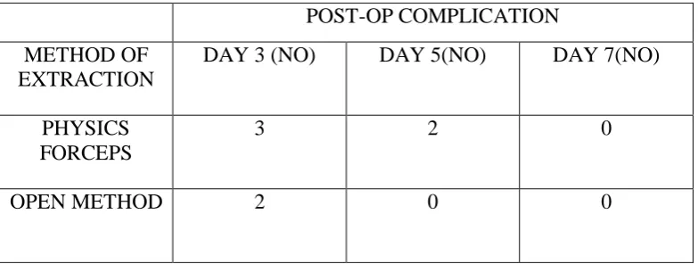
TABLE 8: DISTRIBUTION OF STUDY SUBJECTS ACCORDING TO THE STATUS OF POST-OPERATIVE COMPLICATION REVIEWED
ON DAY 3, DAY5, DAY 7 FROM THE DAY OF EXTRACTION
POST-OP COMPLICATION
METHOD OF EXTRACTION
DAY 3 (NO) DAY 5(NO) DAY 7(NO)
PHYSICS FORCEPS
3 2 0
39
TABLE 9: DISTRIBUTION OF STUDY SUBJECTS ACCORDING TO THE STATUS OF POST-OPERATIVE PAIN RECORDED FOR PATIENTS WHO UNDERWENT EXTRACTION USING PHYSICS
FORCEPS (VAS SCALE) REVIEWED ON DAY 3, DAY5,DAY 7 FROM THE DAY OF EXTRACTION
POST-OP PAIN-VAS ANALOGUE
PAIN PERCENTAGE%
DAY 3 7 27%
DAY 5 1 3.9%
[image:67.595.134.460.570.682.2]DAY 7 0 0%
TABLE 10: DISTRIBUTION OF STUDY SUBJECTS ACCORDING TO THE STATUS OF POST-OPERATIVE PAIN RECORDED FOR
PATIENTS WHO UNDERWENT EXTRACTION WITH TRANS-ALVEOLAR METHOD(VAS SCALE) REVIEWED ON DAY 3,
DAY5,DAY 7 FROM THE DAY OF EXTRACTION
POST-OP PAIN-VAS ANALOGUE
PAIN PERCENTAGE%
DAY 3 3 75%
DAY 5 2 50%
40
TABLE 11: DISTRIBUTION OF STUDY SUBJECTS ACCORDING TO THE HEALING CONDITION RECORDED FOR PATIENTS WHO
UNDERWENT EXTRACTION USING PHYSICS FORCEPS (LANDRY ET AL) REVIEWED ON DAY 3, DAY5,DAY 7 FROM THE
DAY OF EXTRACTION
POST-OP HEALING DAY 3 DAY 5 DAY 7
VERY POOR 0 0 0
POOR 0 0 0
GOOD 26 0 0
VERY GOOD 0 26 0
41
TABLE 12: DISTRIBUTION OF STUDY SUBJECTS ACCORDING TO THE HEALING CONDITION RECORDED FOR PATIENTS WHO
UNDERWENT EXTRACTION WITH TRANSALVEOLAR METHOD (LANDRY ET AL) REVIEWED ON DAY 3, DAY5,DAY 7 FROM THE
DAY OF EXTRACTION
POST-OP HEALING DAY 3 DAY 5 DAY 7
VERY POOR 0 0 0
POOR 4 3 0
GOOD 0 1 0
VERY GOOD 0 0 4
42
STATISTICS:
Inferential statistics was used for comparision between the two groups
(Physics forceps and Transalveolar method of extraction).Chi square and
Mann-Whitney for intergroup comparision; . Kruskal Wallis for within group
comparision; Friedman Test was done for within group comparision at
[image:70.595.86.512.380.643.2]different time intervals
Table 13; Descriptives:Distribution and comparision of study group characteristics at base line.
*Kruskal Wallis ǂ Mann-Whitney Type of procedure Total Subjects Mean Age (yrs)
Sex Time taken Patient comfort
Gingival Trauma
Male Female 0-5 min
5-10 min
>10 min
Yes No Yes No
Physics forcep
26 37.92 ± 14.3
17 9 18 8 - 26 - - 26
Trans- alveolar
4 28.25 ± 6.02
2 2 - - 4 - 4 3 1
43
Table 14; Healing Index:Distribution and comparision of characteristics of study groups at different time intervals.
*Chi Square ǂFriedman Test
Time
Physics Forceps Transalveolar
Very Poor
Poor Good Very Good
Excellent Very Poor
Poor Good Very Good
Excellent P value
3rd DAY
- - 19 7 - - 4 - - - <0.001
5th DAY
- - 6 15 5 - 3 1 - - <0.001
7th
DAY
- - - 13 13 - - 4 - - <0.001
P
44
Table 15; Pain Index: Distribution and comparison of characteristics of study groups at different time intervals
Time
Physics Forceps Transalveolar P
Value*
No Pain
Mild Moderate Severe No Pain Mild Moderate Severe
3rd DAY
19 6 - 1 1 1 2 - .002
5th DAY
25 1 - - 2 2 - - .039
7th
DAY
26 - - - 4 - - - -
P
Valueǂ
0.001 0.082
45 0 2 4 6 8 10 12 14 16 18 20
MALE(19) FEMALE(11)
GRAPH 1: DISTRIBUTION OF STUDY
SUBJECTS ACCORDING TO GENDER
MALE(19) FEMALE(11)
0 2 4 6 8 10 12 14
18-28 29-38 39-49 50 ABOVE
GRAPH 2: DISTRIBUTION OF STUDY
SUBJECTS ACCORDING TO AGE
46 0
5 10 15 20 25
GROSSLY DECAYED (20) FRACTURED TOOTH (3) ROOT CANAL TREATED TOOTH (4)
FRACTURE OF TOOTH BELOW ALVEOLAR CREST
(3)
GRAPH 3: DISTRIBUTION OF STUDY SUBJECTS
ACCORDING TO THE INDICATION FOR
EXTRACTION
GROSSLY DECAYED (20)
FRACTURED TOOTH (3)
ROOT CANAL TREATED TOOTH (4)
[image:74.595.112.529.215.515.2]47 0
5 10 15 20 25 30
GRAPH 4: DISTRIBUTION OF STUDY
SUBJECTS ACCORDING TO THE METHOD
OF EXTRACTION
[image:75.595.109.492.106.359.2]48 0 10 20 30 40 50 60 70 80 90 100
TRAUMA TO GINGIVA - YES (3) TRAUMA TO GINGIVA - NO (27)
GRAPH 5: DISTRIBUTION OF STUDY
SUBJECTS ACCORDING TO THE GINGIVAL
CONDITION AT THE TIME OF EXTRACTION
TRAUMA TO GINGIVA - YES (3) TRAUMA TO GINGIVA - NO (27)
0 2 4 6 8 10 12 14 16 18 20
0-5 MIN (18) 5-10 MIN (8) ABOVE 10 MIN (4)
GRAPH 6: DISTRIBUTION OF STUDY SUBJECTS
ACCORDING TO THE TIME TAKEN TO
COMPLETE THE PROCEDURE
[image:76.595.109.492.105.365.2]49 0
5 10 15 20 25 30
SOCKET INTACT YES (27) SOCKET INTACT NO (3)
GRAPH 7: DISTRIBUTION OF STUDY SUBJECTS
ACCORDING TO THE CONDITION OF ALVEOLAR
BONE(SOCKET INTACT) AT THE TIME OF
EXTRACTION
[image:77.595.111.545.155.426.2]50 0
0.5 1 1.5 2 2.5 3 3.5
PHYSICS FORCEPS OPEN METHOD
GRAPH 8: DISTRIBUTION OF STUDY SUBJECTS
ACCORDING TO THE CONDITION OF
POST-OPERATIVE COMPLICATION REVIEWED ON DAY
3, DAY5,DAY 7 FROM THE DAY OF EXTRACTION
[image:78.595.111.543.103.410.2]51 0
1 2 3 4 5 6 7 8
DAY 3 (7) DAY 5 (1) DAY 7 (0)
GRAPH 9: DISTRIBUTION OF STUDY SUBJECTS
ACCORDING TO THE CONDITION OF
POST-OPERATIVE PAIN RECORDED FOR PATIENTS
WHO UNDERWENT EXTRACTION USING
PHYSICS FORCEPS(VAS SCALE) REVIEWED ON
DAY 3, DAY5,DAY 7 FROM THE DAY OF
EXTRACTION
[image:79.595.110.544.105.478.2]52 0
0.5 1 1.5 2 2.5 3 3.5
DAY 3 (3) DAY 5 (2) DAY 7 (0)
GRAPH 10: DISTRIBUTION OF STUDY
SUBJECTS ACCORDING TO THE CONDITION
OF POST-OPERATIVE PAIN RECORDED FOR
PATIENTS WHO UNDERWENT EXTRACTION
WITH TRANSALVEOLAR METHOD(VAS
SCALE) REVIEWED ON DAY 3, DAY5,DAY 7
FROM THE DAY OF EXTRACTION
[image:80.595.113.521.102.524.2]53 0
5 10 15 20 25 30
VERY POOR POOR GOOD VERY GOOD EXCELLENT
GRAPH 11: DISTRIBUTION OF STUDY SUBJECTS
ACCORDING TO THE HEALING CONDITION
RECORDED FOR PATIENTS WHO UNDERWENT
EXTRACTION USING PHYSICS FORCEPS
(LANDRY ET AL)
REVIEWED ON DAY 3, DAY5,DAY
7 FROM THE DAY OF EXTRACTION
[image:81.595.110.542.117.479.2]54 0
0.5 1 1.5 2 2.5 3 3.5 4 4.5
DAY 1 DAY 2 DAY 3
GRAPH 12: DISTRIBUTION OF STUDY SUBJECTS
ACCORDING TO THE HEALING CONDITION
RECORDED FOR PATIENTS WHO UNDERWENT
EXTRACTION WITH TRANSALVEOLAR METHOD
(LANDRY ET AL)
REVIEWED ON DAY 3, DAY5,DAY
7 FROM THE DAY OF EXTRACTION
55
DISCUSSION
Tooth extraction requires controlled force and fineness for atraumatic
extractions. Various instruments and techniques have been described to aid
atraumatic tooth extraction. The advantage of physics forceps over
conventional forceps and all other extraction techniques is related to their
unique design that can employ a powerful mechanical advantage by
employing an efficient first-class lever. Unlike the conventional forceps
extraction technique, in physics forceps the buccal portion of the beak is a
plastic covered bumper which is placed apically in the vestibule and work by
rotation of the wrist rather than a squeezing movement.
In physics forceps the length of the forceps handle to the bumper is 8
cm and the distance from the bumper to beak is 1 cm so the torque force that is
generated on the tooth, periodontal ligament, and bone is equal to length of
handle (8cm) divided by the distance from the bumper to the beak of the
forceps (1cm), so the force that is applied on the handle attached to the
bumper will therefore increase the force on the tooth, periodontal ligament,
and bone by about 8 times. The force applied by the bumper onto the gingiva
and bone is distributed over a larger surface area and is a compressive force,
so the tooth and alveolus do not fracture. Once the tooth is subluxated it can be
delivered with the help of conventional forceps or a rongeur (19).
56
Physics forceps. This is a major advantage, particularly in cases that require
atraumatic extraction. Yong-Hoon Choi10 stated that Atraumatic safe extraction (ASE) should be considered as reliable extraction method for safe
and successful intentional replantation (IR) and added that even multirooted
molars can be extracted safely without fracture using the physics forceps.
According to khaled K24, physics forceps applied a constant and steady pressure with the wrist only, that helps to decrease the incidence of buccal
bone fracture. In addition the bumper applies a compressive force at the buccal
bone as it was positioned on the buccal alveolar ridge, resulting in holding and
supporting the bone in its place. This result was in agreement with the result of
Kosinski25 who stated that the buccal movement applied by physics forceps was slow and generally insufficient to fracture the buccal bone plate and
concluded that immediate implant placement following tooth extraction using
Physics forceps showed superior results in the immediate post-operative phase
only.
The application of the Physics Forceps involves 6 stages: (1)
Separation of the gingival attachment from the tooth (e.g. using a periosteal
elevator, Molt No 9); (2) Placement of the ‘beak’ onto a secure palatal/lingual
application point (root surface); (3) Placement of the ‘bumper’ (with
single-use plastic sleeve attached) onto the buccal aspect of the alveolus at the level
of the mucogingival junction (this acts as a fulcrum; braces and protects the
buccal plate); (4) Application of a steady slow rotational force (facilitates
57
(5) Occlusal elevation of the tooth a few millimetres from its socket;
(6) delivery of the tooth using a haemostat/rongeurs/conventional forceps.
Time taken to extract a tooth can be considered as a time period from
engaging a tooth by forceps to completely removing it out of socket . In our
study(Table,Graph 13) it was found that the time required to extract using
physics forceps was significantly lesser as compared to that of conventional
forceps (p<0.001). It is in accordance with Mandal S et al 30., in their comparative study also reported the same results with mean extraction time of
139.8 sec using physics forceps and 236 sec using conventional
forcepsWhereas S Hariharan et al 19., in their study did not find significant difference in time taken, with mean extraction time using physics forceps 29.4
sec and conventional forceps 43.5 sec
In our study, all 26 patients had good comfort with physics forceps
while with transalveolar extraction all patients had even postoperative
discomfort which is in accordance with Dr. Soumen Mandal 13 which stated that all the transalveolar extraction complication not only cause post-op
discomfort to the patient but also lead to difficulty in immediate implant
placement. Trauma to gingiva is the common complication that arise due to
traumatic extraction which gives pain and discomfort to the patient
postoperatively hence atraumatic extraction do not cause any damage to
gingiva.
In our study (Table,Graph 5)., Trauma to gingiva was noted in 1 patient
58
forceps extraction noted none(p<0.014). Design of physics forceps is such that
forces are applied on buccal gingiva through bumper, which can contribute to
crushing injury. Bleeding is always associated with trauma. After tooth
extraction the bleeding is usually occurs due to trauma to the investing tissues.
During dental extractions, barring systemic causes, bleeding usually occurs
following shearing of PDL ligaments, microfractures of cortical plates, antral
perforations, presence of granulation tissue and rarely from apical / nutrient
foramen.
Physics forceps have certain advantages over conventional method of
extraction in multi rooted, decayed tooth. , Saumen mandel13 in his study found more gingival laceration with the application of conventional forceps.
Sonune Avinash M46 in his study concluded that Gingival laceration can be avoided by careful retraction immaterial of forceps used. Bleeding with
physics forceps was less owing to less soft and hard tissue trauma during
extraction. Harsh S Patel(20) in his study found significant difference of mean changes in pre extraction and post extraction gingival level using
physics forceps and conventional forceps which suggested that physics forceps
was comparatively less traumatic to the gingival tissues when compared to
conventional forceps. Mandal S et al 30, compared the gingival tissue status following extraction using physics forceps and conventional forceps by
clinically examining for the presence or absence of laceration on marginal
59
52.38% in conventional forceps group and concluded physics forceps can
performs extractions less traumatically than conventional forceps.
Pain is purely a subjective symptom. It can vary from person to person
and in same person at different times. Postoperative pain due to traumatized
hard tissues may be from bruising of bone during instrumentation or from
allowing a bur to overheat during bone removal. Avoidance of these errors of
technique and attention to the smoothing of sharp bone-edges and socket toilet
eliminate this pain. Soft tissues may be damaged in several ways. An incision
which passes through only one layer of the gingiva may lead to the mucous
layer being separated from the periosteum, with the formation of a ragged flap
which heals slowly. If a flap is too small, much traumatic retraction may be
required to secure access, and if the soft tissues are not properly protected they
may become entangled with a bur. All these errors of technique and their
sequelae are preventable.
Postoperative pain measured on VAS scale was recorded on 3rd, 5th,
and 7th post-op day. Out of 26 patients, only 7 reported with pain on 3rd day
with physics forceps. On follow up on 5th day only 1 reported with pain and on
7th day none reported with pain. Mean VAS score (Table 15) within group
(physics foceps) was 0.001.Out of 4 patients who underwent transalveolar
extraction,3 reported with pain on 3rd day. On follow up on 5 th day 2
reported with pain and on 7 th day none reported with pain. Mean VAS
60
Score on 3rd post-op day with physics forceps and transalveolar extraction
were 0.002(Table 15)and on 7 th day 0.039(Table 15) .Any other
complications were also recorded on 3rd post op day. Postoperative pain in
extractions done by physics forceps and conventional forceps using Visual
Analogue Scale (VAS) was also measured by Harsh S Patel 20 in his study and his results suggested that there was no statistically significant difference in
VAS score on 1st and 3rd postoperative days between two groups however
lower mean VAS score was noted in the physics forceps group which may be
attributed to comparatively less traumatic extractions done by physics forceps.
The results are at par with the conclusions of S Hariharan et al 19., who found significantly lesser postoperative pain in physics forceps group on first
postoperative day when compared to conventional forceps. Satish Madathanapalli et al 29 noted statistically significant difference on 3rd day while there was no difference seen on the 5th and 7th post-operative day.
Sonune Avinash M 47 in his study measured postoperative pain on VAS scale recorded from day 0 to day 7. On evaluation pain was observed in
conventional till day 3 and in physics forceps group till day 4 after which pain
was subsided in both the groups. Mean VAS score on 1st post op day using
conventional forceps and physics forceps were 1.56 and 1.68 respectively.
Mean VAS score on 3rd post op day using conventional forceps and physics
forceps were 0.62 and 0.68 respectively. However, the difference in pain score
using either forceps were not statistically significant(p=0.52,0.87) (47) . In our
61
within groups(p=0.001,0.082) and between two groups(p=0.002,0.039)
postoperatively(Table 15).
If the soft tissues were not handled carefully during an extraction
traumatic oedema may delay healing. The use of blunt instruments, the
excessive retraction of badly designed flaps, or a bur becoming entangled in
the soft tissues predispose to this condition. Postoperative healing measured
with HEALING INDEX-LANDRY ET AL and postoperative pain measured
on VAS SCALE .Both were recorded on 3rd ,5th,7th post-op day. In our
study,all 26 patients who underwent extraction using physics forceps had good
healing postoperatively and hence mean healing score w