JOURNAL OFVIROLOGY, Nov. 2006, p. 10931–10941 Vol. 80, No. 22 0022-538X/06/$08.00⫹0 doi:10.1128/JVI.01287-06
Copyright © 2006, American Society for Microbiology. All Rights Reserved.
Characterization of Human Metapneumovirus F Protein-Promoted
Membrane Fusion: Critical Roles for Proteolytic
Processing and Low pH
䌤
Rachel M. Schowalter, Stacy E. Smith, and Rebecca Ellis Dutch*
Department of Molecular and Cellular Biochemistry, University of Kentucky, Lexington, Kentucky 40536-0509
Received 19 June 2006/Accepted 29 August 2006
Human metapneumovirus (HMPV) is a recently described human pathogen of the pneumovirus subfamily within the paramyxovirus family. HMPV infection is prevalent worldwide and is associated with severe respiratory disease, particularly in infants. The HMPV fusion protein (F) amino acid sequence contains features characteristic of other paramyxovirus F proteins, including a putative cleavage site and potential N-linked glycosylation sites. Propagation of HMPV in cell culture requires exogenous trypsin, which cleaves the F protein, and HMPV, like several other pneumoviruses, is infectious in the absence of its attachment protein (G). However, little is known about HMPV F-promoted fusion, since the HMPV glycoproteins have yet to be analyzed separately from the virus. Using syncytium and luciferase reporter gene fusion assays, we determined the basic requirements for HMPV F protein-promoted fusion in transiently transfected cells. Our data indicate that proteolytic cleavage of the F protein is a stringent requirement for fusion and that the HMPV G protein does not significantly enhance fusion. Unexpectedly, we also found that fusion can be detected only when transfected cells are treated with trypsin and exposed to low pH, indicating that this viral fusion protein may function in a manner unique among the paramyxoviruses. We also analyzed the F protein cleavage site and three potential N-linked glycosylation sites by mutagenesis. Mutations in the cleavage site designed to facilitate endogenous cleavage did so with low efficiency, and our data suggest that all three N-glycosylation sites are utilized and that each affects cleavage and fusion to various degrees.
Human metapneumovirus (HMPV) was identified in 2001 by careful analysis of samples from children with respiratory tract disease for which an etiological agent had not been iden-tified (46). Since the discovery of HMPV, clinical studies have demonstrated that it is a causative agent of respiratory tract disease worldwide (17, 23, 27, 32, 38, 39), and examination of serological samples indicates that HMPV has been circulating in human populations since at least 1958 (46). Between 6 and 12% of children under age five with acute respiratory tract infections are positive for HMPV (18, 47, 48), the clinical symptoms of which closely resemble those seen with respira-tory syncytial virus (RSV), ranging from coughing and wheez-ing to bronchiolitis and pneumonia (9, 10, 24). The cytopathic effect of HMPV has been reported to depend on the strain of HMPV, with some strains inducing syncytium formation sim-ilar to that of RSV, while other strains cause focal rounding and cell destruction (20). Sequence analysis places HMPV in thePneumovirinaesubfamily of theParamyxoviridaefamily, as it is closely related to avian pneumovirus, a cause of severe respiratory infection in turkeys, as well as RSV, the most com-mon cause of severe lower respiratory tract infection in infants and the elderly (16, 45).
As it is an enveloped virus, entry of HMPV into host cells requires the fusion of viral and cellular membranes.
Paramyxo-virus entry usually requires two viral glycoproteins, the fusion (F) and attachment (G, H, or HN) proteins (2, 14), and mem-brane fusion promoted by all paramyxovirus glycoproteins that have been examined takes place at neutral pH, with one pos-sible exception (15, 21, 25). The mechanism of SER virus glycoprotein-induced fusion is controversial, as early studies indicated a requirement for low pH (43), but recent work has found efficient SER virus F protein-mediated membrane fu-sion at neutral pH (8). In addition to virus-cell membrane fusion, paramyxovirus glycoproteins also promote cell-cell fu-sion. Multinucleated giant cells, termed syncytia, can be found in tissues that have been infected by a variety of paramyxovi-ruses (26, 34). Cultured cells infected with RSV form syncytia, but examination of primary human airway epithelial cells in-fected with RSV suggests that syncytium formation by this virus may not be a common in vivo occurrence (50). Neverthe-less, cell-to-cell fusion and the formation of syncytia are valu-able tools for the study of fusion protein function. Expression of viral glycoproteins alone in tissue culture cells is sufficient to induce the formation of syncytia, thus permitting the careful examination of glycoprotein function in the absence of any other viral proteins.
The amino acid sequence of the HMPV F protein shares 33% sequence identity with the RSV F protein (45), and it appears to contain each of the features characteristic of type I viral fusion proteins (Fig. 1) (13). However, the HMPV F protein is only 10 to 18% homologous with the F proteins of paramyxoviruses outside the pneumovirus subfamily (45). The HMPV G protein, the putative attachment protein, shares no significant sequence similarity with other paramyxovirus at-tachment proteins, although it contains a high content of
* Corresponding author. Mailing address: Department of Molecular and Cellular Biochemistry, University of Kentucky College of Medi-cine, Biomedical Biological Sciences Research Building, 741 South Limestone, Lexington, KY 40536-0509. Phone: (859) 323-1795. Fax: (859) 323-1037. E-mail: rdutc2@uky.edu.
䌤Published ahead of print on 13 September 2006.
10931
on November 8, 2019 by guest
http://jvi.asm.org/
serine, threonine, and proline residues, which is also a feature of the RSV G protein (45). Whereas the attachment protein plays a key role in fusion of theParamyxovirinaesubfamily of theParamyxoviridaefamily, its function in thePneumovirinae
subfamily now appears to be different. Recombinant HMPV, avian pneumovirus, and human and bovine RSV lacking the G protein can enter cells and proliferate, suggesting that the F protein alone can promote attachment and fusion (7, 31, 42, 44).
Despite this notable distinction between the two branches of theParamyxoviridaefamily, the F proteins of both appear to be structurally similar. Paramyxovirus F proteins are trimeric type I integral membrane proteins (13), and they are modified by N-linked carbohydrates, which are often required for protein folding or modulation of fusion (1, 11, 28, 29, 51). Further-more, F proteins are synthesized as fusogenically inactive pre-cursors (F0) that are subsequently cleaved to form a
biologi-cally active, disulfide-linked heterodimer, F1⫹F2(13, 22, 40).
The great majority of paramyxovirus F proteins are cleaved by ubiquitous cellular proteases, such as furin or cathepsin L (35, 36). However, HMPV, like Sendai virus and human parainflu-enza virus type 1, requires the addition of trypsin to efficiently propagate in cell culture, presumably to cleave the F protein (6, 46). Work performed by Schickli and coworkers suggests that trypsin does indeed process the HMPV F protein into a mature form (41). However, laboratory-expanded strains of HMPV in which the F protein underwent trypsin-independent cleavage were isolated, and the F protein residues that ap-peared to contribute to trypsin-independent cleavage were identified (41). Recently, the furin consensus sequence was introduced into the HMPV F protein cleavage site of a recom-binant virus in order to examine the effects of trypsin-indepen-dent cleavage on tissue tropism and virulence (5). This F pro-tein mutant was cleaved efficiently in the absence of trypsin, but infection was still restricted to the respiratory tract.
We chose to examine cell-cell fusion promoted by HMPV glycoproteins in transiently transfected cells in order to care-fully dissect the basic requirements for membrane fusion pro-moted by this virus. In addition, we made mutations proximal to the putative F protein cleavage site and in potential N-linked glycosylation sites in order to understand the posttrans-lational modifications made to the HMPV F protein and the effects of these mutations on fusion. Using syncytium and
luciferase reporter gene fusion assays, we show that the HMPV F protein can promote fusion in the absence of the G protein and that trypsin is indeed necessary to process the F protein into a fusogenic form. Surprisingly, we also found that fusion could be detected only when cells expressing the F protein were exposed to low pH, suggesting that this viral fusion pro-tein may function in a manner unique among the paramyxo-viruses.
MATERIALS AND METHODS
Cell lines. Vero cells, BHK cells, and BSR cells (provided by Karl-Klaus Conzelmann, Max Pettenkofer Institut) were grown in Dulbecco’s modified Eagle’s medium (DMEM; Gibco Invitrogen, Carlsbad, Calif.) supplemented with 10% fetal bovine serum (FBS) and 1% penicillin and streptomycin. The media of BSR cells was supplemented with 0.5 mg/ml G-418 sulfate (Gibco Invitrogen) every third passage to select for the T7 polymerase-expressing cells.
Plasmids.HMPV F and G genes within the pGEM-3Zf(⫹) and pBluescript SK(⫺) vectors, respectively, were kindly provided by Ursula J. Buchholz (NIAID, Bethesda, Maryland). The HMPV F gene was released from pGEM-3Zf(⫹) and ligated into the pCAGGS mammalian expression vector following digestion with EcoR1 and Sph1. The HMPV G gene was released from pBluescript SK(⫺) and ligated into the pCAGGS mammalian expression vector following digestion with Sma1 and Xho1. HMPV G was moved from pBluescript SK(⫺) to the pGEM-4Z vector, using Pst1 and Sal1 restriction digestion sites. All HMPV F protein mutants were created while the gene was in pGEM-3Zf(⫹), using QuikChange site-directed mutagenesis (Stratagene). Then the F genes with mutations were subcloned into pCAGGS as described for the wild-type gene. All HMPV F, G, and mutant genes used in this study were sequenced in their entirety. The plasmids pCAGGS-SV5 F and pCAGGS-SV5 HN were provided by Robert Lamb (Howard Hughes Medical Institute, Northwestern University).
Antibodies. Antipeptide antibodies (Genemed Synthesis, San Francisco, Calif.) were generated using amino acids 12 to 26 of HMPV G and 524 to 538 of HMPV F.
Syncytium assay.Subconfluent monolayers of BHK or Vero cells in 6-well plates were transiently transfected with a total of 2g of DNA consisting of pCAGGS-HMPV F (1g) or an F protein mutant (1g) and pCAGGS-HMPV G (1 g) or the empty pCAGGS vector (1 g), using Lipofectamine Plus reagents (Invitrogen) according to the manufacturer’s instructions. One excep-tion is the “mock”-transfected BHK cells, which received 2g of the empty pCAGGS vector. The next morning, confluent cell monolayers were washed and incubated at 37°C in Opti-MEM (Gibco) with 0.3g/ml TPCK (L -1-tosylamide-2-phenylethyl chloromethyl ketone)-trypsin for 1 to 2 h (0.2g/ml TPCK-trypsin for BHK cells). Then the cells were rinsed once with phosphate-buffered saline (PBS; pH 7.2) before PBS of the indicated pH, buffered with 10 mM HEPES and 5 mM MES (2-(N-morpholino)ethanesulfonic acid hemisodium salt), was added. Cells were incubated for 4 min at 37°C under the pH conditions indicated in the figure, and then the media was replaced again with Opti-MEM with 0.3g/ml TPCK-trypsin. The pH pulse was repeated three more times (2 to 3 h apart) throughout the day. Vero cells were incubated for either a few hours at 37°C or overnight at 33°C in order to allow final cellular rearrangements to take place. Digital photographs of syncytia were then taken with a Nikon Coolpix995 camera mounted on a Nikon TS100 inverted phase-contrast microscope. Pictures of BHK cells were taken the same day, and BHK cells were maintained in Opti-MEM with TPCK-trypsin for only 1 h prior to each pH treatment. They were maintained in DMEM plus FBS the rest of the time.
Reporter gene fusion assay.Vero cells in 6-cm dishes were transfected with genes of viral glycoproteins or mutants, as indicated in the figure legends, and the T7 control plasmid (Promega) containing luciferase cDNA under control of the T7 promoter, using Lipofectamine Plus reagents (Invitrogen). The following day, Vero cells in one 6-cm dish were lifted from the plate surface with trypsin, resuspended in DMEM plus 10% FBS, and overlaid onto two 35-mm dishes of BSR cells. For pH titration experiments (see Fig. 3C), three 6-cm dishes of Vero cells were mixed and overlaid onto six 35-mm dishes, and each dish was then treated with a different pH solution. BSR cells constitutively express the T7 polymerase; thus, upon membrane fusion and content mixing of effector and target cells, luciferase is expressed. The combined cells were incubated at 37°C for 30 min. The cells were then rinsed once with PBS (pH 7.2) before PBS of the indicated pH buffered with 10 mM HEPES and 5 mM MES was added. The cells were incubated for 4 min at 37°C under the indicated pH conditions, the media was replaced again by Opti-MEM with 0.3g/ml TPCK-trypsin, and the cells
FIG. 1. Schematic of the HMPV F protein. The F protein is pro-teolytically processed into two fragments, F1and F2, which are
disul-fide linked. Arrows point to potential sites of N-linked glycosylation. The amino acid sequence upstream of the cleavage site is indicated along with the mutations made to this site. FP, fusion peptide; HRA and HRB, heptad repeats A and B; TM, transmembrane domain; WT, wild type.
on November 8, 2019 by guest
http://jvi.asm.org/
were incubated at 37°C for 2 h. The cells were treated two additional times with buffered PBS 2 h apart while being maintained in Opti-MEM with trypsin between treatments. After the last pH treatment, the cells were incubated in DMEM with FBS at 37°C for 2 h. Finally, the cell lysates were analyzed for luciferase activity using a luciferase assay system (Promega) according to the manufacturer’s protocol. Light emission was measured using an Lmax luminom-eter (Molecular Devices, Sunnyvale, Calif.).
Expression of proteins, metabolic labeling, and immunoprecipitation.Vero cells were transiently transfected with pCAGGS expression vectors using Lipo-fectamine 2000 (see Fig. 5A and B) or LipoLipo-fectamine Plus reagent (all others). At 18 to 24 h posttransfection, the cells were starved in methionine- and cysteine-deficient DMEM for 45 min and then metabolically labeled with Tran35S-label
(50 to 100Ci/ml; MP Biomedicals) for 1 h. The cells were washed once with PBS, and then incubated in serum-free DMEM⫾0.3g/ml TPCK-trypsin for 2 h. The cells were washed twice with PBS before being lysed with radioimmu-noprecipitation assay buffer containing 100 mM Tris-HCl (pH 7.4), 150 mM NaCl, 0.1% sodium dodecyl sulfate (SDS), 1% Triton X-100, 1% deoxycholic acid, protease inhibitors (1 KalliKrein inhibitory unit of aprotinin, [Calbiochem, San Diego, Calif.] and 1 mM phenylmethylsulfonyl fluoride [Sigma, St. Louis, Mo.]), and 25 mM iodoacetamide (Sigma). The lysates were centrifuged at 136,500⫻gfor 10 min at 4°C, and the supernatants were collected. Antipeptide sera and protein A-conjugated Sepharose beads (Amersham, Piscataway, N.J.) were used to immunoprecipitate the F proteins as previously described (37). The immunoprecipitated F proteins were analyzed via SDS-15% polyacrylamide gel electrophoresis (SDS-PAGE) and visualized using the Typhoon imaging system.
Biotinylation.The cells were transfected in 6-cm dishes, starved, and meta-bolically labeled for 3 h as described above, except that trypsin was added to the labeling media. No chase was performed. The cells were washed three times with cold pH 8 PBS and incubated with 1 mg/ml EZ-Link Sulfo-NHS-Biotin (Pierce, Rockford, Ill.) diluted in pH 8 PBS for 10 min of rocking at 4°C and then 20 min at room temperature. The cells were washed three times again with pH 8 PBS and then lysed in radioimmunoprecipitation assay buffer as above. The proteins were immunoprecipitated as described above, and then beads were boiled twice in 10% SDS for 10 min to release the proteins. Fifteen percent of the total protein was removed for analysis, and the remaining 85% was incubated with immobilized streptavidin (Pierce) for 1 h at 4°C. Then biotin-labeled protein bound to streptavidin was pulled down and analyzed as described above.
PNGase F.Cells were transfected, starved, and metabolically labeled as de-scribed above and chased for 1 h in the absence of trypsin. PNGase F (N-glycosidase F; Sigma) digestion of the immunoprecipitated proteins was
per-formed as previously described (37), except that 0.5 U of enzyme was used in the digestion. Samples were analyzed as described above.
In vitro coupled transcription/translation.A TNT Quick Coupled Transcrip-tion/Translation reaction (Promega) was assembled according to the manufac-turer’s instructions. The master mix, Tran35
S-label, and pGEM-4Z-HMPV G or water (negative control) were combined. The mixture was incubated at 30°C for 60 min, then sample dye was added, and the product was analyzed via SDS-15% PAGE.
RESULTS
Requirements for HMPV F-promoted fusion. Fusion
pro-moted by the HMPV F protein has yet to be examined in the absence of other viral proteins. To determine the basic require-ments for HMPV F protein-promoted fusion, the HMPV F and G genes were subcloned into the pCAGGS vector, which supports a high level of transient expression in mammalian cells from a chicken actin promoter (33). Expression of the fusion and attachment proteins from most paramyxoviruses is sufficient to induce cell-cell fusion, resulting in the formation of syncytia, which are easily visualized microscopically. How-ever, our initial experiments transfecting the HMPV F and G genes into multiple different cell types, including Vero, BHK, and LLC-MK2 cells, resulted in no formation of syncytia (data not shown). Since HMPV has been reported to require trypsin for efficient propagation (46), we repeated these assays in the presence of various concentrations of TPCK-trypsin. However, no reproducible syncytium formation was observed upon tryp-sin treatment of HMPV F- and G-exprestryp-sing cells. Increased temperatures (45°C) and variations in the relative amounts of HMPV F and G transfected into cells also failed to stimulate syncytium formation, regardless of trypsin treatment (data not shown). Cell surface expression of the HMPV F and G pro-teins was confirmed by biotinylation of surface propro-teins
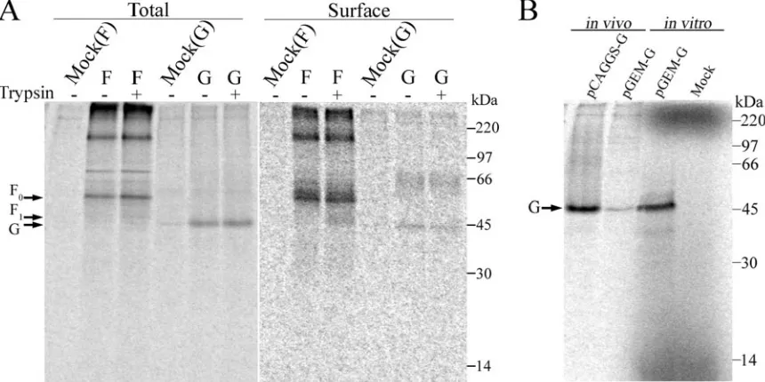
follow-FIG. 2. Expression of HMPV F and HMPV G. (A) Biotinylation. Vero cells in 6-cm dishes were transfected with 4g pCAGGS-HMPV F or pCAGGS-HMPV G. The cells were metabolically labeled for 3 h with Tran35S-label⫾0.3g/ml TPCK trypsin prior to the biotinylation of surface
proteins. Fifteen percent of the immunoprecipitated protein represents the “total” protein. The remaining 85% was subjected to pull down with streptavidin and represents the “surface” protein. The samples were resolved on a 15% SDS-polyacrylamide gel and visualized by autoradiography. Each side of the same gel was differentially contrast enhanced. (B) In vitro coupled transcription/translation of HMPV G (“in vitro”; right side) run side by side on a 15% SDS-polyacrylamide gel with HMPV G immunoprecipitated from transfected Vero cells (“in vivo”; left side).
VOL. 80, 2006 HMPV F PROTEIN-PROMOTED FUSION 10933
on November 8, 2019 by guest
http://jvi.asm.org/
[image:3.585.77.507.71.286.2]ing the metabolic labeling and trypsin treatment of cells ex-pressing either HMPV F or HMPV G. Prior to the separation of the surface protein by streptavidin pull down, 15% of the total population of HMPV F or G, which was immunoprecipi-tated with antipeptide sera, was saved for comparison (Fig. 2A). Analyses of biotin-labeled surface populations of HMPV F confirm that trypsin treatment results in the presence of proteolytically processed F protein on the plasma membrane (Fig. 2A). However, a significant portion of the F protein population remained unprocessed by trypsin. Pulse-chase ex-periments with increasing concentrations of trypsin for various amounts of time did not result in increased processing, sug-gesting that a significant population of the F protein could not be cleaved (data not shown). Immunoprecipitated F protein also contained high-molecular-weight forms that are specific to cells transfected with HMPV F. These aggregates, which may correspond to higher oligomeric forms of the F protein, are quite stable. Increased concentrations of SDS or the addition of 6 M urea in the sample buffer and extended boiling of samples did not decrease the high-molecular-weight bands or increase the intensity of the bands corresponding to the pre-dicted molecular weight of F0(data not shown). The primary
band in the total HMPV G population migrated at approxi-mately 45 kDa, while the cell surface population also included a higher-molecular-weight form migrating at approximately 65 kDa. Both of these forms appeared unaffected by trypsin treat-ment (Fig. 2A). As the size of HMPV G as predicted by its amino acid sequence is considerably smaller than 45 kDa, we verified that the identified band was indeed HMPV G by use of in vitro coupled transcription/translation. HMPV G expressed from a pGEM-4Z plasmid with reticulocyte lysates migrated at a similar position to the product detected in cultured cells, suggesting that this ⬃45 kDa species is indeed a product of HMPV G expression (Fig. 2B). The reason for the higher apparent molecular weight of HMPV G when visualized on an SDS-polyacrylamide gel is unclear. Biacchesi and colleagues noted a ladder of bands ranging from⬃13 to 90 kDa when HMPV G was immunoprecipitated from virions (7). This dif-ference in our results may be due to various forms incorpo-rated into virions or potential proteolysis.
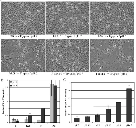
The fusion proteins of some enveloped viruses, such as in-fluenza virus HA, require low pH to induce membrane fusion between cells. Although low pH is normally not required for fusion promoted by paramyxovirus F proteins, we analyzed the HMPV F protein pH requirements by treating HMPV F- and G-expressing Vero cells (maintained the day after transfection in Opti-MEM with 0.3 g/ml TPCK-trypsin, the maximum concentration of trypsin possible without visually impairing cell health) with a brief (4-min) pulse of pH 5-buffered PBS. Un-expectedly, syncytium formation could easily be visualized fol-lowing this treatment, and additional pulses with low pH 18 to 36 h after transfection caused syncytium formation to increase (Fig. 3A). To determine if the HMPV G protein is essential for fusion, Vero cells were transfected with only HMPV F and empty vector. When treated with trypsin and low pH, the HMPV F-expressing cells formed syncytia at least as efficiently as the cells that were cotransfected with HMPV F and G (Fig. 3A). HMPV F did not promote syncytia in the absence of trypsin, even when treated with low pH (Fig. 3A). To verify that the result was not cell type specific, fusion was tested in
BHK cells. Trypsin treatment combined with low pH also stim-ulated fusion by HMPV F in BHK cells, while neither trypsin nor low pH alone promoted syncytium formation above back-ground in this cell type (Fig. 4). In contrast to Vero cells, HMPV G appeared to enhance HMPV F-promoted fusion in BHK cells, suggesting that the effect of G might be cell type specific (Fig. 4). Thus, our results demonstrate that efficient cell-cell fusion promoted by the HMPV F protein occurs only after proteolytic processing and exposure to low pH. Addition-ally, the HMPV G protein is not required for HMPV F-moted membrane fusion, though it may enhance fusion pro-moted by the F protein in certain cell types.
Syncytium assays permit a determination of clear differences in fusion efficiency. However, accurate quantification of fusion in syncytium assays is difficult. Thus, reporter gene fusion as-says were developed to allow quantitative measurements of HMPV F-promoted fusion. Vero cells transfected with viral glycoproteins and luciferase cDNA under control of the T7 promoter were lifted from the plate surface with trypsin (at concentrations that effectively cleave the HMPV F protein), and the cells were gently pelleted, resuspended in DMEM plus 10% FBS, and overlaid onto BSR cells, which constitutively express the T7 polymerase. After 30 min at 37°C to allow cell attachment, the mixed cell population was treated with pH 7-or pH 5-buffered PBS f7-or 4 min. The cells were retreated with trypsin and exposed to pH 7 or pH 5 PBS two more times (2 h apart), the total cell population was lysed, and luciferase ac-tivity was analyzed.
In the reporter gene fusion assay with pH 7 pulses, no difference in fusion could be detected between cells transfected with only HMPV G and those transfected with HMPV F either alone or in combination with HMPV G (Fig. 3B). In contrast, there was a three- to fourfold increase in luciferase activity over background (HMPV G alone, pH 5) when cells trans-fected with HMPV F and G were exposed to pH 5 PBS. Interestingly, HMPV G appeared to have a negative impact on fusion, as cells transfected with only HMPV F induced a sev-enfold increase in fusion over background (Fig. 3B). Biotiny-lation experiments showed that cell surface expression of HMPV F was not modulated by the coexpression of HMPV G (data not shown). To ensure that the increase in luciferase expression was due to the specific effects of low pH on the trigger of membrane fusion by HMPV F, fusion induced by simian virus 5 (SV5) glycoproteins was measured under the same conditions used to examine HMPV fusion. SV5 glyco-protein-induced fusion has been examined extensively, and fusion induced by these viral glycoproteins is not affected by low pH (8). As expected, our data showed that fusion induced by SV5 glycoproteins was not increased by treatment with pH 5 PBS (Fig. 3B), verifying that the effects of pH seen with HMPV were not due to nonspecific effects. Fusion induced by SV5 glycoproteins was always greater than fusion induced by HMPV F, which is to be expected if HMPV F can cause fusion only when treated with low pH, while SV5 F may cause fusion continuously.
Since fusion induced by HMPV F was stimulated by low pH, we next performed a reporter gene fusion assay in which cells were exposed to a range of pH conditions to determine the threshold or optimal pH for fusion induced by HMPV F. Fu-sion induced by HMPV F increased gradually as the pH was
on November 8, 2019 by guest
http://jvi.asm.org/
decreased, with maximum levels of fusion attained at pH 4.5 (Fig. 3C). Lower-pH treatments were not examined due to a lack of physiological relevance. Syncytium assays with HMPV F and G in Vero cells confirmed the results of the reporter gene assay. A gradual increase in fusion was apparent as the pH of the treatment decreased. However, a sharp difference between pH 5 and pH 4.5 in the syncytium assay was less evident (data not shown).
Mutation analysis of the HMPV F cleavage site.As we had
[image:5.585.68.523.78.505.2]established functional fusion assays for the HMPV F protein, we next created a series of mutations proximal to the putative cleavage site in the HMPV F protein to determine the role of the residues in proteolytic processing and fusion. In addition, we created an HMPV F protein with a recognition sequence for the endogenous protease furin to examine the effects of intracellular processing on HMPV F trafficking and membrane
FIG. 3. Analysis of HMPV F-promoted cell-cell fusion. (A) Vero cells were transfected with pCAGGS-HMPV F (1g) and pCAGGS-HMPV G (1g) or the empty pCAGGS vector (1g). After 18 to 24 h, confluent cell monolayers were incubated in Opti-MEM⫾0.3g/ml TPCK-trypsin for 1 to 2 h and then washed and treated with buffered PBS of the indicated pH for 4 min. The cells were again incubated in Opti-MEM⫾trypsin, and the pH pulse was repeated three more times throughout the day before the photographs were taken. (B) Vero cells were transfected with a total of 3.5g of DNA consisting of 1.5 g luciferase cDNA, 1 g pCAGGS-HMPV F or pCAGGS-SV5 F or empty vector, and 1g pCAGGS-HMPV G or pCAGGS-SV5 HN or empty vector. After 18 to 24 h, Vero cells were overlaid on BSR cells expressing the T7 polymerase. The cells were then treated and fusion was analyzed as described in Materials and Methods. The average of the results of four experiments, normalized to the luminosity detected from F-transfected, pH 5-treated cells, is shown. The error bars represent 95% confidence intervals. (C) Six-centimeter dishes of Vero cells were transfected with 1.5g luciferase cDNA and 1.5g pCAGGS-HMPV F. After 18 to 24 h, Vero cells were overlaid on BSR cells expressing the T7 polymerase. The cells were then treated and fusion was analyzed as described in Materials and Methods. The average of the results of four experiments, normalized to the luminosity of pH 5-treated cells, is shown. The error bars represent 95% confidence intervals. F&G, HMPV F and G proteins present.
VOL. 80, 2006 HMPV F PROTEIN-PROMOTED FUSION 10935
on November 8, 2019 by guest
http://jvi.asm.org/
fusion promotion. The wild-type and cleavage site mutant amino acid sequences are displayed in Fig. 1. The cell surface expression of each of these mutants in the presence or absence of trypsin was examined by Tran35S-label metabolic labeling
and biotinylation combined with immunoprecipitation and SDS-PAGE (Fig. 5A and B). Also, the ability of each mutant to induce cell-cell fusion was examined in syncytium and re-porter gene assays (Fig. 6A to C).
The arginine at position 102, after which cleavage was pro-posed to take place, was mutated to an alanine (P1). Trypsin, which cleaves the C-terminals of basic residues, would not be expected to properly process this mutant. Indeed, this mutant was not cleaved in the presence or absence of trypsin (Fig. 5A and B), and no fusion could be detected (Fig. 6A and C). Therefore, the predicted F protein cleavage site is likely the cor-rect site, and like other paramyxovirus F proteins, the HMPV F protein must be cleaved for membrane fusion to occur.
Next, the P2 residue (second amino acid upstream of the cleavage site) was changed from a serine to a proline, as it has recently been reported that certain laboratory-grown strains of HMPV acquired this mutation when propagated in the ab-sence of trypsin (41). However, the F protein used in this study (CAN97-83) is most similar to a strain of HMPV (A2) used in the previous study that did not acquire the P2 mutation and could not grow in the absence of trypsin. Examination of the
P2mutant by SDS-PAGE and in fusion assays showed that it was not cleaved by endogenous proteases (Fig. 5A and 6B), suggesting that other amino acid changes are needed to pro-mote trypsin-independent cleavage of this F protein. Subse-quently, the glutamic acid found 10 residues upstream of the cleavage site was mutated to a lysine, both alone (E93K) and in combination with the P2 mutation (P2/E93K), as this mutation was also reported to enhance cleavage (41). However, these mutants could not promote fusion in the absence of trypsin
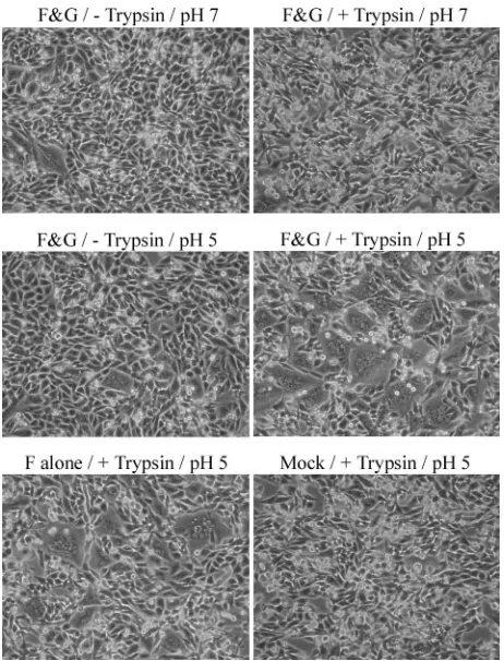
[image:6.585.48.279.67.370.2]FIG. 4. Syncytia promoted by HMPV F in BHK cells. BHK cells were transfected with empty pCAGGS vector (2g) or pCAGGS-HMPV F (1 g) and pCAGGS-HMPV G (1g) or empty vector (1g). After 18 to 24 h, confluent cell monolayers were incubated in Opti-MEM with 0.2 g/ml TPCK-trypsin for 1 h, then washed and treated with PBS of the indicated pH for 4 min. The cells were then incubated in DMEM plus FBS for⬃1 h, then in Opti-MEM plus trypsin for⬃1 h. The pH pulse and change of media were repeated three more times before the photo-graphs were taken. F&G, HMPV F and G proteins present.
FIG. 5. Analysis of the surface expression of HMPV F mutants. Vero cells in 6-cm dishes were transfected with 8g pCAGGS-HMPV F or a mutant F protein. The cells were metabolically labeled for 3 h with Tran35S-label in the absence (A) or presence (B) of 0.5g/ml TPCK trypsin.
Biotinylation and analysis were performed as for Fig. 2A, but only the surface populations are shown. Fwt, wild-type F protein.
on November 8, 2019 by guest
http://jvi.asm.org/
FIG. 6. Fusion promoted by HMPV F cleavage-site mutants. Syncytium assays in the presence (A) or absence (B) of trypsin are shown. Vero cells were transfected with 1g pCAGGS-HMPV F or a mutant F protein and 1g pCAGGS-HMPV G. The cells were treated (each with pH 5 PBS) and analyzed as for Fig. 3A. (C) Reporter gene assay of fusion in the presence of trypsin. Vero cells in 6-cm dishes were transfected with 1.5g luciferase cDNA and 1.5g pCAGGS-HMPV F or a mutant. After 18 to 24 h, the Vero cells were overlaid on BSR cells expressing the T7 polymerase. The cells were then treated and fusion was analyzed as described in Materials and Methods. The average of the results of three experiments is shown, and the error bars represent 95% confidence intervals. The graph is divided into two sections. The mutants on either side were analyzed on different days but always alongside the wild-type (WT) protein, and values were normalized to that of the wild type in each experiment.
VOL. 80, 2006 HMPV F PROTEIN-PROMOTED FUSION 10937
on November 8, 2019 by guest
http://jvi.asm.org/
(data not shown), and they were cleaved only with the addition of trypsin (Fig. 5A and B). Schickli et al. also found that a number of viruses containing the P2 mutation in F acquired an additional glutamine-to-lysine mutation in the P3 position when grown in the absence of trypsin (41). We created this Q-to-K mutation in the P3 position of F, both alone (P3) and in combination with P2 (P2/P3). TheP2/P3mutant promoted the formation of small syncytia in the absence of trypsin when exposed to low pH (Fig. 6B), and a low level of F protein cleavage was detected by surface biotinylation and SDS-PAGE (Fig. 5A). When only the P3 residue was mutated, fusion and cleavage in the absence of trypsin were not detected (Fig. 5A and 6B), suggesting that both the serine-to-proline change at P2 and the glutamine-to-lysine change at P3 were necessary for proteolytic processing by endogenous proteases. Still, cell-cell fusion induced by theP2/P3mutant in the absence of trypsin
was considerably less than that of the trypsin-treated wild-type protein (Fig. 6A and B). Furthermore, cell-cell fusion pro-moted byP2/P3increased in the presence of trypsin (Fig. 6A and B), suggesting that inefficient cleavage rather than changes in F protein stability resulted in the decreased fusion. Finally, a mutant was created in which the second and third residues upstream of the cleavage site were changed to arginines, mak-ing all four residues upstream of the cleavage site arginine. This mutant (furin) is predicted to be cleaved efficiently by thetrans-Golgi-resident enzyme furin, which has strong pref-erence for R-X-K/R-R sequences. Cell surface expression of the cleaved F1protein appeared to be slightly greater for the
furinmutant than theP2/P3mutant (Fig. 5A), although cleav-age of thefurinmutant was still quite low compared to other paramyxovirus F proteins that are normally cleaved by furin. Fusion induced by this mutant in the absence of trypsin was
FIG. 7. Analysis of HMPV F N-linked glycosylation. (A) PNGase F treatment of HMPV F and G. Vero cells in 35-mm dishes were transfected with 2g pCAGGS-HMPV F, pCAGGS-HMPV G, or empty vector. Cells were metabolically labeled with Tran35S-label for 1 h and chased for
1 h. Immunoprecipitated protein was incubated overnight with or without PNGase F, and samples were resolved on a 15% SDS-polyacrylamide gel and visualized by autoradiography. (B) Vero cells were transfected with 2g pCAGGS-HMPV F or a mutant. Cells were metabolically labeled with Tran35S-label for 1 h and chased for 2 h in serum-free DMEM⫾0.3g/ml TPCK-trypsin. Immunoprecipitated protein was analyzed as above.
(C) Syncytium assay. Vero cells transfected with 1g pCAGGS-HMPV F or a mutant and 1g pCAGGS-HMPV G were treated (each with pH 5 PBS) and analyzed as for Fig. 3A. (D) Reporter gene assay. Vero cells in 6-cm dishes were transfected with 1.5g luciferase cDNA and 1.5g pCAGGS-HMPV F or a mutant. After 18 to 24 h, Vero cells were overlaid on BSR cells expressing the T7 polymerase. The cells were then treated and fusion was analyzed as described in Materials and Methods. The average of the results of seven experiments is shown, and the error bars represent 95% confidence intervals.
on November 8, 2019 by guest
http://jvi.asm.org/
similar to that of theP2/P3 mutant (Fig. 6B), and syncytium formation by thefurin mutant was also increased by trypsin treatment (Fig. 6A and B), suggesting that cleavage by endog-enous proteases was less than optimal.
Fusion by each of the mutants, exceptP1, was stimulated by treatment with both trypsin and low pH, and fusion by each under these conditions was similar to or slightly less than that of the wild type (Fig. 6A and C). Notably, fusion induced by the
furinmutant in the presence of trypsin was less than that of the wild type, indicating that, while these amino acid changes per-mitted endogenous cleavage they had a negative effect on fusion promotion. However, this may be a consequence of the reduced levels of cleaved protein on the cell surfaces (Fig. 5B). Only the mutantsP2/P3andfurinpromoted syncytium forma-tion in the absence of trypsin (Fig. 6B and data not shown), but syncytium formation was very low, and fusion by these mutants in the absence of trypsin was not detected over background levels of fusion in a reporter gene assay (data not shown).
Analysis of N-linked glycosylation in HMPV F.The HMPV
F protein contains three sites (N-X-S/T) with the potential to be N-link glycosylated in the endoplasmic reticulum. PNGase F treatment of immunoprecipitated HMPV F suggested that one or more of these sites was utilized, as a large shift in F0was
apparent on a reducing polyacrylamide gel following incuba-tion with PNGase F (Fig. 7A). On the other hand, PNGase F treatment of HMPV G indicated that this protein was not N-link glycosylated. However, there is a higher-molecular-weight form of HMPV G detected only when surface popula-tions are examined (Fig. 2A), which may be the product of carbohydrate modification. However, the decreased G protein left after overnight PNGase treatment was below the limit of detection in our assays. The asparagines of HMPV F with the potential to be glycosylated are found at positions 57, 172, and 353 (Fig. 1), and each of these asparagines was mutated indi-vidually to alanines. These mutants (N57, N172, and N353) were analyzed by SDS-PAGE to reveal changes in mobility, as this would indicate that the site mutated is normally glycosyl-ated. Figure 7B demonstrates that all three potential sites are utilized for glycosylation, as F0of each mutant appears roughly
2 kDa smaller than the wild type F0. Each mutant is expressed
on the cell surface (Fig. 5B), indicating that none of the car-bohydrates individually is required for folding in the endoplas-mic reticulum and subsequent transport to the cell surface.
Removal of the carbohydrate at N57 found within F2
ap-peared to slightly reduce trypsin-mediated proteolytic process-ing compared to that of the wild type (Fig. 5B and 7B). The size of F1does not change, as predicted for a loss of
glycosyl-ation in F2, and we were unable to detect F2by
autoradiogra-phy. This is likely a consequence of low levels of cleavage combined with the presence of only two cysteine and no me-thionine residues in F2. The residues N172 and N353 are
lo-cated between the two heptad repeat regions of F1, with the
carbohydrate on N172 positioned just outside heptad repeat A (Fig. 1). Cleavage of theN172mutant was also reduced, and the level of overall expression detected following immunopre-cipitation and SDS-PAGE was consistently less than that of the wild type (Fig. 5B and 7B). As expected, F1ofN172runs faster
on a reducing gel than F1of wild-type F protein. F1ofN353is
not visible by SDS-PAGE (Fig. 5B and 7B), suggesting that cleavage of theN353 mutant is significantly impaired, which
could result if the protein was not properly folded. However, all three N-glycosylation mutants were detected by biotinyla-tion on the cell surfaces (Fig. 5B), suggesting that the change in structure affected availability of the cleavage site but was not so great as to cause retention of the protein in the endoplasmic reticulum.
Fusion promoted by each of the N-glycosylation mutants was decreased compared to wild type fusion; however, the extent of the reduction depended on which carbohydrate was removed (Fig. 7C and D). Syncytium and reporter gene assays of fusion were generally in agreement, although some variability was observed forN172in the reporter gene assay. Consequently, the reporter gene experiment was repeated seven times, and 95% confidence intervals were calculated (Fig. 7D).N57 pro-moted fusion only slightly less efficiently than the wild type, while fusion promoted by N172 was greatly decreased. The extent of fusion promoted by these mutants may be a reflection of the level of proteolytic processing combined with the level of surface expression (Fig. 5B). As expected, fusion promoted by
N353was very poor. However, a few small syncytia could be found (Fig. 7C), suggesting that at least a small portion of the mutant F protein can be cleaved and promote fusion. How-ever, theN353mutant did not promote significant fusion above background levels in the reporter gene fusion assay (Fig. 7D).
DISCUSSION
The work presented here represents the first analysis of membrane fusion promoted by the F protein of HMPV, a recently identified paramyxovirus. We have shown that low-pH treatment of cells transiently transfected with only the HMPV F protein profoundly enhances cell-cell fusion. Indeed, a grad-ual increase in fusion was observed as the pH to which HMPV F-expressing cells were exposed decreased. Our results indi-cate that proteolytic processing of the F protein is a stringent requirement for fusion. However, the HMPV attachment pro-tein, G, like other pneumovirus attachment proteins, is not needed for HMPV F-promoted fusion. In addition, we have examined the amino acid sequence requirements for trypsin-independent cleavage of the HMPV F protein when transiently expressed in cells in the absence of other viral components, and we have shown that the HMPV F protein is N-link glyco-sylated at three sites, and each of these carbohydrates is im-portant for expression, cleavage, and fusion to various extents. Fusion promoted by the F proteins of all previously exam-ined paramyxoviruses, with only one possible exception (43), takes place efficiently at neutral pH. In vivo syncytium forma-tion by a viral fusion protein is not consistent with a require-ment for low pH to trigger fusion, as fusion takes place at the plasma membrane, where the pH is neutral. Strain-specific differences in the ability of HMPV to form syncytia have been reported (20), and it is not known whether the strain of HMPV from which the F and G genes used in this study originated can promote syncytium formation. Thus it is possible that only some strains of HMPV require low pH to trigger F protein-mediated membrane fusion and that these strains do not pro-mote syncytium formation. While our data demonstrating that low pH stimulates F protein-promoted fusion do not prove that HMPV enters cells via an endosomal, low-pH pathway, further investigation of the mode of entry is clearly warranted.
VOL. 80, 2006 HMPV F PROTEIN-PROMOTED FUSION 10939
on November 8, 2019 by guest
http://jvi.asm.org/
It is possible that a viral protein other than G provides for HMPV in vivo what low pH provides in vitro. Barretto et al. showed that transient transfection of RSV glycoproteins leads to a lack of reproducible syncytium formation in HeLa cells, while RSV infection promotes efficient syncytium formation (3). For RSV glycoproteins, neuraminidase treatment of gly-coprotein-expressing cells greatly increased cell-cell fusion. However, neuraminidase treatment of HMPV F- and G-ex-pressing cells did not alter the requirement for low pH to induce fusion (data not shown), though it should be noted that the optimal pH for neuraminidase activity (approximately pH 5) was not employed, as this would make the analysis of pH effects on fusion difficult. It is also possible that strains of HMPV which form syncytia have F proteins which can be activated in the secretory pathway under certain circum-stances, as was suggested for infectious bronchitis virus (12), since the pH of some secretory vesicles (49) is at the level found to trigger HMPV F-promoted membrane fusion in this study.
Recombinant HMPV lacking the G protein is highly atten-uated in primates (4), but in cell culture deletion of the G protein has no effect on virus titers (7). The RSV G protein enhances binding of virus to cells but has no role in fusion after attachment (44). Our results demonstrate that HMPV F pro-tein-mediated syncytium formation does not require the G protein. Indeed, quantitative analysis of HMPV F-promoted fusion indicated that coexpressing G could reduce fusion levels (Fig. 3B). It remains possible that the HMPV G protein at-taches to a specific cellular receptor but that this receptor is expressed only in certain cell types. This theory may be sup-ported by the fact that syncytium formation in BHK cells was enhanced by the G protein (Fig. 4). However, BSR cells, which serve as the target cell type in the reporter gene assay where G expression was found to inhibit fusion (Fig. 3B), were origi-nally derived from BHK cells.
As with other paramyxovirus F proteins, the HMPV F pro-tein is N-link glycosylated and these modifications differentially affect the production of a mature and fusogenic F protein. Fusion promoted by N-glycosylation-site mutants decreases in the orderWT⬎N57⬎N172⬎N353 (Fig. 7C and D). The levels of cleaved F protein on the cell surfaces also follow this order (Fig. 5B), suggesting that the extent of fusion is at least partially a reflection of the amount of mature F protein on the cell surface. This result is different from that observed for the RSV F protein, which contains two N-linked carbohydrate additions in F2 and one in F1. Mutation of each of these
glycosylation sites had little effect on cleavage or surface ex-pression of the RSV F protein (51). However, removal of one carbohydrate in RSV F2 actually resulted in an increase in
syncytium formation, while removal of the carbohydrate in F1
resulted in a significant decrease in cell-cell fusion (51). Thus, individual N-linked carbohydrates modulate fusion of the RSV F protein but facilitate proper folding of the HMPV F protein such that proteolytic cleavage of the HMPV F protein is di-minished to various extents upon the loss of individual carbo-hydrates.
The cleavage site of HMPV F differs significantly from the cleavage site of RSV F, which is cleaved by furin at two sites 27 amino acids apart (19). Wild-type HMPV F is not cleaved by an endogenous protease when expressed in Vero cells, despite
the presence of a sequence at the cleavage site matching the minimal requirements for furin recognition (R-X-X-R) (5, 30, 41). However, our results clearly demonstrate that proteolytic processing is needed for membrane fusion (Fig. 3A and 6B). Cleavage-site mutations reported to promote or enhance en-dogenous cleavage of the HMPV F protein (41) did not give detectable cleavage in our in vitro system in the absence of trypsin. These differences may be a consequence of strain vari-ation, or they could be due to the method of expression. A recent report examining the strain of HMPV F used in our study and containing the serine-to-proline mutation at P2 found a low but significant level of F protein cleavage when attenuated recombinant virus with the mutation was used to infect Vero cells (5). In contrast, no cleavage or replication of the wild-type virus was detected. Biacchesi et al. also measured a much greater level of cleavage with a mutant containing the same amino acid changes as our furinmutant. We detected inefficient endogenous cleavage of this F protein mutant in the same cell type (Fig. 5A), suggesting that the presence of other virus proteins may affect cleavage.
We discovered that HMPV F protein-promoted fusion re-quires proteolytic cleavage combined with exposure to low pH in order to induce appreciable cell-cell fusion. This raises the possibility that HMPV may be unique among paramyxoviruses, which are primarily thought to enter cells upon receptor en-gagement at the plasma membrane. Furthermore, our data suggest that the HMPV G protein may have a negative impact on F protein-promoted fusion, indicating that this “attach-ment” protein may also have a distinctive role in the viral life cycle. An endocytic/endosomal route of entry for HMPV re-quires further investigation, but our results clearly demonstrate that there are major differences between the F protein of this recently identified paramyxovirus and the paramyxovirus F proteins that have previously been characterized.
ACKNOWLEDGMENTS
We thank Ursula Buchholz (NIAID, Bethesda, Maryland) for pro-viding the HMPV F and HMPV G gene constructs, Robert Lamb (HHMI, Northwestern University) for the pCAGGS-SV5 F and HN expression vectors, Karl-Klaus Conzelmann (Max Pettenkofer Institut) for the BSR cells, and Dan Noonan (University of Kentucky) for the use of his luminometer. We are grateful to Roland Hilgarth for his assistance and members of the Dutch lab for critically reviewing the manuscript.
This study was supported by a research grant from the March of Dimes.
REFERENCES
1.Bagai, S., and R. A. Lamb.1995. Individual roles of N-linked oligosaccharide chains in intracellular transport of the paramyxovirus SV5 fusion protein. Virology209:250–256.
2.Bagai, S., and R. A. Lamb.1995. Quantitative measurement of paramyxovi-rus fusion: differences in requirements of glycoproteins between simian viparamyxovi-rus 5 and human parainfluenza virus 3 or Newcastle disease virus. J. Virol.
69:6712–6719.
3.Barretto, N., L. K. Hallak, and M. E. Peeples.2003. Neuraminidase treat-ment of respiratory syncytial virus-infected cells or virions, but not target cells, enhances cell-cell fusion and infection. Virology313:33–43. 4.Biacchesi, S., Q. N. Pham, M. H. Skiadopoulos, B. R. Murphy, P. L. Collins,
and U. J. Buchholz.2005. Infection of nonhuman primates with recombinant human metapneumovirus lacking the SH, G, or M2-2 protein categorizes each as a nonessential accessory protein and identifies vaccine candidates. J. Virol.79:12608–12613.
5.Biacchesi, S., Q. N. Pham, M. H. Skiadopoulos, B. R. Murphy, P. L. Collins, and U. J. Buchholz.2006. Modification of the trypsin-dependent cleavage activation site of the human metapneumovirus fusion protein to be trypsin
on November 8, 2019 by guest
http://jvi.asm.org/
independent does not increase replication or spread in rodents or nonhuman primates. J. Virol.80:5798–5806.
6.Biacchesi, S., M. H. Skiadopoulos, K. C. Tran, B. R. Murphy, P. L. Collins, and U. J. Buchholz. 2004. Recovery of human metapneumovirus from cDNA: optimization of growth in vitro and expression of additional genes. Virology321:247–259.
7.Biacchesi, S., M. H. Skiadopoulos, L. Yang, E. W. Lamirande, K. C. Tran, B. R. Murphy, P. L. Collins, and U. J. Buchholz.2004. Recombinant human metapneumovirus lacking the small hydrophobic SH and/or attachment G glycoprotein: deletion of G yields a promising vaccine candidate. J. Virol.
78:12877–12887.
8.Bissonnette, M. L., S. A. Connolly, D. F. Young, R. E. Randall, R. G. Paterson, and R. A. Lamb.2006. Analysis of the pH requirement for mem-brane fusion of different isolates of the paramyxovirus parainfluenza virus 5. J. Virol.80:3071–3077.
9.Boivin, G., Y. Abed, G. Pelletier, L. Ruel, D. Moisan, S. Cote, T. C. Peret, D. D. Erdman, and L. J. Anderson.2002. Virological features and clinical manifestations associated with human metapneumovirus: a new paramyxo-virus responsible for acute respiratory-tract infections in all age groups. J. Infect. Dis.186:1330–1334.
10.Bosis, S., S. Esposito, H. G. Niesters, P. Crovari, A. D. Osterhaus, and N. Principi.2005. Impact of human metapneumovirus in childhood: compari-son with respiratory syncytial virus and influenza viruses. J. Med. Virol.
75:101–104.
11.Carter, J. R., C. T. Pager, S. D. Fowler, and R. E. Dutch.2005. Role of N-linked glycosylation of the Hendra virus fusion protein. J. Virol.79:7922–7925. 12.Chu, V. C., L. J. McElroy, V. Chu, B. E. Bauman, and G. R. Whittaker.2006.
The avian coronavirus infectious bronchitis virus undergoes direct low-pH-dependent fusion activation during entry into host cells. J. Virol.80:3180–3188. 13.Dutch, R. E., T. S. Jardetzky, and R. A. Lamb.2000. Virus membrane fusion proteins: biological machines that undergo a metamorphosis. Biosci. Rep.
20:597–612.
14.Dutch, R. E., S. B. Joshi, and R. A. Lamb.1998. Membrane fusion promoted by increasing surface densities of the paramyxovirus F and HN proteins: comparison of fusion reactions mediated by simian virus 5 F, human para-influenza virus type 3 F, and para-influenza virus HA. J. Virol.72:7745–7753. 15.Earp, L. J., S. E. Delos, H. E. Park, and J. M. White.2005. The many
mechanisms of viral membrane fusion proteins. Curr. Top. Microbiol. Im-munol.285:25–66.
16.Easton, A. J., J. B. Domachowske, and H. F. Rosenberg.2004. Animal pneumoviruses: molecular genetics and pathogenesis. Clin. Microbiol. Rev.
17:390–412.
17.Ebihara, T., R. Endo, H. Kikuta, N. Ishiguro, M. Yoshioka, X. Ma, and K. Kobayashi.2003. Seroprevalence of human metapneumovirus in Japan. J. Med. Virol.70:281–283.
18.Falsey, A. R., D. Erdman, L. J. Anderson, and E. E. Walsh.2003. Human metapneumovirus infections in young and elderly adults. J. Infect. Dis.187:
785–790.
19.Gonzalez-Reyes, L., M. B. Ruiz-Arguello, B. Garcia-Barreno, L. Calder, J. A. Lopez, J. P. Albar, J. J. Skehel, D. C. Wiley, and J. A. Melero.2001. Cleavage of the human respiratory syncytial virus fusion protein at two distinct sites is required for activation of membrane fusion. Proc. Natl. Acad. Sci. USA
98:9859–9864.
20.Hamelin, M. E., Y. Abed, and G. Boivin.2004. Human metapneumovirus: a new player among respiratory viruses. Clin. Infect. Dis.38:983–990. 21.Hernandez, L. D., L. R. Hoffman, T. G. Wolfsberg, and J. M. White.1996.
Virus-cell and cell-cell fusion. Annu. Rev. Cell Dev. Biol.12:627–661. 22.Homma, M., and M. Ouchi.1973. Trypsin action on the growth of Sendai
virus in tissue culture cells. III. Structural difference of Sendai viruses grown in eggs and tissue culture cells. J. Virol.12:1457–1465.
23.Jartti, T., B. van den Hoogen, R. P. Garofalo, A. D. Osterhaus, and O. Ruuskanen.2002. Metapneumovirus and acute wheezing in children. Lancet
360:1393–1394.
24.Laham, F. R., V. Israele, J. M. Casellas, A. M. Garcia, C. M. Lac Prugent, S. J. Hoffman, D. Hauer, B. Thumar, M. I. Name, A. Pascual, N. Taratutto, M. T. Ishida, M. Balduzzi, M. Maccarone, S. Jackli, R. Passarino, R. A. Gaivironsky, R. A. Karron, N. R. Polack, and F. P. Polack.2004. Differential production of inflammatory cytokines in primary infection with human meta-pneumovirus and with other common respiratory viruses of infancy. J. Infect. Dis.189:2047–2056.
25.Lamb, R. A.1993. Paramyxovirus fusion: a hypothesis for changes. Virology
197:1–11.
26.Lamb, R. A., and D. Kolakofsky.2001. Paramyxoviridae: the viruses and their replication, p. 1305–1340.InD. M. Knipe and P. M. Howley (ed.), Fields virology, 4th ed. Lippincott-Raven Press, New York, N.Y.
27.Maggi, F., M. Pifferi, M. Vatteroni, C. Fornai, E. Tempestini, S. Anzilotti, L. Lanini, E. Andreoli, V. Ragazzo, M. Pistello, S. Specter, and M. Bendinelli.
2003. Human metapneumovirus associated with respiratory tract infections in a 3-year study of nasal swabs from infants in Italy. J. Clin. Microbiol.
41:2987–2991.
28.McGinnes, L., T. Sergel, J. Reitter, and T. Morrison.2001. Carbohydrate
modifications of the NDV fusion protein heptad repeat domains influence maturation and fusion activity. Virology283:332–342.
29.Moll, M., A. Kaufmann, and A. Maisner.2004. Influence of N-glycans on processing and biological activity of the Nipah virus fusion protein. J. Virol.
78:7274–7278.
30.Molloy, S. S., P. A. Bresnahan, S. H. Leppla, K. R. Klimpel, and G. Thomas.
1992. Human furin is a calcium-dependent serine endoprotease that recog-nizes the sequence Arg-X-X-Arg and efficiently cleaves anthrax toxin pro-tective antigen. J. Biol. Chem.267:16396–16402.
31.Naylor, C. J., P. A. Brown, N. Edworthy, R. Ling, R. C. Jones, C. E. Savage, and A. J. Easton.2004. Development of a reverse-genetics system for avian pneumovirus demonstrates that the small hydrophobic (SH) and attachment (G) genes are not essential for virus viability. J. Gen. Virol.85:3219–3227. 32.Nissen, M. D., D. J. Siebert, I. M. Mackay, T. P. Sloots, and S. J. Withers.
2002. Evidence of human metapneumovirus in Australian children. Med. J. Aust.176:188.
33.Niwa, H., K. Yamamura, and J. Miyazaki.1991. Efficient selection for high-expression transfectants with a novel eukaryotic vector. Gene108:193–199. 34.O’Sullivan, J. D., A. M. Allworth, D. L. Paterson, T. M. Snow, R. Boots, L. J.
Gleeson, A. R. Gould, A. D. Hyatt, and J. Bradfield.1997. Fatal encephalitis due to novel paramyxovirus transmitted from horses. Lancet349:93–95. 35.Ortmann, D., M. Ohuchi, H. Angliker, E. Shaw, W. Garten, and H. D. Klenk.
1994. Proteolytic cleavage of wild type and mutants of the F protein of human parainfluenza virus type 3 by two subtilisin-like endoproteases, furin and Kex2. J. Virol.68:2772–2776.
36.Pager, C. T., and R. E. Dutch.2005. Cathepsin L is involved in proteolytic processing of the Hendra virus fusion protein. J. Virol.79:12714–12720. 37.Paterson, R. G., and R. A. Lamb.1993. The molecular biology of influenza
viruses and paramyxoviruses, p. 35–73.InA. Davidson and R. M. Elliott (ed.), Molecular virology: a practical approach. IRL Oxford University Press, Oxford, England.
38.Peiris, J. S., W. H. Tang, K. H. Chan, P. L. Khong, Y. Guan, Y. L. Lau, and S. S. Chiu.2003. Children with respiratory disease associated with meta-pneumovirus in Hong Kong. Emerg. Infect. Dis.9:628–633.
39.Peret, T. C., G. Boivin, Y. Li, M. Couillard, C. Humphrey, A. D. Osterhaus, D. D. Erdman, and L. J. Anderson.2002. Characterization of human meta-pneumoviruses isolated from patients in North America. J. Infect. Dis.185:
1660–1663.
40.Scheid, A., and P. W. Choppin.1974. Identification of biological activities of paramyxovirus glycoproteins. Activation of cell fusion, hemolysis, and infec-tivity of proteolytic cleavage of an inactive precursor protein of Sendai virus. Virology57:475–490.
41.Schickli, J. H., J. Kaur, N. Ulbrandt, R. R. Spaete, and R. S. Tang.2005. An S101P substitution in the putative cleavage motif of the human metapneu-movirus fusion protein is a major determinant for trypsin-independent growth in Vero cells and does not alter tissue tropism in hamsters. J. Virol.
79:10678–10689.
42.Schmidt, U., J. Beyer, U. Polster, L. J. Gershwin, and U. J. Buchholz.2002. Mucosal immunization with live recombinant bovine respiratory syncytial virus (BRSV) and recombinant BRSV lacking the envelope glycoprotein G protects against challenge with wild-type BRSV. J. Virol.76:12355–12359. 43.Seth, S., A. Vincent, and R. W. Compans.2003. Activation of fusion by the
SER virus F protein: a low-pH-dependent paramyxovirus entry process. J. Virol.77:6520–6527.
44.Techaarpornkul, S., N. Barretto, and M. E. Peeples.2001. Functional analysis of recombinant respiratory syncytial virus deletion mutants lacking the small hydrophobic and/or attachment glycoprotein gene. J. Virol.75:6825–6834. 45.van den Hoogen, B. G., T. M. Bestebroer, A. D. Osterhaus, and R. A.
Fouchier.2002. Analysis of the genomic sequence of a human metapneu-movirus. Virology295:119–132.
46.van den Hoogen, B. G., J. C. de Jong, J. Groen, T. Kuiken, R. de Groot, R. A. Fouchier, and A. D. Osterhaus.2001. A newly discovered human pneumo-virus isolated from young children with respiratory tract disease. Nat. Med.
7:719–724.
47.van den Hoogen, B. G., D. M. Osterhaus, and R. A. Fouchier.2004. Clinical impact and diagnosis of human metapneumovirus infection. Pediatr. Infect. Dis. J.23:S25–S32.
48.Williams, J. V., P. A. Harris, S. J. Tollefson, L. L. Halburnt-Rush, J. M. Pingsterhaus, K. M. Edwards, P. F. Wright, and J. E. Crowe, Jr.2004. Human metapneumovirus and lower respiratory tract disease in otherwise healthy infants and children. N. Engl. J. Med.350:443–450.
49.Wu, M. M., M. Grabe, S. Adams, R. Y. Tsien, H. P. Moore, and T. E. Machen.2001. Mechanisms of pH regulation in the regulated secretory pathway. J. Biol. Chem.276:33027–33035.
50.Zhang, L., M. E. Peeples, R. C. Boucher, P. L. Collins, and R. J. Pickles.
2002. Respiratory syncytial virus infection of human airway epithelial cells is polarized, specific to ciliated cells, and without obvious cytopathology. J. Virol.76:5654–5666.
51.Zimmer, G., I. Trotz, and G. Herrler.2001. N-glycans of F protein differ-entially affect fusion activity of human respiratory syncytial virus. J. Virol.
75:4744–4751.
VOL. 80, 2006 HMPV F PROTEIN-PROMOTED FUSION 10941