0022-538X/08/$08.00⫹0 doi:10.1128/JVI.01520-07
Copyright © 2008, American Society for Microbiology. All Rights Reserved.
Akt Plays a Critical Role in Replication of Nonsegmented
Negative-Stranded RNA Viruses
䌤
Minghao Sun,
1† Sandra M. Fuentes,
1† Khalid Timani,
2Dengyun Sun,
3Chris Murphy,
2Yuan Lin,
2Avery August,
1,2,3,4Michael N. Teng,
1,3,4,5and Biao He
1,2,3,4*
Graduate Program in Pathobiology,1Department of Veterinary and Biomedical Sciences,2Intercollege Graduate Program in
Cell and Developmental Biology,3Center of Molecular Immunology and Infectious Disease,4and Department of
Biochemistry and Molecular Biology,5Pennsylvania State University, University Park, Pennsylvania 16802
Received 11 July 2007/Accepted 10 October 2007
The orderMononegavirales(comprised of nonsegmented negative-stranded RNA viruses or NNSVs) contains many important pathogens. Parainfluenza virus 5 (PIV5), formerly known as simian virus 5, is a prototypical paramyxovirus and encodes a V protein, which has a cysteine-rich C terminus that is conserved among all paramyxoviruses. The V protein of PIV5, like that of many other paramyxoviruses, plays an important role in regulating viral RNA synthesis. In this work, we show that V interacts with Akt, a serine/threonine kinase, also known as protein kinase B. Both pharmacological inhibitors and small interfering RNA against Akt1 reduced PIV5 replication, indicating that Akt plays a critical role in PIV5 replication. Furthermore, treatment with Akt inhibitors also reduced the replication of several other paramyxoviruses, as well as vesicular stomatitis virus, the prototypical rhabdovirus, indicating that Akt may play a more universal role in NNSV replication. The phosphoproteins (P proteins) of NNSVs are essential cofactors for the viral RNA polymerase complex and require heavy phosphorylation for their activity. Inhibition of Akt activity reduced the level of P phosphory-lation, suggesting that Akt is involved in regulating viral RNA synthesis. In addition, Akt1 phosphorylated a recombinant P protein of PIV5 purified from bacteria. The finding that Akt plays a critical role in replication of NNSV will lead to a better understanding of how these viruses replicate, as well as novel strategies to treat infectious diseases caused by NNSVs.
The single-stranded RNA genomes of theMononegavirales
range from⬃11,000 to 18,000 nucleotides in length and con-tain a series of tandemly linked genes separated by nontran-scribed sequences (32, 34). The viral RNA-dependent RNA polymerase (vRNAP) that transcribes the nucleocapsid protein (NP)-encapsidated RNA into 5⬘capped and 3⬘polyadenylated mRNAs minimally consists of two proteins, the phosphopro-tein (P) and the large polymerase prophosphopro-tein (L) (22). The viral RNA polymerase is thought to bind the genomic RNA at a single 3⬘entry site and to transcribe the genome by a sequen-tial and polar process. The same vRNAP also replicates viral RNA genome (1, 2, 21, 28). Regulation of the switch between viral RNA transcription and replication is not well understood. It is thought that the phosphorylation status of the P protein may be critical in regulating RNA synthesis (15, 52). For ve-sicular stomatitis virus (VSV), mutations altering the phosphor-ylation of the P protein that differentially affect RNA transcrip-tion or RNA genome replicatranscrip-tion have been identified (15, 16). Similarly, mutations in the P protein of respiratory syncytial virus (RSV) that affect viral RNA synthesis have been identi-fied (5, 20, 38). While host kinases are thought to phosphory-late nonsegmented negative-stranded RNA virus (NNSV) P proteins, little is known about the identity of the specific host
kinases required. For all paramyxoviruses examined, full activ-ity of RNA synthesis by purified P-L complex with NP-encap-sidated RNA genome (3, 17, 40–42, 44, 46, 58) is achieved only when cell extracts are added, indicating that the P-L complex is essential but not sufficient for viral RNA synthesis and host factors are required for viral RNA synthesis. For some of these viruses, such as Sendai virus (SeV) and measles virus (MeV), it is thought that the host protein tubulin is an important com-ponent of the transcription initiation complex (42, 45). For RSV, actin and profilin play an essential role in viral RNA synthesis (8, 9). While cytoskeletal proteins may be important for viral RNA synthesis, possibly providing a physical anchor for viral RNA synthesis, these proteins are unlikely to regulate the switch of viral RNA synthesis from mRNA transcription to viral genome replication.
Parainfluenza virus 5 (PIV5), formerly known as simian virus 5, is a prototypical member of theRubulavirus genus of the family Paramyxoviridae (11, 32). PIV5 has seven genes but encodes eight known viral proteins (32). The V/P gene of PIV5 is transcribed into both the V mRNA and the P mRNA through a process of pseudotemplated addition of nucleotides, commonly called “RNA editing.” The V mRNA is made when the viral RNA polymerase faithfully transcribes the V/P gene. However, during transcription, the viral RNA polymerase complex recognizes a specific RNA sequence in the V/P gene and inserts two nontemplated G residues at the site to generate the P mRNA. As a result, the V/P gene is transcribed at similar levels into two mRNAs that are translated into two proteins, which share identical N termini but are encoded by different C termini (60). The C terminus of the V protein contains a
* Corresponding author. Mailing address: Center of Molecular Im-munology and Infectious Disease, Department of Veterinary and Bio-medical Sciences, Pennsylvania State University, 115 Henning Bldg., University Park, PA 16802. Phone: (814) 8533. Fax: (814) 863-6140. E-mail: [email protected].
† M.S. and S.M.F. contributed equally.
䌤Published ahead of print on 24 October 2007.
105
on November 8, 2019 by guest
http://jvi.asm.org/
cysteine-rich region that is well conserved among all paramyxo-virus V proteins (61). While P is required for the transcription and replication of a PIV5 minigenome, the addition of V inhibits viral RNA synthesis in this system (36). The mecha-nism by which the V protein regulates viral RNA synthesis is unclear. However, we speculated that a host protein is in-volved. In this work, we show that V interacts with Akt1, and we have investigated the role of this host protein in viral rep-lication.
Akt, also known as protein kinase B (PKB), was first discov-ered in retrovirus AKT8 as a viral protooncogene capable of transforming certain cells (6). Identification and cloning of the Akt gene showed that it has high homology to protein kinases A and C, hence the name PKB. Three mammalian Akt genes (Akt1, -2, and -3, also known as PKB␣, -, and -␥, respectively) have been identified, and all have serine/threonine kinase ac-tivity. The three Akt isoforms each contain a pleckstrin homol-ogy domain, a catalytic domain, and a regulatory domain. There are two major phosphorylation sites within Akt1, amino acid residues Thr308 and Ser473, which are phosphorylated by PDK1 (PI3K-dependent kinase 1) and rictor-mTOR complex, respectively (10, 54). Akt is known to have many downstream targets, including the BCL2 antagonist of cell death, the cyclic AMP response element-binding protein, endothelial nitric ox-ide synthase, I-B kinase ␣, GSK-3, and p21 CIP1 (19). In addition, Akt is a key regulator in the PI3K signaling pathway and plays an important role in many cellular processes, such as cell survival, metabolism, growth, proliferation, and mobility. Akt has been found to be activated in many cancers, and targeting Akt with small molecules has reduced tumor growth in some circumstances (50, 64). While Akt1 and -2 are widely expressed in many organs and cell types, Akt3 is expressed preferentially, but not exclusively, in neuronal cells. All three Akt genes share some redundant functions, but studies using knockout mice and small interfering RNAs (siRNAs) show that each has distinct functions (56). Here, we show that Akt activity is required for optimal replication of a number of paramyxoviruses and that its function may involve regulating the activity of the viral polymerase by phosphorylation of the P protein.
MATERIALS AND METHODS
Plasmids, viruses, and cells.Plasmids expressing V, P, and green fluorescent protein (GFP) have been described before (36). Genes for Akt1 (GenBank accession number NM_001014432), mitogen-activated protein kinase 1 (MAPK1) (accession number NM_002745), and phosphoinositide-binding pro-tein (PIP3E) (accession number NM_015553) were obtained from Invitrogen (Carlsbad, CA) and OriGene Technologies, Inc. (Rockville, MD) and cloned into expression vector pCAGGS with a FLAG or a hemagglutinin (HA) tag, when necessary, by following standard molecular cloning procedures. Akt1 was cloned into pYFP1-Zip to replace the Zip gene to obtain pYFP1-Akt1, and V was cloned into pYFP2-Zip to replace the Zip gene to obtain pYFP2-V. pYFP1-Zip and pYFP2-pYFP1-Zip were from Stephen Michnick (University of Montreal) (51). Generation of rPIV5-R-Luc, which contains a Renilla luciferase (R-Luc) gene between HN and L of PIV5, was similar to the generation of rPIV5-GFP (26), which contains GFP between HN and L of PIV5. PIV5 and rPIV5-R-Luc were grown in MDBK cells as described before (26). Mumps virus (MuV) and MeV were grown and titrated in Vero cells; RSV was grown in HEp-2 cells and titrated in Vero cells. VSV was grown and titrated in BHK cells. SeV was grown in eggs, and titers were determined by HA assay. RSV A2 was a gift of Brian Murphy (National Institute of Allergy and Infectious Diseases), and rRSV-GFP has been previously described (24). HeLa, MDBK, and Vero cells were maintained in Dulbecco’s modified Eagle’s medium (DMEM) (Invitrogen) with 10% fetal
bovine serum (FBS), 100 IU/ml penicillin, and 100g/ml streptomycin at 37°C with 5% CO2. BHK cells were maintained in the same medium plus 10% tryptose
phosphate. BSR/T7 cells were maintained in the same medium as BHK cells plus 0.4g/ml G418 (Geneticin; Invitrogen) to maintain T7 RNA polymerase expres-sion as described by Buchholz et al. (7). After infection, the FBS concentration was reduced to 2%.
Immunoprecipitation.To detect protein interactions, cells were lysed and processed for immunoprecipitation as described before (47, 62). The cells trans-fected with plasmids encoding V, Akt1, or vector were metabolically labeled with [35S]Met and [35S]Cys for 3 h at 24 h posttransfection. The cells were lysed with
whole-cell extraction buffer (50 mM Tris-HCl, pH 8; 280 mM NaCl; 0.5% NP-40; 0.2 mM EDTA; 2 mM EGTA; and 10% glycerol), and the lysates were pre-cleared with 40l Protein A Sepharose beads and 4g mouse immunoglobulin G (Santa Cruz Biotech) for 1 h. The lysates were then precipitated with V-specific antibody (Pk) (49) or anti-Akt1 antibody (Cell Signaling Technology). The precipitated proteins were resolved by 15% sodium dodecyl sulfate-poly-acrylamide gel electrophoresis (SDS-PAGE) and visualized using a Storm PhosphorImager (Molecular Dynamics). For33
P labeling, the cells were infected at a multiplicity of infection (MOI) of 5, and at 1 day postinfection, the infected cells were first starved in the medium lacking phosphate for 30 min and then labeled with [33P]orthophosphate for 4 hours in the presence or absence of Akt
inhibitor. The cells were then lysed and immunoprecipitated as before. BiF.To examine the interaction between V and Akt1, bimolecular fluores-cence (BiF) with split yellow fluorescent protein (YFP) molecules was used. Briefly, potentially interacting proteins are fused to either the N-terminal 160 or the C-terminal 61 amino acids of YFP. Interaction between the two proteins of interest brings the two parts of YFP into proximity, resulting in a functional fluorophore. Akt1 and V were inserted into the plasmids YFP1-ZIP and YFP2-ZIP (51), respectively, resulting in plasmids YFP1-AKT1 and YFP-V. HeLa cells at 70% confluence in six-well plates were transfected with 0.8g of plasmids encoding YFP1-ZIP, YFP2-ZIP, YFP1-AKT1, YFP2-V, YFP1-ZIP plus YFP2-V, YFP1-ZIP plus YFP2-ZIP, or YFP1-AKT1 plus YFP2-V in triplicate, and empty vector pCAGGS was used to maintain the same total amounts of transfected DNA. The cells were incubated with transfection medium (Opti-MEM) at 37°C overnight. The medium was then changed to normal cell growth medium (DMEM with 10% FBS and 1% penicillin-streptomycin) and incubated at 30°C for 24 h. The cells were collected using trypsin digestion and resuspended in 500l of PBS. The YFP fluorescence was examined by flow cytometry.
Detection of viral proteins.For immunoblotting of viral proteins and Akt1, the cells were mock infected or infected with PIV5 at a MOI of 5 for 1 day and then lysed in 500l of protein lysis buffer (2% sodium dodecyl sulfate, 62.5 mM Tris-HCl [pH 6.8], 2% dithiothreitol) and sonicated briefly to shear DNA. Up to 100l of the lysate was resolved in 15% SDS-PAGE and subjected to Western blotting with a rabbit anti-PIV5 antiserum or anti-Akt antibody as described above.
To examine the effects of Akt inhibitors on PIV5 replication, cells were infected with rPIV5-R-Luc at a MOI of 1 and incubated with Akt inhibitors. At the indicated times postinfection, the cells were lysed and assayed for luciferase activity by using a Renilla luciferase assay system (Promega), following the manufacturer’s instructions.
Minigenome assays.A minigenome system of PIV5 containing R-Luc (36) was used to examine the effects of Akt inhibitors on viral RNA synthesis. BSR/T7 cells in 24-well plates were transfected with plasmids encoding NP, P, L, and pMG-R-Luc as well as a firefly luciferase gene (F-Luc) under control of a T7 promoter as a transfection control (Promega). The cells were collected, and dual luciferase assays were carried out using a Dual-Luciferase reporter assay system (Promega) according to the manufacturer’s protocol. Ratios of R-Luc to F-Luc are expressed as relative luciferase activity. To examine the effects of the Akt protein on the minigenome system, a plasmid encoding Akt1 was transfected into the cells with the minigenome and the V plasmid. The amount of the V plasmid was kept constant, and the pCAGGS-GFP plasmid was used to balance the amount of total transfected DNA.
siRNA and inhibitors.siRNA against Akt1 along with control siRNA were purchased from Dharmacon. Cells were transfected with 100 nM of siRNA using Oligofectamine (Invitrogen) following the manufacturer’s protocol. Briefly, cells in 24-well plates at about 30 to 50% confluence were washed once with Opti-MEM and incubated in 400l of Opti-MEM. Five microliters of GL3 siRNA or Akt1 siRNA (10M stock) for each well in a 24-well plate was mixed with 95l of Opti-MEM for 5 min at room temperature. Meanwhile, 2l of Oligo-fectamine was mixed with 10l of Opti-MEM. The mixture was added to the siRNA mixture and incubated for 15 min at room temperature. The mixture of siRNA and Oligofectamine was added to the cells. After 6 h incubation, 250l of DMEM with 30% FBS was added to the cells. At 2 days posttransfection, the
on November 8, 2019 by guest
http://jvi.asm.org/
transfected cells were infected with PIV5 or rPIV5-rLuc at a MOI of 5. The transfected/infected cells were collected for immunoblotting or luciferase assay at 1 day postinfection. Inhibitors against Akt were purchased from Calbiochem, and inhibitors against PI3K (LY294002 and wortmannin) were purchased from Sigma. The compounds were dissolved in dimethyl sulfoxide (DMSO) and used at the concentrations indicated in the figure legends after virus absorption.
Akt kinase assay.Recombinant PIV5 P protein with a hexahistidine tag at the N terminus was generated by subcloning the P gene into the bacterial expression vector pET15b (Novagen) and transforming this plasmid into BL21(DE3)/pLysS cells. P protein expression was induced with isopropyl--D-thiogalactopyranoside (IPTG) after overnight culture, and His-P was purified from cell lysates using a His-Resin (Novagen) column by following the manufacturer’s protocol as de-scribed previously (27). Recombinant Akt1 (400 ng) (Upstate Biotechnology), which is constitutively active, was incubated with 10Ci [␥-32P]ATP (Amersham
Biosciences) and purified His-P protein in a volume of 30l with kinase buffer (25 mM HEPES, 25 mM-glycerophosphate, 25 mM MgCl2, 2 mM
dithiothre-itol, and 0.1 mM NaVO3) following a protocol by Powell et al. (48). Reactions
were incubated at 30°C for 2 h and terminated by the addition of SDS sample dilution buffer. Proteins were separated by 10% SDS-PAGE, and phosphoryla-tion was visualized by phosphorimagery with a Storm PhosphorImager (Molec-ular Dynamics).
RESULTS
Interaction between V and Akt.Previously, we found that the V protein of PIV5 inhibits viral RNA synthesis (36). To study the mechanism of the regulation, we used bioinformatics and traditional molecular biology approaches (e.g., yeast two-hy-brid screening and affinity copurification) to identify V-inter-acting proteins. Screening for motifs within V using Scansite (http://scansite.mit.edu) under high-stringency conditions re-sulted in the identification of three candidate motifs: targeting sites for Akt1, MAPK1, and PIP3E. The Akt1 target site is located at Ser176 (GFHRREYSIGWVGDE [Ser176 is under-lined, and surrounding sequences are shown]). Ile83 within the sequence of PKKPRPKIAIVPADD was found to be a poten-tial MAPK1-binding site. Phe170 within the sequence of RG RDTGGFHRRESI was found to be a good PIP3-E binding site. We have mapped the region that is essential and sufficient for the V-mediated inhibition of viral RNA synthesis to resi-dues 120 to 222 of the V protein (data not shown). Interest-ingly, the target region for Akt1 within V is between residues 169 and 183, which are well conserved in all paramyxovirus V
proteins (61). To examine the interactions between V and Akt1, MAPK1, or PIP3E, expression vectors encoding these human proteins were cotransfected with a V expression plas-mid into HeLa cells, and coimmunoprecipitation analysis was performed. In cells expressing both V and Akt1 proteins, V-specific antibody immunoprecipitated V, together with a smaller amount of Akt1 (Fig. 1a). Conversely, Akt1, together with a smaller amount of V, was immunoprecipitated with an Akt1-specific antibody. Coimmunoprecipitation of V and Akt1 was readily detected, while no interactions between V and MAPK1 or PIP3E were detected (data not shown). To further confirm the interaction between V and Akt1, BiF using a split YFP system was performed (51). Plasmids encoding YFP1-Akt1 and YFP2-V were transfected into cells, and the fluores-cence of transfected cells was examined by flow cytometry. As shown in Fig. 1b, cotransfection of YFP1-Akt1 and YFP2-V gave rise to fluorescence well above background levels, indicating that V and Akt1 interact. As expected, the positive control (YFP1-Zip plus YFP2-Zip) gave rise to significant fluorescence, while trans-fection of YFP1-ZIP plus YFP2-V did not. These data demon-strate that V protein and Akt1 interact intracellularly, although it should be noted that these experiments do not distinguish be-tween direct and indirect interactions.
Akt1 played a critical role in viral protein synthesis. Previ-ously, we have established a vaccinia virus-free minigenome system for PIV5, in which viral RNA synthesis can be studied in the absence of virus infection (36). BSR/T7 cells are trans-fected with a plasmid containing a reporter gene, R-Luc, flanked with the leader and trailer sequences of PIV5 along with plasmids encoding NP, P, and L of PIV5, resulting in replication and transcription of the minigenome, which can be detected in cell lysates by luciferase assay. Interestingly, the expression of the V protein inhibits viral RNA synthesis in the minigenome system (36). We hypothesized that the interaction of V with cellular Akt1 inhibited the function of Akt in viral RNA synthesis and that overexpression of Akt1 may overcome this inhibition. Alternatively, increased levels of Akt1 binding to V may titrate out the amount of V available to inhibit the vRNAP. Therefore, we cotransfected plasmids encoding Akt1
FIG. 1. Interaction of V and Akt1. (a) Coimmunoprecipitation of V and Akt1. HeLa cells were transfected with vector control (empty vector) or plasmids encoding V and/or Akt1. After 24 h, transfected cells were metabolically labeled with [35S]Cys and [35S]Met, lysed, and then subjected to immunoprecipitation with anti-V or anti-Akt1 antibody. The precipitates were resolved by 15% SDS-PAGE, and proteins were visualized by phosphorimagery. Migration of V and Akt1 are indicated on the right, and coprecipitated V or Akt1 are marked with asterisks. (b) BiF analysis of V and Akt1 interaction. HeLa cells were transfected with the following combinations of plasmids: YFP1-Zip and YFP2-V (negative control), YFP1-Zip and YFP2-Zip (positive control), or YFP2-V and YFP1-Akt1. Cells were collected, and fluorescence was measured by flow cytometry. The region considered YFP positive is indicated.
on November 8, 2019 by guest
http://jvi.asm.org/
and V into cells to examine their effect on the minigenome system (Fig. 2). Expression of Akt1 abolished the inhibition of viral RNA synthesis by the V protein in a dose-dependent manner, indicat-ing that Akt1 plays a role in viral RNA synthesis.
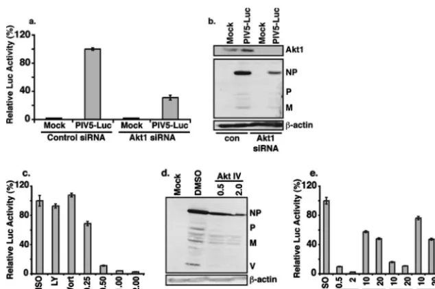
To further confirm the role of Akt1 in viral macromolecule synthesis, siRNA against Akt1 was used to knock down Akt1 expression levels. The effect of the Akt1 siRNA was first ex-amined using a recombinant PIV5 expressing a reporter gene, R-Luc (rPIV5-R-Luc), which was constructed by inserting a R-Luc gene between HN and L of the PIV5 genome; luciferase activity in rPIV5-R-Luc-infected cells thus reflects the level of viral RNA and protein synthesis (data not shown). As shown in Fig. 3a, the siRNA against Akt1 was effective in reducing luciferase activity from rPIV5-R-Luc-infected cells by over 50%. In addition, the effect of siRNA against Akt1 on viral protein expression was examined. As shown in Fig. 3b, siRNA against Akt1 reduced expression of Akt1 and, importantly, PIV5 viral proteins.
[image:4.594.53.272.66.243.2]There are three isoforms of Akt: Akt1, Akt2, and Akt3. Thus, it is possible that Akt2 and Akt3 may compensate for the loss of the Akt1 after siRNA knock-down of Akt1 in HeLa cells. Reverse transcription-PCR analysis of mRNA purified from HeLa cells detected Akt1 as well as Akt2 and Akt3, supporting the possibility that Akt2 and Akt3 may compensate for the loss of Akt1 due to Akt1 siRNA in HeLa cells (data not shown). To examine this possibility, a chemical inhibitor of Akt (AktIV) targeting all three Akt isoforms was used. As shown in Fig. 3c, the addition of the inhibitor reduced the luciferase activity in rPIV5-R-Luc-infected cells in a dosage-dependent manner compared to that in control-treated cells. This inhibi-tion was more potent than that seen with the Akt1 siRNA, suggesting that the other isoforms of Akt could indeed com-pensate for the loss of Akt1 in viral replication. Since Akt is downstream of PI3K, we examined whether PI3K inhibition
[image:4.594.134.451.429.640.2]FIG. 2. Effect of Akt1 expression on V-mediated inhibition of the minigenome system. Expression of Akt abrogates the inhibition of viral RNA synthesis by the V protein. Increasing amounts of an Akt1 expres-sion vector were cotransfected together with a fixed amount of plasmid encoding V into a PIV5 minigenome system (36). The total amount of DNA transfected into the cells was maintained constant by using a plas-mid expressing GFP. F- and R-Luc activities were detected in cell lysates at 24 h postinfection. Normalized luciferase activity of the positive control (no V or Akt1 plasmid; third bar from left) was set as 100%. NC, negative control.
FIG. 3. Role of Akt1 in PIV5 protein expression. (a) Effect of siRNA against Akt1 on PIV5 protein expression. HeLa cells were transfected with siRNA against firefly luciferase (control) or human Akt1 and then infected with rPIV5-R-Luc at a MOI of 5. One day postinfection, luciferase activities (a) and expression levels of Akt1, PIV5 proteins, and actin (b) were determined. Luciferase activity in PIV5-R-Luc cells transfected with control siRNA (con) was set at 100%. (c) Inhibition of luciferase expression from PIV5-R-Luc by Akt inhibitor treatment. HeLa cells were infected with rPIV5-R-Luc at a MOI of 1 and treated with DMSO or increasing concentrations of the AktIV inhibitor (Calbiochem). The cells were collected at 1 day postinfection and assayed for luciferase activity. LY, LY294002 (10M); Wort, wortmannin (1M). (d) HeLa cells were infected with PIV5 and treated with DMSO or Akt inhibitor. The cells were collected 24 h postinfection and subjected to immunoblotting using anti-PIV5 and anti-actin antibodies. (e) Effects of different Akt inhibitors on PIV5 gene expression. The experiments shown in panel c were repeated using different Akt inhibitors. AktV, Akt1/2, and AktXI from Calbiochem were used in micromolar concentrations as indicated.
on November 8, 2019 by guest
http://jvi.asm.org/
could also block virus replication. Interestingly, the PI3K in-hibitors LY294002 and wortmannin had no effect on PIV5 replication (Fig. 3c), suggesting that Akt activation in the con-text of viral infection is not PI3K dependent. Treatment with Akt inhibitor showed similar inhibition of viral structural pro-tein production in virus-infected cells, as determined by immu-noblotting with anti-PIV5 antibodies (Fig. 3d), compared with luciferase. Because of a general concern over the specificity of small-molecule inhibitors, the effects of three additional Akt inhibitors with different chemical structures and specificities were examined; all were found to be effective at blocking the luciferase expression, confirming Akt’s critical role in PIV5 macromolecule synthesis (Fig. 3e).
Akt inhibitor reduced the replication of PIV5, MuV, and MeV. To examine the effect of the Akt inhibitors on virus replication, MDBK cells, the optimal cell type for growing PIV5, were infected with PIV5 at a MOI of 5 and treated with the Akt inhibitor or DMSO. Twenty-four or 48 h postinfection, the supernatants were collected for plaque assays to deter-mine the viral titers. As shown in Fig. 4a, the addition of the Akt inhibitor reduced virus titers over 1,000-fold, indicating that the Akt inhibitor very effectively reduced virus growth. No cell death due to the inhibitor treatment at the inhibitory concentrations was observed by counting trypan blue-stained cells (data not shown). Similar results were obtained when PIV5 growth was analyzed with HeLa cells (Fig. 4b).
MuV is very closely related to PIV5, and the V proteins of PIV5 and MuV are highly homologous (32). To examine whether Akt plays a role in MuV replication, HeLa cells were infected with MuV and treated with the Akt inhibitor. At 24 h postinfection, titers of MuV in the media of infected cells were measured using plaque assays. Treatment with Akt inhibitor
reduced MuV replication approximately 10-fold (Fig. 4c). Sim-ilarly, the Akt inhibitor reduced both cell-associated and re-leased virus in MeV infection (Fig. 4d), with the rere-leased virus being more sensitive to Akt inhibition. The reasons for this difference are not known, though it is possible that the less-dramatic reduction of cell-associated MeV is due to the pres-ence of residual virus from the initial infection. Treatment with the Akt inhibitor also reduced the growth of SeV (data not shown). Together, these data indicate that paramyxoviruses from different subfamilies require the Akt pathway for efficient viral replication.
Inhibition of RSV and VSV replication by the Akt inhibitors.
To study whether Akt plays a more general role in paramyxo-virus replication, A549 cells, a human lung cell line, were infected with a recombinant RSV containing a GFP gene (rRSV-GFP) and then treated with the Akt inhibitor for 24 h. As shown in Fig. 5a, the Akt inhibitor reduced synthesis of GFP in rRSV-GFP-infected cells, suggesting that Akt plays a role in RSV replication. Furthermore, Akt inhibitor treatment blocked the production of infectious RSV virions, indicating that the decreased GFP expression correlates with viral protein and virion production (Fig. 5a, right panel).
To investigate the role of Akt in the replication of NNSVs other than paramyxoviruses, the effect of the Akt inhibitors on rhabdovirus replication was examined. BHK cells were in-fected with VSV at a MOI of 2 and treated with the Akt inhibitor. Treatment with the Akt inhibitor reduced VSV titers by 2 to 3 logs (Fig. 5b). Interestingly, the inhibitor effectively blocked the cytopathic effect induced by VSV (Fig. 5b). Addi-tional Akt inhibitors were also tested and found to be effective in blocking VSV replication, with reductions of viral replica-tion ranging from 10- to 20-fold (data not shown).
Inhibition of phosphorylation of P in PIV5-infected cells by Akt inhibitor.In the experiments described above, the inhibi-tors were added after the virus was incubated with cells, indi-cating that the effect of the inhibitors on the virus life cycle was at a postentry step. The fact that the siRNA against Akt and the inhibitors against Akt reduced viral protein expression (Fig. 2 and 3) indicates that Akt likely plays a role in viral RNA synthesis, since PIV5 protein expression is regulated at the level of transcription. To examine this possibility, we utilized the PIV5 minigenome described above (Fig. 2). We found that the Akt inhibitor blocked reporter gene expression from the minigenome system, indicating that, indeed, Akt plays a critical role in viral RNA synthesis (Fig. 6a). Consistent with the pre-vious results (Fig. 3), the PI3K inhibitor LY294002 did not inhibit reporter gene expression from the minigenome.
[image:5.594.44.284.67.259.2]The fact that Akt inhibitor inhibits viral RNA synthesis in the minigenome system, which does not contain the V protein, indicates that the likely target of Akt is not the V protein itself. In addition, Akt plays a role in the replication of RSV and VSV, which do not encode V protein homologs. Thus, Akt likely targets a component of the viral RNA polymerase com-plex, which is minimally comprised of the P and L proteins for all NNSVs. It is known that the phosphorylation of the P protein plays a critical role in viral RNA synthesis (38). Since Akt is a protein kinase, we examined the effect of the Akt inhibitor on phosphorylation of P in both PIV5- and RSV-infected cells. HeLa (PIV5) or A549 (RSV) cells were RSV-infected for 18 to 24 h and then labeled with either [33P]orthophosphate
FIG. 4. Akt inhibitor inhibited growth of PIV5, MuV, and MeV. MDBK (a) or HeLa (b) cells were infected with PIV5 at a MOI of 5 and treated with DMSO or Akt inhibitor (0.5M AktIV inhibitor or 10M Akt 1/2 inhibitor). The media from the cells were collected at the indicated times postinfection. (c) Inhibition of MuV growth. HeLa cells were infected with MuV at a MOI of 3 and treated with DMSO or AktIV inhibitor (0.5M). (d) Inhibition of MeV growth. Vero cells were infected with MeV at a MOI of 3 and treated with DMSO or AktIV inhibitor (2M). For all samples, virus titers were determined by plaque assay.
on November 8, 2019 by guest
http://jvi.asm.org/
or [35S]Met/[35S]Cys mix in the presence or absence of the Akt
inhibitor. Viral proteins were immunoprecipitated and sepa-rated by SDS-PAGE. Treatment with the Akt inhibitor re-duced phosphorylation of the P protein of PIV5 by about 30% without affecting viral protein translation during the same la-beling period (Fig. 6b). Similarly, the Akt inhibitor reduced phosphorylation of the P protein of RSV by about 50% (Fig. 6c). The partial reduction of phosphorylation of the P proteins suggests that Akt inhibition likely affects some, but not all, of the phosphorylation sites within the P proteins.
Phosphorylation of recombinant P of PIV5 by Akt1 in vitro.
The fact that Akt inhibitor treatment reduced the level of P phosphorylation indicates that Akt plays a role in P phosphor-ylation in infected cells. However, it is not clear whether this effect is direct or indirect. To test whether Akt can directly phosphorylate P, we first purified recombinant PIV5 P protein with a His tag fromEscherichia coliby Ni-affinity chromatog-raphy (27) and then performed an in vitro kinase assay using the recombinant P and activated Akt1. We found that Akt1 phosphorylates the recombinant P protein from bacteria (Fig. 7a). To ensure that the protein phosphorylated by Akt is in-deed P, not a nonspecific bacteria protein copurified with P that has the same electrophoretic mobility as that which mi-grated with the P protein, we immunoprecipitated the purified recombinant P protein with P-specific antibodies (P282 or Pk) and carried out the in vitro kinase assay using the precipitates. The result (Fig. 7b) confirms that Akt 1 can directly phosphor-ylate recombinant P purified from bacteria.
DISCUSSION
We show here that inhibition of Akt by either siRNA or drug treatment results in significant inhibition of PIV5 protein ex-pression and replication. In addition, Akt inhibitor treatment appears to have a broad spectrum of antiviral activity, decreas-ing the replication of NNSV from the rubulavirus, morbillivi-rus, pneumovimorbillivi-rus, and rhabdovirus families. To the best of our knowledge, ours is the first study to report the potential of Akt inhibition as an antiviral therapeutic intervention. Because of the interest in Akt as a target for cancer therapy, there are many good compounds targeting Akt with low toxicity and high efficacy, and some of them have advanced to clinical trials (13). It is possible that some of these compounds will be effective against infections caused by NNSVs in vivo. The development of a broad-spectrum antiviral against NNSVs would allow for therapeutic intervention in a wide array of human diseases.
We have shown that Akt inhibitors can be effective against RSV, the most important etiologic agent of pediatric viral respiratory infection, which remains a major cause of morbid-ity and mortalmorbid-ity among infants and among immunocompro-mised subjects and the elderly (reviewed in reference 14). Importantly, there is no vaccine for RSV, nor are there effec-tive curaeffec-tive treatments for severe RSV disease, although aero-solized ribavirin and prophylactic immunoglobulin therapy are used in the clinical setting. However, the high cost of palivi-zumab prophylaxis due to the need for monthly injections during RSV season raises the question of cost effectiveness
FIG. 5. Inhibition of RSV and VSV growth by the AktIV inhibitor. (a) Inhibition of RSV gene expression by the Akt inhibitor. Left panel, A549 cells were infected with rRSV-GFP at a MOI of 3 and treated with DMSO or AktIV inhibitor (1M). Expression of GFP was monitored by fluorescence microscopy and photographed at 1 day postinfection. Right panel, A549 cells were infected with RSV A2 at a MOI of 3 in triplicate and treated with DMSO or AktIV as described above. The media were collected 1 or 2 days postinfection, and titers of RSV were determined by plaque assay. (b) Inhibition of VSV growth. BHK cells were infected with VSV in triplicate at a MOI of 2 and treated with the AktIV inhibitor (2M). The cells were photographed at 18 h postinfection, and titers of virus from media of infected cells were determined by plaque assays. Graphs shown represent a single experiment. Errors bars show standard deviations of the means.
on November 8, 2019 by guest
http://jvi.asm.org/
relative to health benefits. Therefore, there is a pressing need for safe and effective therapeutic interventions for RSV infec-tion. In addition, Akt inhibitors can block MuV and MeV replication. While MuV and MeV infections have been well controlled in vaccinated population, they still pose a serious health threat in developing counties, where vaccine coverage is poor. VSV, a livestock pathogen, is a rhabdovirus similar to rabies virus, which causes a lethal infection in humans and also lacks an effective antiviral drug. The fact that the Akt inhibitor effectively blocked VSV replication provides evidence that Akt inhibition may be a good strategy for developing anti-rabies virus drugs.
One potential drawback is that the effects of the Akt inhib-itors were variable in different cells for different viruses. For instance, in MDBK cells, a bovine cell line that is optimal for PIV5 growth, the Akt inhibitor had a dramatic effect on virus
[image:7.594.124.463.71.443.2]growth, reducing virus titers by 2 to 3 logs, while the same inhibitor reduced the PIV5 growth by only 1 to 2 logs in HeLa cells. We speculate that the differences are a reflection of how effective the inhibitors are in various cell lines, based on their tissue of origin and level of Akt isoform expression. Further optimization of Akt inhibitor treatment in primary cell culture and animal models of viral disease will enhance their potency for use in humans for therapy of infectious diseases caused by NNSV. While we have not tested the effects of Akt inhibitors on DNA virus replication, we do not expect the inhibitors to be effective against DNA virus. The AktIV inhibitor was originally identified as an inhibitor of the FOXO protein nuclear trans-location, in which FOXO was expressed from a recombinant adenovirus (29). In the report, the inhibitor did not appear to inhibit adenovirus gene expression, suggesting that Akt activa-tion is not necessary for adenovirus replicaactiva-tion.
FIG. 6. Effect of Akt inhibition on minigenome RNA synthesis and P phosphorylation. (a) Inhibition of reporter gene expression from PIV5 minigenome system (36) by the Akt inhibitors. The minigenome system was as described before, and the inhibitors were added to the cells after transfection. (b) Effect of Akt on the phosphorylation status of the P protein of PIV5. HeLa cells were infected with PIV5 at a MOI of 5, and at 1 day postinfection, they were labeled with35S-ProMix or [33P]orthophosphate for 4 h. The cells were lysed and immunoprecipitated with anti-P antibody. The average reduction of phosphorylation of the P protein from five experiments was graphed (P⫽0.02). (c) A549 cells were infected with RSV at a MOI of 3 and, at 1 day postinfection, they were labeled with35S-ProMix or [33P]orthophosphate for 4 h. The cells were lysed and immunoprecipitated with anti-RSV antibody. The average reduction of phosphorylation of the P protein from three experiments was graphed (P⫽ 0.01). MK, mock infection.
on November 8, 2019 by guest
http://jvi.asm.org/
Akt, a serine/threonine kinase, is activated through phos-phorylation at different sites within the protein (6). Since some of the inhibitors we used (e.g., the AktIV inhibitor) are known to inhibit phosphorylation of Akt (29), it is likely that phos-phorylation of Akt plays a role in viral RNA synthesis. Which kinase activates Akt in virus-infected cells and how it is acti-vated itself are not known. It is well established that Akt is one of the major downstream targets for PI3K (6). However, two well-known PI3K inhibitors, wortmannin and LY29002, had no effect on PIV5 replication (Fig. 3a), indicating that PI3K acti-vation is not required for Akt actiacti-vation in virus-infected cells. Therefore, we propose that a kinase downstream of PI3K, or potentially a novel kinase, is responsible for activating Akt in virus-infected cells.
While factors required for its activation remain unclear, our studies provide insight as to the mechanism by which Akt regulates PIV5 RNA synthesis. Chemical inhibition of Akt results in a significant decrease in phosphorylation of the viral P protein. In addition, Akt can phosphorylate P in vitro. The phosphorylation status of P is thought to be a key regulator of the switch by vRNAP from viral mRNA synthesis (transcrip-tion) to a viral RNA replication (12, 15, 55). Previous studies have shown that the P proteins of various NNSV are phosphor-ylated by host kinases (33). For VSV, there are two regions (N terminus region, domain I, and C terminus region, domain II) within the P protein that are phosphorylated. One of the host kinases known to phosphorylate the N-terminal region of VSV P is casein kinase II (CKII) (4). While the host kinase for the C-terminal region has not been identified (16), there is some circumstantial evidence that the host kinase may be Akt. Kim et al. first reported that K-252a, a broad nonspecific kinase inhibitor (isolated from Nocardiopsissp.), and its derivative molecule KT5926 have anti-VSV activities in BHK cells (31). KT5926 does not inhibit CKII (30) but has been shown to target a host kinase that matches the size of Akt (59). In addition, staurosporine inhibits VSV RNA synthesis (53) and
has been shown to target Akt in addition to protein kinases A and C (27).
Like other P proteins of paramyxoviruses, RSV P is a major cofactor for vRNAP and plays an essential role in viral RNA synthesis. Phosphorylation of P plays an essential role in its functions (20). There are five major phosphorylation sites within the P protein (43). Mutating these five phosphorylation sites resulted in a recombinant RSV that is severely attenuated in animals and is reduced in growth in some tissue culture cells (38). Interestingly, this mutant P protein was still phosphory-lated in infected cells, albeit at much lower levels than wild-type P (38). The P protein purified from bacteria can be phos-phorylated by casein kinase II, and this phosphos-phorylated P is as active as the P protein treated with cell extract (presumably containing the host kinase that phosphorylates P) in an in vitro viral RNA synthesis assay, indicating that CKII can phosphor-ylate P (39, 63). However, CKII has not been shown to phos-phorylate P in infected cells. We found that inhibition of Akt reduced the phosphorylation of the P protein, indicating that Akt plays a critical role in the phosphorylation of the P protein in RSV-infected cells.
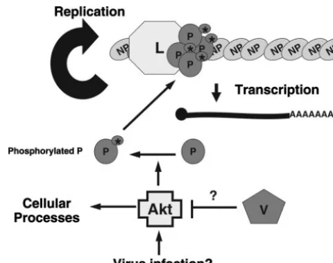
Our data lead us to a specific model for the role of Akt in the replication of NNSV (Fig. 8). Infection of cells by NNSV results in the activation of Akt. While the mechanism by which Akt is activated is unclear, PI3K is unlikely involved, as treat-ment with PI3K inhibitors has no effect on viral replication. Activated Akt then phosphorylates the viral P protein, which is an essential cofactor for vRNAP, regulating viral RNA synthe-sis. Since Akt inhibition causes a significant decrease in viral protein production, it is possible that phosphorylation of P by Akt is responsible for regulating the switch between
[image:8.594.300.540.67.256.2]transcrip-FIG. 7. Phosphorylation of recombinant P by Akt1. Recombinant PIV5 P with a six-His tag at its N terminus was purified from bacteria as described in Materials and Methods and used in in vitro kinase assays with activated Akt1. (a) Phosphorylation of the recombinant P by Akt1 in vitro. P, Akt1, or P plus Akt1 was mixed with [␥-32P]ATP, and the reaction mixtures were resolved by SDS-PAGE. Phosphory-lated proteins were detected by phosphorimagery. Shown on the right are the relative mobilities of Akt1 and P. (b) Phosphorylation of recombinant P immunoprecipitated with P-specific antibody by Akt1. The recombinant P protein purified from bacteria was incubated with P-specific antibody P282, Pk, or control antibody mouse immunoglob-ulin G and washed with immunoprecipitation buffer as described in Materials and Methods. The precipitated products along with the beads were incubated with activated Akt1 and [␥-32P]ATP. The prod-ucts were resolved by SDS-PAGE and visualized as described above.
FIG. 8. A model for the involvement of Akt in viral RNA synthesis. The replication of viral RNA and synthesis of viral mRNA require P and L complex. The phosphorylation status of the P protein plays a critical in viral RNA synthesis. Akt phosphorylates P, thus leading to the activation of P. For viruses encoding a V protein, the V protein can regulate viral RNA synthesis via its interaction with Akt through an unknown mechanism. The V-Akt1 interaction may also contribute to regulation of innate immune responses by the V protein. Activation of Akt1 is often through the PI3K pathway. However, it is not clear what activates Akt1 in virus-infected cells, since PI3K inhibitors had no effect on virus replication.
on November 8, 2019 by guest
http://jvi.asm.org/
[image:8.594.59.268.72.151.2]tion and RNA replication by the vRNAP in favor of transcrip-tion. Increased transcription would then allow for increased viral protein production and, subsequently, genome replication and virion morphogenesis. Inhibition of Akt thus blocks viral replication at an early stage postentry. In the paramyxoviruses which encode V proteins or other accessory proteins, such as W or C, we propose that these proteins regulate viral RNA synthesis by interacting with Akt and inhibiting its function, either by decreasing its kinase activity or by sequestering Akt itself. Thus, we propose that Akt plays a critical role in viral replication by regulating RNA synthesis through the phosphor-ylation of the P protein in infected cells.
The studies presented in this paper arose from our hypoth-esis that the V protein of PIV5 exerts its numerous activities by interacting with host proteins. We initially used a computa-tional method to predict potential V-interacting proteins and then determined empirically that V binds specifically to one of the predicted interactors, Akt1/PKB. This interaction has func-tional consequences in that Akt overexpression alleviates V-mediated inhibition of PIV5 RNA synthesis in a minigenome system. Beyond this effect on the viral polymerase activity, it is possible that the V-Akt interaction plays additional roles in viral infection. Akt plays an important role in many cellular signaling pathways, such as those for apoptosis and interferon signaling and production (23). The V protein of PIV5 is known to inhibit apoptosis induced by viral infection (57). Therefore, the interaction between V and Akt is consistent with the role of V in apoptosis. Furthermore, V has also been shown to block interferon production and interferon signaling as well as to block interleukin-6 expression (18, 25, 37). It will be inter-esting to examine whether the interaction between V and Akt plays a role in these processes. Finally, PIV5 infection slows down the cell cycle, and the V protein of PIV5 is thought to play a critical in the process (35). It is tempting to speculate that the interaction of V and Akt may contribute to the slowing down of the cell cycle by PIV5, since Akt signaling also affects cell cycle control (6). Thus, Akt activation, and interaction with V, may play multiple roles in the replication and pathogenesis of NNSV.
ACKNOWLEDGMENTS
We are grateful to Robert A. Lamb for providing SeV Z strain and VSV, Richard Randall for providing antibodies, Stephen Michnick for the YFP system, and Anthony Schmitt for critical readings of the manuscript. We thank the members of Biao He’s laboratory for helpful discussion and technical assistance. We also thank Kim Tran for as-sistance with generating some of the figures.
The work was supported by grants from the National Institute of Allergy and Infectious Disease to B.H. (AI051372 and K02 AI65795).
REFERENCES
1.Abraham, G., and A. K. Banerjee.1976. Sequential transcription of the genes of vesicular stomatitis virus. Proc. Natl. Acad. Sci. USA73:1504–1508. 2.Ball, L. A., and C. N. White. 1976. Order of transcription of genes of
vesicular stomatitis virus. Proc. Natl. Acad. Sci. USA73:442–446. 3.Barik, S.1992. Transcription of human respiratory syncytial virus genome
RNA in vitro: requirement of cellular factor(s). J. Virol.66:6813–6818. 4.Barik, S., and A. K. Banerjee.1992. Phosphorylation by cellular casein kinase
II is essential for transcriptional activity of vesicular stomatitis virus phos-phoprotein P. Proc. Natl. Acad. Sci. USA.89:6570–6574.
5.Barik, S., T. McLean, and L. C. Dupuy.1995. Phosphorylation of Ser232 directly regulates the transcriptional activity of the P protein of human respiratory syncytial virus: phosphorylation of Ser237 may play an accessory role. Virology213:405–412.
6.Brazil, D. P., and B. A. Hemmings.2001. Ten years of protein kinase B signalling: a hard Akt to follow. Trends Biochem. Sci.26:657–664.
7.Buchholz, U. J., S. Finke, and K. K. Conzelmann.1999. Generation of bovine respiratory syncytial virus (BRSV) from cDNA: BRSV NS2 is not essential for virus replication in tissue culture, and the human RSV leader region acts as a functional BRSV genome promoter. J. Virol.73:251–259. 8.Burke, E., L. Dupuy, C. Wall, and S. Barik.1998. Role of cellular actin in the
gene expression and morphogenesis of human respiratory syncytial virus. Virology252:137–148.
9.Burke, E., N. M. Mahoney, S. C. Almo, and S. Barik.2000. Profilin is required for optimal actin-dependent transcription of respiratory syncytial virus genome RNA. J. Virol.74:669–675.
10.Chan, T. O., S. E. Rittenhouse, and P. N. Tsichlis.1999. AKT/PKB and other D3 regulated kinases: kinase activation by phosphoinositide-dependent phosphorylation. Annu. Rev. Biochem.68:965–1014.
11.Chatziandreou, N., N. Stock, D. Young, J. Andrejeva, K. Hagmaier, D. J. McGeoch, and R. E. Randall.2004. Relationships and host range of human, canine, simian and porcine isolates of simian virus 5 (parainfluenza virus 5). J Gen. Virol.85:3007–3016.
12.Chen, J. L., T. Das, and A. K. Banerjee.1997. Phosphorylated states of vesicular stomatitis virus P protein in vitro and in vivo. Virology228:200–212. 13.Cheng, J. Q., C. W. Lindsley, G. Z. Cheng, H. Yang, and S. V. Nicosia.2005. The Akt/PKB pathway: molecular target for cancer drug discovery. Onco-gene24:7482–7492.
14.Collins, P. L., R. M. Chanock, and B. R. Murphy.2001. Respiratory syncytial virus, p. 1443–1485.InD. M. Knipe and P. M. Howley (ed.), Virology, 4th ed. Raven Press, New York, NY.
15.Das, S. C., and A. K. Pattnaik.2004. Phosphorylation of vesicular stomatitis virus phosphoprotein P is indispensable for virus growth. J. Virol.78:6420– 6430.
16.Das, S. C., and A. K. Pattnaik.2005. Role of the hypervariable hinge region of phosphoprotein P of vesicular stomatitis virus in viral RNA synthesis and assembly of infectious virus particles. J. Virol.79:8101–8112.
17.De, B. P., and A. K. Banerjee.1999. Involvement of actin microfilaments in the transcription/replication of human parainfluenza virus type 3: possible role of actin in other viruses. Microsc. Res. Tech.47:114–123.
18.Didcock, L., D. F. Young, S. Goodbourn, and R. E. Randall.1999. The V protein of simian virus 5 inhibits interferon signalling by targeting STAT1 for proteasome-mediated degradation. J. Virol.73:9928–9933.
19.Du, K., and P. N. Tsichlis.2005. Regulation of the Akt kinase by interacting proteins. Oncogene24:7401–7409.
20.Dupuy, L. C., S. Dobson, V. Bitko, and S. Barik.1999. Casein kinase 2-me-diated phosphorylation of respiratory syncytial virus phosphoprotein P is essential for the transcription elongation activity of the viral polymerase; phosphorylation by casein kinase 1 occurs mainly at Ser215
and is without effect. J. Virol.73:8384–8392.
21.Emerson, S. U.1982. Reconstitution studies detect a single polymerase entry site on the vesicular stomatitis virus genome. Cell31:635–642.
22.Emerson, S. U., and Y.-H. Yu.1975. Both NS and L proteins are required for in vitro RNA synthesis by vesicular stomatitis virus. J. Virol.15:1348–1356. 23.Franke, T. F., C. P. Hornik, L. Segev, G. A. Shostak, and C. Sugimoto.2003.
PI3K/Akt and apoptosis: size matters. Oncogene22:8983–8998.
24.Fuentes, S., K. C. Tran, P. Luthra, M. N. Teng, and B. He.2007. Function of the respiratory syncytial virus small hydrophobic protein. J. Virol.81: 8361–8366.
25.He, B., R. G. Paterson, N. Stock, J. E. Durbin, R. K. Durbin, S. Goodbourn, R. E. Randall, and R. A. Lamb.2002. Recovery of paramyxovirus simian virus 5 with a V protein lacking the conserved cysteine-rich domain: the multifunctional V protein blocks both interferon-beta induction and inter-feron signaling. Virology303:15–32.
26.He, B., R. G. Paterson, C. D. Ward, and R. A. Lamb.1997. Recovery of infectious SV5 from cloned DNA and expression of a foreign gene. Virology 237:249–260.
27.He, B., M. Rong, D. Lyakhov, H. Gartenstein, G. Diaz, R. Castagna, W. T. McAllister, and R. K. Durbin.1997. Rapid mutagenesis and purification of phage RNA polymerases. Protein Expr. Purif.9:142–151.
28.Iverson, L. E., and J. K. Rose.1982. Sequential synthesis of 5⬘-proximal vesicular stomatitis virus mRNA sequences. J. Virol.44:356–365. 29.Kau, T. R., F. Schroeder, S. Ramaswamy, C. L. Wojciechowski, J. J. Zhao,
T. M. Roberts, J. Clardy, W. R. Sellers, and P. A. Silver.2003. A chemical genetic screen identifies inhibitors of regulated nuclear export of a Forkhead transcription factor in PTEN-deficient tumor cells. Cancer Cell4:463–476. 30.Kim, Y. S., and A. Kawai.1998. Studies on the antiviral mechanisms of
protein kinase inhibitors K-252a and KT5926 against the replication of vesicular stomatitis virus. Biol. Pharm. Bull.21:498–505.
31.Kim, Y. S., J. Sagara, and A. Kawai.1995. Studies on the antiviral activity of protein kinase inhibitors against the replication of vesicular stomatitis virus. Biol. Pharm. Bull.18:895–899.
32.Lamb, R. A., and D. Kolakofsky.2001.Paramyxoviridae: the viruses and their replication.InD. M. Knipe, P. M. Howley, D. E. Griffin, R. A. Lamb, M. A. Martin, B. Roizman, and S. E. Straus (ed.), Fields virology, 4th ed. Lippin-cott Williams & Wilkins, Philadelphia, PA.
33.Lenard, J.1999. Host cell protein kinases in nonsegmented negative-strand virus (Mononegavirales) infection. Pharmacol. Ther.83:39–48.
on November 8, 2019 by guest
http://jvi.asm.org/
34.Li, Z., M. Yu, H. Zhang, D. E. Magoffin, P. J. Jack, A. Hyatt, H. Y. Wang, and L. F. Wang.2006. Beilong virus, a novel paramyxovirus with the largest genome of non-segmented negative-stranded RNA viruses. Virology346: 219–228.
35.Lin, G. Y., and R. A. Lamb.2000. The paramyxovirus simian virus 5 V protein slows progression of the cell cycle. J. Virol.74:9152–9166. 36.Lin, Y., F. Horvath, J. A. Aligo, R. Wilson, and B. He.2005. The role of
simian virus 5 V protein on viral RNA synthesis. Virology338:270–280. 37.Lin, Y., M. Sun, S. M. Fuentes, C. D. Keim, T. Rothermel, and B. He.2007.
Inhibition of interleukin-6 expression by the V protein of parainfluenza virus 5. Virology368:262–272.
38.Lu, B., C. H. Ma, R. Brazas, and H. Jin.2002. The major phosphorylation sites of the respiratory syncytial virus phosphoprotein are dispensable for virus replication in vitro. J. Virol.76:10776–10784.
39.Mazumder, B., G. Adhikary, and S. Barik.1994. Bacterial expression of human respiratory syncytial viral phosphoprotein P and identification of Ser237 as the site of phosphorylation by cellular casein kinase II. Virology 205:93–103.
40.Moscona, A., and R. W. Peluso.1996. Analysis of human parainfluenza virus 3 receptor binding variants: evidence for the use of a specific sialic acid-containing receptor. Microb. Pathog.20:179–184.
41.Moyer, S. A., S. C. Baker, and S. M. Horikami.1990. Host cell proteins required for measles virus reproduction. J. Gen. Virol.71:775–783. 42.Moyer, S. A., S. C. Baker, and J. L. Lessard.1986. Tubulin: a factor
neces-sary for the synthesis of both Sendai virus and vesicular stomatitis virus RNAs. Proc. Natl. Acad. Sci. USA83:5405–5409.
43.Navarro, J., C. Lopez-Otin, and N. Villanueva.1991. Location of phosphor-ylated residues in human respiratory syncytial virus phosphoprotein. J. Gen. Virol.72:1455–1459.
44.Ogino, T., M. Iwama, J. Kinouchi, Y. Shibagaki, T. Tsukamoto, and K. Mizumoto.1999. Involvement of a cellular glycolytic enzyme, phosphoglyc-erate kinase, in Sendai virus transcription. J. Biol. Chem.274:35999–36008. 45.Ogino, T., M. Iwama, Y. Ohsawa, and K. Mizumoto.2003. Interaction of cellular tubulin with Sendai virus M protein regulates transcription of viral genome. Biochem. Biophys. Res. Commun.311:283–293.
46.Ogino, T., T. Yamadera, T. Nonaka, S. Imajoh-Ohmi, and K. Mizumoto. 2001. Enolase, a cellular glycolytic enzyme, is required for efficient transcrip-tion of Sendai virus genome. Biochem. Biophys. Res. Commun.285:447–455. 47.Paterson, R. G., and R. A. Lamb.1993. The molecular biology of influenza viruses and paramyxoviruses, p. 35–73.InA. Davidson and R. M. Elliott (ed.), Molecular virology: a practical approach. IRL Oxford University Press, Oxford, United Kingdom.
48.Powell, D. W., M. J. Rane, Q. Chen, S. Singh, and K. R. McLeish.2002. Identification of 14-3-3as a protein kinase B/Akt substrate. J. Biol. Chem. 277:21639–21642.
49.Randall, R. E., and D. F. Young.1988. Comparison between parainfluenza
virus type 2 and simian virus 5: monoclonal antibodies reveal major antigenic differences. J. Gen. Virol.69:2051–2060.
50.Redaelli, C., F. Granucci, L. De Gioia, and L. Cipolla.2006. Synthesis and biological activity of Akt/PI3K inhibitors. Mini Rev. Med. Chem.6:1127– 1136.
51.Remy, I., and S. W. Michnick.2004. A cDNA library functional screening strategy based on fluorescent protein complementation assays to identify novel components of signaling pathways. Methods32:381–388.
52.Richardson, J. C., and R. W. Peluso.1996. Inhibition of VSV genome RNA replication but not transcription by monoclonal antibodies specific for the viral P protein. Virology216:26–34.
53.Rigaut, K. D., Y. Gao, and J. Lenard.1993. Effects of staurosporine on transcription by vesicular stomatitis virus. Virology194:433–440. 54.Sarbassov, D. D., D. A. Guertin, S. M. Ali, and D. M. Sabatini.2005.
Phosphorylation and regulation of Akt/PKB by the rictor-mTOR complex. Science307:1098–1101.
55.Spadafora, D., D. M. Canter, R. L. Jackson, and J. Perrault.1996. Consti-tutive phosphorylation of the vesicular stomatitis virus P protein modulates polymerase complex formation but is not essential for transcription or rep-lication. J. Virol.70:4538–4548.
56.Stambolic, V., and J. R. Woodgett.2006. Functional distinctions of protein kinase B/Akt isoforms defined by their influence on cell migration. Trends Cell Biol.16:461–466.
57.Sun, M., T. A. Rothermel, L. Shuman, J. A. Aligo, S. Xu, Y. Lin, R. A. Lamb, and B. He.2004. Conserved cysteine-rich domain of paramyxovirus simian virus 5 V protein plays an important role in blocking apoptosis. J. Virol. 78:5068–5078.
58.Takeuchi, K., K. Tanabayashi, K. Okazaki, M. Hiahiyama, and A. Yamada. 1993. In vitro transcription and replication of the mumps virus genome. Arch. Virol.128:177–183.
59.Teng, K. K., and L. A. Greene.1994. KT5926 selectively inhibits nerve growth factor-dependent neurite elongation. J. Neurosci.14:2624–2635.
60.Thomas, S. M., R. A. Lamb, and R. G. Paterson.1988. Two mRNAs that differ by two nontemplated nucleotides encode the amino coterminal pro-teins P and V of the paramyxovirus SV5. Cell54:891–902.
61.Tidona, C. A., H. W. Kurz, H. R. Gelderblom, and G. Darai.1999. Isolation and molecular characterization of a novel cytopathogenic paramyxovirus from tree shrews. Virology258:425–434.
62.Ulane, C. M., and C. M. Horvath.2002. Paramyxoviruses SV5 and HPIV2 assemble STAT protein ubiquitin ligase complexes from cellular compo-nents. Virology304:160–166.
63.Villanueva, N., J. Navarro, E. Mendez, and I. Garcia-Albert.1994. Identifi-cation of a protein kinase involved in the phosphorylation of the C-terminal region of human respiratory syncytial virus P protein. J. Gen. Virol.75:555– 565.
64.Yoeli-Lerner, M., and A. Toker.2006. Akt/PKB signaling in cancer: a func-tion in cell motility and invasion. Cell Cycle5:603–605.