EFFECTIVENESS OF 3D FOOT SCANNER
DESIGNED AND FABRICATED CUSTOMIZED
FOOT INSOLE IN THE MANAGEMENT OF
CHILDREN WITH FLAT FOOT
A PROJECT WORK SUBMITTED IN PARTIAL FULFILLMENT
OF THE REQUIREMENTS FOR THE DEGREE OF
MASTER OF OCCUPATIONAL THERAPY
(ADVANCED O.T. IN PAEDIATRICS)
Submitted by
Reg. No. 411613052
JKK MUNIRAJAH MEDICAL RESEARCH FOUNDATION COLLEGE
OF OCCUPATIONAL THERAPY
KOMARAPALAYAM - 638183
Affiliated to
THE TAMILNADU DR. M.G.R. MEDICAL UNIVERSITY,
CHENNAI-600032
EFFECTIVENESS OF 3D FOOT SCANNER
DESIGNED AND FABRICATED CUSTOMIZED
FOOT INSOLE IN THE MANAGEMENT OF
CHILDREN WITH FLAT FOOT
A PROJECT WORK SUBMITTED IN PARTIAL FULFILLMENT OF THE REQUIREMENTS FOR THE DEGREE OF
MASTER OF OCCUPATIONAL THERAPY
(ADVANCED O.T. IN PAEDIATRICS)
Submitted By
Reg. No. 411613052
JKK MUNIRAJAH MEDICAL RESEARCH FOUNDATION
COLLEGE OF OCCUPATIONAL THERAPY
KOMARAPALAYAM-638183.
Affiliated to
THE TAMILNADU DR. M.G.R. MEDICAL UNIVERSITY,
CHENNAI-600032
MAY – 2017
PRINCIPAL EXTERNAL EXAMINER
CERTIFICATE
This is to certify that the research work entitled “EFFECTIVENESS OF 3D FOOT
SCANNER DESIGNED AND FABRICATED CUSTOMIZED FOOT INSOLE IN THE
MANAGEMENT OF CHILDREN WITH FLAT FOOT” was carried out by Reg. No. 411613052,
Final Year student, College of Occupational Therapy under JKK Munirajah Medical Research
Foundation, Komarapalayam – 638183, in partial fulfillment for the award of Degree of “Master
of Occupational Therapy” (Advanced O.T. in Paediatrics) of The Tamil Nadu Dr. M.G.R.
Medical University, Chennai-32.
Mrs. R. RENUCHITRA. M.O.T., MDASLP, Mr. T. JEGADEESAN. M.O.T., M.Sc (Psy) Professor (Paediatrics) Principal
JKKMMRF College of Occupational Therapy, JKKMMRF College of Occupational Therapy,
Komarapalayam. Komarapalayam.
Clinical Guide
Dr. VANATHI BALACHANDRAN MD., Pediatrics, Asst. Professor, JKKMMRF College of Occupational Therapy
CERTIFICATE
This is to certify that the Project work entitled “EFFECTIVENESS OF 3D FOOT
SCANNER DESIGNED AND FABRICATED CUSTOMIZED FOOT INSOLE IN THE
MANAGEMENT OF CHILDREN WITH FLAT FOOT” is a bonafied compiled work carried out
by Reg. No. 411613052, Final Year student, College of Occupational Therapy under JKK
Munirajah Medical Research Foundation, Komarapalayam – 638183, in partial fulfillment for the
award of Degree of “Master of Occupational Therapy”(Advanced O.T. in Paediatrics) of The
Tamilnadu Dr. M.G.R. Medical University, Chennai-32. This work was guided and supervised
by Mrs. R. RENU CHITRA, MOT., (Pediatrics) MDASLP at the Department of Occupational
Therapy, JKKMMRF, Komarapalayam.
Mrs. R. RENU CHITRA. M.O.T., (Paed), MDASLP,
Professor
ACKNOWLEDGEMENT
I am deeply indebted to My LORD ALMIGHTY for his abundant grace,
unseen guidance and ceaseless bestowed upon me in all my endeavours and made my
desire a reality which would have not been a possible otherwise.
I express my sincere thanks to Dr. J.K.K.MUNIRAJAHH, M. Tech
(Bolton), Correspondent J.K.K.MMRF College of Occupational Therapy for
granting me this opportunity to complete my Master of Occupational Therapy course
in their prestigious institution.
I express my deep sense of gratitude to Mr. T. JEGADEESAN, M.O.T,
M.Sc. (Psy), Principal, JKKMMRF College of Occupational Therapy for his
valuable guidance, encouragement and concern at every point which has been the
driving force to complete this dissertation.
It is my great pleasure and privilege to record my sincere thanks to my Guide
Mrs. RENU CHITRA, M.O.T (Paediatrics), MDASLP, Professor, JKKMMRF
College of Occupational Therapy for her patience; constant support, inspiration and
priceless guidance throughout this dissertation which made this study a great success.
My Special thanks to my Clinical Guide Dr. VANATHI
BALACHANDRAN, MD, PAEDIATRICS, Asst. Professor, JKKMMRF College of
Occupational Therapy.
My Sincere thanks to Mr. K. DHANAPAL, Statistician, JKKMMRF
College of Occupational Therapy for the valuable help he offered in Statistical
I extend my thanks to All the College Staffs, Department Faculty Members,
Office Staffs and Librarian, JKKMMRF College of Occupational Therapy who
gave me a helping hand to do this project.
I owe to express my gratitude and sincere thanks to THE LEPROSY
MISSION TRUST INDIA, for funding this project and sponsoring myself for Master
degree.
I have no words to express my thanks to Mr. MANIVANNAN, Programme
Manager, PARTI Project, Mr. KARTHICK, Co-ordinator Research Domain,
Mr. MATHANRAJ David, Programme Manager, CREATE Project,
Dr. MS. RAJU, Research Scientist, Research Domain, Mr. RAJA, Physiotherapist
CFAB project, Mr. SHAM KUNNATHDASS, Prosthetist and Orthotist, CFAB,
The Leprosy Mission Trust India. Mr. SATHISH PAUL, Physiotherapist cum
Podiatrist, Head of Physiotherapy Dept. SLRTC, Karigiri.
My sincere thanks to Dr. CHINNARAJU, Medical Superintendent, Leprosy
Mission Hospital, Vadathorasalur, Dr. JERRY JOSHUA, Head Health, New Delhi,
Mr. MELVIN MORIS, Head HR, New Delhi, Dr. MERCY BERRY, Paediatrician,
Leprosy Mission Hospital, Vadathorasalur, Mr. BINO BERRY, Co-ordinator POID,
Mr. RAM K ROBERT Programme Manager CREAT Project.
And also I express my thanks to my family members especially my daughter
MANSI, Batchmates, Mrs. G.SRINITHYA, Mr. GANAPATHY, Mr. RICHU
JOSEPH and friends for their constant support, encouragement and co-operation.
My special thanks to STAFFS of The Leprosy Mission PARTI Project and
Early Intervention Centre, Chidambaram Block.
Last but not least I thank all the Children and Parents who took part in the
TABLE OF CONTENTS
S.
NO.
CONTENT
PAGE
NO.
Abstract
I
Introduction
1-5
II
Need for the Study
6
III
Aim and Objective
7
IV
Hypothesis
8
V
Significance of the Study
9
VI
Related Literature
10-20
VII
Review Literature
21-31
VIII
Methodology
32-40
IX
Data Analysis and Interpretation
41-57
X
Discussion
58-62
XI
Conclusion
63
XII
Limitations and Recommendation
64
XIII
Bibliography
65-70
LIST OF TABLES
TABLE
NO.
LIST OF TABLES
PAGE
NO.
1
Comparison of foot function index of pre test of
both control and experimental group
42
2
Comparison of oxford ankle foot questionnaire
pre test of both control and experimental group
44
3
Comparison of foot function index between pre
and post test of control group
46
4
Comparison of oxford ankle and foot
questionnaire between pre and post test of
control group
48
5
Comparison of foot function index between pre
and post test of experimental group
50
6
Comparison of oxford ankle and foot
questionnaire between pre and post test of
experimental group
52
7
Comparison of foot function index of post test of
both control and experimental group
54
8
Comparison of oxford ankle foot questionnaire
post test of both control and experimental group
[image:8.595.98.535.92.728.2]LIST OF GRAPHS
GRAPH
NO
LIST OF GRAPHS
PAGE
NO
1
Comparison of foot function index of pre test
of both control and experimental group
43
2
Comparison of oxford ankle foot
questionnaire pre test of both control and
experimental group
45
3
Comparison of foot function index between
pre and post test of control group
47
4
Comparison of oxford ankle and foot
questionnaire between pre and post test of
control group
49
5
Comparison of foot function index between
pre and post test of experimental group
51
6
Comparison of oxford ankle and foot
questionnaire between pre and post test of
experimental group
53
7
Comparison of foot function index of post
test of both control and experimental group
55
8
Comparison of oxford ankle foot
questionnaire post test of both control and
experimental group
ABSTRACT
OBJECTIVE:
The purpose of the study is to evaluate the effectiveness of 3D foot scanner
designed and fabricated customized foot insole in the management of children with
flat foot.
METHODS:
In this study a total of 30 children from the age of 6 to 10 years with flat foot
participated and were divided into 15 each in experimental and control group. The
Foot Function Index (FFI) and Oxford Ankle foot Questionnaire (Parent Version)
were used for both the groups at the start of the intervention and later after 4 months
after intervention. The 3D foot scanner designed and fabricated customized foot
insole was used as an intervention in the management of children with flat foot for 4
months duration and the data were recorded and analysed for statistical analysis.
RESULTS:
In control group Foot Function Index (FFI) pre-test score are56 (5.58) and
post test score was 55.80(5.89) and Oxford Ankle foot Questionnaire Pre-test scores
were 18.73 (5.04) and post test score were 18.73 (5.32). Whereas in Experimental
group Foot Function Index (FFI) pre-test score was 55.40 (8.36) and post test score
was 41(7.92) and Oxford Ankle foot Questionnaire Pre-test scores were 15.80(6.18)
and post test score were 27.60(5.37). The paired t test for the experimental group for
Foot Function Index (FFI) is (p <0.01) and Oxford Ankle foot Questionnaire was
CONCLUSION:
The findings of the study suggest that 3D foot scanner designed and fabricated
customized foot insole is effective in management of children with flat foot.
KEY WORDS:
Flat foot, Heel Valgus, Navicular Height, insole, Medial arch, 3D foot
1
INTRODUCTION
Flat foot (pes planus) is one of the most common conditions observed in
children. The true prevalence of flat foot is unknown, primarily because there is no
consensus on the strict clinical or radiographic criteria for defining a flat foot38.
Flat foot is defined as absence of an arch in the sole of the foot that causes the
foot to lie flat when the person is standing.
Prevalence of flat foot is highly variable in different populations. This
variability is influenced by various factors. Those can be divided into two; internal
and external factors. The internal factors are age, gender, genetics and developmental
milestones while the external factors are type of footwear, environmental conditions
and physical activity.
Prevalence of flat foot among age groups from 6yrs to 10yrs is 26.35% to
11.19% were recorded. Vergera had reported diminished prevalence of flat foot in
children over 6 yrs of age and suggested that the therapeutic measures before this age
are not recommended 33,42.
In addition Gracia had stated the critical age for development of the plantar
arch is 6yrs and consequently, if the prevalence of flat feet is evaluated before this age
the finding will overestimate the problem. Therefore here 6 to 10 aged children were
selected for the study23, 42.
Flat feet is often a complex disorder in which MLA is collapsed/flatted or
depressed sole of the foot comes in contact with ground. In flat feet the progressive
weight bearing/stress produced calcanialvalgacity or flattened arches to drift or four
2
and the body weight forces it downward and medially between the calcanial and
navicular. And the flat feet patient body weight is not uniformly distributed due to
which there is an abnormal stress on the joints of the foot, knee, pelvic, and also
produce the excessive stress on the joint. If a flat foot is not treated progressive stress
will permanently stretch the ligament/tendons and abnormally change the shape of
devolving bone. Ultimately diverse symptoms and varying degree of disability and
deformity will occur.7,23,41
Flat foot signs and symptoms:
Most common signs and symptom of the flat foot are:
1. Pain in the foot, knee, ankle, pelvis, hip, lower back.
2. Stiffness of the joint
3. Sign of tiredness and fatigue on prolonged walking and over standing
4. Flattened MLA on standing
5. Pronated foot
6. Difficulty wearing shoe
7. Shoes having uneven sole due to increase pressure on medial side of the sole of
the foot.1,2,5
The treatment for the flat foot the biomechanics of the normal arch,
rebalancing the forces that act on the arch can improve the function and lessen the
chance for further or subsequent development of the deformity42. Hence these are all
3
The use of 3D surface scanning technologies to produce digitalized
representations of parts of the human anatomy has the potential help to change the
way a wide range of products are designed and fabricated40.
3D surface scanning has the potential to play an important role in the
development of customised products, i.e. devices and apparel that are designed for the
4
OPERATIONAL DEFINITION
Flat foot: Absence of an arch in the sole of the foot that causes the foot to lie
flat when the person is standing.
Heel valgus: Eversion and abducted rear foot is denoted as heel valgus.
Navicular height: It is the height measured as the perpendicular distance
between the supporting surface and the most anterior inferior part of navicular
tuberosity.
Insole: A device placed on the insole of the shoes/footwear to maintain arch
support.
Medial Arch: The medial longitudinal arch is a concave arch that is located
on the medial aspect of the foot between the head of the first metatarsal and the
calcaneal tuberosity.
Voxel care online CAD CAM system (3D foot scanner): Voxel care orthotic
CAD is an easy to use and powerful tool to design any type of orthotic insole. It is a
scanner system.
Voxelcare VCM70 Orthotic CNC Milling Machine: The Voxelcare VCM70
Orthotic CNC Milling Machine is a high-end 1 pair production model where
performance in power as well as accuracy is required.
CONCEPTUAL DEFINITION:
Flat foot: Absence of an arch in the sole of the foot that causes the foot to lie
5
Heel valgus: Eversion and abducted rear foot is denoted as heel valgus, which
is used to confirm the flat foot in children.
Navicular height: It is the height measured as the perpendicular distance
between the supporting surface and the most anterior inferior part of navicular
tuberosity, in children with flat foot the navicular height is reduced which indicates
the medial longitudinal arch collapse.
Insole: A new technology designed and fabricated customised device placed
on the shoes/footwear to maintain arch support, in children with flat foot.
Voxel care online CAD CAM system (3D foot scanner):
Voxel care orthotic CAD is an easy to use and powerful tool to design any
type of orthotic insole. It is a scanner system used in this study for scanning the foot
and to record the foot prints in the software to analyse while fabricating the
customised insole for children with flat foot.
Voxelcare VCM70 Orthotic CNC Milling Machine:
The Voxelcare VCM70 Orthotic CNC Milling Machine is a high-end 1 pair
production model used for making new technology insole for children with flat foot.
KEY WORDS:
Flat foot, Heel Valgus, Navicular Height, Insole, Medial arch, Voxel care 3D
6
NEED FOR THE STUDY
Majority of the children visiting occupational therapy clinics are delayed
developmental children, many children with flat foot have developmental problems
related to walking, jumping, stair climbing and backward walking and playing. These
are the essential activities of daily living and feet play an important role in supporting
the body and maintaining balance during mobility.
A flatfooted child with walking induces excessive foot pronation, which
transfers the weight load to the tibia causing pain in the tibia and the knee. Most of the
children have functional mobility issues due to flat foot. Hence in our present study
7
AIM AND OBJECTIVE
Aim of the study:
The aim of the study is to find out the effectiveness of 3D foot scanner
designed and fabricated customized foot insole in the management of children with
flat foot.
Objective:
1. To identify the children with flat foot of 6 to 10yrs of age by using screening tool.
2. To assess the biomechanics of flat foot by using podiatry assessment
3. To evaluate the effectiveness of 3D foot scanner designed and fabricated
customized foot insole in the management of children with flat foot by using Foot
Function Index short form (FFI) and Oxford Ankle Foot Questionnaire – Parent
8
HYPOTHESIS
Null Hypothesis:
Effect of 3D foot scanner designed customized foot insole will have no
significant effect in the management of flat foot.
Alternate Hypothesis:
Effect of 3D foot scanner designed customized foot insole will have
9
SIGNIFICANCE OF THE STUDY
Much research evidence is seen in the treatment of congenital and acquired
flatfoot using orthotics and proper insoles that reduce muscle activity, provide
comfort, and increase exercise ability.
In our study we are using VOXEL Care online CAD CAM system which
produces a 3D representation of its shape that can be viewed and analysed on a
computer. The Software programs which allow these 3D models to be used as the
basis for creation of foot insole design and integrated with computer controlled
manufacturing systems which will produce the customized foot insole. Later these
insoles fitted to the footwear or shoes for mobility. This insole supports a balanced
weight distribution in the plantar area and arch, and aids in efficient shock absorption,
including the ground reaction generated during walking or running, thus reducing pain
and unstable joint motion.
This new technology has limited application in India. Hence, we want to
introduce this technology to the children with flat foot and benefited by its use
10
RELATED LITERATURE
Background of Foot
Our feet are incredibly specialized structure. It is the base of the body it acts
like a foundation of building. It is unique structure composed of bones supported by
muscles with tendons and ligaments arranged in unique form.
Anatomy of foot
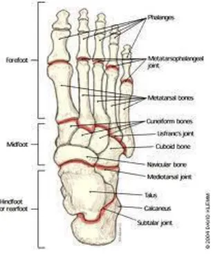
Foot is divided into three parts mainly, hind foot -calcanious, talus, along with
chopart line between calcanious/talus and cuneiform /cuboid, mid foot - navicular,
cuboids, three cuneiforms with Lisfranc joint between tarsal and metatarsal bone and
[image:21.595.240.394.403.587.2]fore foot - five metatarsals and fourteen phalanges.3
Figure 1. Figure shows Anatomy of foot
Function of the foot
The foot support the body weight and provide the leverage for walking and
running segments which enable to adopt the shape on un even surfaces. Foot has a
11
rigid organ during toe off phase of gait. Both feet bear 100% body weight (50% on
each foot), 20% at heel, 17% at head of first metatarsal and 13% at head of fifth
metatarsal. 1.2: Arches of Foot.
Arch is a segmental elevation of the foot. Human foot consist of 3 arches
1. Medial longitudinal arch (MLA)
2. Lateral longitudinal arch (LLA)
[image:22.595.209.416.312.435.2]3. Transverse arch
Figure 2. Figure shows Arches of the foot
Arches of the foot are supported by wedge shape bones, ligament and muscles
with tendons of the foot and leg. Medial longitudinal arch and lateral longitudinal arch
run anterio-posterior of the foot. Medial longitudinal arch consist of talus, calcaneum,
navicular, 3 cuneiforms (medial, intermediate, lateral), 1st three metatarsals, tendons
of tibialis posterior and anterior muscles include (plantar aponeurosis, abductor
hallucis, flexor hallucis longus, flexor hallucis brevis, medial part of flexor hallucis
longus). Ligaments (plantar calcanionavicular or spring ligament, deltoid ligament).
Medial longitudinal arch is clinically more important than other arches. Main feature
of MLA is its elasticity and height (15-18mm), lateral longitudinal arch (LLA) consist
12
calcaneocuboid ligament) LLA is solid and slightly elevated. Transverse arch consist
of 5 metatarsal, cuboid. When there is an excessive stress on the supporting structure
of the arches due to prolong walking, standing or overweight as a result of which
arches may collapse and produce complications.1
[image:23.595.241.392.227.466.2]Biomechanics of Medial longitudinal arch
Figure 3. Figure shows biomechanics of the foot
When we describe the medial longitudinal arch biomechanically it is just like a
suspension bridge. Shape of the bone is like the stones of the bridge in which the
sustenticulumtali hold up the talus, concave proximal surface to the navicular bone
receive rounded head of the talus which is the key stone of the bridge (as shown in
figure) in the center of the arch the inferior edge of the bones are tight together by
stronger plantar ligament for example strong plantar ligament calcanionavicular
ligament or spring ligament and fan like extension of insertion of posterior tibialis
tendon. The end of the arch is tight by the plantar aponeurosis, medial part of flexor
13
digitorum longus, flexor digitorum brevis. From the above the arch is suspended by
tibialis anterior and medial ligament of ankle joint deltoid ligament.3
Flat Feet, Pes Planus, Pronated Feet
Flat feet is often a complex disorder in which MLA is collapsed/ flatted or
depressed sole of the foot comes in contact with ground. In flat feet the progressive
weight bearing/stress produced calcanialvalgacity or flattened arches to drift or fore
feet abduction, pronated or everted foot. The head of the talus is no more supported
and the body weight forces it downward and medially between the calcanial and
navicular. The flat footed individuals body weight is not uniformly distributed due to
which there is an abnormal and excessive stress on the joint of the foot, knee, pelvic.
If the flat foot is not treated, progressive stress will permanently stretch the ligament
and tendons leads to abnormal change in the shape of developing bone. Ultimately
diverse symptoms and varying degree of disability and deformity will occur.2
Types of flat foot
Types of flat foot is classify on the basis of age.
1) Pediatric flat foot (mainly congenital)
2) Adult flat foot (mainly acquired)
1. Pediatric flat foot is a condition occurs in children. It is mostly bilateral,
involving both sides of the foot. Pediatric flat foot either symptomatic or
asymptomatic while the majority of flat foot has asymptomatic condition. The
pediatric flat foot may be flexible or rigid. Flexible flat foot arch is collapsed
in standing (weight bearing) and MLA reappears in sitting position
14
occur in children, adolescent and continues in the adulthood. It is usually
bilateral and asymptomatic but as the age progresses due to increase stresses
from malalignment and improper weight distribution on the foot the soft tissue
of the foot stretched, inflammed or tear then flexible flat foot become
[image:25.595.215.418.218.339.2]symptomatic.
Figure 3. Figure showing flexible flat feet
2. Adult or acquired flat foot is secondary to some disease, injury, illness,
unusual prolonged stress are due to aging process. It is more common in
women’s above 40 years of age (obesity, diabetes, arthritis ), flat foot in
pregnancy is due to increase elastin (elasticity), other causes include trauma,
fracture of foot bones, posterior tibial tendon rupture, polio, malalignment due
to genuvalgum etc.2,5 InRigid flat foot the MLA remains flattened in sitting
(non-weight bearing) as well as in standing (weight bearing). It is more
symptomatic and less asymptomatic.
Causes of the Flat Feet
Flat foot will occur when there is any abnormality in supporting structure of
the MLA like bone ligament muscle/tendon. The causes may classify as congenital
and acquired. Congenital problems are tarsal collation (abnormal connection between
15
bifida, muscular dystrophy, ligament laxity (elardanlose syndrome). Acquired flat feet
is due to trauma, fractures, posterior tibial tendon rupture, arthritis, genu valgum,
metatarsus adductus, tibial torsion, pregnancy obesity diabetes, tight Achilles tendon
age related wear and tear and posterior tibial tendon insufficiency.2,5,7
Sign & symptoms
Flexible flat foot is asymptomatic but in some cases with aging process and
the rigid flat foot is symptomatic.
Most common sign & symptom of the flat foot are:
1. Pain in the foot, knee, ankle, pelvis, hip, lower back pain
2. Stiffness of the joint (restricted movements)
3. Sign of tiredness & fatigue on prolonged walking and over standing
4. Flattened MLA on standing
5. Pronated foot
6. Difficulty in shoe wearing
7. Shoes having uneven sole due to increase pressure on medial side of the sole of the
foot5, 27
Diagnosis of the flat foot
On the basis of visual inspections with special test, we made the diagnosis of
flat foot. It is better to adopt stepwise approach for the diagnosis. It include subjective
history, objective history, examination (look at the shape of the foot, shoes, bones,
skin, muscles condition), feel for the tenderness symmetrically and move the foot
16
examination includes power, sensations, reflexes, vascular examinations which
include pulses, temperature. After these examinations special tests are to be
performed.36
1. Arch height on sitting and standing
2. Tip toe standing test (for rigid or flexible)
3. Single tip toe standing test (for tibialis posterior insufficiency)
4. Standing on heel (for tibialis anterior and contracture of Achilles tendon)
5. Great toe extension (normally elevation of MLA with lateral rotation of tibia)
6. Great toe malalignment
7. Foot prints (normally heel is oval and midline of the heel passes medial to the 2nd toe)
8. Valgus index (for medial or lateral shifting of the malleoli).draw a line from the
mid of the heel to the mid of 3rd toe from the foot prints, then with the help of
setsquare marks the position of the medial & lateral malleoli. Positive index show
medial shifting & negative index shows lateral shifting36
9. Check the joint laxity (note passive hyperextension)
10.Length of calcanial tendon
11. Position of the patella
12.Navicular height10,31,39
13.X-ray for (bone connections), CT for (bony abnormalities & MRI for (soft
17
14.3D foot scanner and milling machine – Newer Technology which is used to
assess and appears to be the most reliable method of obtaining anthropometric
measurements.37
Gait cycle
Gait cycle consist of stance phase (60% of total gait cycle), in this phase the foot
is in contact with ground. The other phase is swing phase (40% of total gait cycle),in
this phase the foot is off the ground. The stance phase is divided into three phases, heel
strike, mid stance and toe off. During stance phase foot has dual function. One is it
serves as a soft organ at heel strike for stress/shock absorption. Another is at the mid
stance(weight acceptance phase) the supination of mid tarsal joint convert the foot from
soft organ to rigid organ which provide rigid lever arm for toe off phase.1,2
Gait cycle flat feet
A person with or without flatfeet at heel strike the ground, foot is pronated
outward and there is inward rolling of the ankle. At the midstance phase the
metatarsus joint supinates as a result, elevation of medial longitudinal arch and foot
changes from soft to rigid organ while in case of flatfoot at heel strike along with
normal pronation there is excessive inward rolling of the ankle, at the midstance phase
while the midtarsal joint supinates and medial longitudinal arch comes in contact with
the ground. The flatfeet person unable to toe off until he will unlock the midtarsal
joint, for this purpose he will pronate the foot as a result the foot is again changes
from rigid to flexible organ and there is excessive pressure on the medial aspect of the
18
Complications of Flatfoot:
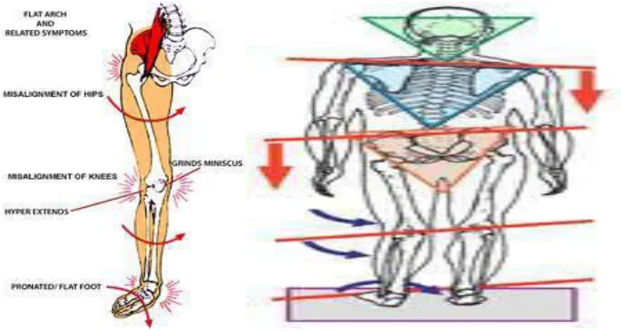
Excessive inward rolling with pronated foot at midstance will lead to inward
rolling of the knee as a result uneven weight or pressure distribution on the foot
especially more pressure on the medial aspect on the foot, there is malalignment on
the lower limb weight, pressure, malalignment exert abnormal stresses on the joint of
the foot, knee, hip and lowerback, which makes the joint hyper mobile and unstable
than it leads to stretching of the supporting tissue of the joint, later on other
complications will produce like (hammertoes, plantar fasciitis, arthritic changes in
foot, knee, hip and backache, shin,Achilles tendinitis, posterior tibial tendinitis,
[image:29.595.147.487.367.549.2]bunions4,5,36
Figure 4. Figures showing complications of flat foot
Treatment Options of the Flatfoot:
Before starting the treatment of the flatfeet, properly diagnose the flatfeet
especially the type of the flatfoot (either flexible/ rigid, symptomatic/ asymptomatic,
congenital/ acquired). Different types of flatfoot require different types of treatment.
Most of the cases conservative treatment is effective but in some cases
19
to correct the deformity, in case of acquired flatfoot it is important to treat a cause,
illness or disease which causes the flatfoot.
The treatment option for flatfoot includes:
1. Activity Modification: Cut down the activities that cause the pain and avoid
prolong standing/walking to give rest to the arches with the arch support.
2. Proper Footwear: Proper shoe size and shoes with arch support are critical for
developing bones because the bones are soft and malleable. Tight constricting shoes
will interfere with normal growth; it may leads to deformity so shoes with arch
support are useful.35
3. Weight Loss: Putting too much weight on arches may aggravate the symptoms.31
4. Medication: NSAIDS or analgesics help to relief from the symptoms.
5. Conventional Orthotic Devices: Majority of the flatfoot resolve with exercise and
proper foot wear in 3-6 year of child and orthotics are seldom need in the early years
of growth if there is an excessive pronation seems to persist beyond the age 6 to 7
responding poorly to home care interventions, the custom made orthotics or arch
support are appropriate. This corrective support will provide to encourage normal
development and prevent future deformity, reduce abnormal kinetic chain stresses and
stresses on the foot, knee, pelvis, spine by keeping the body normally aligned11.17,18
6. 3D Scanned and fabricated newer technology Orthotic devices:
3D surface scanning technologies to produce digitised presentations of parts of
20
appropriately matching with anthropometric measurements to treatment will prevent
future deformity and encourage normal development.27,29,30
Surgery:
There are different surgical techniques for the flat foot. Main purpose of
surgical procedure is to relieve the symptoms, improving the foot function, decrease
pronation in stance phase to provide proper weight bearing and stability to the joint of
the foot.
Some Surgical Techniques are:
1) Rear foot osteotomies (surgical cut of the bone). Mainly in this procedure the
position of the heel is changed to supinated from pronated position. Wedge
osteotomies procedures are important.
2) Medial column stabilization.
3) Tendon transfer. The reattachment of tendon at different suitable location for the
stabilization of the MLA.
4) Tendon lengthening in case of Achilles tendon contracture.
5) Arthrodesis- (Fusion of two or more bones). Major one is triple arthrodesis is
fusion of talocalcanial, talo- navicular, and calcaneo-cuboid, the fusions are held
together by pins or casting to provide immobilization for 2 to 3 months.6
The selection of procedure is on the basis of extent of deformity, x-ray
findings, age, activity level and the duration of the recovery period depend upon the
21
REVIEW OF LITERATURE
Ali K Thabet et al “Dynamic 3D shape of the plantar surface of the foot using
coded structure of light”– 2014.
They stated that the foot provides a crucial contribution to the balance and
stability of the musculoskeletal system, and accurate foot measurements are important
in applications such as designing custom insoles/footwear. In recent years, digitally
acquired scans of the foot have gained popularity among clinicians and foot
specialists. Commercially available foot scanners provide static 3D reconstructions of
the foot, but due to their expensive nature, different research systems have
investigated more cost-efficient solutions, a good summary of 3D foot scanning
methods can be found in the work by Telfer et al.
Shih YF et al “Lower extremity kinematics in children with and without flexible
flat foot”– 2012.
In this study, a high percentage of young children present with flatfeet.
Although the percentage of those with flatfeet declines with age, about 15% of the
population maintains a flat arch. A reduction in longitudinal arch height usually
combines with excessive subtalar joint pronation and may be related to other
musculoskeletal problems of the lower extremity kinetic chain. Hence this study is to
describe and compare the lower extremity kinematics between children with normal
arches and those with flexible flatfeet, with the intent of providing practical
22
Vergara A.E et al “Prevalence of flat foot in school between 3 and 10 years” –
2012.
The study conducted for the prevalence of flat foot in school children between
3 and 10 years among 940 total children, 60% were from Bogoto and remaining from
Barranguilla, they found that diminished prevalence of flat foot in children over 6yrs
of age suggest that therapeutic measures before this age are not recommended.
Angela Mackenzie et al “A review of the evidence for non-surgical interventions
for flexible pediatric flat foot”– 2012.
They stated in his study that the pediatric flat foot is a frequent presentation in
clinical practice, a common concern to parents and continues to be debated within
professional ranks. The available prevalence estimates are all limited by variable
sampling, assessment measures and age groups and hence result in disparate findings
(0.6-77.9%). Consistently, flat foot has been found to normally reduce with age. The
normal findings of flat foot versus children’s age estimates that approximately 45% of
preschool children, and 15% of older children (average age 10 years) have flat feet.
Few flexible flat feet have been found to be symptomatic. Joint hypermobility and
increased weight or obesity may increase flat foot prevalence, independently of age.
Most attempts at classification of flat foot morphology include the arch, heel position
and foot flexibility. Usual assessment methods are footprint measures, X-rays and
visual (scaled) observations. Contemporary management of the pediatric flat foot is
directed algorithmically within this review, according to pain, age, flexibility;
considering gender, weight, and joint hypermobility. When foot orthoses are
indicated, inexpensive generic appliances will usually suffice. Customised foot
23
morphology, or unresponsive cases. Surgery is rarely indicated for pediatric flat foot
(unless rigid) and only at the failure of thorough conservative management. The
assessment of the pediatric flatfoot needs to be considered with reference to the
epidemiological findings, where there is consensus that pediatric flexible flat foot
reduces with age and that most children are asymptomatic. Globally, there is need for
a standard by which the pediatric flat foot is assessed classified and managed. Until
then, assessment should utilize the available evidence-based management model, the
p-FFP Future research needs to evaluate the pediatric flat foot from representative
samples, of healthy and known disease-group children prospectively, and using
validated assessment instruments. He concluded foot exercises along with proper
shoes and footwear were warranted.
Mette kjaergaardNilson, et al “Classification of the height and flexibility of the
medial longitudinal arch of the foot”– 2012.
They stated that the navicular height is an important indicator for denoting flat
foot; among this study they framed a standard level of navicular height which is
highly useful for assessing the level of flat foot and for the provision of orthotic
devices.
Navicular height levels:
NH – less than 2.7 cm – Severely low arch
NH – 2.7 cm to 3.5 cm – Low arch
NH – 3.6 cm to 5.5 cm – Normal arch
NH – 5.6 cm to 6.4 cm - High arch
24
Elly Budiman-Mak, et al “A review of the foot function index and the foot
function index – revised” -2010.
They reviewed in 1991; the Foot Function Index (FFI) was developed as a
self-reporting measure that assesses multiple dimensions of foot function on the basis
of patient-centered values. The FFI consists of 23/17 items divided into 3 subscales
that quantify the impact of foot pathology on pain, disability, and activity limitation in
patients with RA. The FFI was developed using the classical test theory (CTT)
method. It has been found to have good reliability and validity and has had wide
appeal to clinicians and research scientists. In the past 20 years, the FFI has been
widely used by clinicians and investigators to measure pain and disability in various
foot and ankle disorders and its use has expanded to involve children, adults, and
older individuals. Furthermore, the FFI has been widely used in the study of various
pathologies and treatments pertaining to foot and ankle problems such as congenital,
acute and chronic diseases, injuries, and surgical corrections.
Scott Telfer and James Woodburn “3D surface scanning of the foot as an
integral element”– 2010.
They explored in his study, modern 3D surface scanning systems can obtain
accurate and repeatable digital representations of the foot shape and have been
successfully used in medical, ergonomic and footwear development applications.
Finally he was found that the potentiality of the use of 3D surface scanning
technologies play an important role in the development of customised products, i.e.
devices and apparel that are designed for the individual were using their precise
25
Morris C, et al “The Oxford Ankle Foot Questionnaire for children”– 2009.
They designed, studied and found out the reliability and validity of The Oxford
Ankle Foot Questionnaire for Children (OxAFQ-C), created A Guide to the Scoring
System for child patients (aged 5-16) affected by foot and ankle conditions to measure
issues that are considered important to children. The OxAFQ-C (Parent version) has a
total of 15 items, 14 (the first 14 items) of which are used to calculate domain scores,
the three domains are Physical (6 items, 1 - 6), School and Play (4 items, 7 - 10),
Emotional (4 items, 11 - 14) and final item (15) reflect the child concern about can or
cannot wear the footwear they prefer and reported as a single item.
Hawke F, et al “Custom made foot orthotics for the treatment of foot pain”–2008.
Hawke F, et al (2008) stated that, for a number of conditions, customised foot
orthotics have been shown to be more effective at reducing pain and redistributing
pressure than standard "off the shelf" orthotics. Traditionally, customised foot
orthotics are fabricated by taking a plaster cast of the plantar surface of the patient's
foot (the negative cast), making a positive plaster cast of the foot by filling the
negative cast, and then moulding the orthotic around the positive cast to obtain a high
quality fit. The positive cast can be altered either by removing or adding plaster to it
so that, for example, the orthotic will take pressure away from certain areas of the foot
or provide support to the arch.
Deborah Ann George, MS, PT, and Lindsay Elchert, MPT “the Influence of Foot
Orthoses on the Function of a Child with Developmental Delay”– 2007.
They stated lead to improved function when used to control faulty foot
26
foot splints (SFSs) on the function of a child with developmental delay with
hypotonic of 19 month old girl and developmental delay due to hydrocephalus and
congenital absence of the corpus callosum. Modified SFSs as intervention were
created with the child’s feet held in a subtalar neutral position. Five items from the
Peabody Developmental Motor Scale II (rise to stand, standing, lowering, cruising,
and stepping forward) were tracked over three weeks, under three conditions: with
shoes and orthoses, shoes only, and barefoot. The improvement was recorded only
when wearing shoes and orthoses. The outcome indicated that future study of the
modified SFS as an intervention is warranted.
Zhao J, et al “Computerized girth determination for custom footwear
manufacture”– 2007.
In this study they stated that good fitting footwear requires matching not just
the linear dimensions of feet but their girths as well. Footwear fitters have been using
manual measurements for a long time, but the development of computerized
techniques and scanner technologies have now made automatic determination of
different foot dimensions feasible. The resistance to using such computer
measurements has been the lack of trust in the accuracy of the data. This paper
proposes an approach to obtain the necessary girths of feet in order to customize
footwear. The proposed approach attempts to simulate the manual measurement
procedures, and its effectiveness is assessed through an experiment with 15 foot
castings. The results show that the simulated measurements can be within 5 mm of the
manual measurements if the measuring locations can be correctly identified. Linear
regressions show that the differences between the manual measurements and the
simulated measurements can be modeled with the addition of a systematic error term
27
forward for custom shoe manufacturers. They used the device called Foot girth
Custom footwear3D scanning Anthropometry Custom fit.
De pmagalhaes E, et al, “The effect of foot orthosis in rheumatoid arthritis” –
2006.
In this study they stated that, the foot orthoses was effective in 36 patients
with rheumatoid arthritis by using the foot function index (FFI). All the patients were
evaluated 30, 90 and 180 days after the baseline visit. FFI values, daily time of
wearing the orthoses and adverse effects were noted at each appointment. The
Stanford Health Assessment Questionnaire (HAQ) was used at the initial visit to
evaluate the influence of physical condition on FFI response. With the use of foot
orthoses, FFI values decreased in all subscales (pain, disability and activity
limitation). This reduction was noted in the first month and was maintained
throughout the trial. Those using EVA (ethyl-vinyl acetate; n^28) orthoses presented
results similar to those for the total group. Patients wearing made-to-measure orthoses
(n^8) exhibited higher initial FFI values and worse evolution during the trial,
significant for pain and disability but not for activity limitation. Minor adverse
reactions were noted; none required interruption of treatment. He concluded that the
foot orthoses were effective as an adjuvant in the management of rheumatoid foot.
They significantly reduced pain, disability and activity limitation as measured by the
FFI, with minor adverse effects.
Powell M, et al “Efficacy of custom foot orthotics in improving pain and
functional status in children with Juvenile idiopathic arthritis”– 2005.
In his study they compared the clinical efficacy of custom foot orthotics,
28
reducing pain and improving function for children with juvenile idiopathic arthritis
(JIA). Children with JIA and foot pain (n= 40) were randomized to 3 groups. 1 group
of 15 children received custom - made semi rigid foot orthotics with shock absorbing
posts, 2 group of 12 children received off-the-shelf flat neoprene shoe inserts and 3rd
group of 13 children received supportive athletic shoes with a medial longitudinal
arch. Hence this study concluded that custom made semi rigid foot orthotics shows
more efficacies compared with other groups.
Luximon A and Goonetilleke RS “Foot shape modelling”– 2004.
Luximon and Goonetilleke have argued that the foot shape can be modelled
using length, width Identifying bony landmarks on the foot using markers that show
up on the 3D scan appears to be the most reliable method of obtaining accurate girth
measurements and other foot parameters in the case of the foot, quantitative
description of its shape is important for a number of different applications relating to
the ergonomic design of footwear, foot orthotics and insoles otherwise in appropriate
footwear is a major cause of foot illness among the children and elderly.
KyoChulSeo, PhD, et al “Impact of wearing a functional foot orthotic on ankle
joint of frontal surface of young adults with flat foot”– 2004.
They stated in his study investigated the effects of proprietary foot orthotics in
young adults with flatfoot to determine changes in the ankle joint angle in the coronal
plane during the midstance phase. The subjects were 15 college students diagnosed
with flatfoot. Changes in the ankle joint angle in the coronal plane in the midstance
phase were measured using the Vicon Motion System before and after use of the
orthotic. The data were analyzed using Statistical Package for the Social Sciences
29
in the coronal plane during the midstance phase of the gait cycle after use of the
orthotics. However, the difference between the left and right ankle joint angles
showed no significant change, even though the difference increased after use of the
orthotics. Young adults with flatfoot showed increased ankle joint angles after use of
the orthotics. This suggests that orthotic footwear can shape the plantar arch and
affect the ankle joint, and that constant use of orthotics would cause a dynamic
change in normal walking.
Van Boerum DH1 and Sangeorzan BJ “Biomechanics and Pathophysiology of
flat foot”- 2003
There are many terms to describe flat foot, some of the more common ones are
pes planus, planovalgus, calcaneo valgus and fallen arches. The human foot has 26
bones, 10 major extrinsic tendons and their respective muscles, numerous intrinsic
musculo tendinous units and more than 30 joints. These musculoskeletal structures
work together with the neurovascular elements, fat pads and skin to provide a mobile
sensate adaptive foundation during standing and to provide a means of balance and
locomotion during gait. The flat foot describes the common end point of any
abnormality that causes the medial longitudinal arch to collapse. Flat feet can cause
severe symptoms or be asympomatic. Flat foot is now considered a normal variant
assuming it functions in its normal capacity without symptoms. Children have
developmental flat foot, has many causes and may be symptomatic or asymptomatic
flexible or rigid. The cause may be abnormal bone and joint development like with a
tarsal coalition a congenital vertical talus or an accessory navicular bone. Soft tissues
of generalized ligamentous laxity from Marfan’s or Ehlers Danlos can lead to a flat
foot deformity. Adult flat foot may be categorized as either residual flat foot
30
associated with a tight triceps surae or isolated gastronemous tightness, posterior tibial
tendon dysfunction, midfoot laxity abduction of the forefoot, external rotation of the
hindfoot subluxation of the talus traumatic deformities, ruptured plantar fascia,
charcot’s foot and neuromuscular imbalance (polio, cerebral palsy, closed head injury
or following a cerebrovascular accident. It is difficult to define the exact cause of flat
foot in every situation because of the multiple factors which can contribute to the
deformity.
Angela M Evans, et al “Measuring the pediatric foot a criterion validity and
reliability study of navicular height in 4 year old children”– 2003.
They stated in his study indicates that the use of navicular height as a clinical
measure of the foot in 4yr old children among other parameters such as arch index,
foot print analysis, rear foot angle and longitudinal arch angle. They determined the
criterion validity and reliability of NH as a clinical foot measure in young children by
using Sonography.
Garcia, R.A., et al “Flexible flat foot in children, a real problem? – 1999.
They had stated that the critical age for development of the plantar arch is 6
years, and consequently, if the prevalence of flat feet is evaluated before this age, the
finding will overestimate the problem. Therefore, 6-10 aged children were selected as
research subjects in the present study because that is the most appropriate age to
investigate the intervention effectiveness.
Losito JM “Clinical Biomechanics of the lower extremity”– 1996.
He stated in his study about modern scanning systems allow the "positive"
31
(although some handheld systems do allow a cast of the foot to be scanned. A number
of software packages (for example Orthomodel from Delcam PLC, Birmingham, UK;
and Automated Orthotic Manufacturing System, Sharp Shape, CA, USA) have been
developed which have the ability to design foot orthotics based directly on the 3D
representations of the foot obtained by surface scanning. The software, as well as
matching the shape of the foot sole, allows the user to alter the shape and thickness of
the orthotic in a controlled manner, giving greater design freedom than traditional
plaster cast methods. By linking up with computer controlled milling or routing
machines that can manufacture the orthotics, this approach reduces the number of
steps in the process as well as removing many of the sources of human error.
Budiman-Mak E et al “The Foot Function Index”– 1991.
He designed a Questionnaire is called Foot Function Index (FFI). This
questionnaire has been designed to give your therapist information as to how your
foot pain has affected your ability to manage in everyday life. In this, totally 17 items,
would like you to score each question on a scale from 0 (no pain or difficulty) to 10
(worst pain imaginable or so difficult it required help) that best describes your foot
over the past WEEK. Reliability and Validity of the FFI: N = 87, with RA Test-retest
reliability of FFI total and sub-scale scores: 0.87-0.69. Internal consistency: 0.96-0.73
Strong correlation between FFI total and sub-scale scores and clinical measures of
foot pathology supported criterion validity of the index. Number of Items: 23/17
(short forms) 68 (long forms). http://www.proqolid.org/instruments/foot_function
32
METHODOLOGY
RESEARCH DESIGN
Quasi Experimental study
SAMPLE SIZE:
The sample size for the study is 30 children:
15 Children in Control group
15 Children in Experimental group
SAMPLING TECHNIQUE:
Convenient sampling technique
VARIABLES UNDER STUDY:
Independent Variable – 3D designed and Fabricated Customised Insole
Dependent Variable – Children with flat foot.
LOCATION OF STUDY:
The Leprosy Mission Trust India, Community project (PARTI), Cuddalore
district, Tamilnadu. (Early Intervention Centre at PARTI Project).
DURATION OF THE STUDY:
Duration of the study is 1year
33
SCREENING CRITERIA:
Inclusion Criteria:
Age – 6- 10 years
Children with Flat foot
Both Gender
Bilateral involvement
Children with normal cognition and communication skills
Exclusion Criteria:
Recent fractures of lower limb
Those who are not willing to wear foot wear.
Acute inflammatory condition
Plantar ulcers
Plantar warts
Foot Deformity
INSTRUMENTS USED:
34
Voxel care orthotic CAD is an easy to use and powerful tool to design any
type of orthotic insole. It is a scanner system used in this study for scanning the foot
and to record the foot prints in the software to analyse while fabricating the
customised insole for children with flat foot.37,40,45
2. Voxelcare VCM70 Orthotic CNC Milling Machine:
The Voxelcare VCM70 Orthotic CNC Milling Machine is a high-end 1 pair
production model where performance in power as well as accuracy is required. Ideal
for companies that want to start milling EVA orthotics in a clinical environment.
Orthotics can be milled on 2-sides that will save a lot of manual finishing time and
will give a consistent quality product. In the base unit the dust extraction unit and
vacuum pump are integrated. The orthotic milling machine can be controlled from the
supplied tablet as well as from a computer within the network.9, 25
TOOLS USED:
Tools used in this study is
1. Podiatric assessment: In Podiatric assessment, used to collect the information of
biomechanics of foot in subtalar joint neutral position, after calcaneal bisection
foot type of the children in standing position, Navicular height has been
35
[image:46.595.216.416.106.271.2]Foot Type:
Figure 5. Figure shows calcaneal bisection
After calcaneal bisection, position the child is in standing, the examiner stands
exactly behind the child and ask the child to march fast at the same spot for 3 to 4
times and then made to stand in erect posture. This is to stimulate walking and the
joints of the foot will be in the correct positions (Subtalar joint neutral). Explain to the
child to avoid turning back or bending down while doing the assessment and the
examiner examines and categories the foot as being either supinated or pronated.
Criteria that satisfies the Pronatory and Supinatory foot type
Pronated:
- The heel will be in a valgus position (everted)
- There will be a bulge under the medial malleoli
- Achilis tendon may seem to be inserting more laterally.
- The medial arch will be flattened or reduced.
- The foot will probably be abducted (more than 2 toes can be seen lateral to the
leg)
Right foot shows rear foot neutral.
36
Supinated:
- The heel will be in varus position (inverted)
- There will be a slight bulge under lateral malleoli
- There will be a dip under the medial malleoli
- Achilis tendon seem to be medial
- High medial arch
- The foot may be adducted.
Navicular Height : In standing position, Navicular Height (H) was measured as
the perpendicular distance between the supporting surface and the most anterior
inferior part of navicular tuberosity.
NH – less than 2.7 cm – Severely low arch
NH – 2.7 cm to 3.5 cm – Low arch
NH – 3.6 cm to 5.5 cm – Normal arch
NH – 5.6 cm to 6.4 cm - High arch
NH – more than 6.4 cm – Severely high arch 17.31
1. Foot Function Index – Short form:
This questionnaire has been designed to give your information as to how your
foot pain has affected your ability to manage in everyday life. It contains 17 questions
Under this Questionnaire, we used to measure the impact of foot pathology of
functions in terms of pain, disability and activity restriction on the basis of
International classification of Function (ICF)
- Pain – (5items, 1-5)
37
- Activity Restriction – (3items, 15-17)
Scoring : High scores indicates greater the disability.
Reliability : Test-retest reliability of FFI of total and sub scores: 0.87 – 0.69
Validity : Internal consistency: 0.96-0.7313,21
1. Oxford Ankle Foot – Questionnaire - Parent version
The Oxford Ankle foot Questionnaire for Parent version is reported health
status questionnaire is used for children aged from 5 – 16 years affected by foot and
ankle conditions to measure issues that are considered important to children. The
OxAFQ-C parent version has a total of 15 items, 14 (the first 14 items) of which are
used to calculate domain scores, the three domains being:
- Physical (6 items, 1 - 6)
- School and Play (4 items, 7 - 10)
- Emotional (4 items, 11 - 14)
- The final item (item 15 – Has your foot or ankle stopped you wearing any
shoes you wanted to wear?)
Scoring Methodology: Domain scores can be transformed to a percentage
scale (0 – 100) to aid interpretation. A higher score for a domain represents better
functioning.32
MATERIALS USED:
1. EVA (Ethylene Vinyl Acetate) sheet is used for making insole. The width of the
38
soreness, and the remaining upper sheet is black or brown in colour of 30mm
height and soreness which helps in fabricating the stronger medial arch.
PROCEDURE FOR DATA COLLECTION:
A total of 30 Children with flat foot are selected in this study after their
willingness to participate by undertaking informed consent. The sample selected
based on the inclusion and exclusion criteria. Then the participants were divided into
two groups randomly into control and experimental group with a sample size of 15
each.
Initially individual data for paediatric flat foot was done for control and
experimental group using flat foot proforma, immediately to assess the biomechanics
of flat foot among selected children using podiatry assessment manually, followed by
the Tamil translated forms of Foot Function Index short form, Oxford ankle foot
questionnaire- parent version data was collected for both the control and experimental
group. Then 3D Foot scanner assessment was done for experimental group and the
foot prints will be used for the designing and fabrication of customized foot insole.
The parental group education programme conducted before the usage of insole.
Customized 3D foot analysed Foot insole produced by central fabricated by
voxel care milling machine will be given to Experimental group children after the
pretest data collection, the insole to be used by them after fixing on their footwear or
shoes on regular basis of 8 hours of day timing for their functional activity. After a
period of 4 months intervention post evaluation will be done to check the
effectiveness of the foot insole.
During the initial intervention period children’s are advised to wear the insole
39
inflammation. During entire intervention period regular monitoring the use of foot
insole and safety of the children enquired through telephone communication and
follow up house visits.
Whereas the control group children will not receive any special intervention
for the foot apart from occupational therapy, during the study period and the initial
assessments on the parameter will be measured and post evaluation will be done after
a period of 4 months’ time.
After this intervention, data’s are tabulated and dealt with statistically treated
with “t” test.
SAFETY PRECAUTIONS:
1. Should check the skin colour changes periodically.
2. Should keep the footwear inside the house to prevent from dog biting.
3. Should not wash the foot wear.
4. Should not wear the foot wear too tight or too loose.
5. During the initial period recommended to wear continuously for 2 hours then
41
DATA ANALYSIS & INTERPRETATION
DATA SHEET FOR EXPERIMENTAL AND CONTROL GROUP
S. NO IDENTI TY CODIN G
AGE SEX
NAVICULAR HEIGHT (in cms)
FFI PRE SCOR E in %
FFI POST SCORE In % OXFORD PARENT QUESTI ONAIRE PRE SCORE OXFOR D PARENT QUESTI ONAIRE POST SCORE PR E RT FO OT PO ST RT FO OT PRE LT FO OT POS T LT FO OT
1 E1 6yrs Fch 1.6 4.1 1.5 4.0 54 38 11 25
2 E2 7yrs Mch 1.5 4.0 1.5 4.0 53 32 14 28
3 E3 7yrs Mch 2.1 4.6 1.9 4.4 39 27 28 39
4 E4 6yrs Mch 1.8 4.3 1.8 4.3 45 34 20 32
5 E5 8yrs Mch 1.5 4.0 1.5 4.0 52 40 27 37
6 E6 10yrs Mch 1.8 4.3 1.9 4.4 44 30 15 26
7 E7 6yrs Mch 1.9 4.4 1.9 4.4 52 38 14 28
8 E8 7yrs Fch 1.7 4.2 1.7 4.2 60 46 10 23
9 E9 10yrs Mch 1.9 4.4 1.9 4.4 61 45 5 18
10 E10 10yrs Mch 1.9 4.4 2.0 4.5 65 49 14 24
11 E11 8yrs Mch 1.5 4.0 1.6 4.1 68 54 11 24
12 E12 6yrs Fch 1.5 4.0 1.5 4.0 66 51 14 25
13 E13 10yrs Fch 1.7 4.2 1.7 4.2 60 47 18 29
14 E14 10yrs Mch 1.9 4.4 1.9 4.4 55 42 21 30
15 E15 8yrs Mch