Defining the tipping point. A complex
cellular life/death balance in corals in
response to stress
Ainsworth T. D.1, Wasmund K.1, Ukani L.1,2, Seneca F.1,2, Yellowlees D.1, Miller D.1,2&Leggat W.1,2
1ARC Centre of Excellence for Coral Reef Studies, James Cook University Townsville, Australia,2School of Pharmacy and Molecular Sciences, Faculty of Health and Molecular Sciences, James Cook University, Townsville, Australia.
Apoptotic cell death has been implicated in coral bleaching but the molecules involved and the mechanisms by which apoptosis is regulated are only now being identified. In contrast the mechanisms underlying apoptosis in higher animals are relatively well understood. To better understand the response of corals to thermal stress, the expression of coral homologs of six key regulators of apoptosis was studied inAcropora aspera under conditions simulating those of a mass bleaching event. Significant changes in expression were detected between the daily minimum and maximum temperatures. Maximum daily temperatures from as low as 3
6
C below the bleaching threshold resulted in significant changes in both pro- and anti-apoptoticgene expression. The results suggest that the control of apoptosis is highly complex in this eukaryote-eukaryote endosymbiosis and that apoptotic cell death cascades potentially play key roles tipping the cellular life/death balance during environmental stress prior to the onset of coral bleaching.
C
oral bleaching, or the loss of the endosymbiotic dinoflagellates from the tightly-coupled coral symbiosis, is an ecologically devastating global phenomenon occurring on coral reefs in response to environmental stress1,2. The most widely occurring mass bleaching events have resulted from increased sea surface temperatures coupled with high light regimes, which result in stress events that exceed the physiological limit of the symbiotic organism2,3. Previous studies of the bleaching phenomenon point to a range of possible causes and complex molecular networks which drive the disassociation of the symbiosis (for reviews see4–6) but to date we do not understand the mechanisms that govern cellular changes or the initiation of stress responses in this complex eukaryote-eukaryote photosynthetic symbiosis7. Apoptotic cell death however has been proposed as one of several cellular mechanisms driving the breakdown of the symbiosis during thermal sea surface anomalies and coral bleaching events7–9. Understanding the complexity of coral responses to changing environmental conditions is a significant advance in coral biology and in determining the mechanisms which govern whole organism responses to environmental change10.The apoptotic network conveys the interpretation, initiation and regulation of death signals within the cell, whereby an intricate network of proteins function to maintain the balance between life and death within a single cell11–15. Both biotic and abiotic stimuli including exposure to ultraviolet light, starvation, heat stress, and viral and
bacterial pathogens, trigger the regulation of the cell death pathway12,13,16. Specifically the Bcl-2 family members
within the cell death cascade are described as the sensors and regulators of the multitude of stress signals to the cell. The proteins within this family interact to govern the release of cytochrome C and permeabilization of the
outer mitochondrial membrane and the organelle membranes during the onset of cell death17. It has recently been
hypothesized that the type of the cell death signal or stimuli that is interpreted by the cell results in the initiation of a distinct molecular pathways within the cell death network that lead to apoptosis and that these signals may be
specific to cell type18. The control of cell death execution is further regulated within the cell through the actions of
various inhibitors, including members of the Bcl-2 and BIR families. Proteins act to prevent cell death in the cell
through, for example, interactions of anti-apoptotic with pro-apoptotic Bcl-2 family members19–21and the
deactivation of cell death effectors such as caspases22. The role of cell death regulation and the associated
molecules however is highly complex. For example the apoptosis inhibitor BIR which was initially characterised as a suppressor of apoptosis that blocks caspase-9 activity is now also recognised to have a broad range of
functions, including the regulation of mitosis and cellular adaptation to stress23. Since first being described cell
death regulation has been well documented to be significant in homeostasis, development and diseases of higher
organisms (for reviews see14,20,24,25).
SUBJECT AREAS:
GENE EXPRESSION
ENVIRONMENT
ZOOLOGY
PHYSIOLOGY
Received 14 September 2011
Accepted 20 October 2011
Published 17 November 2011
Apoptosis has however also been documented in many lower animals and the apoptotic networks of cnidarians appear to rival
those of mammals in terms of complexity7,15,26. Members of the coral
apoptotic repertoire (both pro- and anti-apoptotic proteins) have
been documented in Hydra27–29, Nematostella15, and corals7,26, and
include Bak-, Bax- and Bcl-2-like members of the Bcl-2 protein
family30. There have been few functional studies on these molecules,
but those that have been carried out are consistent with functional, as well as sequence conservation, with cnidarian and mammalian
pro-teins having the same domain structure. Lasi et al.27demonstrated
strong apoptotic effects of two Hydra Bak-like proteins in cultured mammalian cells, whereas six other Bcl-2-related molecules
inhib-ited camptothecin-induced apoptosis in the same system25. The
pre-dicted apoptotic gene repertoire of the coralAcropora digitiferabased
on the whole genome sequence implies the presence of approxi-mately 10 Bcl-2 family members, six IAPs, as well as a large suite
of caspases31. In the coral,Acropora millepora, a Bcl-2 homolog is
heavily expressed in the aboral ectoderm during metamorphosis,
presumably fulfilling an anti-apoptotic role32. In adult A. millepora,
modest shifts in the relative expression of some pro- and anti-apoptotic Bcl-2 family members have been documented in response to thermal
stress33. Previous studies have also demonstrated Cnidarian apoptosis
occurs during symbiont loss in response to thermal stress34,35. Gates8
first speculated on apoptosis as a key mechanism in thermal induced coral mortality and subsequently substantial evidence for apoptotic
cell death occurring during symbiont loss has been presented33,34.
There is however also morphological evidence for apoptotic cell death occurring within the coral gastrodermal tissue layer (which hosts the
endosymbiotic dinoflagellate) from 3uC below the bleaching
thresh-old7, which is then subsequently only evident in both tissue layers
(epithelial and gastrodermal) during exposure of the coral to bleaching temperatures and the breakdown of the symbiosis. The specific role of the complex array of coral apoptosis related genes in controlling cell death during environmental stress, in response to rapidly changing abiotic stimuli, and ultimately their role in coral mortality in response to climate change has not been widely investigated.
In referring to mammalian systems, Dial and Krosmeyer36state
that cell death operates to maintain homeostatsis and this is espe-cially critical in long-lived animals that must integrate multiple physiological and pathological cell death signals. The coral holobiont is a long-lived colonial organism that is exposed to a myriad of
complex and dynamic environmental and physiological conditions37.
Understanding the control of cell death mechanisms in corals, and particularly their role in stress responses, will provide a greater insight into the biology of these organisms and their capacity to respond and adapt to environmental change. This study investigates the response of the molecular mechanisms governing primary cell death during abiotic stress (temperature and light) of the coral sym-biosis. We selected a suite of regulatory molecules, including four Bcl-2 related proteins and members of the Inhibitors of Apoptosis
(IAP) and Bax-Inhibitor-1 (BI-1) families inAcropora, and studied
the expression of these in a laboratory experiment that simulates conditions occurring during the lead in to a coral bleaching event. The results of this study have implications for determining the mechanisms underlying coral bleaching and the recovery of coral colonies following bleaching events.
Results
Identification of target genes in A. aspera. Coral Bcl-2 family
members, Bcl-2 and Bax have previously been characterised7, and
the current study identifies two additional Bcl-2 family
mem-bers, Bok and Bak from the coral A. aspera based on sequence
homology within conserved BH domains (Table 2). TheAcropora
Bok has similarity to the previously described Bcl-2-like Ovarian killer (Bok), having conserved sequence identity within the
transporter domain, and the BH2 and BH1 conserved domains.
The identifiedAcroporaBak-like protein has conservation within
the BH3, BH2 and BH1 domain regions and the best match
identified within NCBI is to the 188 amino acid Nematostella
predicted Bak-like protein38, the sequence also contains one
identified transmembrane domain. Similarly, we identify 2 key inhibitors of apoptosis, Bax inhibitor 1 (BI-1) and Survivin (Table 2) based on the presence of evolutionarily conserved
domains. The identifiedAcroporaBI-1-like protein is a 238 amino
acid protein containing a highly conserved BI-1-like domain of 206 amino acids and 6 predicted transmembrane domains. The protein
shows highest homology to a putativeNematostellaBI-1-like protein
of 238 amino acids38, although there is significant conservation
within this domain across the phyla. Finally, a highly conserved protein with similarity to the BIR containing Survivin was identified based on conservation within the single 75 amino acid BIR domain which contains the characteristic zinc binding sites at amino acid position Cys58, Cys61, Cys85 and His78.
Thermal stress and the onset of bleaching inAcorpora aspera.
Over the course of 8 days, A. aspera nubbins were subjected to
increasing temperatures, culminating in exposure to 34uC upon
which bleaching (loss of dinoflagellate symbionts or pigments) occurred (Figure 1b). The dark-adapted yields (Fv/Fm) (a standard method to determine the effect of increased temperature on
dinoflagellate symbionts39) were not significantly different between
control and heated corals until the final day when the corals where
exposed to the organisms bleaching threshold40 (Figure 1c). This
decrease in yield is characteristic of thermal damage to PSII and is
a sign of ‘‘algal stress bleaching’’39. This classic bleaching response
is a symptom of chronic photoinhibition and eventual expulsion of Symbiodinium. Using this data we were able to define the
bleaching threshold in the experiment as 34uC on day 8, changes
in gene expression that are seen before day 8 are therefore considered
Table 2 | The identified Bcl-2, IAP and BIR family members of Acropora aspera, regions of domain conservation and gene annotation.
Family Role Conserved domains Gene Citation
Bcl-2 Pro-death, initiator BH3, BH2, BH1 Bok like Bcl-2 Pro-death, effector BH3, BH2, BH1 Bak like Bcl-2 Pro-death, effector BH3, BH2, BH1 Bax like 18 Bcl-2 Pro-Survival, effector BH4, BH3, BH2,
BH1
Bcl-2-like 18
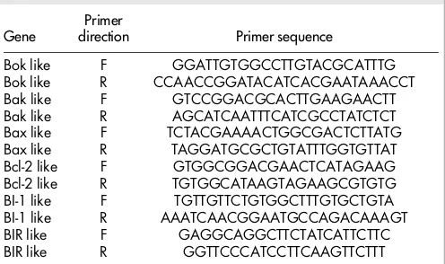
[image:2.595.305.553.68.215.2]IAP Bax Inhibitor-1 Bax inhibitor 1 BI-1 like Table 1 | Primer sequences for use in quantitative PCR.
Gene
Primer
direction Primer sequence
Bok like F GGATTGTGGCCTTGTACGCATTTG
Bok like R CCAACCGGATACATCACGAATAAACCT
Bak like F GTCCGGACGCACTTGAAGAACTT
Bak like R AGCATCAATTTCATCGCCTATCTCT
Bax like F TCTACGAAAACTGGCGACTCTTATG
Bax like R TAGGATGCGCTGTATTTGGTGTTAT
Bcl-2 like F GTGGCGGACGAACTCATAGAAG
Bcl-2 like R TGTGGCATAAGTAGAAGCGTGTG
BI-1 like F TGTTGTTCTGTGGCTTTGTGCTGTA
BI-1 like R AAATCAACGGAATGCCAGACAAAGT
BIR like F GAGGCAGGCTTCTATCATTCTTC
BIR like R GGTTCCCATCCTTCAAGTTCTTT
to be occurring before the onset of ’algal stress bleaching’ and the resultant breakdown of the symbiosis.
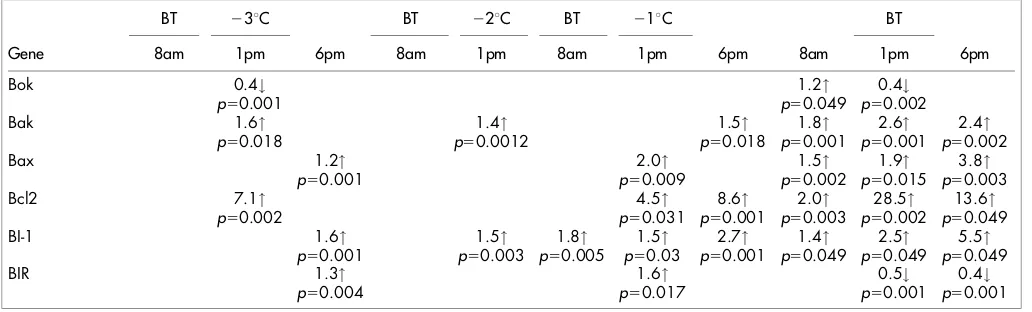
Gene expression patterns during early heat stresses prior to the onset of coral bleaching.The expression of all six genes involved in
apoptosis regulation was found to significantly change inA. asperain
the lead up to a thermal bleaching temperature when compared to control corals, but differed in both direction and scale (Table 3,
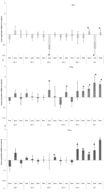
Figure 2, 3). Expression of theAcroporaBok-like protein (Table 3,
Figure 2a), was found to be significantly down regulated in response
to thermal stress at 1 pm at temperatures 3uC below the bleaching
threshold. Also at this temperature, expression of the pro-apoptotic Bcl-2 family members Bak and Bax are found to be significantly upregulated. Significant up-regulation of Bak occurs at midday (cumulative light/temperature stress) whereas Bax gene expression
is significantly up-regulated at 6pm at 3uC below the bleaching
threshold (Figure 2c). Significant changes in gene expression of
both Bak and Bax are also evident at 2uC and 1uC below the
bleaching threshold (Table 3).
In addition there is also up-regulation of the anti-apoptotic Bcl-2, BI-1 and BIR prior to exposure to bleaching thresholds. Bcl-2 was
up-regulated 7.1 fold (p50.002) at 3uC below the bleaching threshold at
1 pm (Table 3, Figure 3a), followed by a consistent up-regulation at
2uC and 1uC below the bleaching threshold. Both the anti-apoptotic
Bax inhibitor-1 and BIR (Figure 3b,c) demonstrate clear patterns
of up-regulation at 3uC below the bleaching threshold and reflects
the increasing cumulative thermal stress. We find there is a 1.6 fold
up-regulation (p50.001) in expression of BI-1 and 1.3 fold
up-regulation of BIR (p50.004) evident at 6 pm at 3uC below the
bleaching threshold. BI-1 expression returns to control levels at 8 am on the following day but is then significantly elevated for the remainder of the pre-bleaching period. During the pre-bleaching period BIR is however subsequently only found to be up-regulated
at 1 pm 1uC below the bleaching threshold.
Gene expression patterns during exposure to the coral bleaching threshold.At bleaching temperatures (the final day of temperature stress, Table 3, Figure 1a) all pro-apoptotic and anti-apoptotic genes were differentially expressed when compared to controls. The pro-apoptotic Bok was up-regulated at 8 am but down-regulated at 1 pm representing a large variation in expression during the day (Table 3, Figure 2a). The significant down regulation of midday Bok-like gene
expression occurs at both bleaching temperatures and 3uC below the
bleaching threshold coinciding with the highest light-temperature stress (Table 3, Figure 1b,c). In contrast, both Bak and Bax gene expression remain significantly up-regulated by between 1.7 and 3.8 fold over the course of the day during the onset of coral bleaching (Table 3).
Both the anti-apoptotic Bcl-2 and BI-1 were up-regulated throughout the onset of bleaching with the highest levels found throughout the experimental period. Bcl-2 expression rose from a significant 2.0 fold up-regulation at 8 am to 28.5 fold, at 1 pm, and a 13.6 fold up-regulation at 6 pm (Table3 , Figure 3a). Similarly BI-1 rose from 1.4 fold up-regulation at 8 am to 2.5, and 5.5 fold upregu-lation during the day (Figure 3b). In contrast, the apoptosis inhibitor BIR (survivin) is found to be significantly (0.2 fold) down-regulated at both 1 pm and 6 pm during the onset of bleaching following exposure to the bleaching threshold (Table 3, Figure 3c).
Discussion
The apoptotic network regulates cellular responses to stress and
death signals and maintains the balance between life and death11–15.
Both biotic (for example starvation and pathogens) and abiotic stim-uli (for example exposure to ultraviolet light and heat stress) initiate
this network12,13,16and during thermal stress events on coral reefs, the
coral animal is exposed to all of these stress signals over long time periods prior to the onset of the observable bleaching stress response. However how the coral organism regulates the cellular response to these signals and controls cell death prior to, and during, the bleach-ing response is largely unknown and we are yet to understand the broader significance of early stress impacts to the coral organism or the reef community.
Tchenov et al.41 recently proposed a model of coral cell death
[image:4.595.42.558.70.226.2]during bleaching, in which they suggest that some populations of cells within the host are irreversibly damaged by dinoflagellate gen-erated ROS, while other cells suppress the cell death cascade, survive the stress event, and are the basis for tissue regeneration. Under the Tchenov model, an up-regulation of genes encoding both pro- and anti-apoptotic proteins would be expected during bleaching, and this was found in the current study. However we also find these changes occurring at temperatures lower than the bleaching threshold and prior to the onset of the bleaching response. Here we present sub-stantial evidence to support the Tchenov model of cellular control of death and recovery from coral bleaching events and further sug-gest that early abiotic stressors, occurring prior to the onset of the Table 3 | Significant gene expression changes for target genes of interest through the thermal stress period. BT, Bleaching threshold.
BT 23uC BT 22uC BT 21uC BT
Gene 8am 1pm 6pm 8am 1pm 8am 1pm 6pm 8am 1pm 6pm
Bok 0.4# 1.2" 0.4#
p50.001 p50.049 p50.002
Bak 1.6" 1.4" 1.5" 1.8" 2.6" 2.4"
p50.018 p50.0012 p50.018 p50.001 p50.001 p50.002
Bax 1.2" 2.0" 1.5" 1.9" 3.8"
p50.001 p50.009 p50.002 p50.015 p50.003
Bcl2 7.1" 4.5" 8.6" 2.0" 28.5" 13.6"
p50.002 p50.031 p50.001 p50.003 p50.002 p50.049
BI-1 1.6" 1.5" 1.8" 1.5" 2.7" 1.4" 2.5" 5.5"
p50.001 p50.003 p50.005 p50.03 p50.001 p50.049 p50.049 p50.049
BIR 1.3" 1.6" 0.5# 0.4#
p50.004 p50.017 p50.001 p50.001
Figure 1| Light (a) and temperature (b) regimes within the aquaria of the experimental setup on outdoor decking at Heron Island Research station from 7uC below the study organisms physiological bleaching threshold, BT, up to exposure to the bleaching threshold, BT, adjacent to the reef flat and the dark apdapted photosynthetic yield of endosymbiotic dinoflagellates in both control and thermal stress conditions (c) throughout the experimental period.
bleaching phenomena also strongly influence the impact of thermal stress events to the coral. In fact the complexity of apoptotic gene expression in thermal stress responses mirrors that of higher organisms and is indicative of the myriad of stress signals being concurrently interpreted.
Upstream apoptotic regulators in the Bcl-2 family have been shown in higher organisms to function as a cellular life/death switch
and to be key sentinels of cell death19 which are up-regulated in
response to cell death signals17. This is the first study of lower
organ-isms to characterize the presence of a Bcl-2 family member with BH and nuclear transporter domains consistent with the upstream apop-tosis regulator, Bok. In mammalian cells, where the function of Bok has been determined, an upregulation in expression is independent of anti-apoptotic Bcl-2 family members and responds to nuclear
damage17. In the present study we find a significant up-regulation
of coral Bok-like expression coinciding with the peak thermal impacts to the coral host, potentially reflecting thermal damage to the nucleus occurring prior to the onset of coral bleaching. However this is the first study to demonstrate a significant down regulation in Bok gene expression, which was observed coinciding with peak light/
temperature interactions 3uC prior to and during the bleaching
threshold. Further investigation of this protein is warranted to deter-mine if it is also a sentinel of nuclear damage and to deterdeter-mine the biological significance of a down regulation of this protein.
Unlike upstream regulators, other Bcl-2 family members however interact with each other to control damage to the cell’s organelles. Pro-apoptotic Bax and Bak, interact with anti-apoptotic Bcl-2 (and Bcl-x) to form heterodimeric proteins and an excess of Bax or Bak within the cytoplasm results in the pro-apoptotic targeting and per-meabilisation of both the mitochondria and the endoplasmic
reticu-lum membranes20,21. In the present study the largest fold gene
expression changes were observed for the anti-apoptotic Bcl-2. Bcl-2 is widely linked to anti-oxidant function in cells and cells expressing Bcl-2 are considered to be resistant to oxidant stress. A 7.1 fold up-regulation in Bcl-2 expression was first observed from
3uC below the bleaching threshold and expression remained
signifi-cantly up-regulated (through to a 28.5 fold up-regulation) until bleaching occurred. This large up-regulation indicates a strong anti-apoptotic and anti-oxidant response from the host prior to the onset of coral bleaching. Importantly, we also find the peak Bcl-2 expression clearly evident during the highest daily cumulative light/temperature stress, lower at the 6 pm temperature stress events (low light but the longest daily thermal stress exposure) and lowest at the 8 am time point following the overnight recovery period. An interaction between light and temperature stress is a necessary deter-minant of coral bleaching, in that without the cumulative impact of light and temperature on the dinoflagellate photosystems the coral
has a higher thermal threshold before mortality occurs39. However
apoptotic cell death is clearly evident within gastrodermal cells (those
holding the endosymbiotic dinoflagellate) 3uC prior to the onset of
bleaching7. Given that the sustained upregulation of Bcl-2 prior to
bleaching is also accompanied by significant up-regulation of both pro-apoptotic Bax and Bak, the anti-oxidant and anti-apoptotic function of Bcl-2 is likely to vary across the organism, have cell and tissue specific regulation and these factors maybe important in determining the capacity of the organism as a whole to control cell death, mortality and ultimately cellular regeneration.
The fine control of cell death in multi-cellular organisms is further regulated through downstream inhibitors. Bax inhibitor-1 (BI-1) is highly conserved and its inhibitory (anti-apoptotic) role has been
demonstrated in both plant and animal species42,43. The protein is
located within the endoplasmic reticulum membrane where it
prevents targeting of the pro-apoptotic Bcl-2 family proteins, confers protection from ER stress, and prevents the generation of ROS within
the cell44. In the present study we find a significant and sustained up
regulation of BI-1 occurring throughout the early thermal stress responses and during bleaching onset. While ROS generation due to ER stress has been proposed as one of the underlying mechanisms
in coral bleaching4,45the prevention of ER damage in some cells
maybe a key mechanism underlying the capacity for coral recovery and regeneration. Over expression of BI-1 is linked to increased cell
adhesion through a direct interaction of the protein with actin46.
Therefore further investigation of the role of BI-1 is warranted to determine if this is a mechanism for maintaining cell and tissue integrity of cells not damaged during coral stress and bleaching. However unlike the anti-apoptotic Bcl-2 and BI-1, the apoptosis inhibitor BIR (survivin) is significantly down regulated only during the onset of coral bleaching. Survivin functions by suppressing both intrinsic and extrinsic apoptotic pathways and blocking caspase-9
function (for review see22). Previous studies in higher organisms
have linked low expression of BIR with an increased sensitivity to
pro–apoptotic stress signals and to cell death execution47,48, an
up-regulation however is considered critical for prevention of the cell
death cycle47. In the current study there is an up-regulation in the
expression of this gene prior to exposure to the bleaching threshold, but a clear down-regulation at the highest temperature exposure and the onset of coral bleaching. If survivin function is analogous to that of higher organisms, a down regulation during bleaching onset pro-vides evidence that stress-affected cells have in fact entered an irre-versible terminal state and there is a tipping of the cellular balance from survival to death.
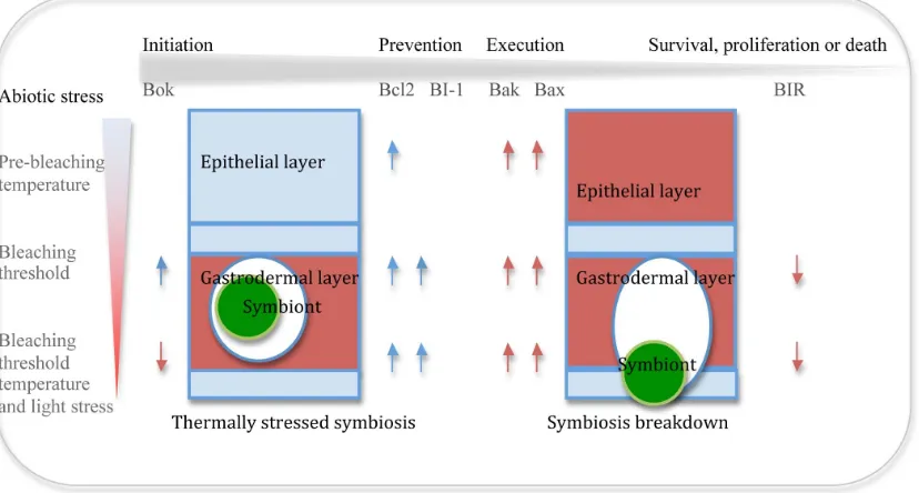
Here we show that the molecular machinery governing cell death in the tightly coupled coral-dinoflagellate symbiosis is highly com-plex and responds significantly to subtle, daily changes in the envir-onment, and at temperatures that are generally considered to have little impact on holobiont function. The kinetics of apoptotic gene expression during thermal stress responses highlights the need to better understand cellular processes occurring prior to and during bleaching events, and the need to determine the mechanisms which underlie coral mortality and recovery in response to environmental stress. Based on the current understanding of coral apoptosis we provide a basic conceptual model of cell death function within the coral symbiotic system during thermal stress (Figure 4) and dem-onstrate that prior to bleaching there is an initiation of the cell death cascade and a potential tipping of the cellular balance from survival to death.
However one major constraint in understanding this complex cell death system is a lack of information on the cell biology, cellular differentiation, tissue function and the cellular recovery processes in coral. It is likely that the regulation of apoptosis found in the current study represents a homogenization of responses across dis-tinct cell and tissue types within a complex, colonial, habitat under
which there is significant biotic and abiotic variation49. All of these
factors likely have significant impacts on the capacity of the coral to regenerate tissues and recover from bleaching events.
Methods
Experimental design and sample collection..Branches from coral colonies of Acropora aspera(cream morph) of at least 7 cm in length (n, 148) were collected from
three adjacent coral patch colonies on the Heron Island reef flat.A. aspera(tan
morph) was selected as the study organism due to its previously documented high
bleaching threshold (34uC) compared to other reef flat corals and species morphs in
the local area50, this species has also been widely used in similar studies of Acroporid
physiology due to this comparatively high thermal threshold(7,50–53). The collected
coral branches were relocated to the adjacent research station, placed upright in stands and held in flow through aquaria within 30 mins of collection from the reef
flat. Handling stress was minimized through the 30 mins collection period and coral branches were transported in low density to prevent branches touching and sloughing mucus, with high volume seawater (40 L) collected from the location of coral collection. Coral branches were randomly assigned to 6 of 60 L experimental aquaria and were held in flow through ambient seawater for 4 days to acclimate and recover from collection. Recovery was evident from tissue re-growth over the collection break at the base of each branch. Following the recovery period three replicate aquaria were designated experimental tanks and three replicate aquaria designated control ambient tanks designated control ambient tanks. Each aquaria was supplied each aquaria where supplied with seawater obtained from the adjacent reef flat through a sand filter system, into 1000 L sump tanks in which internal recirculation heaters were used to adjust the seawater to the daily temperature regimes. High volume sump tanks were used to supply seawater to tanks to allow for variation in daily water temperatures to reflect those experienced within the adjacent reef flat conditions and to prevent variability in thermal regimes between the replicate aquaria. To replicate the conditions found on Heron Island reef flat during periods of thermal stress,
experimental tanks were exposed to increasing daily thermal stress of 1uC above the
previous day’s thermal maximum up to the study organism bleaching threshold
(34uC) (Figure 1). Thermal stress was maintained throughout the daylight period
prior to the temperature being returned to ambient conditions overnight as a recovery period. Aquaria water temperatures were increased daily at 8 am and seawater temperatures gradually increased reaching the daily maximal temperature coinciding with the highest light period at midday (Figure 1). Control tanks were maintained at ambient sea surface temperatures throughout the diurnal period with temperatures fluctuating between the daily thermal maximum and night time thermal minimum of
21–27uC. Coral branches (n, 2) were randomly sampled from each aquaria daily at
8 am following the overnight ambient temperature recovery period, at 1 pm following exposure to the maximum temperature and high light period of the day, and at 6 pm following cumulative exposure to 10 hours daily thermal stress (total of n, 36 coral branches collected per day for 9 consecutive days). Replicate coral branches (n, 6 per tank) were also analysed daily to determine photosynthetic efficiency of
endosymbiotic dinoflagellates54,55using an imaging Pulsed Amplitude Modulated
(iPAM) fluorometer (imaging-PAM, Waltz Gmbh, Germany). At 6 pm each day coral branches were dark adapted for 30 min and the dark adapted quantum yield of
photosystem II determined using the Genty equation Y5(Fm2Fo)/Fm56.
Sequence identification.Two key members of the Bcl-2 family of apoptotic
regulators, Bcl-2 and Bax, have previously been identified fromAcropora millepora33
andA. aspera7. The full length cDNA sequence of two further, previously
unidentified, members of the Bcl-2 family (Bak-like and Bok-like) and two key downstream inhibitors of apoptosis, BIR (survivin) and BI (Bax inhibitor-1), were
identified from theA. milleporaEST and 454 databases57. Newly identified regulators
and inhibitors of apoptosis were aligned and compared to previously identified apoptosis proteins using the NCBI blast server.
Sample processing and quantitative PCR.Upon collection from the aquaria coral
branches were immediately snap frozen in liquid nitrogen and stored at280uC prior
liquid nitrogen prior to RNA extraction and stored at –80uC. mRNA was isolated
from approximately 100 mg of homogenized coral branch using the commercial available Dynabeads Olgio dT kit (Invitrogen, product #610-05) following the
manufacturers instructions (as per58). Approximately 400 ng mRNA was extracted
from each sampled and the integrity of each extraction confirmed using the ND-1000 spectrophotometer (260 nm) (Nanodrop Technologies). cDNA was constructed using Superscript III First Strand Synthesis Supermix for RT-PCR (Invitrogen Cat # 18080-400) following the manufacturer’s instructions and the resultant cDNA was
stored at220uC prior to quantitative PCR (qPCR). Primers for qPCR were design in
PrimerSelect Lasergene 8 (Table 1), PCR confirmed the calculated amplicon sizes (70–90 bp) and sequencing of the generated PCR product confirmed the sequence
homology to higher organism cell death genes withinA. aspera. The melting
temperature curves for each gene amplicon were determined and the efficiency of gene target amplification was established using 4 serial dilutions of cDNA template. Previously determined coral house keeping genes (HKG) appropriate for thermal
stress experiments, L9, S7 and Ado58, were selected and tested for stability within the
current experimental scenario. Each sample was analysed in triplicate to control for technical variability and GOI expression relative to HKG at each time point was
determined using the REST analysis59.
1. Mumby, P. J.&Steneck, R. S. inCoral Reefs: An Ecosystem in Transitioneds Zvy
Dubinsky&Noga Stambler) 509–519 (Springer Netherlands, 2011).
2. Hoegh-Guldberg, O. Climate change, coral bleaching and the future of the world’s
coral reefs.Marine and Freshwater Research50, 839–866 (1999).
3. Brown, B. E. Coral bleaching: causes and consequences.Coral Reefs16, s129–s138
(1997).
4. Weis, V. M. Cellular mechanisms of Cnidarian bleaching: stress causes the
collapse of symbiosis.Journal of Experimental Biology211, 3059–3066 (2008).
5. Baird, A. H., Bhagooli, R., Ralph, P. J.&Takahashi, S. Coral bleaching: the role of
the host.Trends in Ecology&Evolution24, 16–20 (2009).
6. Leggat, W., Whitney, S. M.&Yellowlees, D. Is coral bleaching due to the
instability of the zooxanthellae dark reactions?Symbiosis37, 137–153 (2004).
7. Ainsworth, T. D., Hoegh-Guldberg, O., Heron, S. F., Skirving, W. J.&Leggat,
W. Early cellular changes are indicators of pre-bleaching thermal stress in the
coral host.Journal of Experimental Marine Biology and Ecology364, 63–71 (2008).
8. Gates, R. D., Baghdasarian, G.&Muscatine, L. Temperature stress causes host cell
detachment in symbiotic cnidarians: Implications for coral bleaching.The
Biological Bulletin182, 324–332 (1992).
9. Dunn, S. R., Bythell, J. C., Le Tissier, M. D. A., Burnett, W. J.&Thomason, J. C.
Programmed cell death and cell necrosis activity during hyperthermic
stress-induced bleaching of the symbiotic sea anemoneAiptasiasp.Journal of
Experimental Marine Biology and Ecology272, 29–53 (2002).
10. Levy, O.et al. Complex diel cycles of gene expression in coral-algal symbiosis.
Science331, 175 (2011).
[image:8.595.92.506.55.277.2]11. Elmore, S. Apoptosis: A review of programmed cell death.Toxicologic Pathology
Figure 4|A conceptual model of the cell death and symbiosis breakdown under temperature and light stress in coral. Red coloration indicates morphological evidence for apoptotic cell death; blue block coloration indicates no evidence for apoptotic cell death. Red arrow, significant change in gene expression related to cell death. Blue arrow, significant gene expression change related to cell survival. Grey arrow, indicates progression of the apoptosis cascade.
12. Gross, A., McDonnell, J. M.&Korsmeyer, S. J. BCL-2 family members and the
mitochondria in apoptosis.Genes&Development13, 1899–1911 (1999).
13. Kultz, D. molecular and evolutionary basis of the cellular stress response.Annual
Review of Physiology67, 225–257 (2005).
14. Leber, B., Lin, J.&Andrews, D. Embedded together: The life and death
consequences of interaction of the Bcl-2 family with membranes.Apoptosis12,
897–911 (2007).
15. Zmasek, C. M., Zhang, Q., Ye, Y.&Godzik, A. Surprising complexity of the
ancestral apoptosis network.Genome Biology8, R226 (2007).
16. He, R.et al. Metacaspase-8 Modulates Programmed Cell Death Induced by
Ultraviolet Light and H2O2in Arabidopsis.Journal of Biological Chemistry283,
774–783 (2008).
17. Yakovlev, A. G.et al. BOK and NOXA Are Essential Mediators of p53-dependent
Apoptosis.Journal of Biological Chemistry279, 28367–28374 (2004).
18. Cory, S.&Adams, J. M. The Bcl-2 family: regulators of the cellular life-or-death
switch.Nat Rev Cancer2, 647–656 (2002).
19. Willis, S. N.et al. Apoptosis Initiated When BH3 Ligands Engage Multiple Bcl-2
Homologs, Not Bax or Bak.Science315, 856–859 (2007).
20. Hockenbery, D. M., Oltvai, Z. N., Yin, X.-M., Milliman, C. L.&Korsmeyer, S. J.
Bcl-2 functions in an antioxidant pathway to prevent apoptosis.Cell75, 241–251
(1993).
21. Susnow, N., Zeng, L., Margineantu, D.&Hockenbery, D. M. Bcl-2 family proteins
as regulators of oxidative stress.Seminars in Cancer Biology19, 42–49 (2009).
22. Altieri, D. C. Molecular circuits of apoptosis regulation and cell division control:
The survivin paradigm.Journal of Cellular Biochemistry92, 656–663 (2004).
23. Guha, M.&Altieri, D. C. Survivin as a global target of intrinsic tumor suppression
networks.Cell Cycle8, 2708–2710 (2009).
24. Kerr, J. F., Wyllie, A. H.&Currie, A. R. Apoptosis: A Basic Biological
Phenomenon with Wideranging Implications in Tissue Kinetics.Br J Cancer26,
239–257 (1972).
25. Jacobson, M. D., Weil, M.&Raff, M. C. Programmed Cell Death in Animal
Development.Cell88, 347–354 (1997).
26. Dunn, S. R., Phillips, W. S., Spatafora, J. W., Green, D. R.&Weis, V. M. Highly
conserved caspase and Bcl-2 homologues from the sea anemoneAiptasia pallida:
lower metazoans as models for the study of apoptosis evolution.J Mol Evol63,
95–107 (2006).
27. Lasi, M., David, C.&Bo¨ttger, A. Apoptosis in pre-Bilaterians:Hydraas a model.
Apoptosis15, 269–278 (2010).
28. Galliot, B.&Chera, S. The Hydra model: disclosing an apoptosis-driven generator
of Wnt-based regeneration.Trends in cell biology20, 514–523 (2010).
29. David, C. N.et al. Hydra and the evolution of apoptosis.Integrative and
Comparative Biology45, 631–638 (2005).
30. Mumby, P. J.et al. Unprecedented bleaching-induced mortality in Porites spp. at
Rogiroa Atoll, French Polynesia.Marine Biology139, 183–189 (2001).
31. Shinzato, C.et al. Using theAcropora digitiferagenome to understand coral
responses to environmental change.Natureadvance online publication,
doi:http://www.nature.com/nature/journal/vaop/ncurrent/abs/ nature10249.html#supplementary-information (2011).
32. Grasso, L. C.et al. The biology of coral metamorphosis: Molecular responses of
larvae to inducers of settlement and metamorphosis.Developmental Biology353,
411–419 (2011).
33. Pernice, M.et al. Regulation of Apoptotic Mediators Reveals Dynamic Responses
to Thermal Stress in the Reef Building CoralAcropora millepora.PLoS ONE6,
e16095 (2011).
34. Dunn, S. R., Thomason, J. C., Le Tissier, M. D. A.&Bythell, J. C. Heat stress
induces different forms of cell death in sea anemones and their endosymbiotic
algae depending on temperature and duration.Cell Death Differ11, 1213–1222
(2004).
35. Dunn, S. R.&Weis, V. M. Apoptosis as a post-phagocytic winnowing mechanism
in a coral-dinoflagellate mutualism.Environmental Microbiology11, 268–276
(2009).
36. Danial, N. N.&Korsmeyer, S. J. Cell Death: Critical Control Points.Cell116, 205–
219 (2004).
37. Ainsworth, T. D., Thurber, R. V.&Gates, R. D. The future of coral reefs: a
microbial perspective.Trends in Ecology&EvolutionIn Press, Corrected Proof.
38. Putnam, N. H.et al. Sea Anemone Genome Reveals Ancestral Eumetazoan Gene
Repertoire and Genomic Organization.Science317, 86–94 (2007).
39. Fitt, W. K., Brown, B. E., Warner, M. E.&Dunne, R. P. Coral bleaching:
interpretation of thermal tolerance limits and thermal threshold in tropical corals. Coral Reefs20, 51–65 (2001).
40. Seibt, C.&Schlichter, D. Compatible intracellular ion composition of the host
improves carbon assimilation by zooxanthellae in mutualistic symbioses. Naturwissennschaften88, 382–386 (2001).
41. Tchernov, D.et al. Apoptosis and the selective survival of host animals following
thermal bleaching in zooxanthellate corals.Proceedings of the National Academy
of Sciences(2011).
42. Chae, H.-J.et al. Evolutionarily conserved cytoprotection provided by Bax
Inhibitor-1 homologs from animals, plants, and yeast.Gene323, 101–113 (2003).
43. Watanabe, N.&Lam, E. Arabidopsis Bax inhibitor-1 functions as an attenuator of
biotic and abiotic types of cell death.The Plant Journal45, 884–894 (2006).
44. Xu, C., Bailly-Maitre, B.&Reed, J. C. Endoplasmic reticulum stress: cell life and
death decisions.The Journal of Clinical Investigation115, 2656–2664 (2005).
45. Desalvo, M. K.et al. Differential gene expression during thermal stress and
bleaching in the Caribbean coral Montastraea faveolataMolecular Ecology17,
3952–3971 (2008).
46. Lee, G.-H.et al. Bax Inhibitor 1 Increases Cell Adhesion through Actin
Polymerization: Involvement of Calcium and Actin Binding.Mol. Cell. Biol.30,
1800–1813 (2010).
47. Guo, M.&Hay, B. A. Cell proliferation and apoptosis.Current Opinion in Cell
Biology11, 745–752 (1999).
48. Kobayashi, K., Hatano, M., Otaki, M., Ogasawara, T.&Tokuhisa, T. Expression of
a murine homologue of the inhibitor of apoptosis protein is related to cell
proliferation.Proceedings of the National Academy of Sciences of the United States
of America96, 1457–1462 (1999).
49. Ainsworth, T. D., Thurber, R. V.&Gates, R. D. The future of coral reefs: a
microbial perspective.Trends in Ecology&Evolution25, 233–240 (2010).
50. Dove, S. Scleractinian corals with photoprotective host pigments are
hypersensitive to thermal bleaching.Marine Ecology Progress Series272,99–116
(2004).
51. Leggat, W., Hoegh-Guldberg, O., Dove, S.&Yellowlees, D. Analysis of an EST
library from the dinoflagellate (Symbiodinium sp.) symbiont of reef-building
corals.Journal of Phycology43,1010–1021 (2007).
52. Brown, B. E.&Dunne, R. P. Solar radiation modulates bleaching and damage
protection in a shallow water coral.Marine Ecology Progress Series362, 99–107
(2008).
53. Middlebrook, R., Hoegh-Guldberg, O.&Leggat, W. The effect of thermal history
on the susceptibility of reef-building corals to thermal stress.Journal of
Experimental Biology211, 1050–1056 (2008).
54. Jones, R. J., Kildea, T.&Hoegh-Guldberg, O. PAM chlorophyll fluorometry: a
new in situ technique for stress assessment in scleractinian corals, used to examine
the effects of cyanide from cyanide fishing.Marine Pollution Bulletin38, 864–874
(1999).
55. Jones, R. J.&Hoegh-Guldberg, O. Diurnal changes in the photochemical
efficiency of the symbiotic dinoflagellates (Dinophyceae) of corals:
photoprotection, photoinactivation and the relationship to coral bleaching.Plant,
Cell and Environment24, 89–99 (2001).
56. Genty, B., Briantais, J. M.&Baker, N. R. The relationship between the quantum
yield of photosynthetic electron transport and quenching of chlorophyll
fluorescence.Biochimica et Biophysica acta990,87–92 (1989).
57. Miller, D. J.et al. The innate immune repertoire in Cnidaria - ancestral complexity
and stochastic gene loss.Genome Biol8, R59 (2007).
58. Csaret al. Variation in antioxidant gene expression in the scleractinian coral
Acropora millepora under laboratory thermal stress.Marine Ecology Progress
Series392, 93–102 (2009).
59. Pfaffl, M. W., Horgan, G. W.&Dempfle, L. Relative expression software tool
(REST) for group-wise comparison and statistical analysis of relative expression
results in real-time PCR.Nucleic Acids Research30, e36 (2002).
Author contributions
TA, BL conducted the field experiments and sample collection. BL, FS, KW, LU, TA conducted the sample processing, data collection and data analysis. TA, BL, DM, DY wrote the manuscript.
Additional information
Competing financial interests:The authors declare no competing financial interests.
License:This work is licensed under a Creative Commons
Attribution-NonCommercial-ShareAlike 3.0 Unported License. To view a copy of this license, visit http://creativecommons.org/licenses/by-nc-sa/3.0/