0022-538X/09/$08.00⫹0 doi:10.1128/JVI.00135-09
Copyright © 2009, American Society for Microbiology. All Rights Reserved.
JNK and p38 Mitogen-Activated Protein Kinase Pathways Contribute
to Porcine Circovirus Type 2 Infection
䌤
Li Wei,
1Zhongwu Zhu,
2Jing Wang,
1and Jue Liu
1*
Institute of Animal Husbandry and Veterinary Medicine, Beijing Municipal Academy of Agriculture and Forestry Sciences,
No. 9 Shuguang Garden Central Road, Haidian District, Beijing 100097,1and Hunan Entry-Exit Inspection and
Quarantine Bureau, No. 188 Xiangfu Road, Changsha 410003,2People’s Republic of China
Received 21 January 2009/Accepted 23 March 2009
Infection with a wide variety of viruses often perturbs host cell signaling pathways including the Jun NH2-terminal kinase/stress-activated kinase (JNK/SAPK) and the p38 mitogen-activated protein kinase (p38/
MAPK), which are important components of cellular signal transduction pathways. The present study dem-onstrated for the first time that porcine circovirus type 2 (PCV2), which is the primary causative agent of an emerging swine disease, postweaning multisystemic wasting syndrome, can activate JNK1/2 and p38 MAPK pathways in PCV2-infected PK15 cells. However, PCV2 at an early stage of infection, as well as UV-irradiated PCV2, failed to activate these two MAPK families, which demonstrated that PCV2 replication was necessary for their activation. We further found that PCV2 activated the phosphorylation of JNK1/2 and p38 MAPK downstream targets c-Jun and ATF-2 with virus replication in the cultured cells. The roles of these kinases in PCV2 infection were further evaluated using specific inhibitors: the JNK inhibitor 1 for JNK1/2 and SB202190 for p38. Inhibition of JNK1/2 and p38 kinases by these specific inhibitors did result in significant reduction of PCV2 viral mRNA transcription and protein synthesis, viral progeny release, and blockage of PCV2-induced apoptotic caspase-3 activation in the infected cells. Taken together, these data suggest that JNK/SAPK and p38 MAPK pathways play important roles in the PCV2 replication and contribute to virus-mediated changes in host cells.
Porcine circovirus (PCV) is classified in the genusCircovirus
of the family Circoviridae (52). PCV was first discovered in 1974 as a contaminant of a continuous porcine kidney cell line (PK15) (49). Two genotypes of PCV have been identified. PCV type 1 (PCV1) is known to be nonpathogenic to pigs (1). A high prevalence of anti-PCV1 antibodies has been detected in the swine population, but no disease is correlated with PCV1 infection (33, 50). In contrast, infection with PCV2 occurs in all swine-producing areas of the world, and related PCV2-associ-ated diseases are increasingly recognized as serious threats to global pig production (2, 7, 11, 13, 34, 39, 43). A primary manifestation of PCV2 infection is postweaning multisystemic wasting syndrome (PMWS), which appears in pigs aged 5 to 18 weeks and is clinically characterized by fever, wasting or un-thriftiness, respiratory distress, enlarged lymph nodes and, oc-casionally, jaundice and diarrhea (8, 19, 43). Mortality rates may vary from 1 to 2% up to 30% in complicated cases when coinfections with porcine reproductive and respiratory syn-drome virus, porcine parvovirus, or Mycoplasma
hyopneu-moniae(12). Microscopic lesions are characterized by
lympho-cyte depletion of follicular and interfollicular areas together with macrophage infiltration of lymphoid tissues in PMWS-affected pigs. Several lines of field and experimental evidence
have suggested that severely PMWS affected pigs may develop immunosuppression (44).
PCV genome is a circular single-stranded DNA molecule of ⬃1.7 kb. Two major open reading frames (ORFs) have been recognized for PCV: ORF1, called therepgene, which encodes a protein of 35.7 kDa involved in virus replication (35), and ORF2, called thecapgene, which encodes the major immu-nogenic capsid protein of 27.8 kDa (5, 38). In addition to the replicase ORF1 and the capsid protein ORF2, a novel protein, ORF3, has been detected in PCV2 productive infection and is not essential for PCV2 replication in the cultured cells but is involved in viral pathogenesis via an apoptotic function (30, 31). In a recent report, we demonstrated that PCV2 infection induces NF-B activation in the cultured cells and further found the role of NF-B activation in PCV2 replication and PCV2-induced apoptotic caspase activity (56). However, whether other signaling pathways may also contribute to PCV2 infection in the cultured cells remains unclear.
Mitogen-activated protein kinases (MAPKs), including ex-tracellular signal-regulated kinases (ERK1/2), c-Jun NH2
-ter-minal kinase/stress-activated protein kinase (JNK/SAPK), and p38 MAPK, are central components of signal transduction pathways in the regulation of cell proliferation and differenti-ation, cytokine production, and apoptosis (17). ERK1/2 is pri-marily activated by growth factors, cytokines, and phagocytosis, whereas JNK and p38 are potently induced by proinflam-matory cytokines, bacterial endotoxins, and environmental stresses (17, 26). Although there is coordinated regulation of JNK/SAPK and p38 MAPK, they have corresponding downstream targets. Activated JNK/SAPK and p38 MAPK can phosphorylate numerous substrates, including a variety
* Corresponding author. Mailing address: Institute of Animal Hus-bandry and Veterinary Medicine, Beijing Municipal, Academy of Agri-culture and Forestry Sciences, No. 9 Shuguang Garden Central Road, Haidian District, Beijing 100097, People’s Republic of China. Phone: 86-10-51503548. Fax: 86-10-88433070. E-mail: liujue@263.net.
䌤Published ahead of print on 1 April 2009.
6039
on November 8, 2019 by guest
http://jvi.asm.org/
of transcription factors. The transcription factor c-Jun is specifically phosphorylated by JNK/SAPK, while ATF-2 is phosphorylated by both JNK/SAPK and p38 MAPK (9, 18, 29). Phosphorylation and activation of these downstream substrates ultimately alters gene expression, thereby mani-festing the biological consequences of JNK/SAPK and p38 MAPK activation (57).
It has been shown that viruses are ultimately dependent on the host cell for their replication via altering cellular signal transduction pathways, including MAPK cascades. For in-stance, modulation of JNK and/or p38 MAPK pathways is required for infection and replication of human immunodefi-ciency virus type 1 (27), herpes simplex virus type 1 (HSV-1) (60), Epstein-Barr virus (16), Kaposi’s sarcoma-associated her-pesvirus (41, 58), echovirus 1 (24), Sindbis virus (37), encepha-lomyocarditis virus (21), coxsackievirus B3 (46), and varicella-zoster virus (42, 61). However, there is still no report on the activation of JNK/SAPK and p38 MAPK induced by PCV2 replication and the effects of their activation on PCV2 repli-cation.
The present study was initiated to determine whether JNK/SAPK and p38 MAPK play a role in the replication of PCV2. Here, we report that phosphorylation of JNK/SAPK and p38 MAPK is increased during PCV2 replication and demonstrate that activation of these two MAPK pathways is essential for efficient PCV2 replication. Inhibition of these two MAPK activations reduces viral mRNA transcription and protein synthesis, viral progeny release, and PCV2-induced apoptotic caspase activity. These findings indicate that the JNK/SAPK and p38 MAPK pathways are involved in the PCV2 replication and contribute to virus-mediated changes in host cells.
MATERIALS AND METHODS
Virus and cells.The permanent PK15 cell line, which was free of PCV, was maintained in minimal essential medium supplemented with 5% heat-inactivated fetal bovine serum (FBS), 5%L-glutamine, 100 U of penicillin G/ml, and 100l
of streptomycin/ml at 37°C in a humidified 5% CO2incubator. The PCV2 used
in the study was originally isolated from a kidney tissue sample of a pig with naturally occurring PMWS (strain BJW) (31).
For PCV2 infection, PK15 cells seeded the day before were infected with PCV2 strain BJW at a multiplicity of infection (MOI) of 1 50% tissue culture infective dose (TCID50). Cells were additionally treated with 300 mMD
-gluco-samine at 24 h after infection as described previously (51).
Reagents and antibodies.The JNK peptide inhibitor 1 and SB202190 were purchased from Calbiochem (La Jolla, CA). PK15 cells were treated with either dimethyl sulfoxide (DMSO), which is the solvent for JNK peptide inhibitor 1 and SB202190, or various concentrations (5 to 20M) of these two inhibitors for 1 h prior to infection. After 1 h of virus adsorption, the virus inoculum was removed, and fresh basal medium containing fresh inhibitor was added to the culture. The cytotoxicity of the inhibitors on PK15 cells was determined by trypan blue exclusion dye staining. It was noted that throughout all doses of the inhibitors used in the present study, cell viability assay showed no detectable cell death in the PK15 cells.
Rabbit antibodies against c-Jun, ATF-2, phosphorylated c-Jun (p-c-Jun), phos-phorylated ATF-2 (p-ATF-2), and-actin were purchased from Santa Cruz Biotechnology. Antibodies specific for p38 and JNK, as well as phosphorylated forms of p38 (p-p38) and JNK (p-JNK), were obtained from Cell Signaling Technology. Horseradish peroxidase (HRP)-linked secondary antibodies were purchased from Sigma.
FACE. Fast-activated cell-based enzyme-linked immunosorbent assay (FACE) kits to monitor the levels of JNK1/2 and p38 MAPK activation were obtained from Active Motif. Procedures were performed strictly according to the manufacturer’s instructions. Briefly, PK15 cells seeded in 96-well plates 1 day were infected with PCV2 strain BJW and fixed with 4% formaldehyde in
phosphate-buffered saline (PBS) at the indicated time points after infection. After washing and blocking steps, the cells were reacted overnight with an anti-JNK, anti-phospho-JNK, anti-p38, or anti-phospho-p38 antibody. After incubation with a HRP-conjugated secondary antibody, colorimetric analysis was performed. TheA450was determined by using a plate spectrophotometer. RT-PCR.The splicedcapmRNA during the course of PCV2 infection in the cultured cells was assayed by reverse transcription-PCR (RT-PCR) as described elsewhere (59). Briefly, total cell RNAs were prepared from virus-infected PK15 cells 24 h after being treated with various concentrations of JNK inhibitor 1 or p38 inhibitor SB202190 by using TRIzol RNA extract reagent (Invitrogen) for RT-PCR. The RNA samples were incubated with DNase I for 60 min at 37°C to remove any contaminating viral DNA. A sense primer (5⬘-TTTACTTAGGGG TCATAGGTGAG-3⬘) and an antisense primer (5⬘-AGACTCCCGCTCTCCA ACAAG-3⬘) were used to amplify a 515-bp product from the splicedcapmRNA. cDNAs were reverse transcribed from total RNAs by the use of the antisense primer and the first-strand synthesis system (AMV reverse transcriptase kit; Roche). In addition,-actin gene was used as an internal standard and amplified by using a pair of primers (forward, 5⬘-CCTCCCTGGAGAAGAGCTAC-3⬘; reverse, 5⬘-GATCCACACGGAGTACTTGA-3⬘). The PCR consisted of an ini-tial denaturation at 94°C for 5 min, followed by 94°C for 45 s, annealing at 52°C for 45 s, extension at 72°C for 1 min, and a final extension at 72°C for 10 min. The PCR product was electrophoresed in 1% agarose gel and photographed.
Immunochemical staining (ICS). PK15 monolayer cells seeded in 24-well culture plates were infected with PCV2 strain BJW. At 72 h, the cells were washed with PBS and fixed in 4% paraformaldehyde. After three washes, the cells were incubated with mouse anti-ORF2 antibody (56) diluted in 3% bovine serum albumin-PBS at room temperature for 1 h. After three further washes, cells were incubated with HRP-conjugated anti-mouse immunoglobulin G (Sigma) at room temperature for 1 h and washed with PBS three times. Color development was carried out with 3,3⬘-diaminobenzidine tetrahydrochloride (Pierce) substrate and then stopped with distilled water. The cells were examined under a microscope, and cells positive for PCV2 viral antigens were counted in six fields of view.
Whole-cell lysates.Whole-cell lysate extracts from PK15 cells at various time points after infection were prepared with a nuclear extract kit (Active Motif) according to the manufacturer’s protocol. Protein concentration was determined by using a protein assay (Bio-Rad, Hercules, CA) with bovine serum albumin as a standard.
Western blotting.The whole-cell lysate extracts prepared as described above were diluted in 2⫻sample buffer and boiled for 5 min. Portion (20g) of each extract were resolved by sodium dodecyl sulfate–10% polyacrylamide gel elec-trophoresis (SDS–10% PAGE) and blotted onto nitrocellulose (NC) membranes (Stratagene) with a semidry transfer cell (Bio-Rad). The membranes were blocked for 2 h at room temperature in blocking buffer TBST (20 mM Tris-HCl [pH 7.4], 150 mM NaCl, 0.1% Tween 20) containing 5% skim milk powder to prevent nonspecific binding and then incubated with specific primary antibodies raised against ORF2, JNK1/2, p38, c-Jun, ATF-2, p-JNK1/2, p-p38, p-c-Jun, and p-ATF-2, as well as-actin, at room temperature for 2 h. The membranes were washed three times with TBST buffer and then incubated for 2 h at room temperature with HRP-conjugated secondary antibodies diluted in blocking buffer (1:2,000). Immunoreactive bands were visualized by using an enhanced chemiluminescence system (Amersham Biosciences).
Fluorimetric assay of caspase-3 activity. Spectrofluorometric assays of proteolytic activity were carried out using the synthetic fluorogenic substrate 7-amino-4-trifluoromethyl coumarin (AFC) to measure caspase-3 activity. A BD ApoAlert caspase fluorescent assay kit (Clontech) was used to determine caspase-3 activity. In brief, 80% confluent monolayers of PK15 cells grown on T-25 flasks were infected with PCV2 at an MOI of 1 TCID50with various
amounts of JNK1/2 or p38 MAPK inhibitor treatment. At 72 h postinfection, cells were harvested at 400⫻gfor 5 min. Cells (2⫻106to 3⫻106) were lysed
in 50l of lysis buffer on ice for 10 min and centrifuged at 16,000⫻gfor 10 min, and the supernatant was collected. A 50-l supernatant was added to an equal volume of 2⫻ reaction-dithiothreitol buffer supplemented with caspase-3 substrate DEVD-AFC and then incubated at 37°C for 2 h. The optical densities at 400 nm for caspase-3 were determined. The nanomoles of AFC (released) expressed per hour were calculated from the standard curve.
Statistical analysis.Results are presented as averages⫾the standard devia-tions (SD) or standard errors of the means, as indicated. Statistical comparisons are made by using a Studentttest, and differences between groups were con-sidered significant if thePvalue was⬍0.05.
on November 8, 2019 by guest
http://jvi.asm.org/
RESULTS
Activation of JNK/SAPK and p38 MAPK during PCV2 in-fection.It was reported that JNK/SAPK and p38 MAPK are phosphorylated during various virus infection. In order to as-sess whether activation of these two MAPK signaling pathways occurred in the cultured cells during the course of PCV2 in-fection, the degrees of JNK1/2 and p38 phosphorylation in the PCV2-infected cells were examined by Western blotting. PK15 cells were infected with PCV2 strain BJW at an MOI of 1 TCID50, and whole-cell lysates were prepared at 24, 48, 72, and
96 h after infection. Incubation with PBS served as mock-infected controls. As shown in Fig. 1A, infection with PCV2 led to progressive accumulation of p-JNK1/2 signals over time, and the maximal induction was seen at 72 h postinfection. The kinetics of p-p38 accumulation in the PCV2-infected cells was similar to that of the accumulation of phosphorylated JNK
(Fig. 1A). The increased levels of JNK and p38 MAPK phos-phorylation were concurrent with the expression of viral capsid protein ORF2 in the infected cells (Fig. 1A). In contrast, the protein levels of total amounts of JNK1/2 and p38 remained unchanged in the PCV2-infected cells at various time points after infection compared to that in the mock-infected cells. A loading control,-actin in each sample, was comparable.
To further determine activated JNK and p38 MAPK quan-titatively in the infected cells, we used FACE assays to inves-tigate the levels of ability of JNK and p38 MAPK phosphory-lation at the indicated time points after PCV2 infection. As shown by Fig. 1B and C, there was a time-dependent increase in the JNK1/2 and p38 phosphorylation in the PCV2-infected cells at 72 h postinfection which decreased thereafter. At 72 h after infection, the activations of phosphorylated JNK and p38 showed approximately 4.3- and 3.1-fold increases, respectively,
FIG. 1. PCV2 infection activates JNK1/2 and p38 MAPK signaling pathways in the cultured cells. (A) Whole-cell lysates from PK15 cells after infection with PCV2 strain BJW at an MOI of 1 TCID50. PCV2-infected cells were harvested at 24, 48, 72, and 96 h postinfection, and whole-cell
lysates were prepared and resolved by SDS-PAGE, transferred to nitrocellulose membranes, and immunoblotted. The protein levels of JNK1/2 and p38 and their phosphorylated forms, as well as PCV2 viral capsid protein, were analyzed. The amounts of-actin were also assessed to monitor the equal loadings of protein extracts. (B and C) JNK1/2 (B) and p38 MAPK (C) activation induced by PCV2 infection was determined by using FACE assay. PK15 cells were fixed at the indicated time points with 4% formaldehyde and incubated with antibodies directed against JNK1/2 or p38 or their phosphorylated forms followed by HRP-conjugated immunoglobulin G antibodies. JNK1/2 and p38 and their phosphorylated forms were each assayed in triplicate. Cell numbers were normalized by using crystal violet. These results are representative of three independent experiments. Values are means⫾the SD from triplicate wells. p-, Phosphorylated.
on November 8, 2019 by guest
http://jvi.asm.org/
versus those in the mock-infected cells. In addition, the levels of total JNK and p38 remained unchanged in the PCV2-in-fected cells at various time points after infection compared to that in the mock-infected cells. These data indicate that PCV2 infection induces the activation of JNK and p38 MAPK sig-naling pathways.
PCV2 replication is required for JNK/SAPK and p38 MAPK phosphorylation.It has been shown that replicating virus can be detected in PCV2-infected PK15 cells as early as 14 h postinfection (59). Therefore, we used the FACE assay to determine whether PCV2 replication was required for JNK and p38 MAPK phosphorylation during the early phase of PCV2 infection. As shown in Fig. 2, no significant increase in activation of phosphorylated JNK or p38 MAPK was observed from 0.25 h through 5 h postinfection. In contrast, the levels of JNK and p38 MAPK phosphorylation increased by 1.8- and 2.3-fold, respectively, at 24 h postinfection. The levels of total JNK and p38 remained unchanged in the PCV2-infected cells at various time points after infection compared to that in the mock-infected cells. In addition, no replicating virus was de-tected in the PCV2-inoculated cells from 0.25 h to 5 h post-inoculation, whereas the accumulation of PCV2 cap spliced transcript was observed at 24 h postinoculation (data not shown).
We further used an UV light-irradiated PCV2 to determine whether phosphorylation of JNK1/2 and p38 MAPK was asso-ciated with PCV2 replication. Culture fluid from both PCV2-infected PK15 cells and mock-PCV2-infected PK15 cells was col-lected at 72 h postinfection and exposed to UV light (wavelength, 253 nm) for 20 min. In the UV-irradiated PCV2-infected cells at 72 h postinfection, the level of JNK or p38 phosphorylation fell to its basal level as seen in the mock-infected cells (Fig. 2). These results demonstrated that PCV2 replication was required for JNK and p38 MAPK phosphory-lation and eliminated the possibility that attachment of PCV2
to cell surface receptors alone was responsible for JNK and p38 MAPK phosphorylation in the infected cells.
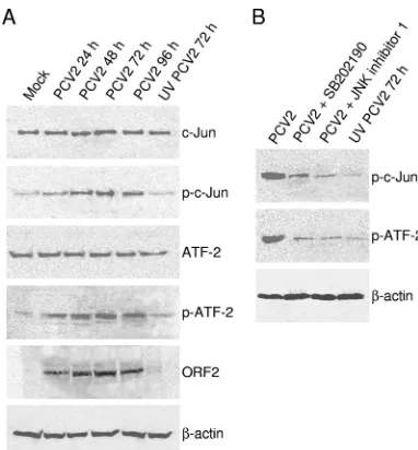
[image:4.585.134.451.68.255.2]Phosphorylated JNK/SAPK and p38 MAPK in infected cells activate downstream targets.Activated JNK and p38 perform Ser/Thr phosphorylation of c-Jun inducing transcription factor and ATF-2, respectively. To confirm that phosphorylation of JNK and p38 MAPK after PCV2 infection truly activated their downstream substrates c-Jun and ATF-2, respectively, the phosphorylation of these two downstream substrates was mea-sured in the infected cells by Western blotting. As shown in Fig. 3A, no activation of c-Jun and ATF-2 was detected in the mock-infected PK15 cells. Consistent with the activation of JNK and p38, a concomitant increase in c-Jun and ATF-2 phosphorylation was observed, which peaked at 72 h postin-fection and then declined (Fig. 3A). In contrast, the protein levels of total amounts of c-Jun and ATF-2 remained un-changed in the PCV2-infected cells at various time points after infection compared to that in the mock-infected cells. Cells exposed to the UV-irradiated virus sample also failed to phos-phorylate c-Jun or ATF-2 (Fig. 3A). The level of-actin, used as a loading control, remained constant.
We then determined the effects of JNK inhibitor (the JNK inhibitor 1) or p38 MAPK inhibitor (SB202190) on the activa-tion of JNK or p38 MAPK downstream substrates. As deter-mined by testing for cell viability by trypan blue exclusion staining, the concentrations (5 to 20M for the JNK inhibitor 1 and SB202190) had no toxic effects on the treated cells (data not shown). In addition, the selected concentrations of these two inhibitors were tested to show their effectiveness in inhib-iting virus-induced JNK or p38 MAPK activity by the FACE assay, which demonstrated that activation of JNK and p38 MAPK decreased dose dependently after treatment with their corresponding inhibitors (data not shown).
We treated the infected cells with the JNK inhibitor 1 or SB202190 at a concentration of 20M and determined the
FIG. 2. PCV2 replication is required for JNK1/2 and p38 MAPK phosphorylation. Monolayer PK15 cells were infected with PCV2 strain BJW at an MOI of 1 TCID50. PCV2-infected cells at 0.25, 1, 3, 5, and 24 h as well as UV-irradiated PCV2-infected cells at 72 h were fixed with 4%
formaldehyde. A FACE assay was used for determination of JNK1/2 (A) or p38 (B) activation. JNK1/2 and p38 and their phosphorylated forms were each assayed in triplicate. Cell numbers were normalized by using crystal violet. These results are representative of three independent experiments. Values are means⫾the SD from triplicate wells. p-, Phosphorylated.
on November 8, 2019 by guest
http://jvi.asm.org/
levels of phosphorylated c-Jun and ATF-2 by Western blotting analysis. As shown in Fig. 3B, the phosphorylation of ATF-2, which is phosphorylated by the p38 MAPK pathway, was blocked by the p38 MAPK-specific inhibitor SB202190, while the activation of ATF-2 was substantially increased after PCV2 infection. In addition, the phosphorylation of c-Jun, which is phosphorylated by the JNK pathway, was also inhibited by
treatment with SB202190 (Fig. 3B). While we examined the phosphorylation of c-Jun and ATF-2 after treatment with the JNK inhibitor 1, we also found that the inhibitor was able to reduce PCV2-induced c-Jun and ATF-2 phosphory-lation (Fig. 3B).
Inhibition of ATF-2 activation by treatment with JNK inhib-itor 1 and p38 inhibinhib-itor SB202190 was due to both JNK and p38 phosphorylated ATF-2 in the PCV2-infected cells, In ad-dition, we also observed that p38 inhibitor SB202190 reduced c-Jun activation, which is only phosphorylated by JNK. This suggests that an indirect inhibition of c-Jun activation might be related to reduction of PCV2 replication when the PCV2-infected cells were treated with p38 inhibition. Therefore, these data suggested that phosphorylation of these two MAPKs as a consequence of PCV2 infection led to the acti-vation of downstream targets.
JNK/SAPK and p38 MAPK regulate PCV2 replication. To determine whether activated JNK/SAPK and p38 MAPK play any role in the replication of PCV2, we examined the effect of these kinases on progeny virus production in the PCV2-in-fected PK15 cells by treatment with these kinase inhibitors. We infected PK15 cells with PCV2 in the presence of JNK inhib-itor 1 (5 to 20M) or the p38 inhibitor SB202190 (5 to 20M) and determined the virus titers in the cell culture supernatant at 72 h postinfection by using an ICS assay. PCV2 replication in PK15 cells was not significantly altered by the presence of JNK or p38 inhibitors at 5M (Fig. 4). At 10 and 20M, the JNK inhibitor reduced PCV2 growth in the cultured cells by 54 and 83%, respectively. Treatment with the p38 inhibitor SB202190 at 10 and 20M reduced PCV2 growth by 59 and 87%, respectively. As expected, the UV-irradiated PCV2 failed to grow compared to the mock-infected cells (Fig. 4). These results suggested that the activation of MAPK pathways in-volving JNK and p38 regulate the efficient replication of PCV2 in the PK15 cells.
[image:5.585.66.257.66.272.2]Effect of JNK/SAPK and p38 MAPK inhibition on PCV2 viral transcription and protein synthesis. Because the JNK inhibitor and SB202190 inhibited PCV2 replication in PK15
FIG. 3. Phosphorylated JNK1/2 and p38 MAPK activate down-stream targets c-Jun and ATF-2 signals. (A) PK15 cells were infected with PCV2 strain BJW at an MOI of 1 TCID50. PCV2-infected cell
lysates at 24, 48, 72, and 96 h, as well as UV-irradiated PCV2-infected cell lysates at 72 h, were harvested and resolved by SDS-PAGE, trans-ferred to nitrocellulose membranes, and immunoblotted. The protein levels of c-Jun and ATF-2 and their phosphorylated forms, as well as of PCV2 viral capsid protein, were analyzed. (B) PK15 cells were infected in the absence or presence of JNK inhibitor 1 (20M) or p38 inhibitor SB202190 (20M). Cell lysates at 72 h postinfection were harvested to examine c-Jun and ATF-2 phosphorylation.-Actin was probed as the loading control. p-, Phosphorylated.
FIG. 4. Inhibition of JNK1/2 and p38 MAPK phosphorylation blocks PCV2 replication. Supernatants of PCV2-infected PK15 cells at 72 h after treatment with various concentrations of JNK inhibitor 1 (A) or SB202190 (B), as well as UV-irradiated PCV2-infected cell supernatants at 72 h, were inoculated on monolayers of PK15 cells. Virus production was assayed by ICS under a microscope. The values represent the means of results for three independent experiments; error bars indicate the SD.
on November 8, 2019 by guest
http://jvi.asm.org/
[image:5.585.111.486.517.688.2]cells, we determined which phase of the replication cycle, viral transcription or protein synthesis, is affected. The PCV2 viral transcription was examined by RT-PCR analysis with RNA extracted from the PCV2-infected PK15 cells 24 h after treat-ment with the JNK inhibitor 1 or p38 inhibitor SB202190 at various concentrations. The abundance of the target fragment corresponding to the splicedcapmRNA was dose dependently decreased in the PCV2-infected cells with these two inhibitors tested (Fig. 5A). In contrast, the fragment was detected in the DMSO-treated infected cells as seen in the DMSO-untreated infected cells (Fig. 5A and data not shown). No product was amplified from RNA extracted from cells infected with the UV-irradiated PCV2, as well as from the mock-infected PK15 cells (Fig. 5A).
To help delineate the effect of JNK and p38 inhibition on virus translation, the expression of PCV2 capsid protein ORF2 in the PK15 cells infected with PCV2 in the presence or ab-sence of the inhibitors was analyzed by ICS. The ORF2 protein expressions were significantly reduced when cells were treated with these two inhibitors, as demonstrated by the increased number of PCV2-positive cells observed in the DMSO-treated infected cells (Fig. 5B). No significant differences were seen in
the ORF2 protein expression between DMSO-treated infected cells and untreated infected cells (data not shown). No ORF2 protein expression was detected in the UV-irradiated PCV2-infected cells, as well as in the mock-PCV2-infected cells (Fig. 5B). These results suggested that inhibitors of these two MAPK pathways decreased PCV2 viral RNA transcription and protein synthesis.
Inhibition of JNK/SAPK and p38 MAPK activation prevents PCV2-mediated caspase activation.PCV2 has been shown to induce apoptosis in cultured cells through the activation of caspase-8, followed by activation of the caspase-3 pathway (31). As demonstrated above, PCV2 replication was strongly prevented after treatment with these kinase inhibitors. There-fore, to determine whether JNK1/2 and p38 MAPK activation is required for PCV2-induced apoptosis, caspase-3 activity was assayed in the PCV2-infected PK15 cells at 72 h postinfection after treatment with the indicated amounts of these inhibitors. As expected, PCV2 alone induced activation of caspase-3 in the infected cells, whereas its activity was significantly reduced when the infected cells were treated with Ac-DEVD-CHO, a peptide inhibitor of caspase-3 activity (Fig. 6). A basal caspase-3 activity was detected in the UV-irradiated
PCV2-FIG. 5. Inhibition of JNK1/2 and p38 MAPK phosphorylation decreases viral transcription and protein synthesis in the PCV2-infected PK15 cells. (A) Effects of JNK and p38 inhibitors on PCV2 splicedcapmRNA synthesis in infected cells. Total RNAs (1g) isolated from PCV2-infected cells at 24 h after treatment with JNK inhibitor 1 or SB202190 at the indicated concentrations were subjected to RT-PCR analysis of PCV2 spliced
capmRNA synthesis. The products were separated on a 1% agarose gel.-Actin was used as an internal control. The results are representative of at least three independent experiments. (B) JNK inhibitor 1 and SB202190 reduce PCV2 protein expression. PCV2-infected PK15 cells at 72 h in the presence of the JNK inhibitor 1 (20M) or SB202190 (20M), as well as UV-irradiated PCV2-infected cells at 72 h, were assayed for the amount of PCV2 viral antigen by ICS. The amounts of PCV2 ORF2 protein expression are shown as percentages of PCV2-positive signals in cells infected with PCV2 alone. The data are mean values⫾the SD based on three independent experiments; error bars show the SD.
on November 8, 2019 by guest
http://jvi.asm.org/
infected cells, as well as in the mock-infected cells (Fig. 6). Furthermore, activation of caspase-3 was dose dependently decreased in the infected cells when treated with increasing amounts of the JNK or p38 inhibitor (Fig. 6). Therefore, we believe that the activation of MAPK pathways involving JNK and p38 is required for PCV2-induced apoptosis, but this can be an indirect effect due to a decrease in virus replication when PCV2-infected cells were treated with JNK or p38 inhibition (Fig. 4).
DISCUSSION
Viral infections are known to activate various cellular sig-naling pathways, which can affect cellular function and virus replication. In this report, we show that JNK1/2 and p38 MAPK pathways were activated during the course of PCV2 infection in PK15 cells and that activation of these two MAPK pathways was required for active replication of PCV2. We further show that inhibitors of these two MAPK pathways lead to a reduction of viral activity, as determined by decreases in viral transcription, virus protein synthesis and progeny produc-tion, and decreased cell death. This study has demonstrated for the first time that the MAPK signaling pathways involving JNK1/2 and p38 are manipulated by PCV2 and that these two pathways play beneficial roles in the PCV2 replication.
The effect of JNK and p38 MAPK on virus replication has been studied in a number of viruses. We determined whether PCV2-induced JNK and p38 MAPK are involved in viral rep-lication by examining the effects of inhibitors of the MAPK pathways on PCV2 production and found that JNK inhibitor 1 and SB202190 significantly reduced the production of progeny virus (Fig. 4). This shows that optimal PCV2 replication re-quires these two MAPK activities and raises the possibility that
PCV2 has acquired the ability to activate these two MAPK kinases to aid its replication. MAPK activation has been shown to be required for the optimal replication of many other vi-ruses. For instance, rotavirus replication was significantly re-duced after JNK and p38 inhibition (22). JNK and p38 activa-tion by HSV requires the expression of particular viral proteins, and blockage of JNK translocation to the nucleus resulted in decreased virus production (10, 20, 36). Coxsackie-virus B3 activates JNK and p38 MAPK, which appears to facilitate virus replication and virion release (46). Treatment of HSV-1-infected cells with p38 inhibitor resulted in a decrease in virus titer (60). Human immunodeficiency virus type 1 pro-duction was increased in p38 MAPK-activated cells (45). Like the above-mentioned viruses, PCV2-induced activation of JNK and p38 MAPK are truly involved in the replication of PCV2 in PK15 cells.
MAPK regulation of viral replication can act at specific steps of the replication cycle, including entry, gene transcription, protein expression, and assembly. In some cases, inhibition of MAPK activation did not affect viral transcription and/or pro-tein synthesis. For instance, inhibition of p38 MAPK by treat-ment of HSV-1-infected cells with inhibitor SB203580 did not affect the viral transcription program (25). Coxsackievirus B3 viral protein synthesis was reported to be unaffected by treat-ment with JNK or p38 inhibitors (46). On the other hand, inhibition of p38 MAPK reduced EMCV replication via inhi-bition of EMC viral protein synthesis, as well as a secondary effect on viral RNA synthesis (21). Reduced murine coronavi-rus production in p38 inhibitor SB203580-treated cells was in part due to a decrease in virus-specific protein synthesis and virus-specific mRNA accumulation (3). Efficient translation in HSV-1-infected cells is regulated by p38-dependent eukaryotic translation initiation factor 4E phosphorylation (54). PCV
rep-FIG. 6. Inhibition of JNK1/2 and p38 MAPK phosphorylation reduces PCV2-induced caspase-3 activity. Whole-cell lysates harvested from PCV2-infected cells at 72 h after treatment with various concentrations of the JNK inhibitor 1 (A) or SB202190 (B), as well as UV-irradiated PCV2-infected cells at 72 h, were assayed for DEVDase activity using the caspase-3 colorimetric DEVD-AFC. In addition, cells infected with PCV2 alone were treated with the caspase-3 inhibitor DEVD-CHO. Mock-infected cells were used as a negative control. The values shown are means from duplicate experiments.
on November 8, 2019 by guest
http://jvi.asm.org/
[image:7.585.113.475.68.288.2]licates via rolling-circle replication involving an intermediate double-stranded replicative form of DNA (6). In the present study, we further determine which step of PCV2 replication is affected by inhibition of JNK or p38 MAPK activation. Our data showed that treatment of cells with JNK inhibitor 1 or p38 inhibitor SB202190 suppressed PCV2 viral RNA transcription and protein synthesis, which is due to that viral transcription and protein synthesis are closely related in the replication of PCV2. Thus, the results suggest that JNK or p38 MAPK inhi-bition reduced the accumulation of both viral mRNA and structural protein, followed by causing a lowered production of infectious PCV2 particles in cells. However, our study does not exclude the possibility that these specific inhibitors have a preferential effect on PCV2 viral protein synthesis and/or RNA transcription.
Apoptosis has been shown to be regulated by the JNK and p38 MAPK cellular stress pathways (28, 48, 53). Virus infection can trigger an apoptogenic kinase cascade, leading to the phos-phorylation and/or activation of JNK and p38 MAPK, which are responsible for inducing apoptotic cell death (47). Hepa-titis B virus X protein-mediated apoptosis was reported to involve sustained activation of JNK and p38 pathways (55). Severe acute respiratory syndrome coronavirus 3a protein-in-duced apoptosis was dependent on the activation of p38 MAPK (40). In the present study, using the specific inhibitor of JNK or p38 MAPK activation, we demonstrated that interfer-ence with the JNK and p38 MAPK pathways leads to reduction of PCV2-induced apoptotic caspase activity (Fig. 6). This result suggests that JNK and p38 MAPK signaling pathways play a role in the mechanism by which PCV2 triggers an apoptotic program in infected cells. In nonstressed cells, nonphosphory-lated JNK is complexed to p53, causing its ubiquitination, followed by proteasomal degradation (14, 15). Upon stimula-tion, the phosphorylated JNK mediates dissociation of p53, followed by its stabilization (14, 15). The role of p38 MAPK kinase in transcription of the p53-regulated Bax and Noxa genes also involves p53 phosphorylation (4, 23). Binding of PCV2 ORF3 protein to porcine p53-induced RING-H2 was reported to interfere with its function of ubiquitination of p53, followed by causing increased p53 level and thus suggesting that the PCV2-induced apoptosis in the cultured cells may be p53 mediated (32). Therefore, activation of JNK and p38 after PCV2 infection may participate in p53 stabilization and phos-phorylation, ultimately leading to apoptosis.
In conclusion, the results reported here establish that PCV2 infection induces the activation of JNK and p38 and their involvement in c-Jun and ATF-2 activation in the cultured cells and demonstrate that the activation of JNK or p38 is required for efficient PCV2 replication, as well as PCV2-induced apop-totic cell death. The role of JNK and p38 activation in PCV2 infection and replication will contribute important information about the molecular mechanism of PCV2 pathogenesis.
ACKNOWLEDGMENTS
This study was supported by grants from the National Natural Sci-ence Foundation (30871866) and the Beijing Municipal SciSci-ence and Technology Contract Project (Z07010501780701), People’s Republic of China.
REFERENCES
1.Allan, G. M., F. McNeilly, J. P. Cassidy, G. A. Reilly, B. Adair, W. A. Ellis, and M. S. McNulty.1995. Pathogenesis of porcine circovirus: experimental infections of colostrum-deprived piglets and examination of pig foetal ma-terial. Vet. Microbiol.44:49–64.
2.Allan, G. M., F. McNceilly, S. Kennedy, B. Daft, E. G. Clark, J. A. Ellis, D. M. Haines, B. M. Meehan, and B. M. Adair.1998. Isolation of porcine circovirus-like viruses from pigs with a wasting disease in the USA and Europe. J. Vet. Diagn. Investig.10:3–10.
3.Banerjee, S., K. Narayanan, T. Mizutani, and S. Makino.2002. Murine coronavirus replication-induced p38 mitogen-activated protein kinase acti-vation promotes interleukin-6 production and virus replication in cultured cells. J. Virol.76:4937–5948.
4.Bulavin, D. V., S. Saito, M. C. Hollander, K. Sakaguchi, C. W. Anderson, E. Appella, and A. J. Fornace, Jr.1999. Phosphorylation of human p53 by p38 kinase coordinates N-terminal phosphorylation and apoptosis in response to UV radiation. EMBO J.18:6845–6854.
5.Cheung, A. K.2003. Transcriptional analysis of porcine circovirus type 2. Virology305:168–180.
6.Cheung, A. K.2006. Rolling-circle replication of an animal circovirus ge-nome in a theta-replicating bacterial plasmid inEscherichia coli. J. Virol.
80:8686–8694.
7.Choi, C., C. Chae, and E. G. Clark.2000. Porcine postweaning multisystemic wasting syndrome in Korean pig: detection of porcine circovirus 2 infection by immunohistochemistry and polymerase chain reaction. J. Vet. Diagn. Investig.12:151–153.
8.Darwich, L., J. Segales, and E. Mateu.2004. Pathogenesis of postweaning multisystemic wasting syndrome caused by porcine circovirus 2: an immune riddle. Arch. Virol.149:857–874.
9.Derijard, B., M. Hibi, I. H. Wu, T. Barrett, B. Su, T. Deng, M. Karin, and R. J. Davis.1994. JNK1: a protein kinase stimulated by UV light and Ha-Ras that binds and phosphorylates the c-Jun activation domain. Cell76:1025– 1037.
10.Diao, L., B. Zhang, C. Xuan, S. Sun, K. Yang, Y. Tang, W. Qiao, Q. Chen, Y. Geng, and C. Wang.2005. Activation of c-Jun N-terminal kinase (JNK) pathway by HSV-1 immediate-early protein ICP0. Exp. Cell Res.308:196– 210.
11.Edwards, S., and J. J. Sands.1994. Evidence of circovirus infection in British pigs. Vet. Rec.134:680–681.
12.Ellis, J., E. Clark, D. Haines, K. West, S. Krakowka, S. Kennedy, and G. M. Allan.2004. Porcine circovirus-2 and concurrent infections in the field. Vet. Microbiol.98:159–163.
13.Fenaux, M., P. G. Halbur, M. Gill, T. E. Toth, and X. J. Meng.2000. Genetic characterization of type 2 porcine circovirus (PCV-2) from pigs with post-weaning multisystemic wasting syndrome in different geographic regions of North America and development of a differential PCR-restriction fragment length polymorphism assay to detect and differentiate between infections with PCV-1 and PCV-2. J. Clin. Microbiol.38:2494–2503.
14.Fuchs, S. Y., V. Adler, T. Buschmann, Z. Yin, X. Wu, S. N. Jones, and Z. Ronai.1998. JNK targets p53 ubiquitination and degradation in nonstressed cells. Genes Dev.12:2658–2663.
15.Fuchs, S. Y., V. Adler, M. R. Pincus, and Z. Ronai.1998. MEKK1/JNK signaling stabilizes and activates p53. Proc. Natl. Acad. Sci. USA95:10541– 10546.
16.Gao, X., H. Wang, and T. Sairenji.2004. Inhibition of Epstein-Barr virus (EBV) reactivation by short interfering RNAs targeting p38 mitogen-acti-vated protein kinase or c-myc in EBV-positive epithelial cells. J. Virol.
78:11798–11806.
17.Garrington, T. P., and G. L. Johnson.1999. Organization and regulation of mitogen-activated protein kinase signaling pathways. Curr. Opin. Cell Biol.
11:211–218.
18.Gupta, S., D. Campbell, B. Derijard, and R. J. Davis.1995. Transcription factor ATF2 regulation by the NJK signal transduction pathway. Science
267:389–393.
19.Harding, J. C.1996. Post-weaning multisystemic wasting syndrome (PMWS): preliminary epidemiology and clinical presentation. Proc. West Can. Assoc. Swine Pract.1996:21.
20.Hargett, D., T. McLean, and S. T. Bachenheimer.2005. Herpes simplex virus ICP27 activation of stress kinases JNK and p38. J. Virol.79:8348–8360. 21.Hirasawa, K., A. Kim, H. S. Han, J. Han, H. S. Jun, and J. W. Yoon.2003.
Effect of p38 mitogen-activated protein kinase on the replication of encepha-lomyocarditis virus. J. Virol.77:5649–5656.
22.Holloway, G., and B. S. Coulson.2006. Rotavirus activates JNK and p38 signaling pathways in intestinal cells, leading to AP-1-driven transcriptional responses and enhanced virus replication. J. Virol.80:10624–10633. 23.Huang, C., W. Y. Ma, A. Maxiner, Y. Sun, and Z. Dong.1999. p38 kinase
mediates UV-induced phosphorylation of p53 protein at serine 389. J. Biol. Chem.274:12229–12235.
24.Huttunen, P., T. Hyypia, P. Vihinen, L. Nissinen, and J. Heino.1998. Echo-virus 1 infection induces both stress- and growth-activated mitogen-activated
on November 8, 2019 by guest
http://jvi.asm.org/
protein kinase pathways and regulates the transcription of cellular immedi-ate-early genes. Virology250:85–93.
25.Karaca, G., D. Hargett, T. I. McLean, J. S. Aguilar, P. Ghazal, E. K. Wagner, and S. L. Bachenheimer.2004. Inhibition of the stress-activated kinase, p38, does not affect the virus transcriptional program of herpes simplex virus type 1. Virology329:142–156.
26.Kujime, K., S. Hashimoto, Y. Gon, K. Shimizu, and T. Horie.2000. p38 mitogen-activated protein kinase and c-Jun-NH2-terminal kinase regulate
RANTES production by influenza virus-infected human bronchial epithelial cells. J. Immunol.164:3222–3228.
27.Kumar, A., S. K. Manna, S. Dhawan, and B. B. Aggarwal.1998. HIV-Tat protein activates c-Jun N-terminal kinase and activator protein-1. J. Immu-nol.161:776–781.
28.Kyriakis, J. M., and J. Avruch.2001. Mammalian mitogen-activated protein kinase signal transduction pathways activated by stress and inflammation. Physiol. Rev.81:807–869.
29.Kyriakis, J. M., P. Banerjee, E. Nikolakaki, T. Dai, E. A. Rubie, M. F. Ahmad, J. Avruch, and J. R. Woodgett.1994. The stress-activated protein kinase subfamily of c-Jun kinases. Nature369:156–160.
30.Liu, J., I. Chen, Q. Du, H. Chua, and J. Kwang.2006. The ORF3 protein of porcine circovirus type 2 is involved in viral pathogenesis in vivo. J. Virol.
80:5065–5073.
31.Liu, J., I. Chen, and J. Kwang.2005. Characterization of a previously un-identified viral protein of porcine circovirus type 2-infected cells and its role in virus-induced apoptosis. J. Virol.79:8262–8274.
32.Liu, J., Y. Zhu, I. Chen, J. Lau, F. He, A. Lau, Z. Wang, A. K. Karuppannan, and J. Kwang.2007. The ORF3 protein of porcine circovirus type 2 interacts with porcine ubiquitin E3 ligase Pirh2 and facilitates p53 expression in viral infection. J. Virol.81:9560–9567.
33.Mahe, D., P. Blanchard, C. Truong, C. Arnauld, P. LeCann, R. Cariolet, F. Madec, E. Albina, and A. Jestin.2000. Differential recognition of ORF2 protein from type 1 and type 2 porcine circoviruses and identification of immunorelevant epitopes. J. Gen. Virol.81:1815–1824.
34.Mankertz, A., M. Domingo, J. M. Folch, P. LeCann, A. Jestin, J. Segale´s, B. Chmielewicz, J. Plana-Dura´n, and D. Soike.2000. Characterization of PCV2 isolates from Spain, Germany, and France. Virus Res.66:65–77. 35.Mankertz, A., J. Mankertz, K. Wolf, and H. J. Buhk.1998. Identification of
a protein essential for replication of porcine circovirus. J. Gen. Virol.79:
381–383.
36.McLean, T. I., and S. L. Bachenheimer.1999. Activation of c-Jun N-terminal kinase by herpes simplex virus type 1 enhances viral replication. J. Virol.
73:8415–8426.
37.Nakatsue, T., I. Katoh, S. Nakamura, Y. Takahashi, Y. Ikawa, and Y. Yoshinaka.1998. Acute infection of Sindbis virus induces phosphorylation and intracellular translocation of small heat shock protein HSP27 and acti-vation of p38 MAP kinase signalling pathway. Biochem. Biophys. Res. Com-mun.253:59–64.
38.Nawagitgul, P., I. Morozov, S. R. Bolin, P. A. Harms, S. D. Sorden, and P. S. Paul.2000. Open reading frame 2 of porcine circovirus type 2 encodes a major capsid protein. J. Gen. Virol.81:2281–2287.
39.Onuki, A., K. Abe, K. Togashi, K. Kawashima, A. Taneichi, and H. Tsun-emitsu.1999. Detection of porcine circovirus from lesions of a pig with wasting disease in Japan. J. Vet. Med. Sci.61:1119–1123.
40.Padhan, K., R. Minakshi, M. A. Towheed, and S. Jameel.2008. Severe respiratory syndrome coronavirus 3a protein activates the mitochondrial death pathway through p38 MAP kinase activation. J. Gen. Virol.89:1960– 1969.
41.Pan, H., J. Xie, F. Ye, and S. J. Gao.2006. Modulation of Kaposi’s sarcoma-associated herpesvirus infection and replication by MEK/ERK, JNK, and p38 multiple mitogen-activated protein kinase pathways during primary in-fection. J. Virol.80:5371–5382.
42.Rahaus, M., N. Desloges, and M. H. Wolff.2004. Replication of
varicella-zoster virus is influenced by the levels of JNK/SAPK and p38/MAPK acti-vation. J. Gen. Virol.85:3529–3540.
43.Segale´s, J., and M. Domingo.2002. Postweaning multisystemic wasting syn-drome (PMWS) in pigs: a review. Vet. Q.24:109–124.
44.Segale´s, J., M. Domingo, F. Chianini, N. Majo´, J. Domínguez, L. Darwich, and E. Mateu.2004. Immunosuppression in postweaning multisystemic wast-ing syndrome affected pigs. Vet. Microbiol.98:151–158.
45.Shapiro, L., K. A. Heidenreich, M. D. Meintzer, and C. A. Dinarello.1998. Role of p38 mitogen-activated protein kinase in HIV type 1 production in vitro. Proc. Natl. Acad. Sci. USA95:7422–7426.
46.Si, X., H. Luo, A. Morgan, J. Zhang, J. Wong, J. Yuan, M. Esfandiarei, G. Gao, C. Cheung, and B. M. McManus.2005. Stress-activated protein kinases are involved in coxsackievirus B3 viral progeny release. J. Virol.79:13875– 13881.
47.Sumbayev, V. V., and I. M. Yasinska.2006. Role of MAP kinase-dependent apoptotic pathway in innate immune responses and viral infection. Scand. J. Immunol.63:391–400.
48.Tibbles, L. A., and J. R. Woodgett.1999. The stress-activated protein kinase pathways. Cell. Mol. Life Sci.55:1230–1254.
49.Tischer, I., H. Glederblom, W. Vettermann, and M. A. Koch.1982. A very small porcine virus with circular single-stranded DNA. Nature295:64–66. 50.Tischer, I., W. Mields, D. Wolff, M. Vagt, and W. Griem.1986. Studies on
epidemiology and pathogenicity of porcine circovirus. Arch. Virol.91:271– 276.
51.Tischer, I., D. Peters, R. Rasch, and S. Pociuli.1987. Replication of porcine circovirus: induction by glucosamine and cell cycle dependence. Arch. Virol.
96:39–57.
52.Todd, D., P. Biagini, M. Bendinelli, S. Hino, A. Mankertz, S. Mishiro, C. Niel, H. Okamoto, S. Raidal, B. W. Ritchie, and G. C. Teo.2005.Circoviridae, p. 327–334.InC. M. Fauquet, M. A. Mayo, J. Maniloff, U. Desselberger, and L. A. Ball (ed.), Virus taxonomy. Eighth report of the International Committee for the Taxonomy of Viruses. Elsevier/Academic Press, New York, NY.
53.Wada, T., and J. M. Penninger.2004. Mitogen-activated protein kinases in apoptosis regulation. Oncogene23:2838–2849.
54.Walsh, D., and I. Mohr.2004. Phosphorylation of eIF4E by Mnk-1 enhances HSV-1 translation and replication in quiescent cells. Genes Dev.18:660–672. 55.Wang, W. H., G. Ge´gori, R. L. Hullinger, and O. M. Andrisani.2004. Sustained activation of p38 mitogen-activated protein kinase and c-Jun N-terminal kinase pathways by hepatitis B virus X protein mediates apoptosis via induction of Fas/FasL and tumor necrosis factor (TNF) receptor 1/TNF-␣expression. Mol. Cell. Biol.24:10352–10365.
56.Wei, L., J. Kwang, J. Wang, L. Shi, B. Yang, Y. Li, and J. Liu.2008. Porcine circovirus type 2 induces the activation of nuclear factorB by IB␣ degra-dation. Virology378:177–184.
57.Weitzman, J. B., and M. Yaniv. 1998. Signal transduction pathways and modulation of gene activity. Clin. Chem. Lab. Med.36:535–539. 58.Xie, J., H. Pan, S. Yoo, and S. J. Gao.2005. Kaposi’s sarcoma-associated
herpesvirus induction of AP-1 and interleukin 6 during primary infection mediated by multiple mitogen-activated protein kinase pathways. J. Virol.
79:15027–15037.
59.Yu, S., S. Carpenter, T. Opriessnig, P. G. Halbur, and E. Thacker.2005. Development of a reverse transcription-PCR assay to detect porcine circo-virus type 2 transcription as a measure of replication. J. Virol. Med.123:
109–112.
60.Zachos, G., B. Clements, and J. Conner.1999. Herpes simplex virus type 1 infection stimulates p38/c-Jun N-terminal mitogen-activated protein kinase pathways and activates transcription factor AP-1. J. Biol. Chem.274:5097– 5103.
61.Zapata, H. J., M. Nakatsugawa, and J. F. Moffat.2007. Varicella-zoster virus infection of human fibroblast cells activates the c-Jun N-terminal kinase pathway. J. Virol.81:977–990.