0022-538X/08/$08.00⫹0 doi:10.1128/JVI.00763-08
Copyright © 2008, American Society for Microbiology. All Rights Reserved.
Targeting Tat Inhibitors in the Assembly of Human Immunodeficiency
Virus Type 1 Transcription Complexes
䌤
†
Iva
´n D’Orso,
1Jocelyn R. Grunwell,
1Robert L. Nakamura,
2Chandreyee Das,
1‡ and Alan D. Frankel
1*
Department of Biochemistry and Biophysics, University of California—San Francisco, 600 16th Street, San Francisco, California 94143-2280,1and Advanced Genetic Systems, San Francisco, California 94143-22802
Received 7 April 2008/Accepted 21 July 2008
Human immunodeficiency virus type 1 (HIV-1) transcription is regulated by the viral Tat protein, which relieves a block to elongation by recruiting an elongation factor, P-TEFb, to the viral promoter. Here, we report the discovery of potent Tat inhibitors that utilize a localization signal to target a dominant negative protein to its site of action. Fusing the Tat activation domain to some splicing factors, particularly to the Arg-Ser (RS) domain of U2AF65, creates Tat inhibitors that localize to subnuclear speckles, sites where pre-mRNA pro-cessing factors are stored for assembly into transcription complexes. A U2AF65 fusion named T-RS interacts with the nonphosphorylated C-terminal domain of RNA polymerase II (RNAP II) via its RS domain and is loaded into RNAP II holoenzyme complexes. T-RS is recruited efficiently to the HIV-1 promoter in a TAR-independent manner before RNAP II hyperphosphorylation but not to cellular promoters. The “preloading” of T-RS into HIV-1 preinitiation complexes prevents the entry of active Tat molecules, leaving the complexes in an elongation-incompetent state and effectively suppressing HIV-1 replication. The ability to deliver inhibitors to transcription complexes through the use of targeting/localization signals may provide new avenues for designing viral and transcription inhibitors.
Dominant negative proteins typically are nonfunctional vari-ants that form inactive oligomers with a wild-type subunit or otherwise compete for functionally essential protein-protein or protein-nucleic acid interactions (21). Transcription complexes have provided prime targets for dominant-negative inhibition due to the large number of interfaces formed during transcrip-tion and the dynamic nature of transcriptranscrip-tion factor interactranscrip-tions during key steps of complex assembly and disassembly (8, 20). However, inhibition typically requires high levels of expression of the mutant protein to inactivate, at least partially, the wild-type protein activity (13, 17, 21, 44). Dominant negative pro-teins have been developed as potential human immunodefi-ciency virus type 1 (HIV-1) therapeutics, including some aimed toward altering viral transcription (19, 38, 48).
In HIV-1, the viral Tat protein is essential for regulating transcription initiation complex assembly (40) and also for recruiting P-TEFb (positive transcription elongation factor b) to a promoter-proximal site on the nascent HIV-1 pre-mRNA (the transactivation response element [TAR]) to assemble elongation-competent, activated transcription complexes (4). Without Tat, RNA polymerase II (RNAP II) complexes are inefficiently converted to the elongating form, which requires phosphorylation of the C-terminal domain (CTD) of the large RNAP II subunit (1, 24). P-TEFb is a heterodimer of cyclin T1 (CycT1) and its associated Cdk9 catalytic subunit and is
re-quired by many, but not all, activators for CTD phosphoryla-tion, either at the promoter or during elongation (3, 18, 37). In the case of HIV-1, the Tat activation domain (AD; residues 1 to 48), in the absence of its RNA-binding domain (RBD), functions as a weak dominant negative that is believed to form inactive complexes with P-TEFb (12, 19, 33, 35). Their poten-tial use in therapeutic strategies has been hindered, in part, by their low potency.
The unusual function of Tat as an RNA-binding transcrip-tion factor has allowed the development of the Tat hybrid assay, in which the Tat AD fused to a heterologous RBD activates an HIV-1 long-terminal-repeat (LTR) reporter con-taining a cognate RNA-binding site in place of TAR (26). In developing the Tat hybrid assay to screen libraries for RNA-protein interactions, we discovered a novel class of highly po-tent dominant negatives, exemplified by fusions with splicing factors, whose potencies appear to be dictated by their efficient recruitment to the HIV-1 promoter. We report that tethering a targeting/localization motif, such as a splicing factor Arg-Ser (RS) domain, to a dominant negative domain strongly en-hances inhibitory activity by facilitating the loading of such an inhibitor into HIV-1 transcription complexes. This recruit-ment-based mechanism effectively co-opts the transcriptional machinery, impairing Tat loading at the promoter, blocking transcription elongation, and inhibiting viral replication.
MATERIALS AND METHODS
Transcriptional activation and inhibition reporter assays.For fluorescence-activated cell sorter analyses, HeLa cells were transfected with green fluorescent protein (GFP) or DsRed reporter plasmids and appropriate amounts of Tat activator and inhibitor plasmids by using PolyFect (Qiagen). Reporter activity was measured 48 h posttransfection by using a Becton-Dickinson FACSCalibur
instrument. Activation (n-fold) was calculated by determining the number of
cells in the appropriate quadrant, multiplied by their average fluorescence, rel-ative to the same values calculated for the reporters alone. All LTR reporter plasmids utilize the HIV-1 LTR and various RNA elements in place of TAR (see
* Corresponding author. Mailing address: Department of Biochem-istry and Biophysics, University of California—San Francisco, 600 16th St., San Francisco, CA 94143-2280. Phone: (415) 476-9994. Fax: (415) 514-4112. E-mail: frankel@cgl.ucsf.edu.
† Supplemental material for this article may be found at http://jvi .asm.org/.
‡ Present address: Dana Farber Cancer Institute, Room D728, 44 Binney St., Boston, MA 02115.
䌤Published ahead of print on 30 July 2008.
9492
on November 8, 2019 by guest
http://jvi.asm.org/
Table 1) and contain an internal ribosome entry site upstream of the firefly
luciferase (FFL) gene to ensure efficient translation irrespective of the 5⬘
un-translated region sequence used. For FFL reporter assays, transcriptional activ-ities in the linear range were obtained by transfecting HeLa cells with 5 ng of Tat activator and 1 or 5 ng of Tat fusion (referred to as T-fusion) plasmids (see Table 2) (1:0.2 and 1:1 ratios of activator to inhibitor). All transfection mixtures
in-cluded an appropriate FFL reporter and pCMV-RL (withRenillaluciferase)
(Promega) to normalize for transfection efficiency, and activities were measured using a Dual-Glo luciferase assay kit (Promega). Activation assays were
per-formed in triplicate, and data are presented as means⫾standard deviations.
RNase protection assay.HeLa cells were transfected with reporter alone or with activator- and inhibitor-expressing plasmids. Total RNA was extracted by
use of TRIzol (Invitrogen), and 15g of each sample was hybridized with
proximal and distal probes corresponding to the HIV-1 LTR promoter and FFL open reading frame regions, respectively. The antisense probes were synthesized from linearized templates using a MaxiScript kit (Ambion), and hybridization
was performed with an [␣-32
P]CTP-labeled probe {in 80% formamide, 40 mM
PIPES [piperazine-N,N⬘-bis(2-ethanesulfonic acid)], 400 mM NaCl, 1 mM
EDTA} incubated at 42°C overnight. RNase digestion was performed for 1.5 h
at 30°C (in 10 mM Tris [pH 8.0], 300 mM NaCl, 5 mM EDTA, 11 units/l of
RNase A, 11 units/l RNase T1), and duplexes were purified. RNAs were
separated on a 6% polyacrylamide–8 M urea gel and visualized and quantified by using a Typhoon phosphorimager (Molecular Dynamics). Experiments were performed in duplicate with similar results.
Expression analyses by Western blotting and fluorescence microscopy.To quantitatively assess relative inhibitor and activator expression levels by Western
blotting (see Fig. 1), HeLa cells were cotransfected with 0.3g of pEGFPN3
(Clontech) and 1.35g of pSV2-HA-tagged T-fusion plasmids in six-well plates.
Nuclear extracts were prepared using a NE-PER kit (Pierce), and samples were separated by sodium dodecyl sulfate-polyacrylamide gel electrophoresis (SDS-PAGE), transferred to nitrocellulose, and probed with antibodies (see Table S1 in the supplemental material). To examine localization of the GFP-tagged T-fusions (see Fig. 4), HeLa cells were grown to 50% confluence on glass cover-slips, transfected with 100 ng of plasmid DNA, fixed in 4% paraformaldehyde in
1⫻phosphate-buffered saline (PBS) buffer (pH 7.4) at 24 h posttransfection,
rinsed twice with PBS, and permeabilized with PBS–0.5% Triton X-100 for 10
min at 4°C. Nonspecific antibody sites were blocked in 1⫻PBS, 3% goat serum,
and 4% bovine serum albumin for 1 h at room temperature, and cells were incubated with primary antibodies for 1 h at room temperature, washed three times with PBS, incubated with appropriate Alexa 488-coupled secondary anti-bodies (Molecular Probes), and washed three times with PBS. Coverslips were
then mounted on DAPI (4⬘,6-diamidino-2-phenylindole)-containing Vectashield
slides (Vector Labs) and examined using an LSM510 confocal microscope (Zeiss).
Selection and characterization of HIV-1 LTR reporter-transfected HeLa cell lines.To generate cell lines with an integrated reporter for chromatin immuno-precipitation (ChIP) analyses, HeLa cells were transfected with an Ssp1-linear-ized pcDNA3.1 (Invitrogen) template bearing either the HIV-1 LTR-RREIIB
(Rev response element, stem-loop IIB)-FFL or HIV-1 LTR-⌬TAR-FFL
re-porter (Table 1) by use of PolyFect (Qiagen). Clones were selected for more than four weeks in Dulbecco’s modified Eagle’s medium–10% fetal bovine serum with
750g/ml of Geneticin (Gibco). Twenty clones selected from the transfection
with HIV-1 LTR-RREIIB-FFL were analyzed for transcription activation with T-Rev (Table 2), and those with reproducible activity levels were selected. From those, a single active clone with the promoter integrated at a single locus was
chosen for the ChIP analyses (see Fig. 5). To generate the HIV-1 LTR-⌬
TAR-FFL-containing cell line, 12 clones were selected and a clone with a single
integration site was chosen based on its response to tumor necrosis factor alpha
(TNF-␣) (53). The number of integration sites was determined by quantitative
PCR (ABI) using genomic DNA extracted with Flexigene (Qiagen) and digested with PacI/EcoRI, which liberates the promoter and amplified with HIV-1 LTR primers (see Table S2 in the supplemental material). Analyses were done using a standard curve derived from the amplification of different amounts of pcDNA– HIV-1 LTR-FFL template and the following formula: (transgene mass/genomic
DNA mass)⫽(transgene bp/haploid genome bp).
ChIP assay.The reporter cell lines were transfected with Tat and the indicated
T-fusion plasmids (5g each) by using calcium phosphate, incubated for 36 h,
and washed in PBS. Chromatin was cross-linked with 1% formaldehyde for 5 min at room temperature, and the reaction was stopped by the addition of glycine (150 mM). Cells were washed with PBS and harvested in radioimmunoprecipi-tation assay (RIPA) buffer (46), and samples were sonicated to generate
⬍500-bp DNA fragments. For immunoprecipitation, 1 mg of protein extract was
precleared for 2 h with 40l of a 1:1 protein A/G-agarose mixture (Santa Cruz)
before the addition of 2g of antibody (see Table S1 in the supplemental
material). Reaction mixtures were incubated overnight at 4°C in the presence of
40l of protein A/G beads preblocked with 1 mg/ml bovine serum albumin and
0.3 mg/ml of salmon sperm DNA. Beads were washed twice with RIPA buffer, four times with ChIP wash buffer (100 mM Tris-HCl [pH 8.5], 500 mM LiCl, 1% [vol/vol] Nonidet P-40, 1% [wt/vol] deoxycholic acid), twice with RIPA buffer,
and twice with 1⫻Tris-EDTA. Immunocomplexes were eluted for 10 min at
65°C with 1% SDS, and cross-linking was reversed by adjusting the solution to 200 mM NaCl and incubating it for 5 h at 65°C. A fraction of the DNA was used as the template in PCRs, which were performed in the exponential range of amplification (25 to 32 cycles, depending on the primer combination and
anti-body used). To ensure linearity, parallel control PCRs were performed at (⫺1)
cycle with twice the amount of the sample and at (⫹1) cycle with half the amount
[image:2.585.40.279.79.183.2]of the sample. Amplification products (200 to 250 bp) were electrophoresed in 2% agarose gels and visualized by use of ethidium bromide. PCR primer se-quences are shown in Table S2 in the supplemental material. Relative quantifi-cation of PCR products was performed with Scion Image1.63 software by using PCR products from known amounts of input DNA as standards.
TABLE 1. Reporter nomenclature
Reporter Promoter/RNA element Reporter
protein
HIV-1 LTR-HTAR-FFL HIV-1 LTR/HIV-1 TAR FFL HIV-1 LTR-BTAR-FFL HIV-1 LTR/BIV TAR FFL HIV-1 LTR-RREIIB-FFL HIV-1 LTR/HIV-1 RREIIB FFL HIV-1 LTR-⌬TAR-FFL HIV-1 LTR/no TAR FFL HIV-1 LTR-BPS-FFL HIV-1 LTR/intronic BPS FFL HIV-1
LTR-BTAR-DsRed
HIV-1 LTR/BIV TAR DsRed
HIV-1 LTR-BPS-GFP HIV-1 LTR/intronic BPS GFP
TABLE 2. T-fusion nomenclature
Fusion Tat portion/fusion partner(s) Tag(s) used
T-BIV HIV-1 Tat AD/BIV Tat RBD HA T-SF1 HIV-1 Tat AD/splicing factor 1 HA T-U2AF65 HIV-1 Tat AD/U2AF65 HA, GFP
T(K41A)-U2AF65
HIV-1 Tat AD K41A/U2AF65 HA, GFP
T-U2AF65⌬RS HIV-1 Tat AD/U2AF65⌬RS domain
GFP
Tat-U2AF65 HIV-1 Tat/U2AF65 HA
T HIV-1 Tat AD/no fusion GFP
T-Rev HIV-1 Tat AD/HIV-1 Rev RBD (residues 2–73)
HA, GFP
T-NLS HIV-1 Tat AD/SV40 T-Ag NLS GFP, HF T-RS HIV-1 Tat AD/U2AF65 RS
domain
HA, GFP, HF
T(K41A)-RS HIV-1 Tat AD K41A/U2AF65 RS domain
HA, GFP
T-MS2cp HIV-1 Tat AD/MS2 coat protein HA
T-U2AF35 HIV-1 Tat AD/U2AF35 HA
T-RS25-63 HIV-1 Tat AD/U2AF65 RS
dipeptides of residues 25–63 HA
T-RE HIV-1 Tat AD/U2AF65 RS dipeptides of residues 25–63 mutated to RE
HA
T-RG HIV-1 Tat AD/U2AF65 RS dipeptides of residues 25–63 mutated to RG
HA
T-GS HIV-1 Tat AD/U2AF65 RS dipeptides of residues 25–63 mutated to GS
HA
T-BIV-U2AF65 HIV-1 Tat AD/BIV Tat RBD and U2AF65
on November 8, 2019 by guest
http://jvi.asm.org/
[image:2.585.300.542.81.382.2]Protein coimmunoprecipitation and GST pull-down assays.To examine the association of dominant negative inhibitors with RNAP II (see Fig. 6), HeLa cells were transiently transfected with GFP-tagged T-fusion proteins and protein extracts were prepared with RIPA buffer. Half of the extract was used directly for
immunoprecipitation, and the remaining half was treated first with 10g of
RNase A, which was sufficient to quantitatively digest the RNA from 106HeLa
cells. RNAP II was immunoprecipitated using agarose-conjugated 8WG16 or H14 antibodies overnight at 4°C with mild shaking. Similarly, GFP- and hemag-glutinin (HA)-tagged proteins were immunoprecipitated using agarose-conju-gated primary antibodies (see Table S1 in the supplemental material).
Com-plexes were dissociated by boiling for 10 min in 2⫻SDS loading buffer, and
proteins were resolved by SDS-PAGE, transferred to nitrocellulose, and ana-lyzed by Western blotting. Interactions between the HA-tagged U2AF65 RS
domain (1g) and glutathioneS-transferase (GST)–CTD (2 to 5g) prebound
to glutathione-S-Sepharose beads were assessed by incubating both proteins in
500l buffer containing 20 mM HEPES (pH 8.0), 100 mM NaCl, and 0.5%
NP-40 for 2 h at 4°C, extensively washing the beads, and analyzing them by SDS-PAGE and Western blotting.
Holo-Pol II complex purification and analyses.A transcription factor IIS (TFIIS) affinity resin was used to purify polymerase II holoenzyme (holo-Pol
II) complexes (34, 42). To prepare the resin,Escherichia coliBL21(DE3) cells
(Stratagene) expressing GST or GST-TFIIS(N) (a TFIIS N-terminal frag-ment) (34) were lysed by sonication in Tris-buffered saline buffer containing protease inhibitors and clarified supernatants were bound to a glutathione-agarose resin (Sigma). Holo-Pol II-containing HeLa total extracts were pre-pared as described previously (34) and further purified by gel filtration on a Sepharose CL-2B column (Amersham).
Selection of inducible T-fusion cell lines.We constructed T-NLS and T-RS (Table 2) inducible cells lines by cloning the genes into a pcDNAT04 vector (Invitrogen) with a C-terminal His-FLAG (HF) tag and transfecting the plasmids into HeLa T-Rex cells (Invitrogen) that express the tetracycline repressor. A
stable cell population was selected in 200g/ml zeocin for almost two months,
and basal and inducible protein expression were characterized by incubation of
cells in Dulbecco’s modified Eagle’s medium containing 1g/ml doxycycline for
16 h and analysis of nuclear extracts by Western blotting with an M2 anti-FLAG antibody (Sigma). The selected populations were used to purify Holo-Pol II complexes (see Fig. 6) as described above.
Dominant negative-expressing SupT1 cells and viral replication kinetics.
Plasmids expressing Tat and/or U2AF65 fusions were constructed in a pBMN retroviral vector (kindly provided by G. Nolan) carrying a simian virus 40 promoter. Plasmids were transfected into øNX packaging cells by using Poly-Fect (Qiagen), and the retrovirus-containing supernatant was used to
trans-duce human CD4⫹SupT1 cells. Populations of stable integrants were
se-lected by growing cells in 2 mg/ml G418 (Invitrogen) for at least 4 weeks. Each stable SupT1 population was infected with an HIV-1 Tat-TAR-depen-dent or bovine immunodeficiency virus (BIV) Tat-TAR-depenTat-TAR-depen-dent virus (54) to determine the specificity of inhibition. Supernatant samples were har-vested at different intervals following infection, and the amount of viral replication was monitored by p24 antigen expression using enzyme-linked immunosorbent assay (Immuno Diagnostics). Each experiment was per-formed in duplicate, and mean values for p24 were calculated.
Genomic DNA extraction from SupT1-infected cells and viral genome se-quencing.SupT1-T-U2AF65-, SupT1-T-BIV-U2AF65-, and
SupT1-Tat-U2AF65-infected populations (about 1⫻106
cells) were harvested 30 days postinfection and genomic DNA was extracted using Flexigene (Qiagen). DNA was amplified by PCR
usingTaqpolymerase and primer pairs specific to regions of the HIV-1 promoter
and surrounding the Tat coding sequence. PCR-amplified DNA was gel purified and cloned into a TOPO vector (Invitrogen). Eight clones from each cell population were sequenced, and sequences were compared to the original HXB2 viral isolate by using the NCBI BLAST algorithm.
Expression levels of Tat domains or fusions from stable SupT1 cell lines.
SupT1 cells (1⫻108) were harvested by centrifugation, resuspended in lysis
buffer (50 mM Tris-HCl [pH 7.5], 150 mM NaCl, 0.1% NP-40, 0.2 mM EDTA, protease inhibitor cocktail [Roche]), and lysed by sonication on ice using a Branson Sonifier 250 with five 30-s pulses. The lysate (1 ml) was precleared with agarose for 1 h and then incubated with an anti-Tat antibody (MMS-116, 1:100 dilution; Covance) coupled to protein G-agarose (Sigma) for 3 h at 4°C. A
parallel control immunoprecipitation was done with an anti--actin antibody
(Santa Cruz). Samples were washed four times in 1 ml lysis buffer, and proteins were eluted with 0.1 M glycine-HCl (pH 3) and neutralized with 1 M Tris-HCl (pH 10). Half of each sample was electrophoresed on an 8 to 16% gradient gel (Bio-Rad) and probed with the relevant antibody.
RESULTS
A potent dominant negative fusion protein identified in a reporter assay.Tat can activate transcription through a heter-ologous RNA site when fused to a corresponding RNA-bind-ing protein (43, 45). We have been developRNA-bind-ing variants of this Tat hybrid assay to screen for new RNA-binding proteins (26, 47). While modifying the assay to simultaneously monitor two pairs of reporters and T-fusions, we found that certain fusion proteins, particularly those including splicing factors, are po-tent dominant negatives of Tat activation. Here, we report the exploration of the efficacies of these inhibitors and their mech-anisms of action.
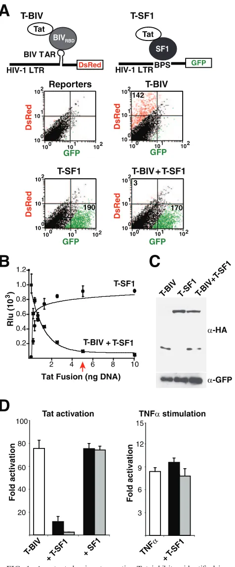
To initially calibrate a dual-reporter Tat hybrid assay in a format for fluorescence, we made use of two HIV-1 LTR reporters engineered with corresponding RNA-binding sites (refer to Table 1 for reporter nomenclature) and two previ-ously characterized T-fusions (refer to Table 2 for T-fusion nomenclature). T-BIV, a fusion of the HIV-1 Tat AD and the RBD of BIV Tat, was used to activate an HIV-1 LTR-BIV TAR (BTAR)-DsRed reporter (Fig. 1A), while T-SF1, a fu-sion of Tat to human splicing factor 1, was used to activate an HIV-1 LTR-branch point sequence (BPS)-GFP reporter (36). When transfected on their own, both T-fusions strongly acti-vated only their cognate RNA reporters (Fig. 1A). Strikingly, activation by T-BIV was strongly inhibited when cotransfected with T-SF1 (3-fold activation), whereas activation by T-SF1 was unaffected (170-fold activation) (Fig. 1A).
Using a more-quantitative luciferase reporter assay, we found that inhibition was remarkably potent, with a stoichio-metric amount of T-SF1 plasmid sufficient to almost com-pletely block T-BIV-mediated activation (Fig. 1B). The dose-response curve corresponding to inhibition by T-SF1 mirrors that corresponding to the activation of its cognate BPS re-porter (Fig. 1B), demonstrating that T-SF1 is a fully functional activator. The high potency observed by transfection accurately reflects relative protein stoichiometries (Fig. 1C) and requires the fusion of Tat and SF1, as SF1 alone does not inhibit (Fig. 1D) and the Tat AD alone is only a very weak dominant negative (see below). Inhibition is specific to Tat-mediated activation, as T-SF1 does not inhibit TNF-␣-induced transcrip-tion (Fig. 1D), prompting us to examine the effects on Tat mechanism in more detail.
Specificity of inhibition by T-fusions and RNA-binding re-quirements. Given that several splicing factors interact with RNAP II- or CTD-associated factors (2, 24, 25, 42), we hy-pothesized that the SF1 moiety targets the T-fusion to RNAP II. If the targeting hypothesis is correct, then T-fusions with other RNAP II-localized splicing factors, such as U2AF65, might show a similar phenotype. To also assess whether the RBD of Tat contributes to the dominant negative activity, we generated fusions of U2AF65 with full-length Tat (Tat-U2AF65) or the Tat AD alone (T-(Tat-U2AF65) (Table 2) and measured their effects by using an HIV-1 LTR-BTAR-FFL reporter recognized by the T-BIV activator (Fig. 2A). Indeed, both Tat-U2AF65 and T-U2AF65 inhibited activation more than 10-fold at substoichiometric DNA levels relative to acti-vator (Fig. 2A) and were even more potent than T-SF1, prompting us to focus on the U2AF65 fusions. In contrast, the Tat AD and full-length Tat showed little inhibition, even
on November 8, 2019 by guest
http://jvi.asm.org/
though the Tat AD and T-U2AF65 are expressed at compa-rable levels (Fig. 2B), consistent with previous reports of their weak dominant negative activity (7, 12, 19). U2AF65 fusions with full-length Tat and with the AD alone are equally potent, showing that TAR RNA-binding activity is dispensable for inhibition.
To test whether T-U2AF65 can inhibit activation when Tat is delivered to RNA sites other than BIV TAR, we measured its activity by using a previously described HIV-1 Tat-Rev fusion (T-Rev) (Table 2) and HIV-1 LTR-RREIIB FFL re-porter (45), as well as a wild-type HIV-1 Tat and HIV-1 LTR-FFL reporter pair. Activity was in both cases inhibited potently by T-U2AF65 but not by unfused U2AF65 (Fig. 2C), indicating that inhibition does not rely on the nature or affinity of the RNA-protein interaction used to recruit the activator and con-sistent with the dispensability of TAR-binding activity for in-hibition. Since U2AF65 is itself an RNA-binding protein, we asked whether simply appending any RBD would contribute to dominant negative activity. This does not appear to be the case, as T-fusions with U2AF35 (T-U2AF35) or MS2 coat protein (T-MS2cp) (Table 2) are not inhibitors yet are expressed at levels comparable to those of T-U2AF65 (Fig. 2D). Because Tat works primarily at the elongation level, unlike most other activators, we examined the step in transcription affected by the inhibitor. RNase protection assays (Fig. 2E) indicate that T-U2AF65 blocks transcription elongation, but not initiation, at the HIV-1 promoter and that the Tat AD alone is a very weak inhibitor. Specificity of inhibition was confirmed further by the observation that only Tat-mediated activation of the HIV-1 promoter was inhibited and not basal or activated tran-scription by other activators on their cognate promoters, in-cluding the P-TEFb-dependent promoters HLA-DRA (a class II major histocompatibility complex [MHC]) and interleukin-8 (IL-8) (28) (Fig. 3A and B). As well, expression levels from a variety of endogenous promoters were not affected (Fig. 3C).
[image:4.585.44.282.73.647.2]Domains required for inhibition and subnuclear localiza-tion.Splicing factors often are localized to subnuclear compart-ments, where they are recruited to sites of active transcription and splicing (32). If the T-fusions are targeted to RNAP II, like splic-ing factors are, we would anticipate similar localization patterns. Indeed, T-U2AF65-GFP showed a striking subnuclear speckle pattern, like U2AF65-GFP (Fig. 4A) or U2AF65 itself (14). In contrast, T-fusions to GFP without or with a nuclear localization
FIG. 1. A potent dominant negative Tat inhibitor identified in a reporter assay. (A) The top illustrations represent a schematic for a dual-reporter fluorescence assay in which T-BIV is used to activate an HIV-1 LTR-BTAR-DsRed reporter. The T-SF1 fusion protein is used to activate an HIV-1 LTR-BPS-GFP reporter (Tables 1 and 2). The graphs shows results for HeLa cells cotransfected with both reporters and T-BIV and/or T-SF1 activators as indicated and sorted by flow cytometry. Expression of GFP is shown in green, and expression of
DsRed is shown in red. Numbers in each quadrant represent activation (n-fold). (B) Dose-response curves of T-SF1 activation on an HIV-1 LTR-BPS-FFL reporter and T-SF1-mediated inhibition of T-BIV ac-tivity on an HIV-1 LTR-BTAR-FFL reporter. Rlu, relative luciferase units. The arrow indicates stoichiometric DNA concentrations of in-hibitor and activator. (C) Western blot of HeLa cells cotransfected with HA-tagged versions of the T-BIV activator and/or the T-SF1 inhibitor along with a GFP expressor to normalize for transfection efficiency. ␣, anti. (D) The left panel shows results for HeLa cells transiently transfected with the HIV-1 LTR-BTAR-FFL reporter plas-mid and different ratios of T-BIV activator to T-SF1 or unfused SF1 plasmids (1:0.2, black bars; 1:1, gray bars). The right panel shows results for HeLa cells transiently transfected with the HIV-1 LTR-BTAR-FFL reporter plasmid and two amounts of T-SF1 plasmid (1 ng, black bars; 5 ng, gray bars), and transcription was stimulated 16 h posttransfection by incubation with TNF-␣(10 ng/ml) for 4 h.
on November 8, 2019 by guest
http://jvi.asm.org/
signal (NLS) (T-GFP or T-NLS-GFP), which had weak dominant negative activity (Fig. 4A), showed very different localization pat-terns. T-GFP was distributed mostly in the cytoplasm and nu-cleus, like unfused GFP, whereas⬎95% of T-NLS-GFP is nu-clear, and T-NLS-GFP is absent from the nucleolus (Fig. 4A and C), indicating that nuclear localization is not the major factor contributing to potency.
Previous results implicate splicing factor RS domains in a subnuclear localization (32). To test whether the U2AF65 RS domain is responsible for T-U2AF65 localization and potent inhibition activity, we generated a T-fusion lacking this portion (T-U2AF65⌬RS, which contains U2AF65 residues 91 to 475) and a second with the RS domain alone (T-RS, which contains U2AF65 residues 2 to 73). Of these, T-RS remained a potent inhibitor and showed a speckle pattern, whereas T-U2AF65⌬RS was a weak inhibitor and showed a different nuclear pattern with nucleolar exclusion (Fig. 4B and C). The RS domain alone with-out the Tat AD or the Tat AD alone is a poor inhibitor and is not
localized to speckles. Thus, both the Tat AD and RS domain portions of T-RS are needed for speckle localization and inhibi-tion activity. To confirm the importance of the Tat AD, we gen-erated T-U2AF65 and T-RS mutants with a Lys41-to-Ala substi-tution (K41A) in the AD that abrogates the P-TEFb interaction and therefore Tat function (15). Both fusions are inactive as inhibitors despite having the same localization patterns as the nonmutant versions, indicating that a functional AD is important (Fig. 4B and C). We hypothesize that the RS domain efficiently delivers the Tat AD into compartments where RNAP II com-plexes assemble for recruitment to the HIV-1 promoter.
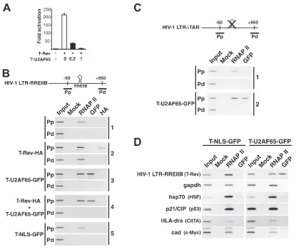
Recruitment of the inhibitor to the HIV-1 promoter. We next used ChIP assays to test the hypothesis that T-U2AF65 is efficiently targeted to the HIV-1 promoter. To assess complex assembly in the context of integrated chromatin, we generated a stable HeLa cell line carrying a single integrated HIV-1 LTR-RREIIB-FFL reporter. This cell line was activated by T-Rev and inhibited by T-U2AF65 in a dose-responsive
man-FIG. 2. Tat RNA-binding activity is dispensable for transcription elongation inhibition.␣, anti. (A) Dose-response curves indicating inhibition of T-BIV-mediated activation on an HIV-1 LTR-BTAR-FFL reporter by the Tat AD, Tat, T-U2AF65, and Tat-U2AF65 (Table 2). The arrow indicates the position corresponding to stoichiometric DNA concentrations (5 ng) of inhibitor and activator. (B) HeLa cells were cotransfected with HA-tagged versions of the Tat AD and T-U2AF65, and total cell extracts were probed for expression levels with anti-HA antibody or with antiactin to control for protein loading. (C) Activation by HIV-1 Tat and T-Rev (Tat AD fused to Rev) (Table 2) measured in the absence (white bars) or presence (black bars) of T-U2AF65 or unfused U2AF65 by using the HIV-1 LTR-FFL and HIV-1 LTR-RREIIB-FFL reporters indicated. Black bars are 1:0.2 and gray bars are 1:1 ratios of activator to T-U2AF65 or unfused U2AF65. (D) The left panel shows results for activity of T-Rev (white bar) or T-Rev cotransfected with T-U2AF65, T-U2AF35, or T-MS2cp at 1:0.2 (black bars) and 1:1 (gray bars) ratios of T-Rev activator to T-fusion by using the HIV-1 LTR-RREIIB-FFL reporter. The right panel shows results for expression of the T-fusions in nuclear extracts, probed with anti-HA or antiactin to control for protein loading. (E) T-U2AF65 blocks transcription elongation. Cells were transfected with HIV-1 Tat, the Tat AD, or T-U2AF65 as indicated, and RNase protection was performed with a Pp probe directed to the HIV-1 LTR and a Pd probe directed to the FFL open reading frame to quantify transcription rates. The experiment was performed in duplicate with similar results.
on November 8, 2019 by guest
http://jvi.asm.org/
[image:5.585.114.472.67.383.2]ner (Fig. 5A). In the absence of T-Rev activator, RNAP II was detected in the promoter-proximal (Pp) region but not in the promoter-distal (Pd) region (Fig. 5B, panel 1), implying a block to elongation, while RNAP II was seen in both regions following activator expression (panel 2). The level of RNAP II detected in the Pp region increased⬃5-fold in the presence of the activator, consistent with its proposed role in transcription complex assembly (40). The T-Rev activator also was detected in the Pp region (panel 2), but notably, the T-U2AF65-GFP inhibitor showed an even higher level of occupancy (panel 3), confirming that the targeting/localization moiety facilitates re-cruitment to the HIV-1 promoter. To more directly evaluate competition between the activator and inhibitor, we cotrans-fected activator and inhibitor plasmids together and analyzed promoter occupancy. We observed a high level of occupancy of the promoter by T-U2AF65-GFP, but not by T-Rev (panel 4), suggesting that the inhibitor is able to displace or efficiently competes with the Tat activator for binding to the viral pro-moter. Furthermore, the AD alone (T-NLS-GFP) was not detectable at the promoter (panel 5), nor was the
T(K41A)-U2AF65 mutant (data not shown), indicating the importance of the modular composition (U2AF65 RS domain and Tat AD) for transcription complex assembly.
Because inhibition does not require TAR and occurs when different RNA sites are used to deliver Tat (Fig. 2), we rea-soned that the inhibitor does not use TAR to load into the HIV-1 promoter. To test this hypothesis, we generated a stable HeLa cell line carrying a single integrated copy of an HIV-1 LTR reporter with a deletion of TAR (HIV-1 LTR-⌬ TAR-FFL) and examined bound factors by ChIP (Fig. 5C). In the absence of Tat activator, RNAP II is associated with the viral promoter (Fig. 5C, panel 1), and more importantly, T-U2AF65 is recruited to the HIV-1 promoter in a TAR-independent manner (panel 2).
This TAR-independent assembly prompted us to measure T-U2AF65 occupancy at non-HIV-1 promoters by ChIP. We found that of five cellular promoters analyzed, including the P-TEFb-dependent MHC class II and hsp70 promoters, none showed detectable T-U2AF65-GFP (Fig. 5D). Thus, the ChIP experiments support the hypothesis that T-U2AF65 is
specifi-FIG. 3. Promoter specificity of the T-U2AF65 fusion. (A) Mammalian cells were transiently transfected with reporter plasmid and different ratios of activator to T-U2AF65 plasmids (from left to right, 1:0, 1:0.2, 1:1, and 1:2). Activities were normalized to activation by HIV-1 Tat alone and to a cotransfected pCMV-RL internal control. To measure heat shock response, endogenous HSF1 was activated 24 h posttransfection by treatment of HeLa cells with 50M AsNO2for 12 h. The activity of transfected p53 was measured in SAOS2 cells. (B) HeLa cells were transiently
transfected with the indicated luciferase reporter plasmids (with MHC class II and IL-8) and different ratios of activator to T-U2AF65 plasmids (1:0.2, black bars; 1:1, gray bars). Transcription was stimulated 16 h posttransfection by incubation with TNF-␣(10 ng/ml) for 4 h. (C) Relative expression levels of endogenous transcripts in SupT1 cells lines stably expressing the Tat AD or T-U2AF65 using quantitative real-time PCR of total RNA extracted to monitor housekeeping genes (those encoding-actin [actin], glyceraldehyde-3-phosphate dehydrogenase [gapdh], and eukaryotic translation elongation factor 1 gamma [EF1G]), regulatory factor genes (those encoding heterogeneous nuclear ribonucleoprotein 1 [hnRNPA1], TBP, hypoxanthine phosphoribosyltransferase 1 (HPRT1)]), and P-TEFb regulated genes (those encoding HLA-DQA1 MHC class II [HLA-DQA], IL-8, and androgen receptor [AR]).
on November 8, 2019 by guest
http://jvi.asm.org/
[image:6.585.112.477.70.375.2]FIG. 4. Domains that contribute to inhibition and subcellular localization. (A) HeLa cells were transiently cotransfected with an HIV-1 LTR-RREIIB-FFL reporter plasmid, T-Rev activator, and various inhibitors at 1:0.2 (black bars) or 1:1 (gray bars) ratios of activator to inhibitor. Activation levels are plotted as relative levels of activation, normalized to the level of activation without inhibitor (85-fold). Confocal images of each GFP-tagged fusion, including images with magnification of⫻3 (lower images, representing the boxed cells shown in the upper images) to highlight the subcellular compartments, are shown below the graph. T-NLS-GFP contains the eight-amino-acid NLS (PPKKKRKV) of simian virus 40 T-Ag (Table 2). (B) Relative activities of T-U2AF65 deletion mutants and Tat AD variants, as determined in experiments whose results are shown in panel A. Corresponding confocal anti-HA (␣-HA) immunofluorescence images are shown. T-U2AF65⌬RS-HA has a deletion of the first 90 amino acids of U2AF65, and T-RS contains only residues 2 to 73. (C) Subnuclear localization of active and inactive Tat inhibitors. HeLa cells were transfected with pEGFP-N3 plasmids expressing GFP fused to U2AF65, T-U2AF65 (dominant negative), T(K41A)-U2AF65 (inactive dominant negative), RS (U2AF65 RS domain only), T-RS (active dominant negative), T(K41A)-RS (inactive dominant negative), U2AF65⌬RS, and T-U2AF65⌬RS. Spk indicates the position of nuclear speckles; No indicates nucleoli.
on November 8, 2019 by guest
http://jvi.asm.org/
cally recruited to the HIV-1 promoter, blocking entry of the Tat activator.
The RS domain of the inhibitor interacts with the CTD of RNAP II.Because splicing factor RS domains can interact with the RNAP II CTD (10, 32) and because the RS domain of T-U2AF65 is important for inhibition, we tested whether T-U2AF65 interacts with RNAP II complexes by coimmuno-precipitating with the nonphosphorylated (RNAP IIa) form of the CTD. T-U2AF65-GFP formed complexes with RNAP IIa in an RNA-independent manner (Fig. 6A) but interacted only weakly with T-NLS-GFP lacking the U2AF65 moiety (data not shown). Immunofluorescence further confirmed the interac-tion, showing partial colocalization of T-U2AF65-GFP with RNAP IIa (Fig. 6B and data not shown). Furthermore, a direct interaction was observed using the recombinant CTD and the U2AF65 RS domain (Fig. 6C). To further corroborate the hypothesis that the inhibitor is loaded into transcription com-plexes via an interaction with the CTD, we cotransfected
T-U2AF65-GFP into HeLa cells along with an ␣ -amanitin-resistant Rpb1 mutant containing either a full-length CTD (52 heptad repeats) or a CTD deletion mutant (5 heptad repeats) (11, 16) and coimmunoprecipitated the complexes (Fig. 6D). Both the full-length and deletion-containing Rpb1 forms were expressed, but T-U2AF65 immunoprecipitated only with the full-length protein.
[image:8.585.81.496.64.414.2]To more precisely determine which RNAP II complexes interact with the inhibitor, we purified polymerase holoenzyme complexes by using a GST-TFIIS affinity column, in which the TFIIS elongation factor selectively binds transcription com-plexes that have not yet assembled into elongation comcom-plexes (23, 34). We generated HeLa cell lines inducibly expressing T-NLS or T-RS (Table 2) and purified holo-Pol II by using the GST-TFIIS column (Fig. 6E). The isolated complexes were purified further by gel filtration and migrated as a single 2- to 4-MDa peak (data not shown). Both the unfused Tat AD (T-NLS) and the T-RS inhibitor were found in holo-Pol II
FIG. 5. Recruitment of the dominant negative to the HIV-1 promoter via RNAP II. (A) Transcription activation and dose-responsive inhibition of T-Rev in the integrated HIV-1 LTR-RREIIB-FFL reporter-containing cell line used for the ChIP assays, with the molar ratios of the T-U2AF65-GFP inhibitor to T-Rev activator indicated. (B) ChIP from mock-transfected HeLa cells with HIV-1 LTR-RREIIB-FFL (panel 1) or cells transfected with the indicated tagged proteins (panels 2 to 5), using the antibodies shown to monitor occupancy in the Pp and Pd regions. Mock lanes used normal rabbit immunoglobulin G for the immunoprecipitation as a specificity control, and input refers to PCRs from isolated chromatin samples. (C) ChIP from mock-transfected HeLa cells (transfected with HIV-1 LTR⌬TAR-FFL; panel 1) or cells transfected with T-U2AF65 (panel 2), using the antibodies shown to monitor occupancy in the Pp and Pd regions. (D) ChIP assays were carried out for the HIV-1 LTR-RREIIB-FFL-transfected HeLa cell line transfected with T-NLS-GFP or T-U2AF65-GFP and using primers to amplify the indicated promoters. Known transcription factors that activate each promoter are indicated in parentheses.
on November 8, 2019 by guest
http://jvi.asm.org/
complexes along with nonphosphorylated RNAP II and CycT1 (Fig. 6E), among other expected proteins, such as TATA-box-binding protein (TBP) and Cdk7 (34) (data not shown). T-RS was particularly abundant, presumably due to a combination of RS domain-CTD- and Tat AD-mediated interactions.
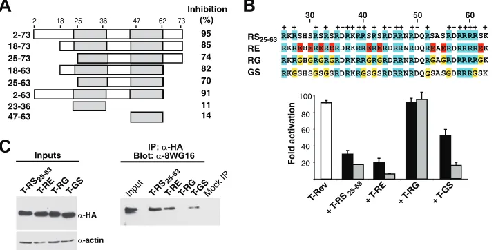
Amino acid requirements of the RS domain.We wished to more precisely pinpoint residues in the U2AF65 RS domain important for inhibitor function and RNAP II interaction. The domain has a negatively charged N terminus (residues 2 to 22) followed by a region containing nine RS dipeptides (residues 25 to 63). Mutants with deletions within the domain from residues 2 to 73 of T-RS showed a progressive loss of inhibitor potency, but a minimal fragment spanning the RS dipeptides (T-RS25-63) still retained significant activity (Fig. 7A). To test the importance of the Arg and Ser residues, we replaced the nine RS dipeptides in T-RS25-63 with Glu (T-RE), Arg-Gly (T-RG), or Arg-Gly-Ser (T-GS). Interestingly, T-RE was as
potent as T-RS, whereas T-RG was inactive and T-GS showed intermediate potency (Fig. 7B). To further test the hypothesis that the RS domain-RNAP II CTD interaction is important for inhibition, we sought to determine whether complex formation between the dipeptide mutants and RNAP II correlated with their inhibition activities. Indeed, immunoprecipitating RNAP IIa pulled down substantial amounts of T-RS25-63 and T-RE but no T-RG and an intermediate amount of T-GS (Fig. 7C), correlating well with inhibitor potency.
[image:9.585.81.502.65.412.2]The fact that the U2AF65 RS domain also directly contacts the pre-mRNA during splicing (22, 50) prompted us to evalu-ate whether RNA binding might contribute to the dominant negative effect. We performed RNA-binding assays with T-RS25-63, T-RE, T-RG, T-GS, and the unfused Tat AD by using U2 snRNA and HIV-1 RRE RNA coupled with beads, and we found that T-RS25-63, T-RE, and T-RG bound the two forms of RNA equally well, T-GS bound 50% less efficiently,
FIG. 6. Interactions with the CTD and colocalization with RNAP II.␣, anti. (A) GFP-tagged T-U2AF65 and T-NLS proteins were immuno-precipitated from cell extracts and analyzed by Western blotting using the indicated antibodies (Ab). (B) Confocal microscopy of HeLa cells transfected with GFP-tagged T-U2AF65 and immunostained with H14 antibody. (C) In vitro pull-down assays for HA-tagged U2AF65 RS domain with GST-CTD or with GST alone as a control. Inputs show the purified Coomassie blue-stained recombinant proteins used in the pull-down assays. (D) HeLa cells were transfected with the HA-Rpb1 or HA-Rpb1⌬CTD constructs shown (top), and expression levels were assessed by Western blot analysis relative to antiactin antibody (bottom left). Cells were then cotransfected with T-U2AF65-GFP, and extracts were immunoprecipitated (IP) with anti-HA and probed for T-U2AF65-GFP by Western blotting with anti-GFP (bottom right). (E) The left panel shows doxycycline (DOX)-induced expression of HF-tagged Tat AD (T-NLS-HF) and T-RS-HF in stable HeLa T-Rex cell lines analyzed in whole-cell extracts with anti-FLAG. The right panel shows extracts bound and eluted from a GST-TFIIS column that were probed with the antibodies indicated. The asterisk indicates a cross-reacting protein.
on November 8, 2019 by guest
http://jvi.asm.org/
and the Tat AD showed no appreciable binding (data not shown). Thus, RNA binding appears to correlate with arginine content, as previously proposed (50), but not with inhibition activity, consistent with the need for the RS domain for CTD targeting.
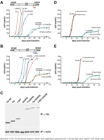
T-RS inhibits viral replication and generates a chronically replicating cell line.The high potency of the Tat dominant negatives and the requirement of Tat for viral replication sug-gested that the dominant negatives might be effective viral inhibitors. Thus, we generated SupT1 lymphocyte cell lines stably expressing T-U2AF65, Tat-U2AF65, or T-BIV-U2AF65 dominant negatives or the nonfusion controls Tat AD, Tat, T-BIV, or U2AF65 and monitored viral replication rates by using viruses engineered with either the HIV-1 Tat-TAR or BIV Tat-TAR interaction (54). Only viruses containing a cog-nate Tat-TAR pair will efficiently replicate, and this allowed us to examine a matched set of viruses with RNA-binding speci-ficity controls in the replication assays.
We observed striking specificity of the dominant negative proteins, as replication was inhibited only in viruses driven by a noncognate RNA-protein interaction. Expression of T-U2AF65, lacking a TAR RBD, markedly suppressed rep-lication of both viruses compared to the Tat AD or U2AF65 controls, with no p24 antigen detectable until 18 to 20 days after infection. Expression of Tat-U2AF65 or T-BIV-U2AF65 inhibited replication of the noncognate virus to an extent similar to that of T-U2AF65 and only slightly inhib-ited the cognate virus (Fig. 8A and B). Interestingly, expres-sion of Tat or T-BIV activators accelerated replication of the cognate viruses but not the noncognate viruses, suggest-ing that Tat levels in these viruses may be limitsuggest-ing. The strong levels of inhibition are notable, given that expression levels of the inhibitors are low in these cell lines, as judged by Western blot analysis of samples immunoprecipitated
with a Tat antibody (Fig. 8C) and reverse transcription-PCR and reporter assays (data not shown), explaining why virus expression ultimately is seen.
Viruses that emerged from the inhibitor cell lines after 18 to 20 days displayed slow replication kinetics and reached a low plateau of p24 expression that remained constant for at least 110 days without producing cytopathic effects. Reinfection ex-periments with Tat AD- and T-U2AF65-expressing cell lines indicate that the viruses do not acquire resistance mutations during this time period but rather grow poorly under these conditions of dominant negative inhibitor expression and con-tinuously suppress viral replication (Fig. 8D and E).
DISCUSSION
[image:10.585.111.470.70.251.2]Previously described dominant negative Tat proteins con-taining the Tat AD alone have shown relatively modest levels of inhibition (6, 19, 35). The extraordinarily potent proteins described here represent a new mechanistic class of transcrip-tional inhibitor, and a splicing factor (U2AF65 or SF1) or short RS domain acts as a targeting/localization moiety for the teth-ered Tat AD. The localization function provided by this tar-geting moiety allows the inhibitor to function at stoichiometric levels, without the need for substantial overexpression typically required by dominant negatives that operate by squelching or other simple competition mechanisms (13, 17, 21). Ptashne and Gann proposed the concept of “regulated localization,” activity and specificity being imposed by simple binding inter-actions between a locator (transcription factor), the transcrip-tional machinery, and the DNA (39). Such localization can include subcellular compartmentalization, in which molecular crowding can enhance the assembly of large macromolecular complexes (30, 31), in our case, favoring assembly into holo-Pol II complexes. We further suggest that combining multiple
FIG. 7. Inhibition activities and RNAP II interactions of RS domain mutants. (A) Various T-RS domain deletion mutants were tested for dominant negative inhibition on an HIV-1 LTR-RREIIB-FFL reporter, and the values shown represent percent inhibition at a 1:1 activator/ inhibitor plasmid ratio relative to activation without inhibitor. The data are mean values from three independent experiments. (B) Sequences of RS dipeptide substitutions and their inhibition activities with a T-Rev activator (white bar) and HIV-1 LTR-RREIIB-FFL reporter at 1:0.2 (black bars) and 1:1 (gray bars) ratios of activator to inhibitor. (C) HeLa cells were transfected with HA-tagged versions of the T-RS25-63domain or
mutants (expression levels shown on the left top panel) with loading controls (levels shown on left bottom panel). Amounts of associated RNAP IIa were assessed by immunoprecipitation (IP) with anti-HA (␣-HA) and Western blotting with the 8WG16 (␣-8WG16) antibody (right panel).
on November 8, 2019 by guest
http://jvi.asm.org/
targeting functions within a single polypeptide provides an entropic benefit, allowing T-RS to load efficiently into early transcription complexes through CTD and CycT1 interactions, thereby blocking the entry of wild-type Tat.
[image:11.585.85.500.65.622.2]It is especially interesting that RNA binding is not required for the delivery of T-RS to the HIV-1 promoter, raising a number of questions about the mechanisms and timing of T-RS and Tat recruitment into HIV-1 transcription complexes
FIG. 8. Expression of the Tat dominant negative blocks viral replication and generates a latency-like state. SupT1 cells stably expressing the Tat domains or fusion proteins indicated were infected with either HIV-1 Tat-TAR-dependent (A) or BIV Tat-TAR-dependent (B) virus (54), and the kinetics of p24 antigen expression were monitored by enzyme-linked immunosorbent assay. (C) Expression levels of Tat domains or fusions analyzed by immunoprecipitation followed by Western blotting using an anti-Tat (␣-Tat) antibody, with an anti--actin (␣-actin) antibody control. (D) HIV-1 Tat-TAR-dependent viruses emerging from the T-U2AF65 inhibitor-expressing cell lines was harvested at day 30 (arrows) and used to reinfect SupT1 cells expressing Tat AD and T-U2AF65. (E) Reinfection using the BIV Tat-TAR-dependent virus.
on November 8, 2019 by guest
http://jvi.asm.org/
and also whether other dominant negative transcription factors can be recruited efficiently to their promoters by a similar cotranscriptional tethering strategy. Preliminary experiments with HSF1, CIITA, and GAL4-VP16 activators show little or no effect of an appended RS domain (data not shown), sug-gesting that Tat assembly may represent a special case, perhaps related to its role in elongation or dependence on RNA bind-ing. The Tat AD itself associates directly with holo-Pol II complexes (9; this work), perhaps explaining why the U2AF65 RS moiety, known to associate with preinitiation or early tran-scription complexes (42, 49), enhances T-RS assembly. The proposed targeting function of the RS domain to the CTD is consistent with the observations that U2AF65 is found in early transcription complexes (42, 49) and is recruited to the HIV-1 promoter (5). Our experiments demonstrate a direct interac-tion between the U2AF65 RS domain and CTD in vitro and a CTD-dependent interaction in vivo (Fig. 6), agreeing with the observation that splicing factors copurify with nonphosphory-lated RNAP II (10). Mutagenesis of T-RS indicates that the serines of the RS dipeptides are important for CTD binding and dominant negative activity, whereas the positively charged arginines contribute to nonspecific RNA binding, as previously proposed (50), but have little effect on inhibition (Fig. 7).
Still, the CTD-tethering hypothesis is insufficient to explain the specificity of recruitment to the HIV-1 promoter, given that T-RS, which requires interactions with CycT1 in addition to the CTD, would be expected to bind to and inhibit activation of other P-TEFb-dependent promoters, yet this is not the case (Fig. 3 and 5). Perhaps the transient nature or precise timing of splicing factor-CTD interactions (29), interactions of the Tat AD or P-TEFb with other HIV-1 promoter-specific factors, or the assembly of TBP-associated-factor-less TBP complexes (40) differ among promoters and determine whether stable “ChIPable” complexes can form. Irrespective of the details of assembly, it is clear that mechanisms used to cotranscription-ally load RNA-processing factors at promoters (2, 41) can be co-opted to deliver the dominant negative Tat AD to the HIV-1 promoter and thereby generate an extraordinarily po-tent inhibitor.
Given the high inhibitor potency, viral replication is substan-tially inhibited by low-level expression of the dominant nega-tive in stable cell lines, even without optimizing and selecting for lines with high activity. It is interesting that these cells establish a chronic infection without cytopathic effects, remi-niscent of other cellular environments that may resemble la-tent stages of HIV-1 infection (27). The amount of Tat clearly affects viral replication rates (51) and also can drive phenotypic diversity (52), and here, we show that expression of the dom-inant negative provides another means to alter Tat function. It will be interesting to examine mechanisms by which resistance to the Tat dominant negative might arise and to evaluate its therapeutic potential.
ACKNOWLEDGMENTS
I.D. dedicates this work to Ileana Cuevas and to the memory of Agustı´n “Guity” Mairal. We thank R. Andino for critical discussions and K. Yamamoto, A. Johnson, C. Guthrie, V. Calabro, M. Daugherty, and other members of the Frankel laboratory for critical readings of the manuscript. We thank M. Green, R. Tjian, R. Voellmy, R. Kings-ton, J. Ca´ceres, A. Kornblitth, W. Wang, J. Greenblatt, and B. M. Peterlin for sharing reagents and advice.
I.D. is a postdoctoral fellow of the Human Frontier Science Program and Fundacio´n Antorchas. J.R.G. was a postdoctoral fellow of the Damon-Runyon Foundation. C.D. was supported by a predoctoral fellowship from the American Foundation for Pharmaceutical Educa-tion. This work was supported by NIH grant AI29135 to A.D.F. R.L.N. and A.D.F. are founders of Advanced Genetic Systems, a biotechnol-ogy company with an interest in developing antiviral therapeutics.
REFERENCES
1.Ahn, S. H., M. Kim, and S. Buratowski.2004. Phosphorylation of serine 2 within the RNA polymerase II C-terminal domain couples transcription and
3⬘end processing. Mol. Cell13:67–76.
2.Bentley, D. L.2005. Rules of engagement: co-transcriptional recruitment of
pre-mRNA processing factors. Curr. Opin. Cell Biol.17:251–256.
3.Boehm, A. K., A. Saunders, J. Werner, and J. T. Lis.2003. Transcription factor and polymerase recruitment, modification, and movement on dhsp70
in vivo in the minutes following heat shock. Mol. Cell. Biol.23:7628–7637.
4.Brady, J., and F. Kashanchi.2005. Tat gets the “green” light on transcription
initiation. Retrovirology2:69.
5.Bre`s, V., N. Gomes, L. Pickle, and K. A. Jones.2005. A human splicing factor, SKIP, associates with P-TEFb and enhances transcription elongation
by HIV-1 Tat. Genes Dev.19:1211–1226.
6.Caputo, A., M. P. Grossi, R. Bozzini, C. Rossi, M. Betti, P. C. Marconi, G. Barbanti-Brodano, and P. G. Balboni.1996. Inhibition of HIV-1 replication and reactivation from latency by tat transdominant negative mutants in the
cysteine rich region. Gene Ther.3:235–245.
7.Carroll, R., B. M. Peterlin, and D. Derse.1992. Inhibition of human
immu-nodeficiency virus type 1 Tat activity by coexpression of heterologoustrans
activators. J. Virol.66:2000–2007.
8.Cramer, P.2004. RNA polymerase II structure: from core to functional
complexes. Curr. Opin. Genet. Dev.14:218–226.
9.Cujec, T. P., H. Cho, E. Maldonado, J. Meyer, D. Reinberg, and B. M. Peterlin.1997. The human immunodeficiency virus transactivator Tat
inter-acts with the RNA polymerase II holoenzyme. Mol. Cell. Biol.17:1817–1823.
10.Das, R., J. Yu, Z. Zhang, M. P. Gygi, A. R. Krainer, S. P. Gygi, and R. Reed.
2007. SR proteins function in coupling RNAP II transcription to pre-mRNA
splicing. Mol. Cell26:867–881.
11.Fong, N., G. Bird, M. Vigneron, and D. L. Bentley.2003. A 10 residue motif at the C-terminus of the RNA pol II CTD is required for transcription,
splicing and 3⬘end processing. EMBO J.22:4274–4282.
12.Fraisier, C., D. A. Abraham, M. van Oijen, V. Cunliffe, A. Irvine, R. Craig, and E. A. Dzierzak.1998. Inhibition of Tat-mediated transactivation and HIV replication with Tat mutant and repressor domain fusion proteins.
Gene Ther.5:946–954.
13.Friedman, A. D., S. J. Triezenberg, and S. L. McKnight.1988. Expression of a truncated viral trans-activator selectively impedes lytic infection by its
cognate virus. Nature335:452–454.
14.Gama-Carvalho, M., R. D. Krauss, L. Chiang, J. Valcarcel, M. R. Green, and M. Carmo-Fonseca.1997. Targeting of U2AF65 to sites of active splicing in
the nucleus. J. Cell Biol.137:975–987.
15.Garber, M. E., P. Wei, V. N. KewalRamani, T. P. Mayall, C. H. Herrmann, A. P. Rice, D. R. Littman, and K. A. Jones.1998. The interaction between HIV-1 Tat and human cyclin T1 requires zinc and a critical cysteine residue
that is not conserved in the murine CycT1 protein. Genes Dev.12:3512–
3527.
16.Gerber, H. P., M. Hagmann, K. Seipel, O. Georgiev, M. A. West, Y. Liting-tung, W. Schaffner, and J. L. Corden.1995. RNA polymerase II C-terminal
domain required for enhancer-driven transcription. Nature374:660–662.
17.Gill, G., and M. Ptashne.1988. Negative effect of the transcriptional
activa-tor GAL4. Nature334:721–724.
18.Gomes, N. P., G. Bjerke, B. Llorente, S. A. Szostek, B. M. Emerson, and J. M. Espinosa.2006. Gene-specific requirement for P-TEFb activity and RNA polymerase II phosphorylation within the p53 transcriptional program.
Genes Dev.20:601–612.
19.Green, M., M. Ishino, and P. M. Loewenstein.1989. Mutational analysis of HIV-1 Tat minimal domain peptides: identification of trans-dominant
mu-tants that suppress HIV-LTR-driven gene expression. Cell58:215–223.
20.Hahn, S.2004. Structure and mechanism of the RNA polymerase II
tran-scription machinery. Nat. Struct. Mol. Biol.11:394–403.
21.Herskowitz, I.1987. Functional inactivation of genes by dominant negative
mutations. Nature329:219–222.
22.Hertel, K. J., and B. R. Graveley.2005. RS domains contact the pre-mRNA
throughout spliceosome assembly. Trends Biochem. Sci.30:115–118.
23.Kim, B., A. I. Nesvizhskii, P. G. Rani, S. Hahn, R. Aebersold, and J. A. Ranish.2007. The transcription elongation factor TFIIS is a component of RNA polymerase II preinitiation complexes. Proc. Natl. Acad. Sci. USA
104:16068–16073.
24.Komarnitsky, P., E. J. Cho, and S. Buratowski.2000. Different phosphory-lated forms of RNA polymerase II and associated mRNA processing factors
during transcription. Genes Dev.14:2452–2460.
on November 8, 2019 by guest
http://jvi.asm.org/
25.Kornblihtt, A. R., M. de la Mata, J. P. Fededa, M. J. Munoz, and G. Nogues.
2004. Multiple links between transcription and splicing. RNA10:1489–1498.
26.Landt, S. G., R. Tan, and A. D. Frankel. 2000. Screening RNA-binding libraries using Tat-fusion system in mammalian cells. Methods Enzymol.
318:350–363.
27.Lassen, K., Y. Han, Y. Zhou, J. Siliciano, and R. F. Siliciano.2004. The
multifactorial nature of HIV-1 latency. Trends Mol. Med.10:525–531.
28.Luecke, H. F., and K. R. Yamamoto.2005. The glucocorticoid receptor blocks P-TEFb recruitment by NFkappaB to effect promoter-specific
tran-scriptional repression. Genes Dev.19:1116–1127.
29.Mabon, S. A., and T. Misteli.2005. Differential recruitment of pre-mRNA
splicing factors to alternatively spliced transcripts in vivo. PLoS Biol.3:e374.
30.Minton, A. P.2000. Implications of macromolecular crowding for protein
assembly. Curr. Opin. Struct. Biol.10:34–39.
31.Misteli, T.2007. Beyond the sequence: cellular organization of genome
function. Cell128:787–800.
32.Misteli, T., and D. L. Spector.1999. RNA polymerase II targets pre-mRNA
splicing factors to transcription sites in vivo. Mol. Cell3:697–705.
33.Orsini, M. J., and C. M. Debouck.1996. Inhibition of human immunodefi-ciency virus type 1 and type 2 Tat function by transdominant Tat protein
localized to both the nucleus and cytoplasm. J. Virol.70:8055–8063.
34.Pan, G., T. Aso, and J. Greenblatt.1997. Interaction of elongation factors TFIIS and elongin A with a human RNA polymerase II holoenzyme capable of promoter-specific initiation and responsive to transcriptional activators.
J. Biol. Chem.272:24563–24571.
35.Pearson, L., J. Garcia, F. Wu, N. Modesti, J. Nelson, and R. Gaynor.1990. A transdominant tat mutant that inhibits tat-induced gene expression from the human immunodeficiency virus long terminal repeat. Proc. Natl. Acad.
Sci. USA87:5079–5083.
36.Peled-Zehavi, H., J. A. Berglund, M. Rosbash, and A. D. Frankel.2001. Recognition of RNA branch point sequences by the KH domain of splicing factor 1 (mammalian branch point binding protein) in a splicing factor
complex. Mol. Cell. Biol.21:5232–5241.
37.Peterlin, B. M., and D. H. Price.2006. Controlling the elongation phase of
transcription with P-TEFb. Mol. Cell23:297–305.
38.Plavec, I., M. Agarwal, K. E. Ho, M. Pineda, J. Auten, J. Baker, H. Matsu-zaki, S. Escaich, M. Bonyhadi, and E. Bohnlein.1997. High transdominant RevM10 protein levels are required to inhibit HIV-1 replication in cell lines and primary T cells: implication for gene therapy of AIDS. Gene Ther.
4:128–139.
39.Ptashne, M., and A. Gann.1998. Imposing specificity by localization:
mech-anism and evolvability. Curr. Biol.8:R812–R822.
40.Raha, T., S. W. Cheng, and M. R. Green.2005. HIV-1 Tat stimulates transcription complex assembly through recruitment of TBP in the absence
of TAFs. PLoS Biol.3:e44.
41.Reed, R., and E. Hurt.2002. A conserved mRNA export machinery coupled
to pre-mRNA splicing. Cell108:523–531.
42.Robert, F., M. Blanchette, O. Maes, B. Chabot, and B. Coulombe.2002. A human RNA polymerase II-containing complex associated with factors
nec-essary for spliceosome assembly. J. Biol. Chem.277:9302–9306.
43.Selby, M. J., and B. M. Peterlin.1990. Trans-activation by HIV-1 Tat via a
heterologous RNA binding protein. Cell62:769–776.
44.Shamah, S. M., and C. D. Stiles.1995. Transdominant negative mutations.
Methods Enzymol.254:565–576.
45.Southgate, C., M. L. Zapp, and M. R. Green.1990. Activation of transcrip-tion by HIV-1 Tat protein tethered to nascent RNA through another
pro-tein. Nature345:640–642.
46.Szak, S. T., D. Mays, and J. A. Pietenpol.2001. Kinetics of p53 binding to
promoter sites in vivo. Mol. Cell. Biol.21:3375–3386.
47.Tan, R., and A. D. Frankel.1998. A novel glutamine-RNA interaction iden-tified by screening libraries in mammalian cells. Proc. Natl. Acad. Sci. USA
95:4247–4252.
48.Trono, D., M. B. Feinberg, and D. Baltimore.1989. HIV-1 Gag mutants can
dominantly interfere with the replication of the wild-type virus. Cell59:113–
120.
49.Ujva´ri, A., and D. S. Luse.2004. Newly initiated RNA encounters a factor involved in splicing immediately upon emerging from within RNA
polymer-ase II. J. Biol. Chem.279:49773–49779.
50.Valca´rcel, J., R. K. Gaur, R. Singh, and M. R. Green.1996. Interaction of U2AF65 RS region with pre-mRNA branch point and promotion of base
pairing with U2 snRNA. Science273:1706–1709.
51.Verhoef, K., M. Koper, and B. Berkhout.1997. Determination of the mini-mal amount of Tat activity required for human immunodeficiency virus type
1 replication. Virology237:228–236.
52.Weinberger, L. S., J. C. Burnett, J. E. Toettcher, A. P. Arkin, and D. V. Schaffer.2005. Stochastic gene expression in a lentiviral positive-feedback
loop: HIV-1 Tat fluctuations drive phenotypic diversity. Cell122:169–182.
53.Williams, S. A., L. F. Chen, H. Kwon, C. M. Ruiz-Jarabo, E. Verdin, and W. C. Greene.2006. NF-kappaB p50 promotes HIV latency through HDAC
recruitment and repression of transcriptional initiation. EMBO J.25:139–
149.
54.Xie, B., M. A. Wainberg, and A. D. Frankel.2003. Replication of human immunodeficiency viruses engineered with heterologous Tat-transactivation
response element interactions. J. Virol.77:1984–1991.