Antibodies to the Conserved CD4-Binding Site
Xueling Wu,aCharlene Wang,aSijy O’Dell,aYuxing Li,cBrandon F. Keele,dZhongjia Yang,aHiromi Imamichi,bNicole Doria-Rose,b James A. Hoxie,eMark Connors,bGeorge M. Shaw,eRichard T. Wyatt,cand John R. Mascolaa
Vaccine Research Center,a
Laboratory of Immunoregulation,b
NIAID, NIH, Bethesda, Maryland, USA; IAVI Center for Neutralizing Antibodies at TSRI, Department of Medicine and Microbial Science, The Scripps Research Institute, La Jolla, California, USAc
; AIDS and Cancer Virus Program, SAIC-Frederick, Inc., NCI-Frederick, NIH, Frederick, Maryland, USAd
; and Department of Medicine, University of Pennsylvania, Philadelphia, Pennsylvania, USAe
The monoclonal antibody (MAb) VRC01 was isolated from a slowly progressing HIV-1-infected donor and was shown to
neu-tralize diverse HIV-1 strains by binding to the conserved CD4 binding site (CD4bs) of gp120. To better understand the virologic
factors associated with such antibody development, we characterized HIV-1 envelope (Env) variants from this donor and five
other donors who developed broadly neutralizing antibodies. A total of 473
env
sequences were obtained by single-genome
am-plification, and 100 representative
env
clones were expressed and tested for entry and neutralization sensitivity. While VRC01
neutralizes about 90% of the genetically diverse heterologous HIV-1 strains tested, only selective archival Env variants from the
VRC01 donor were sensitive to VRC01 and all of the Env variants derived from the donor plasma were resistant, indicating
strong antibody-based selection pressure. Despite their resistance to this broadly reactive MAb that partially mimics CD4, all
Env variants required CD4 for entry. Three other CD4bs MAbs from the same donor were able to neutralize some VRC01 escape
variants, suggesting that CD4bs antibodies continued to evolve in response to viral escape. We also observed a relatively high
percentage of VRC01-resistant Env clones in the plasma of four of five additional broadly neutralizing donors, suggesting the
presence of CD4bs-directed neutralizing antibodies in these donors. In total, these data indicate that the CD4bs-directed
neu-tralizing antibodies exert ongoing selection pressure on the conserved CD4bs epitope of HIV-1 Env.
A
pproximately 25% of HIV-1-infected individuals develop
cross-reactive neutralizing antibodies, and the serum of some
donors potently neutralizes most HIV-1 strains (11, 12, 24, 43, 53,
62, 66, 67, 71). Serum mapping studies and subsequent
monoclo-nal antibody (MAb) isolation have defined several conserved
neu-tralization epitopes on the HIV-1 Env trimer (2, 8, 11, 24, 27, 43,
45, 63, 69–71, 75, 77, 80, 81). One such epitope is the initial site of
gp120 attachment to the cellular receptor CD4 (38, 81). We
pre-viously described the CD4 binding site (CD4bs) MAb VRC01,
isolated from a slowly progressing subtype B-infected donor,
which neutralized 91% of the genetically diverse HIV-1 isolates
tested (75). Biophysical characterization of VRC01 suggested that
it partially mimics the interaction of CD4 with gp120, and the
liganded crystal structures of VRC01 defined specific similarities
in the heavy chain of VRC01 and domain 1 of CD4 as related to
their binding interaction with gp120 (80). Recently, additional
potently neutralizing CD4bs antibodies were identified in a total
of six donors (63, 77), indicating that other HIV-1-infected
do-nors can make similarly potent CD4bs antibodies.
Previous studies have shown that HIV-1 encounters a genetic
bottleneck during transmission, often resulting in a genetically
homogenous population of initial plasma viremia (10, 33, 46, 47,
61, 73). Partially due to the host adaptive immunity, the early
circulating virus evolves into a diverse population over time, with
genetic diversity of up to 10% in the
env
gene within an infected
individual (36, 65). Autologous virus-neutralizing antibodies
gen-erally develop within the first months or year of infection, and
there is a well-documented antibody-based selection process that
results in ongoing viral escape from autologous neutralizing
anti-bodies (1, 3, 4, 18–20, 26, 40, 50, 51, 54, 57, 58, 72). Thus,
circu-lating plasma viral Env variants are poorly neutralized by
concur-rent serum but are often potently neutralized by serum samples
from later time points. Importantly, the early autologous
neutral-izing antibody response is usually highly strain specific; these
an-tibodies do not appear to target conserved regions of Env and do
not neutralize most heterologous viral isolates (25, 48, 52, 54, 58).
Until recently, few broadly reactive HIV-1-neutralizing
antibod-ies had been isolated (5, 55, 68, 82); thus, the Env quasispecantibod-ies in
donors with such antibodies have not been characterized.
The isolation of VRC01 and other related MAbs from donor 45
provided the opportunity to study the interaction of these MAbs
with the circulating viral quasispecies. It was not clear if
circulat-ing virus would escape from a neutralizcirculat-ing antibody targetcirculat-ing a
relatively conserved epitope or if broadly reactive antibodies
would evolve in relation to viral escape. To better understand
these events, we performed detailed studies of the plasma viral
quasispecies obtained from donor 45 at three time points (2001,
2006, and 2009). For comparison, we also studied the viral
quasi-species in five additional donors with broadly reactive serum
neu-tralizing antibodies. We used single-genome amplification (SGA)
to derive viral Env sequences and expressed representative
se-quences as Env pseudoviruses to assess their
in vitro
neutralization
sensitivity to VRC01, as well as several other MAbs isolated from
donor 45. In total, this analysis revealed strong selection pressure
by VRC01 on the circulating viral quasispecies and ongoing viral
Received16 December 2011Accepted4 March 2012
Published ahead of print14 March 2012
Address correspondence to John R. Mascola, [email protected]. Copyright © 2012, American Society for Microbiology. All Rights Reserved.
doi:10.1128/JVI.07139-11
on November 7, 2019 by guest
http://jvi.asm.org/
escape and evolution of the antibody response to the CD4bs of
HIV-1 gp120.
MATERIALS AND METHODS
Study subjects.The plasma, serum, and peripheral blood mononuclear cell (PBMC) samples described in this study were from HIV-1-infected individuals enrolled in clinical protocols approved by the appropriate institutional review board of the National Institute of Allergy and Infec-tious Diseases or the University of Pennsylvania. Donor 45 has never been on antiretroviral treatment (ART), and all other donor samples were from time points prior to or after withdrawal from ART. All donors except B7B5 were considered slow progressors with peripheral blood CD4 T-cell levels of⬎350 cells/l and plasma viral RNA levels of⬍36,000 copies/ml. The clinical status of donor B7B5 was not fully characterized, but he was in the clinically asymptomatic phase of HIV-1 infection.
Antibodies and cells.The anti-CD4bs MAbs VRC01 and VRC03 have been described previously (75). The CD4-Ig plasmid construct was pro-vided by Joseph Sodroski (Dana-Farber Cancer Institute), and the fusion protein was expressed and purified as described previously (75). The anti-CD4bs MAb b12 was provided by Dennis Burton and Ralph Pantophlet (The Scripps Research Institute, La Jolla, CA). The donor 45 serum IgG was purified using protein G UltraLink columns (Thermo Scientific, Rockford, IL) and following the manufacturer’s instructions. The concen-trations of purified serum IgG or IgG MAbs were determined using Nano-Drop (Thermo Scientific) with 1.40 as the extinction coefficient. The TZM-bl cells were obtained from the NIH AIDS Research and Reference Reagent Program, as contributed by John Kappes and Xiaoyun Wu. The Cf2th-CD4.CCR5 and Cf2th-synCCR5 cell lines, canine thymocyte lines stably transfected to express both human CD4 and CCR5 or only human CCR5, respectively, were obtained from the NIH AIDS Research and Ref-erence Reagent Program, as contributed by Tajib Mizabekov and Joseph Sodroski (Dana-Farber Cancer Institute).
Viral RNA extraction, cDNA synthesis, and genomic DNA extrac-tion.A 140-l volume of each plasma or serum sample was used to extract viral RNA using the QIAamp viral RNA minikit (Qiagen, Valencia, CA). The RNA was eluted in 50l of elution buffer and subjected to first-strand cDNA synthesis immediately using the SuperScript III reverse transcrip-tase (Invitrogen Life Technologies, Grand Island, NY). cDNA synthesis was done in a final volume of 100l including 50l viral RNA, 5l of a deoxynucleoside triphosphate (dNTP) mixture (each at 10 mM), 1.25l antisense primer envB3out (5= -TTGCTACTTGTGATTGCTCCATGT-3=) at 20M, 20l 5⫻first-strand buffer, 5l dithiothreitol at 100 mM, 5l RNaseOUT (Invitrogen), and 5l SuperScript III reverse transcrip-tase. RNA, primers, and dNTPs were heated at 65°C for 5 min and then chilled on ice for 1 min, and then the entire reaction mixture was incu-bated at 50°C for 60 min, followed by 55°C for an additional 60 min. Finally, the reaction was heat inactivated at 70°C for 15 min and then treated with 1l RNase H at 37°C for 20 min. The QIAamp DNA Blood minikit (Qiagen) was used to extract the donor 45 PBMC genomic DNA. The synthesized cDNA and the extracted genomic DNA were subjected to the first-round PCR immediately or stored frozen at⫺80°C.
SGA.The nested PCR of HIV-1envSGA was described previously (33, 41, 61). Briefly, the synthesized cDNA or genomic DNA was serially di-luted and distributed in replicates of 12 to 16 PCRs in ThermoGrid 96-well plates (Denville Scientific, Metuchen, NJ) to identify a dilution where PCR-positive wells constituted about 30% of the total number of reac-tions. At this dilution, most of the wells contain amplicons derived from a single cDNA or DNA molecule. Additional PCR amplifications were per-formed using this dilution in full 96-well plates. PCR amplification was carried out using the PlatinumTaqHigh Fidelity PCR system (Invitro-gen). The final 20-l reaction volume was composed of 2l 10⫻buffer, 0.8l MgSO4, 0.4l dNTP mixture (each at 10 mM), 0.2l each primer at 20M, 0.1l PlatinumTaqHigh Fidelity polymerase, and 1l tem-plate DNA. The primers for the first-round PCR were envB5out (5=-TAG AGCCCTGGAAGCATCCAGGAAG-3=) and envB3out (5=-TTGCTACT
TGTGATTGCTCCATGT-3=). The primers for the second-round PCR were envB5in (5=-CACCTTAGGCATCTCCTATGGCAGGAAGAAG-3=) and envB3in (5=-GTCTCGAGATACTGCTCCCACCC-3=). The cycler parameters were 94°C for 2 min, followed by 35 cycles of 94°C for 15 s, 55°C for 30 s, and 68°C for 4 min and by a final extension of 68°C for 10 min. The product of the first-round PCR (1l) was subsequently used as the template in the second-round PCR under the same conditions but with a total of 45 cycles. The amplicons were inspected on a precast 1% agarose gel (Embi Tec, San Diego, CA). All PCR procedures were carried out in a designated PCR clean hood using procedural safeguards against sample contamination.
DNA sequencing.Amplicons were directly sequenced by BigDye Ter-minator chemistry (Applied Biosystems, Foster City, CA) by ACGT, Inc. (Wheeling, IL). Both DNA strands were sequenced using partially over-lapping fragments. Individual sequence fragments for each amplicon were assembled and edited using Sequencher 5.0 (Gene Codes, Ann Arbor, MI). All chromatograms were inspected for sites of mixed bases (double peaks), which would be evidence of priming from more than one template or the introduction of PCR error in early cycles. Any sequence with evi-dence of double peaks was excluded from further analysis.
Sequence alignments, diversity, and phylogenetic analysis.Theenv
sequences containing unproductive mutations (such as those that cause stop codons and frameshifts) or large deletions (⬎24 nucleotides) in conserved regions were removed from subsequent sequence analysis andenvcloning. The 28 PBMC-derived donor 45envsequences were checked for hypermu-tation using Hypermut (59) at the Los Alamos HIV sequence database (http: //www.hiv.lanl.gov/content/sequence/HYPERMUT/hypermut.html), and none was hypermutated using the intradonor plasma-derived sequence 45_01A14 as a reference. A total of 473envsequences from the study donors suitable for full analysis were used to generate a neighbor-joining tree as fol-lows. The 473 donor gp160 protein sequences together with the subtype B reference sequence HXB2 were aligned using MUSCLE for multiple-se-quence comparison by log expectation (13, 14). The protein distance matrix was calculated by protdist using the Jones-Taylor-Thornton model (32). The tree was constructed by Neighbor using the neighbor-joining method (37). The nucleotide sequences were analyzed as follows. The donor gp160 nucle-otide sequences, together with HXB2, were initially aligned with ClustalW (39) and then hand checked using BioEdit (http://www.mbio.ncsu.edu /bioedit/bioedit.html) to improve the alignments according to the codon translation. Each donor nucleotide sequence alignment was submitted to dnadist for calculation of the pairwise nucleotide sequence distance. The F84 model (17, 34) of nucleotide substitution was used for these calculations. After gap stripping, each donor nucleotide sequence alignment was evaluated for potential natural recombination using the recombination analysis tool at
http://cbr.jic.ac.uk/dicks/software/RAT/(15). Of the total of 473 sequences evaluated, none was scored as recombinant using a lower threshold of 80% and an upper threshold of 92% sequence identity. Each donor nucleotide sequence alignment with HXB2 was also submitted to PAUP-easy with built-in MODELTEST (56) for maximum-likelihood analysis. For the 186 donor 45 gp160 sequences aligned with HXB2, the likelihood model selected was GTR⫹G⫹I and the settings were as follows. There were six substitution types. The substitution rates were as follows: A↔C, 1.2361; A↔G, 2.9959; A↔T, 0.4754; C↔G, 0.3293; C↔T, 2.9959; G↔T, 1.0000. The nucleotide frequencies were as follows: A, 0.35390; C, 0.18020; G, 0.25000; T, 0.21590. The among-site rate variation was as follows: proportion of invariable sites, 0.5152; distribution of rates at variable sites, gamma (discrete approxima-tion); shape parameter (alpha), 0.5707; number of rate categories, 4; repre-sentation of average rate for each category, mean; molecular clock, not en-forced. All phylogenetic analysis was performed at using software programs implemented at the NIAID Biocluster. The trees were displayed with Dendro-scope (30).
V1V2 length and gp120 glycosylation analysis.The V1V2 length and the number of putative N-linked glycosylation sites (PNGS) were deter-mined for all of the 473 Env sequences from the six study donors. For comparison, a set of prealigned 897 subtype B Env protein sequences
on November 7, 2019 by guest
http://jvi.asm.org/
(B_database) was downloaded (with the file name HIV1_FLT_2010_ env_PRO) from the Los Alamos HIV database (http://www.hiv.lanl.gov /content/sequence/HIV/mainpage.html). The criteria for this data set were subtype B, intact gp120 sequences, and one sequence per donor. Because all of the study donors were chronically infected, another set of 2,121 chronic Env sequences (B_chronic) isolated by SGA and used in a recent study (21) at the Los Alamos National Laboratory (LANL) was also included, with one sequence removed because of V1V2 deletion (Gen-Bank accession numberHQ217183). These sequences were downloaded from the journal websitehttp://www.plospathogens.org/article/info%3 Adoi%2F10.1371%2Fjournal.ppat.1002209#s5 (with the file names journal.ppat.1002209.s012.doc and journal.ppat.1002209.s013.doc). The criteria for this data set were subtype B, isolated by SGA, not on ART, and a minimum of 2 years of infection time.
Cloning of HIV-1envgenes.Representativeenvsequences from the study donors were selected for cloning from theenvphylogenetic trees. Because only 12envsequences were amplified from donor B7B5, we cloned all 12envsequences from that donor. The second-roundenvPCR products containing full-length rev and env genes were directionally cloned into the expression vector pcDNA3.1D (Invitrogen Life Technol-ogies) under the control of the T7 promoter. Eachrevandenvexpression plasmid was maxiprepped (Qiagen), and its sequence was verified. Of a total of 33envsequences cloned from donor 45, 29 (88%) were functional in mediating virus entry and all 71envsequences cloned from other do-nors were functional in mediating virus entry.
Viral stocks and neutralization assay.The HIV-1 Env pseudovirus stocks were generated and titrated as described previously (42). Briefly, viral stocks were prepared by transfecting 293T cells (6⫻106in 50 ml growth medium in a T-175 culture flask) with 10g ofrevandenv ex-pression plasmid and 30g of anenv-deficient HIV-1 backbone vector (pSG3⌬env) using Fugene 6 transfection reagents (Invitrogen). The pseu-dovirus-containing culture supernatants were harvested 2 days after transfection, filtered (0.45m), and stored at⫺80°C. The 50% tissue culture infectious dose (TCID50) of a single thawed aliquot of each batch of pseudovirus was determined with TZM-bl cells. Briefly, 11 serial 5-fold dilutions of pseudovirus were made in quadruplicate wells in 96-well cul-ture plates to infect TZM-bl cells. Virus infectivity was measured 2 days later by luciferase activity in cell lysates (Promega, Madison, WI). Wells producing luminescence (measured in relative luminescence units [RLU])⬎3⫻the background were scored as positive. The TCID50was calculated as described previously (31).
Neutralization was measured using HIV-1 Env pseudoviruses to infect TZM-bl cells as described previously (42, 64, 76). Briefly, 40l of virus was incubated for 30 min at 37°C with 10l of serially diluted test anti-body or CD4-Ig in duplicate wells of a 96-well flat-bottom culture plate. To keep assay conditions constant, sham medium was used in place of antibody in specified control wells. The antibody and CD4-Ig concentra-tions were defined at the point of incubation with virus supernatant. The virus input was set at a multiplicity of infection of 0.01 to 0.1, which generally results in 100,000 to 400,000 RLU in the luciferase assay. Neu-tralization curves were fitted by nonlinear regression using a five-param-eter Hill slope equation programmed into JMP 5.1 statistical software (SAS Institute Inc., Cary, NC). The 50% inhibitory concentration (IC50) was reported as the antibody concentration required to inhibit infection by 50%. The IC50cutoff was 50g/ml for MAbs and CD4-Ig and 1,000 g/ml for serum IgG because these were the highest concentrations tested. CD4-independent entry assay.To assess CD4-dependent and -inde-pendent viral entry, Env pseudoviruses were generated by small-scale 293T transfections using the HIV-1 pNL4-3⌬Env backbone containing a luciferase reporter gene. Briefly, 2⫻105293T cells were plated in 3 ml growth medium in a 6-well culture plate 1 day before transfection. Trans-fection was carried out with 1g ofrevandenvexpression plasmid and 3 g of pNL4-3⌬Env luciferase backbone using Fugene 6 transfection re-agents (Invitrogen). The culture medium was replaced with 2 ml fresh medium on the following day. The pseudovirus-containing culture
super-natants were harvested 1 day later and used freshly to infect Cf2th-CD4.CCR5 and Cf2th-synCCR5 cells. Briefly, 100l each of undiluted or serially 2-fold diluted viral stock in triplicate wells was incubated with 20 l medium containing 1⫻104Cf2th-CD4.CCR5 or Cf2th-synCCR5 cells at 37°C. The culture was fed with 80l fresh complete medium on the following day. Virus entry was measured 1 day later by determining the luciferase activity in cell lysates. The previously reported CD4-indepen-dentenvmutant ADA.⌬197 (35) and its parental wild-typeenvADA were used as controls.
Statistical analysis.To compare the V1V2 lengths and the numbers of gp120 PNGS of Env sequences, a one-way analysis of variance (ANOVA) was used, followed by Dunnett’s multiple-comparison test using either B_database or B_chronic as the control group. The nonparametric equiv-alent tests (Kruskal-Wallis one-way ANOVA followed by Dunn’s multi-ple-comparison test) gave the same statistical significance. Unpairedttest was used to compare the means of two groups. The Pearson and Spearman tests were used to evaluate potential correlations between two variables. All statistical analyses were performed using GraphPad Prism version 5.0 (GraphPad Software Inc.).
Nucleotide sequence accession numbers.The 473 full-lengthenv nu-cleotide sequences determined in this study are available in GenBank un-der accession numbersJQ609684toJQ610156.
RESULTS
Study subjects.
The main goal of this study was to assess the viral
quasispecies of donor 45, from whom the broadly neutralizing
MAbs VRC01 and VRC03 were isolated (75). We had previously
shown that a major fraction of donor 45 serum neutralizing
activ-ity was directed to the CD4bs (43), and this was confirmed by the
isolation of the VRC01 and VRC03 CD4bs neutralizing MAbs. We
also isolated two additional MAbs from donor 45 (VRC06 and
VRC06b) that are clonal relatives of VRC03. Donor 45 plasma
samples from three time points (2001, 2006, and 2009) were used
for Env isolation. For comparison to donor 45, five additional
donors with potent cross-reactive serum neutralizing activity were
studied (Table 1). The neutralizing activity of these serum samples
has been previously described (12, 43, 45). The serum samples
from donors 1 and B7B5 were previously shown to contain a
ma-jor fraction of neutralizing antibodies directed to the CD4bs of
gp120 (43, 45); hence, these two donors were chosen as being
generally similar to donor 45. Donor 1 plasma samples from 1995,
2001, and 2006 were studied. Donor B7B5 serum from only one
time point (1988) was available for study. The neutralizing
anti-body specificities of serum samples 18, N26, and N90 could not be
clearly defined, but most of their neutralizing activity did not
ap-pear to be directed to the CD4bs. A sample taken at one time point
from each of these three donors was studied.
Analysis of
env
diversity.
A total of 473 full-length
env
genes
encoding gp160 were amplified and sequenced from viral RNA
present in plasma or serum or from proviral DNA integrated in
PBMC genomic DNA (median of 73 sequences per donor; range,
12 to 186). All sequences belonged to HIV-1 subtype B. The
rela-tionship of these sequences was examined by phylogenetic
analy-sis. In a neighbor-joining phylogenetic tree (Fig. 1A), the viral Env
protein sequences formed distinct donor-specific lineages with no
evidence of cross-donor contamination. The maximum
within-donor
env
distance at any time point ranged from 3.4% to 9.4% or
from 0.23 to 1.45%/year based on the estimated infection time
(Table 1). These values reflect the
env
sequence diversity and
di-versification rate in these donors and are within the range
re-ported previously (65). In donor 1, a predominant
env
sequence,
representing 20 (51%) of the 39 sequences in 1995, was identified
on November 7, 2019 by guest
http://jvi.asm.org/
(light blue in Fig. 1A), but this sequence was not found at later
time points. The viral quasispecies from donor 45 PBMC obtained
in 2001 and from plasma samples obtained in 2001, 2006, and
2009 was studied. A maximum-likelihood tree with a total of 186
env
sequences from donor 45 was constructed (Fig. 1B). Most of
the sequences from the two 2001 time points (red and green in Fig.
1B) intermingled, with a few integrated proviral DNA sequences
(shown in red) clustering toward HXB2, which was displayed as
the outgroup root. Most of the sequences derived from the 2006
plasma (shown in blue) formed a separate major branch of the
tree. The sequences derived from the 2009 plasma sample (shown
in purple) were within the same major tree branch as those from
2006, but most of these formed a distinct subcluster. These data
indicate a continuous
env
evolutionary pattern in donor 45 during
the 9-year time span between 2001 and 2009.
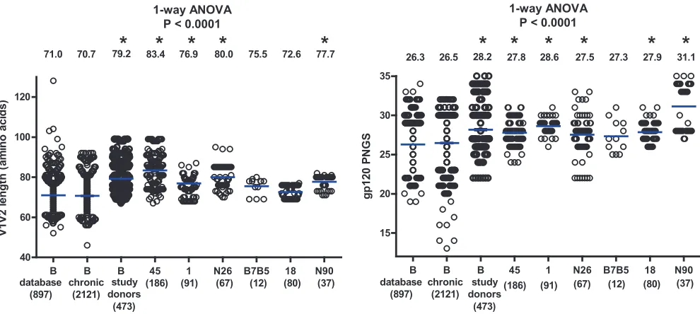
Analysis of gp120 N-linked glycosylation sites and V1V2
lengths.
During the course of infection, N-linked glycosylation
sites are often added to the variable regions of gp120, sometimes as
a consequence of length additions in V1V2 (6, 7, 9, 10, 60). In
some cases, these changes in Env were paired with increased viral
resistance to autologous or heterologous antibody neutralization
(6, 10, 60, 72, 74, 78). Therefore, increased numbers of N-linked
glycosylation sites and increased V1V2 lengths have been
sug-gested as part of the viral mechanisms for immune evasion. The
V1V2 length and the number of PNGS were also determined for
all of the 473 Env sequences from the six study donors (Fig. 2). For
comparison, we used a set of 897 subtype B sequences in the HIV
database (B_database, described in Materials and Methods).
Be-cause this set contained sequences from donors with unknown
infection times and included sequences from cultured viruses, we
added another set of 2,121 chronic subtype B infection sequences
(B_chronic, described in Materials and Methods) isolated by SGA
and used in a recent study at the LANL (21). Notably, the average
V1V2 length was comparable between the B_database and
B_chronic sequences; however, the average V1V2 length of Env
variants from four of the six broadly neutralizing study donors
was significantly greater than that in both the B_database and
B_chronic groups (
P
⬍
0.0001, one-way ANOVA, Fig. 2, left). The
average number of PNGS was also comparable between the
B_da-tabase and B_chronic sequences; however, the average number of
PNGS among Env variants from five of the six study donors was
significantly higher than in both the B_database and B_chronic
groups (
P
⬍
0.0001, one-way ANOVA, Fig. 2, right). Because an
extended V1V2 region with an increased number of glycosylation
sites in gp120 has been described as a characteristic of Env
se-quences from chronically infected individuals (9, 60), our data
suggest that donors with broadly reactive serum neutralizing
an-tibodies are further enriched for longer V1V2 regions with more
gp120 N-linked glycans.
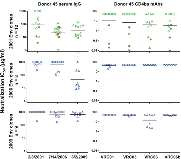
Neutralization sensitivity of Env variants from the VRC01
donor.
Representative
env
genes from the VRC01 donor were
cloned and expressed as Env pseudoviruses to test for
neutraliza-tion sensitivity. We first examined the concurrent serum IgG
neu-tralization of Env variants amplified from 2001, 2006, and 2009
plasma samples (Fig. 3, left panels; Table 2). Most of the 2001 Env
clones were only weakly neutralized by the concurrent serum IgG
(IC
50,
⬎
100
g/ml) but were potently neutralized by serum IgGs
from 2006 and 2009. Similarly, the 2006 Env clones were poorly
neutralized by the concurrent and earlier 2001 serum IgG but were
potently neutralized by the 2009 serum IgG. Thus, even in a donor
with a broadly reactive serum neutralizing antibody such as
VRC01, there is ongoing autologous virus escape from serum
neu-tralizing antibodies, similar to the pattern described for more
strain-specific autologous neutralizing antibodies (25, 48, 57,
58, 72).
We then examined the neutralization sensitivity of donor 45
Env variants to the CD4bs MAbs isolated from this donor
(autol-ogous MAbs) (Fig. 3, right panels; Table 2). Almost all of the
plasma-derived Env variants from each time point were highly
resistant to VRC01, with only two 2006 clones (45_06A3 and
45_06C7) showing moderate sensitivity (IC
50s of
⬎
10
g/ml).
[image:4.585.39.549.79.273.2]Three archival Env clones, 45_01dG5, 45_01dH1, and 45_01dH5,
derived from proviral DNA of a 2001 PBMC sample and chosen
based on phylogenetic analysis (Fig. 1B) were fully sensitive to
VRC01. These three sequences were not found in the tested donor
plasma by our method of SGA and DNA Sanger sequencing. One
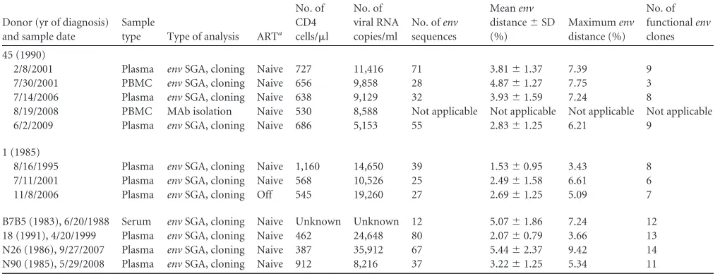
TABLE 1Characteristics of the donor samples included in this studyDonor (yr of diagnosis) and sample date
Sample
type Type of analysis ARTa
No. of CD4 cells/l
No. of viral RNA copies/ml
No. ofenv
sequences
Meanenv
distance⫾SD (%)
Maximumenv
distance (%)
No. of functionalenv
clones
45 (1990)
2/8/2001 Plasma envSGA, cloning Naive 727 11,416 71 3.81⫾1.37 7.39 9
7/30/2001 PBMC envSGA, cloning Naive 656 9,858 28 4.87⫾1.27 7.75 3
7/14/2006 Plasma envSGA, cloning Naive 638 9,129 32 3.93⫾1.59 7.24 8
8/19/2008 PBMC MAb isolation Naive 530 8,588 Not applicable Not applicable Not applicable Not applicable
6/2/2009 Plasma envSGA, cloning Naive 686 5,153 55 2.83⫾1.25 6.21 9
1 (1985)
8/16/1995 Plasma envSGA, cloning Naive 1,160 14,650 39 1.53⫾0.95 3.43 8
7/11/2001 Plasma envSGA, cloning Naive 568 10,526 25 2.49⫾1.58 6.61 6
11/8/2006 Plasma envSGA, cloning Off 545 19,260 27 2.69⫾1.25 5.09 7
B7B5 (1983), 6/20/1988 Serum envSGA, cloning Naive Unknown Unknown 12 5.07⫾1.86 7.24 12
18 (1991), 4/20/1999 Plasma envSGA, cloning Naive 462 24,648 80 2.07⫾0.79 3.66 13
N26 (1986), 9/27/2007 Plasma envSGA, cloning Naive 387 35,912 67 5.44⫾2.37 9.42 14
N90 (1985), 5/29/2008 Plasma envSGA, cloning Naive 912 8,216 37 3.22⫾1.25 5.34 11
aAll donors were either ART naïve or off ART at the time that blood specimens were collected.
on November 7, 2019 by guest
http://jvi.asm.org/
on November 7, 2019 by guest
http://jvi.asm.org/
2001 Env clone (45_01A14) was fully sensitive to VRC03, but
other Env variants from all time points were resistant to VRC03.
In contrast, five of the nine 2001 Env clones were sensitive to
VRC06 or VRC06b, suggesting that these closely related MAbs
may have evolved after the 2001 time point. By 2006, all tested Env
sequences were resistant to VRC06 and VRC06b, with three 2009
sequences sensitive to VRC06. Because the 2009 serum IgG was
potent against the 2006 Env clones and this neutralization activity
was not recapitulated by the known donor MAbs, these data
sug-gest that there are unidentified antibodies in the 2009 serum that
potently neutralized the 2006 Env variants.
Neutralization sensitivity of Env variants from other donors
to VRC01.
In addition to donor 45, we cloned representative
env
sequences from five other subtype B-infected donors who
devel-oped broadly neutralizing antibodies (Table 1). We expressed
rep-resentative Env clones as Env pseudoviruses and examined the
viral neutralization sensitivity to VRC01 (Fig. 4A) and to VRC03,
VRC06, VRC06b, and b12 (Table 3). Prior serum mapping studies
indicated that donors 1 and B7B5 had a major fraction of
neutral-izing antibodies to the CD4bs, and we observed that 81% and 42%
of the Env clones, respectively, were resistant to VRC01. In
con-trast, all of the Env clones from neutralizing donor N90 were
VRC01 sensitive. Somewhat surprisingly, a substantial percentage
of the sequences from donor N26 (64%) and donor 18 (38%) were
also VRC01 resistant.
VRC01 escape Env variants are still sensitive to CD4-Ig
neu-tralization and require CD4 for efficient entry.
As previously
noted, the heavy chain of MAb VRC01 partially mimics the
bind-ing interaction of CD4 with viral gp120. The VRC01 bindbind-ing
epitope largely overlaps the binding surface of CD4 in the outer
domain of gp120; however, VRC01 makes fewer contacts than
CD4 in the inner domain and bridging sheet regions (80). We
therefore asked if mutations that confer VRC01 resistance would
compromise the CD4-gp120 interaction and lead to reduced
CD4-Ig sensitivity or to the evolution of Env clones that could
enter cells without CD4, as previous studies have reported rare
HIV-1 strains that are capable of CD4-independent entry (29, 35,
79). To test these possibilities, we first looked at the Env variants’
sensitivity to CD4-Ig neutralization (Tables 2 and 3; Fig. 4B).
De-spite high levels of Env resistance to VRC01 in five of the six study
donors, most of the donor Env variants were sensitive to CD4-Ig.
Comparison of the VRC01-senstive (
n
⫽
40) and
VRC01-resis-tant (
n
⫽
60) Env variants did not reveal any difference in CD4-Ig
sensitivity (
P
⫽
0.89, unpaired
t
test, Fig. 4C, left). In addition, the
VRC01 and CD4-Ig neutralization IC
50s did not reveal any
corre-lation (
P
⫽
0.97, Pearson test; Fig. 4C, right). To test if the
VRC01-FIG 1Phylogenetic trees of HIV-1 envelope sequences. (A) Neighbor-joining tree showing the phylogenetic clustering of Env protein sequences from six HIV-1-infected donors. A total of 473 gp160 sequences from the six subtype B-infected donors were aligned with the reference sequence HXB2. The tree was constructed based on sequence distance and unrooted and then rooted at HXB2 for visualization. The gp160 sequences were derived at a single time point for donors 18, N90, N26, and B7B5; at three time points for donor 1; and at four time points for donor 45. The donor identification is indicated at each donor’s major branch node. Representative sequences indicated by a dot were cloned and expressed. A predominant sequence from donor 1 is shown in light blue from the 1995 time point (indicated by a bracket); one such sequence was cloned as indicated with an identification number. (B) Maximum-likelihood tree of envelope sequences from donor 45. A total of 186 gp160 nucleotide sequences from four temporal samples were aligned with the reference sequence HXB2. The tree was constructed as unrooted and then rooted at HXB2 for visualization. The gp160 sequences are color coded as follows: red, 2001 provirus; green, 2001 plasma; blue, 2006 plasma; purple, 2009 plasma. The major branch colors follow the color of most of the sequences on the branch. Representative sequences indicated by a dot and an identification number were cloned and expressed. The horizontal branch scale is indicated for each tree.
FIG 2Comparisons of V1V2 lengths (left) and numbers of gp120 PNGS (right) among the subtype B sequences from the HIV-1 database (B_database), from chronic infection (B_chronic), and from the six study donors as one group or individually. The blue horizontal bars indicate the group means, and the actual mean values are indicated at the top of the columns. The number of sequences for each group or individual is indicated in parentheses below the group or donor identification. One-way ANOVAPvalues are indicated, with the asterisks indicating groups that are significantly different (P⬍0.05) from both the B_database and B_chronic groups in subsequent Dunnett’s multiple-comparison tests.
on November 7, 2019 by guest
http://jvi.asm.org/
[image:6.585.45.540.65.287.2]resistant Env variants can enter cells in a CD4-independent
man-ner, we made luciferase-containing Env pseudoviruses of all 29
donor 45
env
clones and examined their entry into CD4
⫺CCR5
⫹cell line Cf2th-synCCR5 (22) and its CD4
⫹CCR5
⫹counterpart
Cf2th-CD4.CCR5 (28, 49). All of the Env variants entered the
CD4
⫹cell line Cf2th-CD4.CCR5 efficiently but not the CD4
⫺cell
line Cf2th-synCCR5, indicating that these Env variants were able
to escape VRC01 neutralization yet retain efficient interaction
with the viral cellular receptor CD4. Data from nine
env
clones
from the 2009 plasma of donor 45 are shown in Fig. 5.
DISCUSSION
The CD4bs of gp120 is an attractive target for HIV-1 vaccine
de-sign because it is functionally conserved and contains epitopes of
potently neutralizing antibodies. The isolation of VRC01 and
sim-ilar CD4bs MAbs demonstrates that the immune system is capable
of generating antibodies to this region of gp120. To characterize
the circulating Env variants that coexist with VRC01 and to
un-derstand how the HIV-1
env
gene evolves under the selection
pres-sure of a broadly reactive antibody, we isolated Env variants from
the VRC01 donor and studied their neutralization sensitivities.
While numerous published studies have shown that autologous
plasma virus is often resistant to concurrent serum neutralization
(4, 19, 26, 40, 50, 54, 57, 58, 72), there had not previously been a
detailed assessment of the Env quasispecies in a donor with a
broadly reactive serum neutralizing antibody such as VRC01.
Using SGA followed by DNA Sanger sequencing, we found that
plasma-derived Env variants from donor 45 from three time
points, spanning from 2001 to 2009, displayed almost uniform
resistance to VRC01 neutralization, indicating a strong selection
pressure on the viral Env quasispecies by VRC01 and related
au-tologous CD4bs antibodies. While the Env SGA and Sanger
se-quencing approach has an inherent limitation in the overall depth
of the sequences identified, our data show that the vast majority of
the viruses circulating in plasma were VRC01 resistant. Donor 45
was first found to be HIV-1 infected in 1990; thus, the time points
studied here were in excess of 10 years after HIV-1 infection. The
VRC01 MAb was isolated from IgG
⫹memory B cells from a 2008
PBMC sample (Table 1), but since memory B cells may circulate
for a long time, it is not clear when VRC01 first developed. Our
finding that all nine plasma Env variants from 2001 were VRC01
resistant suggests that VRC01 arose prior to 2001. It was only
through the isolation of HIV-1
env
sequences from proviral DNA
that we were able to identify archival Env clones that are highly
sensitive to VRC01, with IC
50s of
⬍
1
g/ml. Thus, it seems
un-likely that donor 45 was infected with a VRC01-resistant virus but
rather likely that the Env variants evolved to escape from this
potently neutralizing antibody.
The neutralization sensitivity of some of the VRC01 escape
variants to MAbs VRC03, VRC06, and VRC06b suggests the
pos-sibility of continuous evolution of CD4bs neutralizing antibodies
in response to viral escape. VRC03 was isolated from the same
FIG 3Analysis of neutralization sensitivity of Env variants taken from donor 45 at three time points to autologous serum IgG (left panels) and autologous CD4bs MAbs VRC01, VRC03, VRC06, and VRC06b (right panels). The horizontal bars indicate the group geometric means. The three symbols highlighted in red in the2001 Env plots indicate the archival Env variants derived from proviral DNA.
on November 7, 2019 by guest
http://jvi.asm.org/
[image:7.585.112.473.63.381.2]PBMC samples as VRC01, and its liganded structure has been
solved (75, 77). While the VRC03 heavy chain derives from the
same IGHV1-2*02 allele as VRC01, it has a different light chain,
indicating that it arose from a different B-cell clone. The crystal
structure of VRC03 in complex with a gp120 core revealed that the
antibody forms contacts with the bridging sheet between the inner
and outer domains of gp120, which is part of the coreceptor
bind-ing site (77). Therefore, we speculate that the donor immune
sys-tem responded to VRC01 escape variants by generating antibody
specificities extending from the VRC01 epitope toward the
core-ceptor binding region and resulting in efficient neutralization of
some VRC01 escape variants. MAbs VRC06 and VRC06b are
clonal relatives of VRC03 and will be described in detail in a
sep-arate report (Yuxing Li et al., unpublished data). The sensitivity of
some 2001 viral Env clones (which are all resistant to VRC01) to
VRC03, VRC06, and VRC06b suggests that these MAbs might
have evolved later than VRC01. Indeed, suboptimal 454
pyrose-quencing of the IgG
⫹memory B-cell heavy-chain transcripts
from the 2008 PBMC sample identified a population of antibody
sequences with
⬎
90% identity to VRC03 but not VRC01-related
sequences (77), suggesting the presence of greater numbers of
memory B cells for VRC03 than VRC01 in 2008. We are currently
optimizing PCR and sequencing conditions to analyze donor 45
longitudinal samples to determine the dynamics of these
neutral-izing antibody clones.
[image:8.585.39.544.76.521.2]Although MAbs VRC03, VRC06, and VRC06b were able to
neutralize VRC01 escape Env variants in donor 45, the
neutraliza-tion breadth of these MAbs against heterologous viruses (
⬍
60%)
was less than that of VRC01 (90%) (75). Therefore, the
develop-ment or maintenance of these MAbs after VRC01 in donor 45 was
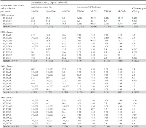
TABLE 2Neutralization IC50s of autologous serum IgG, autologous CD4bs MAbs, and CD4 surrogate protein CD4-Ig for donor 45 Env variantsenvisolation time, source, andenvclone or parameter
Neutralization IC50(g/ml) or breadth
Autologous serum IgG Autologous CD4bs MAbs
CD4 surrogate CD4-Ig
2/8/2001 7/14/2006 6/2/2009 VRC01 VRC03 VRC06 VRC06b
2001, provirus
45_01dG5 3.8 10.9 9.5 0.020 0.010 0.019 0.010 0.256
45_01dH1 58.6 61.4 77.0 3.6 2.2 6.9 3.9 0.870
45_01dH5 11.6 18.4 16.8 0.049 0.028 0.149 0.207 ⬎50
Breadtha(n⫽3) 3 (100) 3 (100) 3 (100) 3 (100) 3 (100) 3 (100) 3 (100) 2 (67)
2001, plasma
45_01A8 310 47.6 13.0 ⬎50 ⬎50 ⬎50 ⬎50 1.3
45_01A10 ⬎1,000 26.2 12.3 ⬎50 ⬎50 0.248 0.925 3.6
45_01A14 121 39.8 21.0 ⬎50 0.453 ⬎50 1.5 1.7
45_01A21 ⬎1,000 22.9 65.6 ⬎50 ⬎50 ⬎50 ⬎50 4.4
45_01B14 ⬎1,000 14.1 68.2 ⬎50 ⬎50 ⬎50 ⬎50 5.3
45_01E8 11.0 0.823 15.9 ⬎50 ⬎50 9.4 ⬎50 0.265
45_01E11 201 129 146 ⬎50 ⬎50 2.3 1.8 0.698
45_01E17 109 33.0 83.2 ⬎50 ⬎50 3.6 3.3 1.8
45_01F4 47.3 54.2 83.2 ⬎50 ⬎50 7.8 10.7 0.533
Breadth (n⫽9) 6 (67) 9 (100) 9 (100) 0 (0) 1 (11) 5 (56) 5 (56) 9 (100)
2006, plasma
45_06A1 689 ⬎1,000 17.2 ⬎50 ⬎50 ⬎50 ⬎50 3.4
45_06A2 ⬎1,000 ⬎1,000 9.0 ⬎50 ⬎50 ⬎50 ⬎50 2.8
45_06A3 ⬎1,000 ⬎1,000 9.6 17.5 ⬎50 ⬎50 ⬎50 2.5
45_06A4 542 109 225 ⬎50 ⬎50 ⬎50 ⬎50 6.0
45_06A8 254 ⬎1,000 22.4 ⬎50 ⬎50 ⬎50 ⬎50 0.614
45_06A10 640 ⬎1,000 14.5 ⬎50 ⬎50 ⬎50 ⬎50 5.3
45_06C7 ⬎1,000 ⬎1,000 ⬎1,000 31.2 ⬎50 ⬎50 ⬎50 3.3
45_06C8 ⬎1,000 165 205 ⬎50 ⬎50 ⬎50 ⬎50 1.4
Breadth (n⫽8) 4 (50) 2 (25) 7 (88) 2 (25) 0 (0) 0 (0) 0 (0) 8 (100)
2009, plasma
45_09A5 ⬎1,000 462 879 ⬎50 ⬎50 ⬎50 ⬎50 ⬎50
45_09A6 ⬎1,000 447 905 ⬎50 ⬎50 5.5 38.4 ⬎50
45_09A10 ⬎1,000 ⬎1,000 ⬎1,000 ⬎50 ⬎50 ⬎50 ⬎50 5.2
45_09B3 ⬎1,000 ⬎1,000 456 ⬎50 35.7 3.2 ⬎50 4.0
45_09C5 ⬎1,000 ⬎1,000 564 ⬎50 ⬎50 ⬎50 ⬎50 2.7
45_09C8 ⬎1,000 ⬎1,000 743 ⬎50 ⬎50 ⬎50 ⬎50 ⬎50
45_09C20 211 178 209 ⬎50 ⬎50 ⬎50 ⬎50 0.809
45_09C26 ⬎1,000 ⬎1,000 ⬎1,000 ⬎50 ⬎50 ⬎50 ⬎50 5.5
45_09TA1 ⬎1,000 785 560 ⬎50 ⬎50 2.3 ⬎50 9.1
Breadth (n⫽90) 1 (11) 4 (44) 7 (78) 0 (0) 1 (11) 3 (33) 1 (11) 6 (67)
a
Breadth is shown as the number (percentage) of Env variants sensitive to the corresponding serum IgG, MAb, or CD4-Ig.
on November 7, 2019 by guest
http://jvi.asm.org/
specific to the relevant autologous viruses but not necessarily
as-sociated with better neutralization breadth of “irrelevant”
heter-ologous viruses circulating globally. This highlights the
complex-ity of underlying factors that influence the broadening of antibody
neutralization (16, 23). Although most of the donor 45 Env
vari-ants from 2001 plasma was neutralized by VRC03, VRC06, and
VRC06b, Env variants from 2006 plasma were resistant,
suggest-ing that the virus was able to escape these newly developed
anti-body variants. Note that the 2006 Env variants remained highly
sensitive to the 2009 serum IgG, indicating the presence of
un-identified antibody specificities, including the possibility of those
outside the CD4bs, in this serum that likely arose in response to
the escaped variants.
We also analyzed Env variants from five additional subtype
B-infected individuals who developed strong broadly neutralizing
antibody responses. We previously reported that the serum
sam-ples of donors 1 and B7B5 contained a major fraction of
CD4bs-directed neutralizing antibodies (43, 45); thus, we hypothesized
that Env sequences from these two donors would contain some
VRC01-resistant clones. However, we were surprised to find a
majority of Env clones in donor N26 also resistant to VRC01, as
our mapping did not definitively indicate CD4bs neutralizing
an-tibodies in this donor. This was in contrast to donor N90, whose
Env isolates were fully sensitive to VRC01 neutralization,
suggest-ing a lack of VRC01-like antibody pressure in this donor. Overall,
the rather high level of VRC01-resistant clones in four of five
donors (other than donor 45) suggests that the CD4bs-directed
neutralizing antibodies similar to VRC01 may not be uncommon
in donors whose serum samples are broadly neutralizing.
Lastly, we observed that viral variants fully resistant to VRC01
FIG 4Comparisons of VRC01 (A) and CD4-Ig (B) neutralization sensitivities of Env variants derived from the six study donors indicated on thexaxis. The blue horizontal dashed line indicates an IC50of 50g/ml, which was used as the cutoff for neutralization sensitivity. The values below the donor designations are thenumbers and percentages of Env variants resistant to VRC01 or CD4-Ig neutralization. (C) Plot of log-transformed CD4-Ig neutralization IC50s for
VRC01-sensitive (n⫽40) and VRC01-resistant (n⫽60) Env variants (left). The mean of each group is indicated by a blue horizontal bar. The log-transformed VRC01 and CD4-Ig neutralization IC50s are plotted for each individual Env variant (right,n⫽100). ThePvalues and the corresponding statistical analyses are indicated.
on November 7, 2019 by guest
http://jvi.asm.org/
[image:9.585.135.454.61.482.2]TABLE 3Neutralization IC50s of heterologous CD4bs MAbs and CD4 surrogate protein CD4-Ig for Env variants from five donors
Donor, yr, andenvclone or parameter
Neutralization IC50or breadth
VRC01 VRC03 VRC06 VRC06b b12 CD4-Ig
1 1995
1_95A21 ⬎50 ⬎50 ⬎50 ⬎50 ⬎50 ⬎50
1_95C1 ⬎50 ⬎50 ⬎50 ⬎50 ⬎50 13.6
1_95C5 0.017 0.020 3.3 0.016 0.023 ⬎50
1_95C10 ⬎50 ⬎50 ⬎50 ⬎50 0.214 3.7
1_95TC1 ⬎50 ⬎50 ⬎50 ⬎50 ⬎50 ⬎50
1_95TC6 ⬎50 ⬎50 ⬎50 ⬎50 ⬎50 ⬎50
1_95TC13 ⬎50 ⬎50 ⬎50 ⬎50 0.773 1.4
1_95TC14 14.6 5.0 26.2 7.1 ⬎50 28.0
2001
1_01A10 ⬎50 ⬎50 ⬎50 ⬎50 ⬎50 38.6
1_01A19 ⬎50 ⬎50 ⬎50 ⬎50 ⬎50 1.9
1_01A20 11.5 12.0 ⬎50 ⬎50 ⬎50 2.6
1_01B3 ⬎50 ⬎50 ⬎50 ⬎50 0.920 1.4
1_01B5 0.041 0.111 ⬎50 0.128 0.032 2.6
1_01TB5 ⬎50 ⬎50 ⬎50 ⬎50 ⬎50 3.2
2006
1_06B1 ⬎50 ⬎50 ⬎50 ⬎50 6.1 0.995
1_06B3 ⬎50 0.841 13.1 2.5 ⬎50 19.3
1_06B7 ⬎50 ⬎50 ⬎50 ⬎50 ⬎50 22.8
1_06B9 ⬎50 ⬎50 ⬎50 ⬎50 ⬎50 0.218
1_06C3 ⬎50 ⬎50 ⬎50 ⬎50 0.289 7.8
1_06C5 ⬎50 ⬎50 ⬎50 ⬎50 50.0 2.8
1_06D29 ⬎50 ⬎50 ⬎50 ⬎50 6.3 1.7
Breadtha(n⫽21) 4 (19) 5 (24) 3 (16) 4 (19) 9 (47) 17 (81)
N26, 2007
N26_07A10 ⬎50 ⬎50 ⬎50 ⬎50 ⬎50 9.5
N26_07A14 ⬎50 ⬎50 ⬎50 ⬎50 ⬎50 0.317
N26_07A16 ⬎50 ⬎50 ⬎50 ⬎50 ⬎50 2.8
N26_07A21 1.6 1.7 ⬎50 10.9 0.640 2.9
N26_07A22 ⬎50 ⬎50 ⬎50 ⬎50 ⬎50 ⬎50
N26_07A38 0.591 ⬎50 ⬎50 ⬎50 1.1 ⬎50
N26_07B9 ⬎50 ⬎50 ⬎50 ⬎50 7.2 3.9
N26_07B10 ⬎50 ⬎50 ⬎50 ⬎50 7.9 ⬎50
N26_07B18 19.0 ⬎50 ⬎50 ⬎50 ⬎50 0.755
N26_07B22 ⬎50 ⬎50 ⬎50 ⬎50 ⬎50 2.9
N26_07B36 ⬎50 ⬎50 ⬎50 ⬎50 ⬎50 3.1
N26_07B41 ⬎50 ⬎50 ⬎50 ⬎50 ⬎50 12.9
N26_07TC39 19.3 ⬎50 ⬎50 ⬎50 ⬎50 44.4
N26_07TC40 0.089 ⬎50 ⬎50 ⬎50 10.4 ⬎50
Breadth (n⫽14) 5 (36) 1 (7) 0 (0) 1 (7) 5 (36) 10 (71)
B7B5, 1988
B7B5_88A1 17.0 38.7 ⬎50 ⬎50 ⬎50 1.5
B7B5_88A2 2.3 6.9 ⬎50 15.5 0.452 25.3
B7B5_88A3 3.1 12.6 ⬎50 ⬎50 0.426 13.7
B7B5_88A4 ⬎50 ⬎50 ⬎50 ⬎50 0.731 0.119
B7B5_88A5 ⬎50 20.0 ⬎50 ⬎50 ⬎50 6.9
B7B5_88A6 3.5 7.5 ⬎50 19.1 0.141 0.970
B7B5_88A7 ⬎50 ⬎50 ⬎50 ⬎50 36.1 9.6
B7B5_88A10 25.9 ⬎50 ⬎50 ⬎50 ⬎50 7.3
B7B5_88TB1 ⬎50 ⬎50 ⬎50 ⬎50 ⬎50 11.4
B7B5_88TB2 9.6 2.2 5.3 1.6 ⬎50 8.8
B7B5_88TB3 11.4 2.7 8.7 1.6 4.4 6.9
(Continued on following page)
on November 7, 2019 by guest
http://jvi.asm.org/
retained their sensitivity to CD4-Ig and required CD4 for entry.
These results suggest that VRC01 escape mutations do not
sub-stantially impair the gp120 interaction with CD4, at least not
enough to have a major impact on viral entry. Our prior studies of
VRC01-resistant viruses demonstrated that mutations in the
D-loop and V5 and regions of gp120 were often responsible for
VRC01 resistance and that some of these mutations were not
[image:11.585.39.546.80.433.2]con-tact sites for CD4 (44, 80). It remains possible that initial escape
from VRC01 is associated with some loss of binding to CD4 and
that compensatory mutations restore viral entry and replication.
More systematic studies of viral fitness and the affinity of
interac-tion with CD4 are in progress. Donor 45 is a slow progressor, as
are several other donors from whom broadly neutralizing CD4bs
MAbs have been isolated (63, 77). More systematic and unbiased
TABLE 3(Continued)Donor, yr, andenvclone or parameter
Neutralization IC50or breadth
VRC01 VRC03 VRC06 VRC06b b12 CD4-Ig
B7B5_88TB6 ⬎50 ⬎50 ⬎50 ⬎50 ⬎50 2.4
Breadth (n⫽12) 7 (58) 7 (58) 2 (17) 4 (33) 6 (50) 12 (100)
18, 1999
18_99A27 0.661 ⬎50 ⬎50 ⬎50 ⬎50 ⬎50
18_99A34 3.5 1.9 ⬎50 ⬎50 ⬎50 ⬎50
18_99A38 ⬎50 ⬎50 ⬎50 ⬎50 ⬎50 ⬎50
18_99B1 4.8 ⬎50 ⬎50 ⬎50 9.6 13.5
18_99B15 1.9 1.5 ⬎50 ⬎50 ⬎50 ⬎50
18_99B19 0.855 ⬎50 ⬎50 ⬎50 ⬎50 43.1
18_99B27 ⬎50 ⬎50 ⬎50 ⬎50 ⬎50 10.2
18_99B39 ⬎50 ⬎50 ⬎50 ⬎50 ⬎50 28.8
18_99B46 1.6 ⬎50 ⬎50 ⬎50 ⬎50 ⬎50
18_99B48 ⬎50 ⬎50 ⬎50 ⬎50 ⬎50 3.1
18_99B51 1.2 ⬎50 ⬎50 ⬎50 ⬎50 ⬎50
18_99TA3 ⬎50 ⬎50 ⬎50 ⬎50 ⬎50 7.2
18_99TB13 1.1 ⬎50 ⬎50 ⬎50 ⬎50 ⬎50
Breadth (n⫽13) 8 (62) 2 (15) 0 (0) 0 (0) 1 (8) 6 (46)
N90, 2008
N90_08A13 2.5 0.364 19.4 0.349 5.8 0.618
N90_08A15 1.0 0.311 8.1 0.333 1.5 0.679
N90_08A16 1.3 0.354 11.0 0.462 3.5 0.545
N90_08A19 0.372 0.128 6.2 0.354 ⬎50 7.8
N90_08A25 1.3 0.350 5.6 0.413 ⬎50 0.752
N90_08B2 0.703 0.338 3.9 0.361 5.7 0.394
N90_08B4 2.9 0.236 ⬎50 0.380 2.5 2.6
N90_08B6 0.644 0.293 5.3 0.434 3.0 0.255
N90_08B11 0.143 0.055 4.7 0.063 1.6 0.036
N90_08B16 0.328 0.196 7.0 0.259 2.5 0.472
N90_08B17 1.7 0.651 14.5 0.488 3.0 1.7
Breadth (n⫽11) 11 (100) 11 (100) 10 (91) 11 (100) 9 (82) 11 (100)
a
Breadth is shown as the number (percentage) of Env variants sensitive to the corresponding corresponding MAb or CD4-Ig or CD4-Ig.
FIG 5Entry of nine donor 45 Env variants derived from the 2009 plasma into CD4⫹cell line Cf2Th-CD4.CCR5 (left) and CD4⫺cell line Cf2Th-synCCR5 (right).
on November 7, 2019 by guest
http://jvi.asm.org/
[image:11.585.92.497.556.700.2]sampling is required to know if CD4bs neutralizing MAbs are
more commonly found in donors with slow progression.
In summary, our data show that neutralizing antibodies to the
conserved CD4bs exert selection pressure on HIV-1 Env and that
the viruses evolve to escape from such neutralization. The B-cell
response to the CD4bs also evolves by generating antibodies that
neutralize viral escape mutants. Hence, even for this functionally
conserved region of gp120, there is ongoing viral evolution
matched by antibody evolution. The facts that escape from
VRC01-like CD4bs antibodies appears to occur commonly within
a donor but that the large majority of heterologous viral strains are
sensitive to VRC01 suggest some constraint on resistance to such
CD4bs antibodies, although whether there is any measurable
fit-ness cost to VRC01 escape remains to be determined. Finally, the
study of Env species in donors with broadly neutralizing
antibod-ies may reveal clues about the viral antigenic stimulus that led to
the development of this antibody. Such conclusions are difficult in
the case of donor 45, who had been infected for more than 10 years
prior to the time when the samples we studied were taken. Similar
studies of known seroconverter donors with longitudinal
sam-pling could reveal the relationship between the early circulating
Env sequences and the early B-cell clones that ultimately develop
into mature broadly neutralizing antibodies.
ACKNOWLEDGMENTS
We thank Kim-Truc Pham for technical assistance with SGA ofenvfrom donor 1 plasma samples.
Support for this work was provided by the Intramural Research Pro-gram of the Vaccine Research Center, NIAID, NIH. G.M.S. was supported by grants from the NIH (AI067854) and the Bill & Melinda Gates Foun-dation Grand Challenges Program (37874).
REFERENCES
1.Albert J, et al.1990. Rapid development of isolate-specific neutralizing antibodies after primary HIV-1 infection and consequent emergence of virus variants which resist neutralization by autologous sera. AIDS4:107– 112.
2.Bonsignori M, et al.2011. Analysis of a clonal lineage of HIV-1 envelope V2/V3 conformational epitope-specific broadly neutralizing antibodies and their inferred unmutated common ancestors. J. Virol. 85:9998 – 10009.
3.Bosch KA, Rainwater S, Jaoko W, Overbaugh J.2010. Temporal analysis of HIV envelope sequence evolution and antibody escape in a subtype A-infected individual with a broad neutralizing antibody response. Virol-ogy398:115–124.
4.Bunnik EM, Pisas L, van Nuenen AC, Schuitemaker H.2008. Autolo-gous neutralizing humoral immunity and evolution of the viral envelope in the course of subtype B human immunodeficiency virus type 1 infec-tion. J. Virol.82:7932–7941.
5.Burton DR, et al.1994. Efficient neutralization of primary isolates of HIV-1 by a recombinant human monoclonal antibody. Science266: 1024 –1027.
6.Cheng-Mayer C, Brown A, Harouse J, Luciw PA, Mayer AJ. 1999. Selection for neutralization resistance of the simian/human immunodefi-ciency virus SHIVSF33A variant in vivo by virtue of sequence changes in the extracellular envelope glycoprotein that modify N-linked glycosyla-tion. J. Virol.73:5294 –5300.
7.Chohan B, et al.2005. Selection for human immunodeficiency virus type 1 envelope glycosylation variants with shorter V1-V2 loop sequences oc-curs during transmission of certain genetic subtypes and may impact viral RNA levels. J. Virol.79:6528 – 6531.
8.Corti D, et al.2010. Analysis of memory B cell responses and isolation of novel monoclonal antibodies with neutralizing breadth from HIV-1-infected individuals. PLoS One5:e8805.
9.Curlin ME, et al.2010. HIV-1 envelope subregion length variation during disease progression. PLoS Pathog.6:e1001228.
10. Derdeyn CA, et al.2004. Envelope-constrained neutralization-sensitive HIV-1 after heterosexual transmission. Science303:2019 –2022. 11. Dhillon AK, et al.2007. Dissecting the neutralizing antibody specificities
of broadly neutralizing sera from human immunodeficiency virus type 1-infected donors. J. Virol.81:6548 – 6562.
12. Doria-Rose NA, et al.2010. Breadth of human immunodeficiency virus-specific neutralizing activity in sera: clustering analysis and association with clinical variables. J. Virol.84:1631–1636.
13. Edgar RC.2004. MUSCLE: a multiple sequence alignment method with reduced time and space complexity. BMC Bioinformatics5:113. 14. Edgar RC.2004. MUSCLE: multiple sequence alignment with high
accu-racy and high throughput. Nucleic Acids Res.32:1792–1797.
15. Etherington GJ, Dicks J, Roberts IN.2005. Recombination analysis tool (RAT): a program for the high-throughput detection of recombination. Bioinformatics21:278 –281.
16. Euler Z, et al.2012. Longitudinal analysis of early HIV-1-specific neu-tralizing activity in an elite neutralizer and in five patients who developed cross-reactive neutralizing activity. J. Virol.86:2045–2055.
17. Felsenstein J, Churchill GA.1996. A hidden Markov model approach to variation among sites in rate of evolution. Mol. Biol. Evol.13:93–104. 18. Flynn NM, et al.2005. Placebo-controlled phase 3 trial of a recombinant
glycoprotein 120 vaccine to prevent HIV-1 infection. J. Infect. Dis.191: 654 – 665.
19. Frost SD, et al.2005. Neutralizing antibody responses drive the evolution of human immunodeficiency virus type 1 envelope during recent HIV infection. Proc. Natl. Acad. Sci. U. S. A.102:18514 –18519.
20. Gilbert PB, et al.2005. Correlation between immunologic responses to a recombinant glycoprotein 120 vaccine and incidence of HIV-1 infection in a phase 3 HIV-1 preventive vaccine trial. J. Infect. Dis.191:666 – 677. 21. Gnanakaran S, et al.2011. Recurrent signature patterns in HIV-1 B clade
envelope glycoproteins associated with either early or chronic infections. PLoS Pathog.7:e1002209.
22. Gorry PR, et al.2002. Increased CCR5 affinity and reduced CCR5/CD4 dependence of a neurovirulent primary human immunodeficiency virus type 1 isolate. J. Virol.76:6277– 6292.
23. Gray ES, et al.2011. The neutralization breadth of HIV-1 develops in-crementally over four years and is associated with CD4⫹T cell decline and high viral load during acute infection. J. Virol.85:4828 – 4840.
24. Gray ES, et al.2009. Broad neutralization of human immunodeficiency virus type 1 mediated by plasma antibodies against the gp41 membrane proximal external region. J. Virol.83:11265–11274.
25. Gray ES, et al.2011. Isolation of a monoclonal antibody that targets the alpha-2 helix of gp120 and represents the initial autologous neutralizing-antibody response in an HIV-1 subtype C-infected individual. J. Virol. 85:7719 –7729.
26. Gray ES, et al.2007. Neutralizing antibody responses in acute human immunodeficiency virus type 1 subtype C infection. J. Virol.81:6187– 6196.
27. Gray ES, et al.2009. Antibody specificities associated with neutralization breadth in plasma from human immunodeficiency virus type 1 subtype C-infected blood donors. J. Virol.83:8925– 8937.
28. Haim H, et al.2009. Soluble CD4 and CD4-mimetic compounds inhibit HIV-1 infection by induction of a short-lived activated state. PLoS Pathog. 5:e1000360.
29. Haim H, et al.2011. Contribution of intrinsic reactivity of the HIV-1 envelope glycoproteins to CD4-independent infection and global inhibi-tor sensitivity. PLoS Pathog.7:e1002101.
30. Huson DH, et al.2007. Dendroscope: an interactive viewer for large phylogenetic trees. BMC Bioinformatics8:460.
31. Johnson VA, Byington RE.1990. Infectivity assay (virus yield assay), p 71–76.InAldovani A, Walker BD (ed), Techniques in HIV research. Stockton Press, New York, NY.
32. Jones DT, Taylor WR, Thornton JM.1992. The rapid generation of mutation data matrices from protein sequences. Comput. Appl. Biosci. 8:275–282.
33. Keele BF, et al.2008. Identification and characterization of transmitted and early founder virus envelopes in primary HIV-1 infection. Proc. Natl. Acad. Sci. U. S. A.105:7552–7557.
34. Kishino H, Hasegawa M.1989. Evaluation of the maximum likelihood estimate of the evolutionary tree topologies from DNA sequence data, and the branching order in hominoidea. J. Mol. Evol.29:170 –179.
35. Kolchinsky P, Kiprilov E, Bartley P, Rubinstein R, Sodroski J.2001. Loss of a single N-linked glycan allows CD4-independent human
on November 7, 2019 by guest
http://jvi.asm.org/
nodeficiency virus type 1 infection by altering the position of the gp120 V1/V2 variable loops. J. Virol.75:3435–3443.
36. Korber B, et al.2000. Timing the ancestor of the HIV-1 pandemic strains. Science288:1789 –1796.
37. Kuhner MK, Felsenstein J.1994. A simulation comparison of phylogeny algorithms under equal and unequal evolutionary rates. Mol. Biol. Evol. 11:459 – 468.
38. Kwong PD, et al.1998. Structure of an HIV gp120 envelope glycoprotein in complex with the CD4 receptor and a neutralizing human antibody. Nature393:648 – 659.
39. Larkin MA, et al.2007. Clustal W and Clustal X version 2.0. Bioinfor-matics23:2947–2948.
40. Li B, et al.2006. Evidence for potent autologous neutralizing antibody titers and compact envelopes in early infection with subtype C human immunodeficiency virus type 1. J. Virol.80:5211–5218.
41. Li H, et al.2010. High multiplicity infection by HIV-1 in men who have sex with men. PLoS Pathog.6:e1000890.
42. Li M, et al.2005. Human immunodeficiency virus type 1 env clones from acute and early subtype B infections for standardized assessments of vac-cine-elicited neutralizing antibodies. J. Virol.79:10108 –10125. 43. Li Y, et al.2007. Broad HIV-1 neutralization mediated by CD4-binding
site antibodies. Nat. Med.13:1032–1034.
44. Li Y, et al.2011. Mechanism of neutralization by the broadly neutralizing HIV-1 monoclonal antibody VRC01. J. Virol.85:8954 – 8967.
45. Li Y, et al.2009. Analysis of neutralization specificities in polyclonal sera derived from human immunodeficiency virus type 1-infected individuals. J. Virol.83:1045–1059.
46. Long EM, et al.2000. Gender differences in HIV-1 diversity at time of infection. Nat. Med.6:71–75.
47. Long EM, Rainwater SM, Lavreys L, Mandaliya K, Overbaugh J.2002. HIV type 1 variants transmitted to women in Kenya require the CCR5 coreceptor for entry, regardless of the genetic complexity of the infecting virus. AIDS Res. Hum. Retroviruses18:567–576.
48. Lynch RM, et al.2011. The B cell response is redundant and highly focused on V1V2 during early subtype C infection in a Zambian serocon-verter. J. Virol.85:905–915.
49. Madani N, et al.2008. Small-molecule CD4 mimics interact with a highly conserved pocket on HIV-1 gp120. Structure16:1689 –1701.
50. Mahalanabis M, et al.2009. Continuous viral escape and selection by autologous neutralizing antibodies in drug-naive human immunodefi-ciency virus controllers. J. Virol.83:662– 672.
51. Montefiori DC, et al.1991. Homotypic antibody responses to fresh clin-ical isolates of human immunodeficiency virus. Virology182:635– 643. 52. Moore PL, et al.2008. The c3-v4 region is a major target of autologous
neutralizing antibodies in human immunodeficiency virus type 1 subtype C infection. J. Virol.82:1860 –1869.
53. Moore PL, et al.2011. Potent and broad neutralization of HIV-1 subtype C by plasma antibodies targeting a quaternary epitope including residues in the V2 loop. J. Virol.85:3128 –3141.
54. Moore PL, et al.2009. Limited neutralizing antibody specificities drive neutralization escape in early HIV-1 subtype C infection. PLoS Pathog. 5:e1000598.
55. Muster T, et al.1993. A conserved neutralizing epitope on gp41 of human immunodeficiency virus type 1. J. Virol.67:6642– 6647.
56. Posada D, Crandall KA.1998. MODELTEST: testing the model of DNA substitution. Bioinformatics14:817– 818.
57. Richman DD, Wrin T, Little SJ, Petropoulos CJ.2003. Rapid evolution of the neutralizing antibody response to HIV type 1 infection. Proc. Natl. Acad. Sci. U. S. A.100:4144 – 4149.
58. Rong R, et al.2009. Escape from autologous neutralizing antibodies in acute/early subtype C HIV-1 infection requires multiple pathways. PLoS Pathog.5:e1000594.
59. Rose PP, Korber BT.2000. Detecting hypermutations in viral sequences with an emphasis on G¡A hypermutation. Bioinformatics16:400 – 401.
60. Sagar M, Wu X, Lee S, Overbaugh J.2006. Human immunodeficiency virus type 1 V1-V2 envelope loop sequences expand and add glycosylation sites over the course of infection, and these modifications affect antibody neutralization sensitivity. J. Virol.80:9586 –9598.
61. Salazar-Gonzalez JF, et al.2008. Deciphering human immunodeficiency virus type 1 transmission and early envelope diversification by single-genome amplification and sequencing. J. Virol.82:3952–3970.
62. Sather DN, et al.2009. Factors associated with the development of cross-reactive neutralizing antibodies during human immunodeficiency virus type 1 infection. J. Virol.83:757–769.
63. Scheid JF, et al.2011. Sequence and structural convergence of broad and potent HIV antibodies that mimic CD4 binding. Science333:1633–1637. 64. Seaman MS, et al.2010. Tiered categorization of a diverse panel of HIV-1 Env pseudoviruses for neutralizing antibody assessment. J. Virol.84: 1439 –1452.
65. Shankarappa R, et al.1999. Consistent viral evolutionary changes asso-ciated with the progression of human immunodeficiency virus type 1 in-fection. J. Virol.73:10489 –10502.
66. Simek MD, et al.2009. Human immunodeficiency virus type 1 elite neutralizers: individuals with broad and potent neutralizing activity iden-tified by using a high-throughput neutralization assay together with an analytical selection algorithm. J. Virol.83:7337–7348.
67. Stamatatos L, Morris L, Burton DR, Mascola JR. 2009. Neutralizing antibodies generated during natural HIV-1 infection: good news for an HIV-1 vaccine? Nat. Med.15:866 – 870.
68. Trkola A, et al.1996. Human monoclonal antibody 2G12 defines a dis-tinctive neutralization epitope on the gp120 glycoprotein of human im-munodeficiency virus type 1. J. Virol.70:1100 –1108.
69. Walker LM, et al.2011. Broad neutralization coverage of HIV by multiple highly potent antibodies. Nature477:466 – 470.
70. Walker LM, et al.2009. Broad and potent neutralizing antibodies from an African donor reveal a new HIV-1 vaccine target. Science326:285–289. 71. Walker LM, et al.2010. A limited number of antibody specificities
me-diate broad and potent serum neutralization in selected HIV-1 infected individuals. PLoS Pathog.6:e1001028.
72. Wei X, et al.2003. Antibody neutralization and escape by HIV-1. Nature 422:307–312.
73. Wolinsky SM, et al.1992. Selective transmission of human immunode-ficiency virus type-1 variants from mothers to infants. Science255:1134 – 1137.
74. Wu X, et al.2006. Neutralization escape variants of human immunode-ficiency virus type 1 are transmitted from mother to infant. J. Virol.80: 835– 844.
75. Wu X, et al.2010. Rational design of envelope identifies broadly neutral-izing human monoclonal antibodies to HIV-1. Science329:856 – 861. 76. Wu X, et al.2009. Mechanism of human immunodeficiency virus type 1
resistance to monoclonal antibody B12 that effectively targets the site of CD4 attachment. J. Virol.83:10892–10907.
77. Wu X, et al.2011. Focused evolution of HIV-1 neutralizing antibodies revealed by structures and deep sequencing. Science333:1593–1602. 78. Wyatt R, Sodroski J.1998. The HIV-1 envelope glycoproteins: fusogens,
antigens, and immunogens. Science280:1884 –1888.
79. Zhang PF, et al.2002. A variable region 3 (V3) mutation determines a global neutralization phenotype and CD4-independent infectivity of a human immunodeficiency virus type 1 envelope associated with a broadly cross-reactive, primary virus-neutralizing antibody response. J. Virol.76: 644 – 655.
80. Zhou T, et al.2010. Structural basis for broad and potent neutralization of HIV-1 by antibody VRC01. Science329:811– 817.
81. Zhou T, et al.2007. Structural definition of a conserved neutralization epitope on HIV-1 gp120. Nature445:732–737.
82. Zwick MB, et al.2001. Identification and characterization of a peptide that specifically binds the human, broadly neutralizing anti-human im-munodeficiency virus type 1 antibody b12. J. Virol.75:6692– 6699.
on November 7, 2019 by guest
http://jvi.asm.org/