RESEARCH NOTE
Identification phenotypic and genotypic
characterization of biofilm formation
in
Escherichia coli
isolated from urinary tract
infections and their antibiotics resistance
Elnaz Davari Abad
1, Amin Khameneh
2and Leila Vahedi
3*Abstract
Objective: Urinary tract infections (UTIs) are the most common infectious diseases, and Escherichia coli is the most common pathogen isolated from patients with UTIs. The products of sfa, afa and foc genes are important for binding of the bacterium to urinary tract epithelium. Our aim was to investigate these genes in E. colis isolated from patients with UTIS. The frequencies of the genes were determined using PCR. Biofilm formation and antibiotic resistance rates were determined using microtiter plate and disk diffusion methods, respectively. The P < 0.05 was considered statisti-cally significant.
Results: The frequencies of sfa, afa and foc were 75.3%, 17.5% and 22.5%, respectively showing a significantly higher prevalence of the sfa gene. The most effective antibiotics against the E. colis were nitrofurantoin and amikacin. The highest microbial resistance rates were also observed against amoxicillin and ampicillin. Furthermore, 12.7%, 6.3%, 74.7% and 6.3% of the isolates showed strong, moderate, weak capacities and no connections to form biofilms, respectively. The expression of the sfa gene was significantly associated with forming strong biofilms. Regarding the variabilities in the characteristics of E. coli strains associated with UTIs, it seems reasonable to adjust diagnostic and therapeutic methods according to the regional microbial characteristics.
Keywords: Escherichia coli, Urinary tract infection, sfa gene, afa gene, foc gene, Biofilm formation, Resistance antibiotics
© The Author(s) 2019. This article is distributed under the terms of the Creative Commons Attribution 4.0 International License (http://creat iveco mmons .org/licen ses/by/4.0/), which permits unrestricted use, distribution, and reproduction in any medium, provided you give appropriate credit to the original author(s) and the source, provide a link to the Creative Commons license, and indicate if changes were made. The Creative Commons Public Domain Dedication waiver (http://creativecommons.org/ publicdomain/zero/1.0/) applies to the data made available in this article, unless otherwise stated.
Introduction
Urinary tract infections (UTIs) are the most common human infectious disease affecting the bladder, kid-neys and urinary tracts [1]. Overall, 150 million people are affected by UTIs worldwide [2, 3]. The incidence of UTIs is higher in women, and it has been estimated that 40–50% of adult women experience at least one UTI during their lifespans [1]. Kidney stones, diabetes, weak immune system can increase the risk of UTIs [3].
Escherichia coli, a Gram-negative bacterium, is respon-sible for more than 85% of all UTIs [4].
Biofilm formation ability is one of the important factors increasing the pathogenicity of bacteria and their resist-ance to antimicrobial agents. Biofilms are communities of microorganisms and their microbial products assist-ing bacteria to attach to uroepithelial cells. The products of sfa, afa and foc genes are particularly involved in these interactions [5]. In fact, the pili (Pap) and s fimbrial adhe-sion (sfa) which are encoded within the “operon” region of sfa gene confer resistance to uropathogenic bacteria against the host’s immune system and a wide range of antibiotics [3].
Particularly, some strains of E. coli called the extended—spectrum beta-lactamases (ESBL), have
Open Access
*Correspondence: Vahedi.l49@gmail.com
3 Liver and Gastrointestinal Disease Research Center, Tabriz University
of Medical Sciences, Tabriz, Iran
shown resistance to many antibiotics such as ampicillin and tetracycline [4–7]. The identification of these drug resistant microorganisms is essential for choosing proper antibiotics to avoid the waste of time and money and the development of multi-drug resistant bacteria [7, 8].
Polymerase Chain Reactions (PCR) now provides a sensitive and precise method for timely diagnosis of microbial infections. Furthermore, different genotypic and phenotypic detection methods (e.g. microtiter plate assays) are used to evaluate bacterial biofilm formation [6–8]. In the present study, we aimed to investigate the frequencies of sfa, afa and foc genes using PCR in E. coli
isolated from patients with UTIs. We also assessed the ability of the isolated bacteria to form biofilms by micro-titer plate assay and the tolerance rate of E. colis to antibi-otics depending on specific genes.
Main text Methods
Patients and collecting samples
This research was a cross-sectional research where 150 urine samples were gathered from patients with the symptoms of urinary tract infection who had been admit-ted at Amir Almomenin’s Hospital, Central Laboratory of the University of Medical Sciences, and Private and Medical Dinesh’s Laboratory through census method in Maragheh/Iran in 2018. To confirm infection with E. coli, the samples were cultured in the microbiology sec-tion in the EMB Agar and Blood Agar and were identified by the Gram, Indole, Citrate and MR-VP tests. Duplicate patient samples were excluded from this study. Finally, 79 samples for E. coli were recognized. The laboratory crite-ria of acute urinary tract infection with E. coli included one positive culture of colonies with a minimum num-ber of 105 colonies per 1 ml of urine [9]. This study was
conducted on Azeri Turks, who are members of one of the largest ethnic groups in Iran [10].
Genotypic study
DNA extraction In the current study, the boiling method was used for DNA extraction from the urine sam-ples [11]. Specifically, several fresh bacteria colonies were mixed in 200 µl of buffer TE (Tris HCL 10 Mm + EDTA 1 Mm). Then, some water was boiled and after reaching the boiling point, the above sample was placed on a piece of unileet placed on the surface of water for boiling over 10 min. Finally, the sample was centrifuged at 10,000 rpm for 10 min and upper liquid was used for PCR.
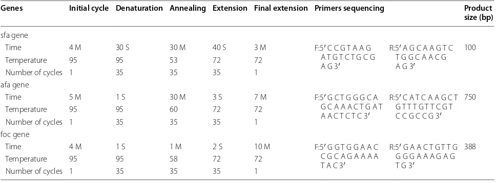
PCR reaction Specific primers were used to amplify the sequences of the sfa, afa, and foc genes [12, 13] (Table 1). As indicated in Table 1, the PCR assay was carried out in a total volume of 25 µl of mixture containing 22 µl master mix, 1 µl DNA sample, 1 µl forward primer, 1 µl reverse primer, and 0.2 µl tag polymerase. The PCR timetable pro-gram for sfa, afa, and foc genes is presented in Table 1.
Once analyzed by 2% agarose gel electrophoresis, the PCR products were stained with ethidium bromide and photographed.
Assessment of biofilm formation via phenotype method For investigating the ability of E. coli isolates to produce biofilms, biofilm test was performed in labora-tory based on the Microtiter Plates Assay as follow as:
The microtiter plate method was used for evaluating the formation of UPEC biofilm. The Microtiter Plates Assay method, such as ELISA, the color-producing chro-mogen in this technique is fuchsin, whose color intensity is directly related to the concentration of biofilm.
Table 1 Primers and PCR timetable program for sfa, afa, and foc genes in UPEC strain
Genes Initial cycle Denaturation Annealing Extension Final extension Primers sequencing Product size (bp)
sfa gene
Time 4 M 30 S 30 M 40 S 3 M F:5′ C C G T A A G
A T G T C T G C G A G 3′
R:5′ A G C A A G T C T G G C A A C G A G 3′
100
Temperature 95 95 53 72 72
Number of cycles 1 35 35 35 1
afa gene
Time 5 M 1 S 30 M 3 S 7 M F:5′ G C T G G G C A
G C A A A C T G A T A A C T C T C 3′
R:5′ C A T C A A G C T G T T T G T T C G T C C G C C G 3′
750
Temperature 95 95 60 72 72
Number of cycles 1 35 35 35 1
foc gene
Time 4 M 1 S 1 M 2 S 10 M F:5′ G G T G G A A C
C G C A G A A A A T A C 3′
R:5′ G A A C T G T T G G G G A A A G A G T G 3′
388
Temperature 95 95 58 72 72
[image:2.595.56.541.548.727.2]Initially, one loop full of bacteria colony was infused into one tube, including 5 ml nutrient broth where this tube was heated at 37 °C for 18 to 24 h. Then, 1 ml of bacterial suspension was injected into the tube, including 10 ml sterile nutrient broth, with this tube being heated at 37 °C for a duration of 18 to 24 h. Regarding the injec-tion only the sterile culture environment of nutrient broth into control well, microtiter plate was heated at 37 °C for 24 h. After the draining and washing of the wells three times by a sterile physiology serum, the plates were vigorously shaken to eliminate the disconnected cells. For stabilizing the cells, 200 µl ethanol 96% was added to wells. After 15 min, the wells were drained, dried and stained by 200 µl fuchsin for 5 min. After 5 min, the wells were washed slowly by urban water and were filled with 200 µL acetic acid 33% as solvent. After plate incuba-tion for 15 min at 37 °C, the Optical Density of the wells stained with fuchsin was screened by ELISA at a wave-length 492 nm. All measurements were repeated three times and the culture environment was used as negative control. A standard deviation larger than the negative control optical absorption was used as Cut-off. The abil-ity of biofilm formation was calculated using the follow-ing formulas [14] that shown in the table. In the current study, OD ≥ 0.1, 0.07 ≤ OD ≤ 0.09, 0.01 ≤ OD ≤ 0.06 and OD ≤ 0.009 were considered strong, average, weak and no connection, respectively.
Assessment of antibiotic resistance in E. coli The antibi-otic sensitivity was examined by Kirby Beuer method (disk diffusion) [14]. The diameter of the zones of inhibition was measured by a ruler in millimeter. In this research, the antibiotics utilized against E. coli pathogens included Imi-penem, Ciprofloxacin (cp5), Tobramycin (TOB10), Ampi-cillin, Tetracycline (TE30), Amikacin (AN30), Amoxicil-lin (AMX25), Nalidixic Acid (NA30), Nitrofurantoin, Cefepime, Gentamycin (GM10), Ceftazidime (CAZ30), Chloramphenicol, and Ceftriaxone (CRO30).
Data analysis
Data were analyzed using SPSS 23 for the frequency and percentage. The comparison between variables was ana-lyzed by a Chi square or Fisher’s exact tests. P < 0.05 was considered statistically significant.
Results
In this study, the PCR reaction was done on 79 samples from urine specimens with UTI symptoms and suspected to have E. coli, after confirmation (Additional file 1: Fig. S1).
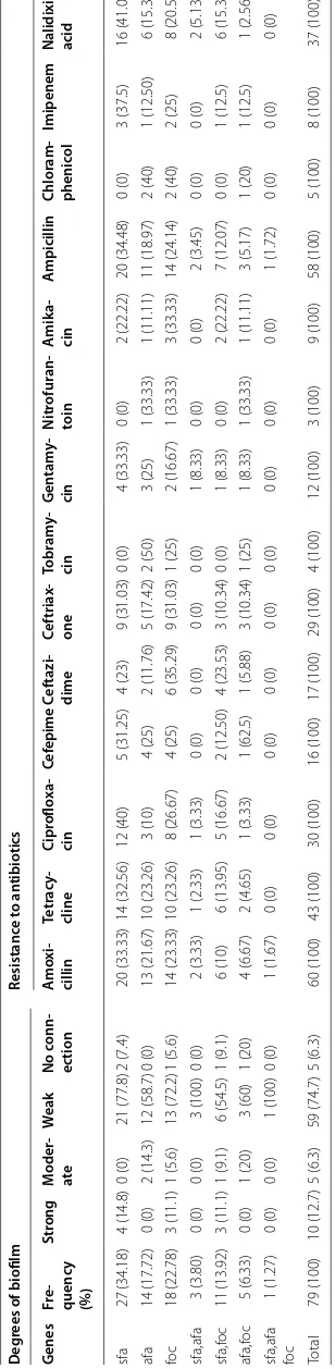
The PCR results indicated that the highest and lowest frequency of fimbrial genes was associated with sfa and
sfa-afa-foc genes (Table 2). Then, the frequency of sfa
gene was compared to other genes. There was a signifi-cant difference between sfa with afa, foc, sfa-afa, sfa-foc,
afa-foc, and sfa-afa-foc with 0.04, 0.001, 0.009, 0.001 and 0.00 P-values, respectively.
Results of microtiter plate biofilm formation
Among 79 E. coli isolates from urinary tract infections, 10 isolates (12.7%), 5 isolates (6.3%), 59 isolates (74.7%) and 5 isolates (6.3%) showed strong, moderate, weakly and no connection biofilm formation ability, respectively.
The capacity of biofilm formation were compared between genes, where there was no significant correla-tion between them; however, the strongest and the weak-est had related to sfa and afa genes, respectively (Table 2). Of note, afa even has reduced ability of biofilm formation in combination with other genes.
In the antibiotic resistance, the most resistant strains were related to the amoxicillin and ampicillin antibiotics, while the greatest sensitivity was associated with nitro-furantoin and amikacin (Tables 2 and 3).
Discussion
E. coli is the most common causative agent of UTIs in both outpatients and inpatients. If left untreated, UTIs may culminate in serious consequences such as renal fail-ure. Pyelonephritis usually develops following a simple bladder infection (i.e. cystitis) [5]. The ability of patho-genic bacteria to adhere to the urinary tract epithelium using pili (fimbriae) is the most important pathogenic feature leading to UTIs [15]. Various genes encoding pili can be identified by molecular techniques such as PCR [16].
Our results revealed a relatively high frequency of sfa
gene compared with the afa and foc in E. coli strains iso-lated from patients with UTIs. This probably indicates the essential role of this gene in the development of UTIs. Various studies have reported the role of sfa gene in encoding adherence molecules involved in the patho-physiology of pyelonephritis caused by E. coli [3]. In line with our finding, Jalali et al. [17] reported that 32% of UTIs patients expressed the sfa gene. In another study among children suffering from UTI caused by E. coli, a high frequency of the sfa gene was reported [18]. Like-wise, the frequency of the afa gene reported in another study was similar to the present report [19].
In the present study, strong and weak biofilm-forming capabilities were associated with the expressions of the
sfa and afa genes, respectively. Reduction of biofilm for-mation ability was observed in combination of afa gene with other genes.
Table 2 T he char ac teristics of genes of
sfa, afa, and
foc Deg rees of biofilm Resistanc e t o an tibiotics G enes Fr e-quenc y (%) Str ong M oder -ate W eak No c onn-ec tion A mo xi -cillin Tetr ac y-cline Cipr oflo xa -cin Cef epime C ef tazi -dime C ef triax -one Tobr am y-cin G en tam y-cin N itr ofur an -toin A mik a-cin A mpicillin Chlor am -phenic ol Imipenem
Nalidixic acid
sfa 27 (34.18) 4 (14.8) 0 (0) 21 (77.8) 2 (7.4) 20 (33.33) 14 (32.56) 12 (40) 5 (31.25) 4 (23) 9 (31.03) 0 (0) 4 (33.33) 0 (0) 2 (22.22) 20 (34.48) 0 (0) 3 (37.5) 16 (41.03) afa 14 (17.72) 0 (0) 2 (14.3) 12 (58.7) 0 (0) 13 (21.67) 10 (23.26) 3 (10) 4 (25) 2 (11.76) 5 (17.42) 2 (50) 3 (25) 1 (33.33) 1 (11.11) 11 (18.97) 2 (40) 1 (12.50) 6 (15.38) fo c 18 (22.78) 3 (11.1) 1 (5.6) 13 (72.2) 1 (5.6) 14 (23.33) 10 (23.26) 8 (26.67) 4 (25) 6 (35.29) 9 (31.03) 1 (25) 2 (16.67) 1 (33.33) 3 (33.33) 14 (24.14) 2 (40) 2 (25) 8 (20.51) sfa,afa 3 (3.80) 0 (0) 0 (0) 3 (100) 0 (0) 2 (3.33) 1 (2.33) 1 (3.33) 0 (0) 0 (0) 0 (0) 0 (0) 1 (8.33) 0 (0) 0 (0) 2 (3.45) 0 (0) 0 (0) 2 (5.13) sfa,f oc 11 (13.92) 3 (11.1) 1 (9.1) 6 (54.5) 1 (9.1) 6 (10) 6 (13.95) 5 (16.67) 2 (12.50) 4 (23.53) 3 (10.34) 0 (0) 1 (8.33) 0 (0) 2 (22.22) 7 (12.07) 0 (0) 1 (12.5) 6 (15.38) afa,f oc 5 (6.33) 0 (0) 1 (20) 3 (60) 1 (20) 4 (6.67) 2 (4.65) 1 (3.33) 1 (62.5) 1 (5.88) 3 (10.34) 1 (25) 1 (8.33) 1 (33.33) 1 (11.11) 3 (5.17) 1 (20) 1 (12.5) 1 (2.56)
sfa,afa foc
1 (1.27) 0 (0) 0 (0) 1 (100) 0 (0) 1 (1.67) 0 (0) 0 (0) 0 (0) 0 (0) 0 (0) 0 (0) 0 (0) 0 (0) 0 (0) 1 (1.72) 0 (0) 0 (0) 0 (0) Total 79 (100) 10 (12.7) 5 (6.3) 59 (74.7) 5 (6.3) 60 (100) 43 (100) 30 (100) 16 (100) 17 (100) 29 (100) 4 (100) 12 (100) 3 (100) 9 (100) 58 (100) 5 (100) 8 (100) 37 (100) The t
otal of it
ems w
er
e c
onsider
ed as fr
equenc
y (per
cen
[image:4.595.187.341.97.726.2]important during 24 h after the colonization of bacteria in the bladder [20]. In another study, Goetz et al. [21] described the role of papG gene in augmenting the ability of bacteria to connect to surfaces. In the report of Pon-nusamy et al. [22], only 23.6% of E. coli strains were able to from strong biofilms. These reports highlight the roles of P fimbriae (pap), afa, hemolysin (hly), and sfa/foc in biofilm formation by bacteria causing human infections. These molecules help bacteria to colonize and dam-age tissues and debilitate the host’s defense mechanisms which ultimately lead to clinical manifestations [23].
In the present study, the highest resistance rates of the isolated E. coli strains were observed against amoxicillin and ampicillin. This was while the greatest sensitivities were related to nitrofurantoin and amikacin antibiotics. In the study of Gazmoh et al. in Ethiopia, Adimi et al. in Nigeria and Abdollahi et al. in Tehran, E. coli strains from patients with UTIs demonstrated a similar resistance pat-tern to the present study [24–26].
Escherichia coli is considered as an important cause of UTI in patients referring to health centers. The severity of the infection depends on both the host’s immune com-petency and the distribution of virulence factors among pathogenic bacteria. UPEC isolates are genetically heter-ogeneous bacteria which have variable capacities for bio-film formation, colonization, invasion, and proliferation in the urinary tract [27, 28].
In another study by Jha et al. [29] on 244 patients with UTIs in Japan, E. coli was reported as the most frequent causative agent, and the least resistance rate was related to ciprofloxacin. In contrary to our results; however, Keikha et al. [30] in their study on 87 urinary E. coli iso-lates reported the highest antibiotic resistance against
cotrimoxazole. These inconsistencies can be related to parameters such as sample sizes, the accuracies of sam-pling and testing methods, as well as different geographi-cal locations [27–30].
Generally, E. coli strains causing UTIs represent increasing rates of antibiotic resistance, especially against the first (e.g. ampicillin) [31] the third (e.g. cephalospor-ins and aminoglycosides) generations of broad-spectrum antibiotics [32]. These high rates of antibiotic resistance may be due to the unprescribed availability and uncon-trolled usage of antibiotics, especially in developing countries. Therefore, selecting antibiotics for treating bacterial infections should be according to the results of urine culture, antibiotic susceptibility, and biofilm forma-tion analyses.
Conclusion
There were differences between the characteristics of UPEC in this area and different regions in terms of fre-quency, formation of biofilm, and drug resistance. These differences were even observed among strains. By collect-ing the characteristics of UPEC strains in each region, the epidemiological characteristics of native isolates were distinguished. Therefore, it is possible to diagnose this condition earlier and offer appropriate treatments.
Limitation
This study was limited by short duration and moderate sample. Ultimately, it is recommended to performing of the other studies on other genes between control and case groups.
Supplementary information
Supplementary information accompanies this paper at https ://doi. org/10.1186/s1310 4-019-4825-8.
Additional file 1: Fig S1.Escherichia coli strains including of sfa, afa and
foc genes on agarose gel.
Abbreviations
E. coli: Escherichia coli; UTIs: urinary tract infections; UPEC: uropathogenic
Escherichia Coli; Pap: pyelonephritis associated pili; sfa: s fimbrial adhesion; OD: optical density; NM: nanometer; µL: microliter; ML: milliliter; C: centigrade.
Acknowledgements
We would like to thank staffs of Amir Almomenin’s Hospitalˏ Central Labora-tory of the University of Medical Sciences and Private and Medical Dinesh’s Laboratory in Maragheh/Iran that corporate in the progression this study.
Authors’ contributions
ED and LV: conceived the study. ED: participated in the acquisition of data and sampling. ED and LV: participated in the design of the study and performed the statistical analysis. LV: interpreted the data. ED: obtained ethical clearance and permission for study. ED: performing tests. ED and AK: preparing image. ED and AK: preparing tables. LV, ED and AK: Drafting the article or revisiting it critically for important intellectual content. LV and AK: manuscript submitting.
Table 3 Results of antibiogram for genes of UPEC strain
*The total of items were considered as frequency (percentage)
Antibiotics Resistant* Intermediate* Sensitive*
[image:5.595.57.289.101.292.2]LV, ED and AK: manuscript revising. AK: English editing. All authors read and approved the final manuscript.
Funding Not applicable.
Availability of data and materials
The datasets used and analyzed during the current study are available from the corresponding author and first author on not allowed from the centers. Private and Medical Dinesh’s Laboratory in Maragheh and Liver and Gastroin-testinal Disease Research Center of Tabriz University of Medical Sciences are where supporting our findings.
Ethics approval and consent to participate
This study was approved by the Ethics Committee of the Islamic azad Uni-versity, Urmia, Iran (NO = 10330507942022). The participation in the research was voluntary the following informed consent obtained from the patients as written and that they will be free to discontinue participation.
Consent for publication Not applicable.
Competing interests
The authors declare that they have no competing interests.
Author details
1 Department Of Microbiology, Faculty of Basic Science, Islamic Azad
Univer-sity, Urmia, Iran. 2 Student of Medicine, Faculty of Medicine, Tabriz University
of Medical Sciences, Tabriz, Iran. 3 Liver and Gastrointestinal Disease Research
Center, Tabriz University of Medical Sciences, Tabriz, Iran.
Received: 9 August 2019 Accepted: 26 November 2019
References
1. Tarchouna M, Ferjani A, Ben-Selma W, Boukadida J. Distribution of uropathogenic virulence genes in Escherichia coli isolated from patients with urinary tract infection. Int J Infect Dis. 2013;17(6):e450-e3. https :// doi.org/10.1016/j.ijid.2013.01.025.
2. Sahib AS, Mohammed IH, Hamdan SJ. Use of aqueous extract of corn silk in the treatment of urinary tract infection. J Complement Med Res. 2012;1(2):93–6. https ://doi.org/10.5455/jice.20120 52512 3150. 3. Emody L, Kerenyi M, Nagy G. Virulence factors of uropathogenic
Escheri-chia coli. Int J Antimicrob Agents. 2003;22:29–33. https ://doi.org/10.1016/ S0924 -8579(03)00236 -X.
4. Oloketuyi SF, Khan F. Strategies for biofilm inhibition and virulence attenuation of foodborne pathogen-Escherichia coli O157: H7. Curr Micro-biol. 2017;74(12):1477–89. https ://doi.org/10.1007/s0028 4-017-1314-y. 5. Lee J, Subhadra B, Son YJ, Kim D, Park H, Kim J, et al. Phylogenetic group
distributions, virulence factors and antimicrobial resistance properties of uropathogenic Escherichia coli strains isolated from patients with urinary tract infections in South Korea. Appl Microbiol. 2016;62(1):84–90. https :// doi.org/10.1111/lam.12517 .
6. Brandström P, Hansson S. Long-term, low-dose prophylaxis against uri-nary tract infections in young children. Pediatr Nephrol. 2015;30(3):425– 32. https ://doi.org/10.1007/s0046 7-014-2854-z.
7. Qu Y, Li R, Jiang M, Wang X. Sucralose increases antimicrobial resistance and stimulates recovery of Escherichia coli mutants. Curr Microbiol. 2017;74(7):885–8. https ://doi.org/10.1007/s0028 4-017-1255-5. 8. Bryce A, Hay AD, Lane IF, Thornton HV, Wootton M, Costelloe C. Global
prevalence of antibiotic resistance in paediatric urinary tract infections caused by Escherichia coli and association with routine use of antibiotics in primary care: systematic review and meta-analysis. BMJ. 2016;352:i939.
https ://doi.org/10.1136/bmj.i939.
9. Terlizzi ME, Gribaudo G, Maffei M. UroPathogenic Escherichia coli (UPEC) infections: virulence factors, bladder responses, antibiotic, and non-antibiotic antimicrobial strategies. Front Microbiol. 2017;8:1566. https :// doi.org/10.3389/fmicb .2017.01566 .
10. Vahedi L, Jabarpoor-Bonyadi M, Ghojazadeh M, Hazrati H, Rafeey M. Asso-ciation between outcomes and demographic factors in an Azeri Turkish population with cystic fibrosis: a cross-sectional study in Iran from 2001 through 2014. IRCMJ. 2016. https ://doi.org/10.5812/ircmj .29615 . 11. Shan Z, Zhou Z, Chen H, Zhang Z, Zhou Y, Wen A, et al. PCR-ready
human DNA extraction from urine samples using magnetic nanopar-ticles. J Chromatogr. 2012;881:63–8. https ://doi.org/10.1016/j.jchro mb.2011.11.042.
12. Gilbert NM, Lewis AL. Covert pathogenesis: transient exposures to microbes as triggers of disease. PLoS Pathog. 2019;15(3):e1007586. https ://doi.org/10.1371/journ al.ppat.10075 86.
13. Malekzadegan Y, Khashei R, Ebrahim-Saraie HS, Jahanabadi Z. Distribution of virulence genes and their association with antimicrobial resistance among uropathogenic Escherichia coli isolates from Iranian patients. BMC Infect Dis. 2018;18(1):572. https ://doi.org/10.1186/s1287 9-018-3467-0. 14. Nourbakhsh F, Momtaz H. Evaluation of phenotypic and genotypic
biofilm formation in Staphylococcus aureus isolates isolated from hospital infections in Shahrekord, 2015. Evaluation. 2016;19(109):69–79 (In Persian).
15. Bahalo S, Tajbakhsh E, Tajbakhsh S, Momeni M, Tajbakhsh F. Detection of some virulence factors of Escherichia coli isolated from urinary tract infec-tion isolated of children in Shahrekord Iran by multiplex PCR. Middle East J Sci Res. 2013;14(1):29–32.
16. Yazdi M, Bouzari M, Ghaemi EA. Detection of fim, pap, sfa and afa adhesin-encoding operons in Escherichia coli strains isolated from urinary tract infections. Mod Med Lab. 2018;12(5):10–5. http://mlj.goums .ac.ir/ artic le-1-1114-en.html.
17. Jalali HR, Pourbakhsh A, Fallah F, Eslami G. Genotyping of virulence factors of uropathogenic Escherichia coli by PCR. Novel Biomed. 2015;3(4):177–81.
18. Abana CO, Bingham BS, Cho JH, Graves AJ, Koyama T, Pilarski RT, et al. IL-6 variant is associated with metastasis in breast cancer patients. PLoS ONE. 2017;12(7):e0181725. https ://doi.org/10.1371/journ al.pone.01817 25. 19. Yun KW, Kim HY, Park HK, Kim W, Lim IS. Virulence factors of
uropatho-genic Escherichia coli of urinary tract infections and asymptomatic bacte-riuria in children. J Microbiol Immunol Infect. 2014;47(6):455–61. https :// doi.org/10.1016/j.jmii.2013.07.010.
20. Lane MC, Simms AN, Mobley HL. Complex interplay between type 1 fimbrial expression and flagellum-mediated motility of uropatho-genic Escherichia coli. J Bacteriol. 2007;189(15):5523–33. https ://doi. org/10.1128/jb.00434 -07.
21. Goetz G, Mahmood A, Hultgren S, Engle M, Dodson K, Alpers DJI, et al. Binding of Pili from Uropathogenic Escherichia coli to membranes secreted by human colonocytes and enterocytes. Infect Immun. 1999;67(11):6161–3.
22. Ponnusamy P, Natarajan V, Sevanan MJ. In vitro biofilm formation by uropathogenic Escherichia coli and their antimicrobial susceptibility pattern. Asian Pac J Trop Med. 2012;5(3):210–3. https ://doi.org/10.1016/ S1995 -7645(12)60026 -1.
23. Tajbakhsh E, Ahmadi P, Abedpour-Dehkordi E, Arbab-Soleimani N, Khamesipour F. Biofilm formation, antimicrobial susceptibility, serogroups and virulence genes of uropathogenic E. coli isolated from clinical samples in Iran. Antimicrob Resist Infect Control. 2016;5:1. https ://doi. org/10.1186/s1375 6-016-0109-4.
24. Adeyemi AO, Evbaziegbere Gideon E, Eromosele IH, Evelyn Chimerenma A, Sunday Oladokun O, Eunice Ogochukwu U, et al. Antibiotics suscep-tibility patterns of some uropathogens to nitrofurantoin and nalidixic acid among pregnant women with urinary tract infections in federal medical centre, Bida, Niger-State, North Central, Nigeria. Am J Epidemiol. 2014;2(4):88–92. https ://doi.org/10.12691 /ajeid -2-4-1.
25. Abdolahi AR, Mehr Azma M. Evaluation of antibiotic susceptibility and resistance in urinary infections, Imam Khomeini Hospital, Tehran. J Jah-rom Univ Med Sci. 2009;7(9):59–66.
26. Gezmu T, Regassa B, Manilal A, et al. Prevalence, diversity and antimi-crobial resistance of bacteria isolated from the UTI parents of Arba Minch Province, Southern Ethiopia. Transl Biomed. 2016;7:3. https ://doi. org/10.21767 /2172-0479.10008 1.
Lancet Infect Dis. 2010;10(9):597–602. https ://doi.org/10.1016/S1473 -3099(10)70143 -2.
28. Karlowsky JA, Kelly LJ, Thornsberry C, Jones ME, Sahm DF. Trends in antimicrobial resistance among urinary tract infection isolates of
Escherichia coli from female outpatients in the United States. Antimi-crob Agents Chemother. 2002;46(8):2540–5. https ://doi.org/10.1128/ AAC.46.8.2540-2545.2002.
29. Jha N, Bapat SJ. A study of sensitivity and resistance of pathogenic micro organisms causing UTI in Kathmandu valley. KUMJ. 2005;3(2):123–9. 30. Keikha M, Rava M. Evaluation of antibiotic resistance of Escherichia coli
strains isolated from urinary tract infections in outpatients referring to Nabi Akram Hospital in Zahedan. J Paramed Sci Rehabilit. 2017;6(4):73–8.
31. Jan N, Meshram SU, Kulkarni A. Plasmid profile analysis of multidrug resistant E. coli isolated from UTI patients of Nagpur city, India. Rom Biotechnol Lett. 2009;14(5):4635–40.
32. Novakova I, Kacaniova M, Hascik P, Pavlicova S, Hleba L. The resistance to antibiotics in strains of E. coli and enterococcus sp. isolated from rectal swabs of lambs and calves. Lucrari Stiintifice Zootehnie Sibiotehnologii. 2009;42(2):322–6.
Publisher’s Note