MANAGEMENT OF GARTLAND TYPE III SUPRACONDYLAR FRACTURE OF HUMERUS IN CHILDREN BY OPEN REDUCTION AND INTERNAL FIXATION USING CROSSED K – WIRES.
- A SHORT TERM FOLLOW UP STUDY
Dissertation submitted for
M.S. DEGREE EXAMINATION
BRANCH – II ORTHOPAEDIC SURGERY Department of Orthopaedics and Traumatology
Thanjavur Medical College Thanjavur
THE TAMILNADU DR.M.G.R. MEDICAL UNIVERSITY
CHENNAI, TAMILNADU.
CERTIFICATE
This is to certify that DR.G.A.RAJMOHAN, postgraduate (2006-2008) in the Department of Orthopaedics and Traumatology,
Thanjavur Medical College and Hospital, Thanjavur, has done this
dissertation on “MANAGEMENT OF GARTLAND TYPE III
SUPRACONDYLAR FRACTURE OF HUMERUS IN CHILDREN BY OPEN REDUCTION AND INTERNAL FIXATION USING CROSSED K – WIRES - A SHORT TERM FOLLOW UP STUDY" under my guidance and supervision in partial fulfilment of the regulation laid down by the TamilNadu DR.M.G.R. Medical University, Chennai for MS (Orthopaedics)
degree examination to be held on March 2008.
PROF.DR.R.RATHINASABAPATHY,
M.S.Ortho, D.Ortho, Professor and Head of the Department, Department of Orthopaedics and Traumatology,
ACKNOWLEDGEMENT
I owe my sincere and grateful acknowledgement to my beloved
Chief, Prof.Dr.R.Rathinasabapathy M.S.Ortho., D.Ortho., Professor
and Head of the Department of Orthopaedics and Traumatology,
Thanjavur Medical College and Hospital for his kind help,
encouragement and invaluable guidance rendered to me in preparing
this dissertation.
I am grateful to my beloved teacher
Prof.Dr.M.GulamMohideen M.S.Ortho., D.Ortho., for his constant
help, advice and guidance rendered to me in preparing this
dissertation.
I express my gratitude to Prof.Dr.K.Anbalagan M.S.Ortho.,
D.Ortho., for his help and motivation during this study.
I sincerely acknowledge and owe much to Dr.S.Kumaravel
M.S.Ortho., D.Ortho., for his suggestions, untiring help and
I thank my teachers Dr.A.Bharathy, Dr.P.Venkatesan,
Dr.M.S.Manoharan, Dr.V.Jayabalan, Dr. D.Thirumalai Pandiyan,
Dr.R.Vasantharaman and my colleagues for their help and
motivation in doing this study.
My sincere thanks to the Dean, Thanjavur Medical College
and Hospital, Thanjavur for permitting me to utilize the hospital
facilities for this study.
I sincerely thank Dr.K.Thennarasu Ph.D., NIMHANS,
Bangalore for helping me in the statistical analysis.
I express my gratitude to the children and their parents, for
their kind cooperation during the study.
My heart full thanks to my dear wife Dr.S.Jayalakshmi MD
(OG) and my loving daughter for their help, patience and whole
hearted support in bringing out this study.
CONTENTS
S.No TITLE Page No.
1. Introduction 1
2. Aim 4
3. Historical Review and Literature 5
4. Materials and Methods 32
5. Surgical Technique 34
6. Observations and Results 36
7. Discussion 45
8. Conclusion 50
9. Bibliography 10. Appendices
Sir Robert Jones said “The difficulty experienced by
an orthopaedic surgeon is mainly an accurate diagnosis, the
facilities with which serious blunders can be made in
prognosis and treatment, and the fear shared by so many, of
the injuries, of the neighbourhood of elbow less attractive
than they might have otherwise proved.”
Nowhere else this is more illustrated than in
supracondylar fracture of humerus which is one of the
commonest elbow injuries in paediatric age group. The
functional results following supracondylar fracture of
humerus with conservative treatment are satisfactory but the
cosmetic results are poor. These are relevant even in modern
era.
The presentation of a child with a swollen elbow still
brings some feeling of anxiety to the treating orthopaedic
Between the mid 20th century, an early report of
pinning distal humerus fractures first appeared, after that the
treatment of supracondylar fractures has evolved
tremendously.
Blount’s1 caution against operative management has
given way to modern concepts of treatment involving skeletal
stabilisation and soft tissue management which have greatly
improved outcomes.
Problems of neurovascular compromise Volkmann’s
ischemic contracture and deformity have been greatly
decreased but not eliminated.
The complications can be minimised by achieving good
anatomical reduction. There are various modalities of
treatment available like closed reduction and Plaster of Paris
immobilization, various types of traction, closed reduction
and percutaneous pinning and open reduction and internal
Closed reduction and application of casts with the elbow
in flexion is one of the oldest and most widely used methods
of treatment. There are concerns about the dangers and
difficulties of this method especially the risk of Volkmann’s
ischemic contracture and high incidence of cubitus varus.
These complications can be prevented by achieving
anatomical reduction by open reduction and internal fixation
with crossed K-wires. This is relatively a simple procedure
The aim of this study is to evaluate the outcome
of management of Gartland type III supracondylar
fracture of humerus in children by open reduction and
internal fixation using crossed K – wires.
HISTORICAL REVIEW
Supracondylar fractures were described in the early
writings of Hippocrates during the 3rd and 4th century A.D.
But it was not until 1700s that much was written about
supracondylar fractures in the classic medical literature.
Dupuytren mentioned the findings of crepitus with the
fracture. Malgaigne demonstrated that there was preservation
of the Olecranon –Humeral condylar relationship with the
fracture but not with the dislocation.
Most of the discussions during 1700s and 1800s were
directed towards the controversy regarding the correct
position of immobilisation.
Desault from Paris in 1800 said that poor results were
due to poor management and not inevitable with this type of
fracture. He demonstrated better results with this prompt
Jones and Thomas propounded treatment in flexed
position, which we follow while Listen and Allis were in
favour of extended position.
At the beginning of 20th century, treatment began to
change from these simple passive methods to more
aggressive and active methods. Scientific reason and study
began to alter the methods of treatment and open reduction
and internal fixation came into vogue.
Herzenberg and co-workers conducted in-vitro
studies of pin stability and found the 5/64” Steinmann pins
placed from medial to lateral entrance points proved the best
stability.
Ziont’s and co-workers2 demonstrated the resistance
of various patterns to rotational stresses.
Cheng J.C, LamT.P, Shen W.Y and co-workers
in treatment of Gartland type III extension fractures with a
high success rate and minimal complications.
Mohammed.S and Rymaszewski.L.A35 in a study
conducted at the Glasgow Royal Infirmary between June1990
and September 1992, on 32 displaced supracondylar fractures
of humerus in children concluded that open reduction and
internal fixation with two K–wires gave the best results.
Aronson D.C, Van Vallendhoven E, Meeuwis J.D in
a study conducted on 11 children with supracondylar
fractures of humerus treated with open reduction and K wire
fixation by a ventral approach concluded that K wire fixation
of supracondylar fractures in children gives excellent results.
Furrer.M, Mark.G and Ruedi.T3 did a open reduction
and crossed K-wire fixation on 33 children with displaced
supracondylar fractures of the humerus and recommended
ANATOMY
Elbow is a complex joint composed of three
individual joints. Articulation occurs between the trochlea
and capitulum of the humerus with the trochlear notch
of the ulna and the head of the radius. In proximal
radioulnar joint articulation occurs between the
circumference of head of radius, the annular ligament and
the radial notch. The articular surfaces are covered with
hyaline cartilage.
DISTAL END OF HUMERUS:
The distal end of humerus is divided into medial and
lateral columns. Each of the columns are roughly triangular
and is bound on its anterior border by supracondylar ridge.
From the structural and functional stand points the
components each containing an articulating and non
articulating portion. Included in the non-articulating portion
are the epicondyles which are the terminal portion of the
supracondylar ridge. The lateral epicondyle contains a
roughened anterolateral surface from which the superficial
forearm extensor muscles arise. The medial epicondyle is
larger than the lateral counter part and serves as the origin of
the forearm flexor muscles.
The posterior distal portion of the medial epicondyle is
smooth and in contact with the ulnar nerve as it crosses the
elbow joint.
When a condyle losses continuity from its supporting
column, as in a fracture displacement the nerve can get
injured by direct compression by the fracture fragments. This
should be well born in mind while treating supracondylar
The articulating surface of the medial epicondyle, the
trochlea, is more cylindrical or spool like.
It has a very prominent medial and lateral ridges.
Between these ridges is a central groove that articulate with
the greater sigmoid notch of proximal ulna. The groove
originates anteriorly in the coronoid fossa and terminates in
the posterior surface of the trochlea. The groove is directed
slightly laterally.
This obliquity of the trochlear groove produces the
valgus carrying angle of the forearm when the elbow is
extended.
Proximal to the condyles on the anterior surface of the
humerus lie the coronoid and radial fossa. They articulate
with the coronoid of ulna and the radial head, respectively
when the elbow is flexed. Posteriorly the olecranon fossa is
a deep hollow for the reception of olecranon process, making
that separates this anterior and posterior fossa is extremely
thin and translucent4.
UPPER END OF RADIUS:
The proximal end of radius consists of the disc shaped
head, the neck and the radial tuberosity.
The head and part of the neck lie within the joint. The
shallow concavity of the head articulates with the capitulum.
UPPER END OF ULNA:
The proximal end consists of the olecranon and
coronoid process, which together form the semilunar notch.
The triceps inserts by a broad tendinous insertion into the
olecranon posteriorly. On the anterior surface the brachialis
muscles inserts into the coronoid process.
The triceps play an important role in maintaining the
reduction of supracondylar fracture and its integrity is
COLLATERAL LIGAMENTS:
The collateral ligaments supplement the natural stability
of the elbow joint. The triangular radial collateral ligament
is attached by its apex to lateral epicondyle of humerus
and by its base to the upper margin of annular ligament.
The thicker and stronger ulnar collateral ligament consists of
three bands. The anterior band passes from medial
epicondyle of humerus to medial margin of coronoid
process. The posterior band passes from medial
epicondyle of humerus to medial side of olecranon. The
transverse band passes between the ulnar attachments of
Medial Collateral Ligament
CAPSULE:
Anteriorly it is attached above to the humerus along
the upper margins of the coronoid and radial fossae and
to the front of medial and lateral epicondyles and below
to the margin of coronoid process of the ulna and to the
annular ligament
Posteriorly it is attached above to the margins of
olecranon fossa of humerus and below to the upper
margin and sides of the olecranon process of ulna and to
the annular ligament.
NEUROVASCULAR ANATOMY OF THE ELBOW :
The orthopaedic surgeon should be well versed with the
neurovascular arrangement in the elbow to operate on the
young patients.
The brachial artery is the most important artery in the
anterior aspect of the distal end of the humerus5. This is the
most common vascular structure to be involved in extension
type of supracondylar fracture.
The median nerve lies medial to the brachial artery
which can also be injured. The radial nerve may be
injured, if the spike is displaced laterally5,6.
The ulnar nerve passes posteriorly to the medial
epicondyle in the ulnar tunnel and emerge in the anterior
aspect winding around the medial epicondyle, this may be
MOVEMENTS:
Elbow joint is capable of flexion and extension.
Proximal radioulnar joint involves in pronation and
supination. Flexion is performed by the brachialis , biceps
brachii, brachioradialis and pronator teres muscle. Extension
is performed by the triceps and anconeus muscles.
IMPORTANT RELATIONS
Anteriorly : The brachialis, the tendon of biceps,
the median nerve and brachial artery.
Posteriorly : Triceps, a small bursa intervening.
Medially : Ulnar nerve passes behind the medial
epicondyle and crosses the medial ligament of the joint .
Laterally : The common extensor tendon and the
OSSIFICATION PROCESS:
In the above mentioned data it is quoted that the olecranon ossifies earlier than the trochlea.
SUPRACONDYLAR AREA OF HUMERUS IN CHILDREN:
There is a considerable difference in the bony
architecture of the supracondylar area of child and adult.
At the age of peak incidence of supracondylar fractures,
the bone in the supracondylar area is undergoing remodelling
with a decrease in both anteroposterior and lateral Sequence and timing of ossification in the elbow7
Girls (y) Boys(y) Capitellum 1.0 1.0 Radial head 5.0 6.0 Medial epicondyle 5.0 7.5
Olecranon 8.7 10.5 Trochlea 9.0 10.7
dimensions. It is less cylindrical than in adult. The
metaphysis of the child extends just distal to two fossae.
Because this is a newly formed bone, the trabeculae are less
defined and thinner and the cortex is very slender.
In the lateral projections the anterior cortices of the
medial and lateral epicondylar column do not project as far
anteriorly thus producing an anterior defect in the area of
coronoid fossa.
LIGAMENTOUS LAXITY :
Ligamentous laxity with hyperextension of joints is
normal in younger children. Thus as the younger child falls
with the arm outstretched, the elbow is more likely to be
hyperextended at the time of the fall.
Thus the local bony and ligamentous anatomy is a
major factor in producing supracondylar fracture during the
BIOMECHANICS AND MECHANISM OF INJURY :
The bone in the supracondylar area is weaker during the
last part of first decade of life because it is undergoing
metaphyseal remodelling. The thinnest portion occurs at the
depth of the olecranon fossa posteriorly and the coronoid
fossa anteriorly4. It is because of this distinct distal humeral
anatomy that a supracondylar fracture is so unstable.
Supracondylar fractures generally occur as a result of
fall onto the outstretched hand with elbow in full extension.
The olecranon in its fossa in the distal humerus acts as a
fulcrum, whereas the capsule transmits an extension force to
the distal humerus just proximal to the physis as the elbow
hyperextends.
In addition, the large amount of elastic epiphyseal and
articular cartilage in the distal portion can serve as a buffer to
transfer the force of hyperextension injury to supracondylar
Posterior displacement of the distal fragment occurs
with proximal fragment impaling the anterior soft tissue
structures. The fracture in the sagittal plane extends obliquely
from anterior and distal to posterior and proximal.
As the distal fragment displaced posteriorly the anterior
periosteum fails and tears away from it. Intact medial or
lateral periosteal hinge provides stability after reduction11,12.
Posteromedial displacement of the distal fragment is
more common than posterolateral displacement. The biceps
tendon insertion and axis of muscle pull lies medial to the
shaft of humerus, this anatomic location of muscle pull
created a force that tend to displace the distal fragment
CLASSIFICATION:
Classification of fracture type is useful only if it enables
the physician to make a decision about treatment or provide
some type of prognosis.
Since extension type of supracondylar fracture is the
commonest type, numerous attempts have been made to
classify this type of fracture. These initial type of
classifications have been based on two factors :
1. The type and location of fracture line.
2. The degree of displacement.
Classification of extension supracondylar fractures by Gartland9:
Type I Undisplaced.
Type II Displaced (with intact posterior cortex)
Type III Displaced (no cortical contact )
A. Posteromedial
Type I fractures have no displacement. It often
manifests only by a posterior fat pad sign10.
Type II fractures have a green stick fracture pattern.
The distal fragment displaced posteriorly with intact posterior
cortex. The presence of intact posterior cortex provides
sufficient internal stability.
Type III fractures have complete destruction of
posterior cortex, and the distal fragment is displaced
posteriorly. The triceps tends to displace the distal fragment
proximally. The distal fragment may be either posteromedial
DIAGNOSIS
The diagnosis is relatively simple. Sometimes there is
difficulty in classifying the fracture. Apart from the classical
signs and symptoms of fractures namely
• Pain
• Tenderness
• Abnormal mobility
• Inability to use the limb
The diagnosis was based on the following criteria
1. Deformity.
2. Pucker sign.
3. Differentiation from dislocation of elbow.
4. Anconeous soft spot.
5. X-ray diagnosis
• Standard AP view
• Lateral view
Deformity
Radiological diagnosis was difficult in type I and minimally
displaced type II fractures
The standard indicators were
1. AP view
• Baumann’s angle4
• Humeral - Ulnar angle
• Metaphyseal-Diaphyseal angle
2. Lateral view
• Shaft condylar angle
• Anterior humeral line
CONTEMPORARY MANAGEMENT
A neurological evaluation and vascular assessment
should be done initially.
Type I fractures:
These are managed by simple immobilization. The limb
is placed in a posterior splint applied at 60 to 90 degrees of
elbow flexion with side supports. X-rays are obtained after
seven days of fracture to document lack of displacement and
a long arm cast can then be applied. The duration of
immobilization is 3-4 weeks.
Type II fractures:
In this incomplete osseous separation with intact
posterior cortex, good stability can be obtained with closed
reduction. Significant swelling, obliteration of pulse with
column collapse are indicators for pin stabilization. If pin
stabilization is used, pins are removed at 3-4 weeks.
Type III fractures:
These are completely displaced fractures.
Management of type III can be broadly classified into:
1. Closed reduction and cast immobilization.
2. Traction.
3. Closed reduction and percutaneous pinning.
4. Open reduction and internal fixation.
Closed reduction and cast immobilization11,12:
The patient is anaesthetised, under image control the
affected arm is extended. Longitudinal traction is applied first
to dislodge the proximal fragment. Once the length is
restored medial or lateral translation is corrected next.
Rotation is corrected simultaneously, but in general
and lateral alignment is corrected, due to the effect of
surrounding soft tissue. A flexion reduction manoeuvre is
performed with pressure of the thumb over the condyles of
the humerus. The reduction can be felt. The elbow is flexed
to 120 degrees and forearm is pronated13 if the initial
displacement is medial. Figure of eight cast is used to
maintain reduction. It should be worn for 3-4 weeks. After
cast removal the limb is placed in a sling until the patient is
comfortable.
Traction14:
Both skin and skeletal traction has been used for many
years to achieve and maintain reduction. In Dunlop’s side
arm traction the forearm is held in supination which tends to
force the distal fragment into varus. In olecranon pin traction
either smooth K wire or a vertical skeletal screw is used.
Although traction is an effective method of treating severely
provides many more advantages and is becoming a more
acceptable alternative.
Closed reduction and Percutaneous pinning:
After closed reduction the reduction is maintained by
percutaneous pinning. This can be done either as a crossed
pinning or passing 2 parallel K wires parallel to each other
from the lateral epicondyle. In posteromedial fracture pattern
the medial pin should be placed first. Likewise the lateral pin
should be placed first for posterolateral fracture.
For a successful outcome near anatomic reduction and
adequate pin placement are needed. Use of power drills and
image intensifier are mandatory. Closed reduction of the
fracture and maintaining it during pinning needs
Pinning by Open reduction with crossed K wires:
Open reduction is indicated in displaced supracondylar
fractures where irreducibility results from proximal fragment
being buttonholed16 through soft tissue or interposition of
biceps or neurovascular structures. Approaches for open
reduction are anterior, posterior, medial and lateral. Posterior
approach through triceps muscle and tendon has been used
with excellent results. Posterior approach23,24,25 is easier when
comparing with other approaches. K wires 1.5mm to 2mm
are used in crossed pin technique. After reducing the fracture
visually lateral pin is inserted first and second pin is placed
medially. Both the pins should have a purchase on the
opposite cortex. Elbow is immobilised in flexion with
forearm in neutral position using above elbow posterior slab.
Open reduction and internal fixation with two K
does not need a C- arm which is ideal in our setup in
COMPLICATIONS
Neurologic Injury5,6 :
Neural injuries occur in 5% to 19% of the displaced
fractures. Anterior interosseous nerve is injured most often.
The radial nerve, median nerve and ulnar nerve are also
injured in that order. Posterolateral displacement of the distal
fragment is highly associated with median nerve injury.
Posteromedial displacement of the distal fragment is
associated with radial nerve injury. Ulnar nerve is injured
most often in flexion type of fracture than in extension type.
Ulnar nerve may also injured in percutaneous medial
pinning. Examination for nerve palsy prior to reduction is
important because entrapment of nerve especially median
nerve can occur during reduction. Most of the palsies resolve
spontaneously, routine exploration is not recommended.
Exploration is recommended for palsies that do not recover
Vascular Injury5,6:
Vascular insufficiency is reported in 3% to 12% of
displaced supracondylar fractures. Fortunately, less than 1%
have a significant risk of sequelae from vascular
compromise. Presence of pulse and perfusion of hand should
be documented. Initial approach to the pulseless limb is to
immobilise in less than 90 degree of flexion and should be
monitored. Perfusion is estimated by colour, warmth and
capillary refill. Exploration of the brachial artery is advocated
only when there is absent pulse with signs of ischemia after
reduction. Diligent observation and appropriate treatment
Cubitus Varus26,27:
It is the most common complication of
supracondylar fracture of the humerus. Cubitus varus occurs
after poor reduction or loss of reduction. Malunion of the
fracture occurs in three planes: internal rotation in the
horizontal plane, medial rotation in the coronal plane, and
extension in the sagittal plane. It is more of a cosmetic
deformity than functional. But patients having cubitus varus
deformity find difficult in throwing sports, push-ups and
swimming. In patients who do not tolerate the deformity
corrective osteotomy should be considered,
MATERIALS AND
METHODS
In our institution we selected 33 cases of Gartland type
III paediatric supracondylar fractures for this prospective
study. The age group varied from a minimum of 3 years to a
maximum of 13 years. The duration of study was from June
2006 to October 2007
Of the 33 children 26 were males and 7 were females.
Of the side right was involved in 8 cases and left was
involved in 25 cases.
SELECTION CRITERIA:
1. Age of the child between 3 years and 13 years
2. Extension- Gartland type III supracondylar fractures
EXCLUSION CRITERIA :
1. Gartland type I and II fractures
2. Fractures with impending compartment syndrome
3. Open fractures
4. Fractures with neurovascular compromise
5. Cases taken up for surgery later than 24 hours post
injury (unfit for anaesthesia post injury due to lower
respiratory tract infection, anaemia etc.)
On admission in our emergency service, detailed
clinical examination of the case was carried out. After ruling
out neurovascular compromise above elbow posterior slab
was applied to immobilize without compromising vascularity
and limb was elevated to reduce oedema.
All cases were given preoperative antibiotic
(inj.Cefotaxime – 50 mg/kg)
All cases were taken up for surgery within 24 hours
SURGICAL TECHNIQUE
Under general anaesthesia the patient in lateral position
the affected limb was hanged over the sand bag with elbow in
flexion. Tourniquet was applied after elevating the limb for 5
minutes. The Campbell’s posterior approach was used. First
step was to identify the ulnar nerve and safeguard it. After
raising triceps aponeurosis tongue flap and splitting of
triceps, the fracture ends were identified and reduced under
vision. Lateral K- wire was introduced from lateral
epicondyle, crossing the physis and always engage medial
cortex proximally. Medial pin was inserted through medial
epicondyle with a precaution not to damage the ulnar nerve
and engage the opposite cortex. Wires were bent and cut.
They were kept inside the wound to prevent accidental
removal of pins post operatively. Wound was closed in layers
keeping suction drain insitu. Tourniquet was released,
posterior above elbow slab was applied with elbow in
flexion. Check X ray was taken and reduction was judged by
with weekly review for upto 5th week. Then the patient was
reviewed monthly once. Posterior slab was discarded at the
end of 3rd week. Active elbow mobilisation exercises was
advised. K- wire removal was done after confirmation of
solid union of fracture by observing callus formation. Usually
K – wires were removed as a out patient procedure at the end
of 3rd , 4th or 5th week depending upon union. Patients were
advised against massaging and passive mobilisation. At the
follow up elbow range of movements and carrying angle
were noted according to Flynn’s criteria.
Flynn’s Criteria28
Results Cosmetic factor- loss of
carrying angle(degree)
Functional factor-loss
of motion (degree)
Excellent 0-5 0-5
Good 6-10 6-10
Fair 11-15 11-15
Surgical Technique Position of the patient
Skin Incision Ulnar Nerve Safeguarding
Triceps Tongue Flap
Haematoma Evacuation
Triceps Resuturing
OBSERVATIONS AND
Out of thirty three children who presented with extension
Gartland type III supracondylar fracture , twenty six (
78.8%) were males and seven (21.2%) were females. Elbow
on the left side was involved in twenty five (75.6%) patients
and the right side was involved in eight (24.4%) patients.
Age range was from three to thirteen years with maximum
patients received between six to nine years. Thirty one
patients (93.9%) were taken up for surgery within twelve
hours of injury and remaining two (6%) patients within
twenty four hours. Twenty eight (84.8%) patients were
having no complications in post-operative period. Two
(6.1%) patients had pin tract infection which were settled
after the K – wire removal and appropriate antibiotics. Three
patients (9.1%) had superficial wound infection which were
settled after administering appropriate antibiotics. K- wires
weeks for twenty three (70%) patients and in five weeks for
four (12%) patients.
According to Flynn’s criteria thirty (90%) patients were
found to have excellent outcome two (6%) patients turned out
with good outcome and one (3%) patient turned out with fair
outcome. In our study three patients had more than five
degrees of range of motion loss compared to the other side.
All these patients had superficial wound infections in the
post - operative period. For these patients loss of carrying
angle could not be measured due to persistant fixed flexion
deformity. Neurovascular compromise was not seen in any
of our patients.
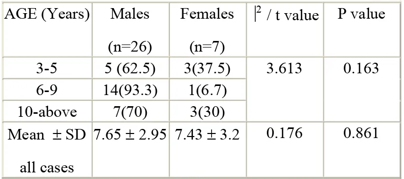
Table I: Distribution and comparison of age by gender
AGE (Years) Males (n=26)
Females (n=7)
⎟2 / t value P value
3-5 5 (62.5) 3(37.5) 3.613 0.163 6-9 14(93.3) 1(6.7)
10-above 7(70) 3(30) Mean ± SD
all cases
7.65 ± 2.95 7.43 ± 3.2 0.176 0.861
There are 26 (78.8%) males and 7 (21.2%) female
young patients in the study. The over all mean age of the
patients is 7.61 ± 2.96 years. The minimum age is 3 years
and maximum is 13 years. Eight patients (24.2%) are in less
than six years age group, 15(45.6%) are in 6-9 years age
group and 10(30.2%) are in greater than 9 years age group.
Table I shows there is no statistical significant (P>0.05)
association between age and gender of the patients who
Table II: Distribution and comparison of age based on the side of the fractured limb
AGE (Years) Left side (n=25)
Right side (n=8)
⎟2 /t value P value
3-5 5 (62.5) 3(37.5) 1.011 0.603
6-9 12(80) 3(20)
10-above 8(80) 2(20) Mean ± SD
all cases
7.80 ± 2.78 7.0 ± 3.59 0.660 0.514
From Table II it can be seen that there are 25(75.6%)
patients had left side fractured limb and 8(24.4%) had right
side fractured limb. Irrespective of age group left side
fracture is more predominant. In 3-5 years age group a
maximum of 3 out of 8 patients (37.5%) had right side
fracture as compare to other age groups. The distribution of
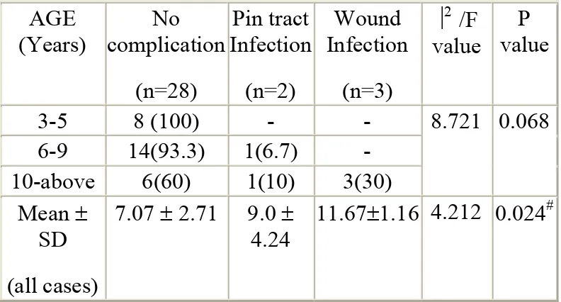
Table III : Distribution and comparison of age based on the complication AGE (Years) No complication (n=28) Pin tract Infection (n=2) Wound Infection (n=3)
⎟2 /F
value
P value
3-5 8 (100) - - 8.721 0.068
6-9 14(93.3) 1(6.7) - 10-above 6(60) 1(10) 3(30)
Mean ± SD (all cases)
7.07 ± 2.71 9.0 ± 4.24
11.67±1.16 4.212 0.024#
P<0.05:- # No complication Vs Wound Infection
In the study 28(84.8%) patients did not have any
complication, 2 (6.1%) patients had pin tract infection and 3
(9.1%) patients had wound infection. None of the subjects
developed complication in 3-5 years age group, 1(6.7%)
patient developed complication in 6-9 years age group and 4
(40%) patients developed complication in more than 10
years age group. The mean age of patients with out
complication is 7.07 ± 2.71 years, Pin tract infection is 9.0 ±
Table IV : Distribution and comparison of age by K wire removal time
AGE
(Years) 3 week(n=6) 4 weeks(n=23) 5 weeks(n=4) ⎟
2 /F
value P value
3-5 6 (75) 2(25) 32.354 0.001
6-9 - 15(100)
10-above - 6(60) 4(40) Mean ± SD
(all cases)
3.67 ± 0.82
7.83 ± 2.06
12.25 ± 0.96
27.24 0.001† ф ¶
P<0.05: †: 3weeks Vs 4 weeks, ф: 3 weeks Vs 5 weeks, ¶: 4 weeks Vs 5 weeks
Table V : Distribution and comparison of age based on FLYNN criteria AGE (Years) Excellent (n=30) Good (n=2) Fair (n=1)
⎟2 /t
value
P value
3-5 8 (100) - - 8.721 0.068
6-9 15(100) - -
10-above 7(70) 2(20) 1(10) Mean ± SD
all cases
7.2 ± 2.77 12.0±1.42 11.0 2.74§ 0.010
Figures in brackets are percentages, §: Excellent vs good+ fair
In this study the age of the patient shown statistically
significant (P=0.010) difference in terms of the outcome
based on Flynn criteria. The mean age of the 30 subjects
who had excellent Flynn criteria is 7.2 ± 2.77, which was
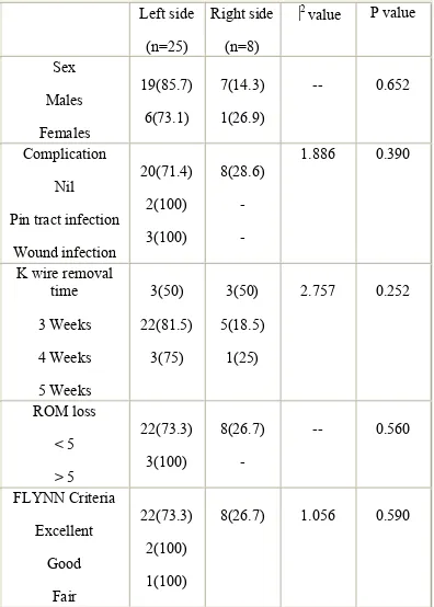
Table VI : Association of side of the fractured limb with gender, complication, K wire removal time, ROM loss
and FLYNN criteria.
Left side
(n=25)
Right side (n=8)
⎟2 value P value
Sex Males Females 19(85.7) 6(73.1) 7(14.3) 1(26.9)
-- 0.652
Complication Nil
Pin tract infection Wound infection 20(71.4) 2(100) 3(100) 8(28.6) - - 1.886 0.390
K wire removal time 3 Weeks 4 Weeks 5 Weeks 3(50) 22(81.5) 3(75) 3(50) 5(18.5) 1(25)
2.757 0.252
ROM loss < 5 > 5 22(73.3) 3(100) 8(26.7) -
-- 0.560
FLYNN Criteria Excellent Good Fair 22(73.3) 2(100) 1(100)
Distribution according to sex
0 10 20 30 40 50 60 70 80 90
male female
Gender
Percen
ta
g
e
Distribution according to age
0 5 10 15 20 25 30 35 40 45 503-5yrs 6-9yrs 10-above
Distribution according to side of
fracture
0 10 20 30 40 50 60 70 80RIGHT LIMB LEFT LIMB
Distribution according to time of surgery and post injury
Distribution according to time of K wire removal 0 10 20 30 40 50 60 70 80 90 100 3-4weeks 4-6weeks
Time of removal
Distribution according to
complication
0 1 2 3 4 5 6 7 8 9 10Pin track inf Wound inf
Complication Pe rc e n ta ge
Results according to FLYNN criteria
All the 33 patients were followed up with mean
follow-up period of 8.39±3.14 months. The minimum
follow-up was 4 months and maximum was 16 months. The
follow-up duration was comparable across gender, age
groups, complication, Range of motion loss and
Flynn-criteria outcome. The difference between groups were not
Preoperative Post operative
Follow up
Preoperative Post operative
Preoperative
Post operative
Follow up
Complications
Wound Healed by Secondary Intension
Loss of ROM
Complications
Wound infection healed by secondary intension
Loss of ROM
Supracondylar fractures of humerus in children are still difficult
to handle because of the age group involved and difficulty in
maintaining near anatomic reduction by closed means.
Dameron29 and Green5 explained, holding a reduction with
rotational deformity of supracondylar humerus was like
balancing two knives on one another.
In our study of thirty three patients with Gartland type III
supracondylar fracture of humerus, the mean age is seven years30.
Boys have had a higher incidence than the girls with a ratio of 3:2.
Non-dominant side or left side is involved more than the
dominant or right side30 .
In our study we advocated open reduction and crossed K-
wire fixation for the primary treatment of type III fractures.
Wilkins K E and Rosemont I L31 in their study cite the
accurate fracture reduction in open reduction and internal
fixation.
Basom WC et al17, Shifrin p et al18 and Wieland A et al19
in their studies showed excellent results with the use of open
reduction.
Two relatively recent reports by Archibald D A et al20 and
Cramer K et al21 showed excellent results in most patients with
the use of open reduction.
In a comparison study between closed reduction with
percutaneous pinning and open reduction with pinning by
Kaewpornsawan K et al32, the author concludes that both
treatments gave good results.
In most of the studies of closed reduction and pinning33,34,
they cite the risk of development of cubitus varus. But in open
complication. In our study even though it is very early to
observe, we had no cases of cubitus varus deformity.
In our study we used Campbell’s approach for fixation.
We had three cases of range of movement loss >5 degree out of
thirty three patients mainly due to wound infection in
post-operative period.
Sibly T et al25 in their study cited that there was no
increase in loss of movement in open reduction when compared
with closed reduction and percutaneous pinning.
Kasser J et al24 also found no loss of motion in elbows
operated using triceps splitting approach.
Gruber and Healey23 also recommended the posterior
approach to elbow for open reduction.
In our study we used crossed K- wires for internal fixation.
Zionts L E et al2 in their study compared the torsional
entry pinning , found out that crossed pinning is more stable than
lateral entry pinning.
In our study we had two cases of pin tract infection and
three cases of post operative wound infection. Which were
settled after appropriate care. Three cases of wound infection
result in loss of range of motion of > 5 degree mainly due to
poor post operative mobilisation of elbow by the patient.
In a study conducted by Muhall K J et al36 in 2000 on 16
children with supracondylar fractures, had 13 patients in
excellent group ,2 patients in good category and one in fair
group according to Flynn’s criteria. He suggested open reduction
and internal fixation is an effective and safe method of primary
treatment and is also associated with good outcomes.
Srivastava S37 in his study conducted during 2000 , which
consists of 42 children of type III supracondylar fractures using
showed excellent results in 81% , good in 17% and pin tract
infection in 14% of cases.
For closed reduction and percutaneous pinning C- arm and
power drill is mandatory#. Closed reduction and maintaining the
reduction during pinning needs experience15.
In our hospital where the emergency theatre at present
does not have a C-arm, open reduction and internal fixation
using crossed K- wires are being carried out routinely.
Limitations of this study:
1. It is not a comparative study
In conclusion of our study about the management of
Gartland type III supracondylar fracture of humerus in
children by open reduction and internal fixation using
crossed K – wires, we propound that this method of
management is ideal for our set up for the following reasons
1. Immediate anatomic reduction of fracture was carried
out there by eliminating the complications like
Volkmann’s ischemic contracture and cubitus varus
deformity.
2. Soft tissue damage was less as compared to the
repeated manipulation during closed reduction there
by reducing the risk of myositis ossificans.
3. Technically not very difficult to execute when
compared to the closed reduction and percutaneous
4. Crossed K – wires provides stability for early
mobilization of the elbow and there by avoiding the
soft tissue contracture.
5. The anxiety of the parents on seeing their child with
gross deformity and child’s agony of pain were
pacified by taking up for surgery at the earliest and
providing moral support to them.
6. Early management of the fracture reduces the period
of absenteeism in school for the child.
Results of our study is comparable with the earlier
studies, which advocate open reduction internal fixation
using crossed K – wires is the ideal treatment for Gartland
type III supracondylar fractures of humerus in children .
1. Blount W P: Fractures in children – Baltimore,
Williams and Wilkins 1955.
2. Zionts L E, McKellop H A, Hathaway R: Torsional
strength of pin configuration used to fix supracondylar
fracture of humerus in children , Journal of Bone and
Joint Surgery (Am) 1994;76:253-256.
3. Furrer M, Mark G, Ruedi T : Management of displaced
supracondylar fractures of humerus in children , Injury
1991: 22(4); 259-262.
4. Aronson D D, Prager B I: Supracondylar fractures of
the humerus in children- A modified technique for
closed pinning, Clinical Orthopaedics 1987; 219: 174 –
184.
5. Green N E : Fractures and Dislocations about the
elbow, In Green N E, ed . Skeletal trauma in children,
6. Campbell C C, Waters P M, Emans J B, et al:
Neurovascular injury and displacement in type III
supracondylar fractures, Journal of Pediatric
Orthopaedics 1995: 15: 47-52.
7. Chen J C, Wing-Man K, Shen W Y, et al: A new look
at the sequential development of elbow ossification
centers in children. Journal of Pediatric Orthopaedics
1998;18:161-167.
8. Nork S E, Hennrikes W L, Loncarich D P et al:
Relationship between ligamentous laxity and the site of
upper extremity fractures in children: Extension
supracondylar fracture verses Distal forearm fractures.
Journal of Pediatric Orthopaedics 1998 B 8:90-92.
9. Gartland J J: Management of supracondylar fracture of
humerus in children . Surg Gynecol Obstet
10. DeBeaux A C, Beattie T , Gilbert F : Elbow fat
pad sign; implication for clinical management. J R Coll
Surgery. Edinberg 1992 . 37; 205-206.
11. Rang M, Moseley C F, Roberts J M et al.
Symposium: Management of displaced supracondylar
fractures of humerus . Contemp Orthopaedics 1989
18;497-535,
12. Griffin P P: Supracondylar of humerus . Paeditric
Clinics of North America 1975 ; 22:477-486.
13. Arnold, J.A.; Nasca, R.J.; Nelson, C.L.
Supracondylar fractures of the humerus. Journal of
Bone Joint Surgery( Am) 59:589–595,1977.
14. Dunlop J: Transcondylar fractures of humerus in
childhood . Journal of Bone and Joint Surgery (Am)
1939, 21:59-73.
15. deBuy Roessing A S, Reiberg O: Open or closed
pinning for distal humerus fracture in humerus. Swiss
16. Archibeck M J, Scott S M et al : Brachialis
muscle entrapment in displaced supracondylar fracture:
a technique of closed reduction and report of initial
reports 1997. Journal of Pediatric Orthopaedics
17(3):298-302.
17. Basom, W.C. Supracondylar and transcondylar
fractures in children. Clin Orthop 1:43–48, 1953
18. Shifrin, P.; Gehring, H.; Iglesias, L. Open
reduction and internal fixation of displaced
supracondylar fractures of the humerus in children.
Orthopaedic Clinics of North America 7:573–581,
1976.
19. Weiland, A.; Meyer, S.; Tolo, V.T.; et al. Surgical
treatment of displaced supracondylar fractures of the
humerus in children. Analysis of fifty-two cases
followed for five to fifteen years. Journal of Bone and
20. Archibald, D.A.; Roberts, J.A.; Smith, M.G.
Transarticular fixation for severely displaced
supracondylar fractures in children. Journal of Bone
and Joint Surgery( Br) 73:147–149, 1991.
21. Cramer, K.; Devito, D.P.; Green, N.E.
Comparison of closed reduction and percutaneous
pinning versus open reduction and percutaneous
pinning in displaced supracondylar fractures of the
humerus in children. Journal of Orthopaedic Trauma
6:407–412, 1992.
22. Kotwal P T, Mani G V et al: Open reduction and
internal fixation of displaced fractures: International
Surgery 1989; 74(2):119-122.
23. Gruber, M.A.; Healey, W. The posterior approach
to the elbow revisited. Journal of Pediatric Orthopaedic
16:215–219, 1996.
24. Kasser, J.; Richards, K.; Millis, M. The
humeral fractures in adolescents: A Cybex evaluation
of triceps function and motion. Journal of Pediatric
Orthopaedics 10:93–96, 1990.
25. Sibly, T.; Briggs, P.; Gibson, M. Supracondylar
fractures of the humerus in childhood: Range of
movement following posterior approach to open
reduction. Injury 22:456–458, 1991.
26. Ippolito, E.; Moneta, M.; D'Arrigo, C.
Post-traumatic cubitus varus: Long-term follow-up of
corrective humeral osteotomy in children. Journal of
Bone Joint Surgergy( Am) 72:757–765, 1990.
27. Barrett I R, Bellemore M C, Kwon Y M.
Cosmetic results of supracondylar osteotomy for
correction of cubitus varus. Journal of Pediatric
Orthopaedics 1998;18:445-447.
28. Flynn, J.C.; Matthews, J.G.; Benoit, R.L. Blind
humerus in children. Journal of Bone Joint
Surgery(Am) 56:263–273, 1974.
29. Dameron, T.B. Transverse fractures of distal
humerus in children. Instructional Course Lecture
30:224–235, 1981.
30. Baratz M, Micucci C et al: Pediatric
supracondylar humerus fracture: Hand Clinics
2006;22(1):69-75.
31. Wilkins K E, ed. Operative management of upper
extremity fractures in children. Rosemont I L:
American Academy of Orthopaedic
Surgeons;1994:75-86.
32. Kaewpornsawan K : Comparison between closed
reduction with percutaneous pinning and open
reduction with pinning in children with closed totally
displaced supracondylar humeral fractures: a
randomized controlled trail . Journal of Pediatric
33. Arino, V.L.; Lluch, E.E.; Ramirez, A.M.; et al.
Percutaneous fixation of supracondylar fractures of the
humerus in children. Journal of Bone Joint
Surgery(Am)59:914–916, 1977.
34. Fahey, J.J. Fractures of the elbow in children.
Instrutional Course Lecture 17:13–46, 1960.
35. Mohammed S, Rymaszewski L A: Supracondylar
fracture of distal humerus in children. Injury 1995 ;
26(7): 487-489.
36. Mulhall K J, Abuzakuk T et al: Displaced
supracondylar fracture of humerus children.
International Orthopaedics 2000 ;24(4):221-223.
37. Srivastava S :The results of open reduction and
pin fixation in displaced supracondylar fractures of
humerus. Medical Journal of Malasyia 2000: 55 suppl
Flynn’s Criteria28
Results Cosmetic factor- loss of
carrying angle(degree)
Functional factor-loss
of motion (degree)
Excellent 0-5 0-5
Good 6-10 6-10
Fair 11-15 11-15
Consent Proforma
Title : Management of Gartland type III supracondylar
fracture of humerus in children by open reduction and
internal fixation using crossed K-wires
Aim : . The aim of this study is to evaluate
the outcome of management of Gartland type III
supracondylar fracture of humerus in children by open
reduction and internal fixation using crossed K – wires
Consent : I have been explained about the nature of my child’s
injury, methods of treatment, potential complications
and need of regular follow up visits in my own
vernacular language.
I hereby give my consent for including my child in this
study.
CLINICAL PROFORMA 1. Name
2. Age
3. Sex
4. In-Patient no. 5. Mode of injury
6. Side of injury 7. Dominant side 8. Gartland Type 9. Associated injury
10. Associated complications 11.Time and Date of injury 12. Time and Date of surgery
13. Post operative complication 14. Date of elbow mobilization 15. Date of suture removal
16. Date of K-wire removal
17. First follow up after K-removal:
18. Further follow ups:
Date ROM CA
19. Late complications
20. Date of last follow up 21. Flynn Criteria score
MASTER CHART
Sl.
no
Name Age Sex Side
T im ing of sur ger y ( post injury)
Procedure Complication Time
o f K W ire re mo va l
(in weeks) ROM LOSS Loss of carry
in
g
angle FLYNN
CRITERIA Follow Up(
m
onths)
1 Gana 10 F L 12 ORIF 4 0 0 E 16 2 Arav 7 M L 12 ORIF 4 0 0 E 15 3 Pras 8 M L 12 ORIF 4 0 0 E 12 4 Vinn 7 M L 12 ORIF 4 0 0 E 12 5 Kala 7 M L 12 ORIF 4 0 0 E 12 6 Nisa 6 M L 12 ORIF Pin
tract inf 4 0 0 E 12
7 Raj 10 M R 12 ORIF 4 0 0 E 11 8 Suba 3 M R 12 ORIF 3 0 0 E 11 9 Gaya 5 F L 12 ORIF 4 0 0 E 11 10 Vetr 7 M L 12 ORIF 4 0 0 E 10 11 Kart 11 F L 12 ORIF Wound
inf 4 15 0 F 10
12 Ajit 7 M L 12 ORIF 4 0 0 E 10 13 Akas 3 M L 12 ORIF 3 0 0 E 9 14 Vetr 11 F L 12 ORIF Wound
inf 4 10 0 G 9
15 Prag 11 M L 12 ORIF 4 0 0 E 9 16 Anan 6 M L 12 ORIF 4 0 0 E 8 17 Mani 4 F L 12 ORIF 3 0 0 E 8 18 Man 11 M L 24 ORIF 5 0 0 E 8 19 Siva 8 M L 12 ORIF 4 0 0 E 8 20 Kann 9 M R 12 ORIF 4 0 0 E 6 21 Siva 4 F R 12 ORIF 3 0 0 E 6 22 Prak 13 M L 12 ORIF Wound
inf 5 10 0 G 6
23 Sidd 3 M R 12 ORIF 3 0 0 E 6 24 Vima 7 M R 12 ORIF 4 0 0 E 6 25 Shar 6 M L 12 ORIF 4 0 0 E 6 26 Kris 12 M L 12 ORIF Pin
tract inf 5 0 0 E 6
KEY TO MASTER CHART
SEX: M- MALE S- SEX
SIDE OF INJURY: R- RIGHT L- LEFT ORIF: OPEN REDUCTION
AND INTERNAL FIXATION inf: INFECTION
ROM: RANGE OF MOTION FLYNN CRITERIA:
E- EXCELLENT G- GOOD