0022-538X/95/$04.0010
Copyright 1995, American Society for Microbiology
Arginine-Rich Regions Succeeding the Nuclear Localization Region
of the Herpes Simplex Virus Type 1 Regulatory Protein ICP27
Are Required for Efficient Nuclear Localization
and Late Gene Expression
MICHELE K. HIBBARDANDROZANNE M. SANDRI-GOLDIN*
Department of Microbiology and Molecular Genetics, College of Medicine, University of California, Irvine, California 92717-4025
Received 23 March 1995/Accepted 2 May 1995
The herpes simplex virus type 1 (HSV-1) immediate-early protein ICP27 is an essential regulatory protein that localizes to the nuclei of infected cells. The strong nuclear localization signal (NLS) of ICP27 was identified recently and shown to reside in the amino-terminal portion of the polypeptide from residues 110 to 137 (W. E. Mears, V. Lam, and S. A. Rice, J. Virol. 69:935–947, 1995). There are also two arginine-rich regions directly succeeding the NLS. The first of these arginine-rich sequences (residues 141 to 151), together with the NLS, has been shown by Mears et al. to form the nucleolar localization signal. Arginine-rich motifs are common in domains involved in nuclear localization and RNA binding. To analyze the role of the arginine-rich regions in ICP27, we constructed stably transformed cell lines containing ICP27 mutants with deletions of all or parts of the NLS and arginine-rich regions. We also constructed mutants in which these regions were replaced with heterologous NLSs or RNA-binding domains. Characterization of these mutants indicated that the arginine-rich regions were required but not sufficient for wild-type localization of ICP27. More importantly, the NLS and arginine-rich regions were also essential to the function of ICP27. Mutants lacking these sequences were defective in late gene expression during infection even when ICP27 was properly localized to the nucleus by substitution of the NLS from simian virus 40 large T antigen. Further, the defect in late gene expression could not be overcome by replacement with the highly basic RNA-binding domain of human immunodeficiency virus type 1 Tat. The deficiency in late gene expression was independent of ICP27’s role in stimulating viral DNA replication. In addition, localization of the HSV-1 proteins ICP4, ICP0, and ICP8 was unaffected by ICP27 mutants in this region. These results suggest that the arginine-rich regions are required for efficient nuclear localization and for the regulatory activity of ICP27 involved in viral late gene expression.
Herpes simplex virus type 1 (HSV-1) lytic infection exhibits a tightly regulated pattern of viral gene expression and DNA replication leading to virion assembly (79). There are four kinetic classes of genes, which are temporally regulated.
Im-mediate-early (a) gene expression occurs immediately
follow-ing infection (30, 31). The early (b) genes are expressed next,
and these genes primarily encode products necessary for viral DNA replication. Following early gene expression, DNA rep-lication begins, and these events stimulate expression of the late genes. The late genes can be separated into two distinct
classes, the leaky late (g-1) genes, whose expression can be
observed prior to viral DNA replication but does not reach maximum levels until DNA synthesis has begun, and the strict
late (g-2) genes, whose expression is dependent on the onset of
viral replication (28, 29, 32, 40, 68).
The immediate-early protein ICP27 (IE63) is one of the four immediate-early proteins of HSV-1 involved in regulated gene expression (41, 55, 63). It is also required for efficient viral DNA replication (41, 55, 63) and plays a role in the shutoff of host protein synthesis (22, 63). ICP27 is a 512-amino-acid, 63-kDa phosphoprotein which localizes to the nuclei of in-fected cells (1, 36, 82). It functions in the switch from imme-diate-early and early gene expression to late gene expression,
which results in a down regulation of early expression and an activation of late expression. Thus, in cells infected with ICP27 temperature-sensitive or deletion mutants, late gene expres-sion is dramatically decreased and some immediate-early and early genes are overexpressed (41, 55, 63, 70). Transfection experiments with different target genes have demonstrated that ICP27 can both positively and negatively affect the expres-sion of different target genes (5, 23, 44, 55, 58, 72, 76). We have termed the positive regulation by ICP27 the activator function and the negative regulation the repressor function (23). These terms may not refer necessarily to activities of ICP27 at the transcription level because in transfection studies we have demonstrated that regulation by ICP27 occurs at least in part at the level of RNA processing (66). The activator function of
ICP27 correlates with its ability to stimulate 39RNA
process-ing at selected poly(A) sites (5, 42, 66). The repressor function correlates with genes containing introns (66) and is related to the effect of ICP27 in the shutoff of host protein synthesis (22). We have shown that there is a reduction in cellular mRNA levels and an accumulation of pre-mRNA during infection in the presence of ICP27 (22). We also demonstrated that HSV-1 infection impairs splicing in vitro and that ICP27 is required for this inhibition (24). This splicing impairment could ulti-mately affect protein synthesis and thus be the means by which ICP27 contributes to host shutoff. In addition, ICP27 temper-ature-sensitive and deletion mutants are defective in viral DNA replication during infection (9, 41, 55, 63). Although the regulation of late gene expression is dependent on viral DNA
* Corresponding author. Mailing address: Department of Microbi-ology and Molecular Genetics, College of Medicine, University of California, Irvine, CA 92717-4025. Phone: (714) 824-7570. Fax: (714) 824-8598. Electronic mail address: [email protected].
4656
on November 9, 2019 by guest
http://jvi.asm.org/
replication and ICP27 has been implicated in both activities, these effects appear to be independent of one another. Some ICP27 mutants are defective in viral replication as well as late gene expression, whereas others exhibit only an effect on late gene expression and still replicate DNA at wild-type levels (56, 57), suggesting a function for ICP27 in gene regulation sepa-rate from viral DNA replication.
Numerous studies have been done to define the regions of ICP27 important for the many activities in which it is involved. These studies have indicated that it is the carboxy-terminal half of ICP27 which is required for its activation and repression functions (5, 23, 44, 55, 58, 72, 76). In addition, one study has implicated the acidic amino-terminal portion of ICP27, from amino acids 12 to 63, in the repressor activity (57). This region is also required for an essential lytic function, as viral recom-binants containing a deletion of this region were defective for growth (57). The carboxy-terminal repressor region is also the region required for ICP27’s effect on splicing (22, 24, 66). Thus, with the exception of the acidic region, no other function has been attributed to the amino-terminal portion of ICP27 until recently. Mears et al. (45) have identified the strong nuclear signal (NLS) and nucleolar localization signal (NuLS) of ICP27 in the amino-terminal half of the polypeptide from amino acids 110 to 137 and 110 to 152, respectively, with one or more weak NLSs mapping to the carboxy-terminal portion of the protein. It is notable that the amino-terminal half of ICP27 also contains two arginine-rich regions which are similar to those found in a number of functional domains. These sequences resemble motifs common to both NLSs and RNA-binding domains by virtue of their high arginine content. In addition, the region encompassing the strong NLS and the NuLS, which includes a portion of the arginine-rich region, has been implicated in an important in vivo function because of the inability of recombinant virus carrying mutations in these re-gions to replicate efficiently (45). This finding suggests an ad-ditional role for this region in the function of ICP27. Yet, aside from the proper localization of ICP27, a specific function for the domain encoding the strong NLS and arginine-rich regions of ICP27 has not been determined.
The focus of this study was to further examine the amino-terminal portion of ICP27 with respect to its role in the local-ization and function of ICP27. We wanted to determine whether the NLS and/or arginine-rich regions of ICP27 were required for the sole purpose of properly localizing ICP27 to the nuclei of infected cells or whether they were further in-volved in the activity of ICP27. Therefore, we constructed stably transformed cell lines which contained ICP27 mutants with deletions of the NLS and/or various portions of the argi-nine-rich regions or in which this region of ICP27 was replaced with well-defined, highly basic functional domains of other heterologous viral proteins. Detailed characterization of these cell lines indicated that the arginine-rich regions of ICP27 were involved in its proper localization. However, this region of ICP27 was important for more than the appropriate localiza-tion of ICP27 because mutants with a delelocaliza-tion of the strong NLS and arginine-rich regions could be localized to the nu-cleus with heterologous signals yet were unable to support viral growth. Specifically, ICP27 mutants of this region were defi-cient in late gene expression, and this defect was independent of DNA replication.
MATERIALS AND METHODS
Bacteria and plasmids.Escherichia coli K-12 strain 1100 derivative DH-1 (21) was used as the host for propagation of all chimeric plasmids. Plasmid pD2DS5 was constructed by combining two previously described ICP27 mutations, D2 and S5 (23). The mutant D2 has a deletion of a serine residue at position 103 and an
insertion of four amino acids, PEFR. The mutant S5 contains a 25-amino-acid deletion of residues 153 to 178 and an insertion of four amino acids, PEFR (23). Plasmids containing these mutations in the context of the BamHI-to-SstI frag-ment of ICP27, with the BamHI site designated nucleotide (nt) 1 and the SstI site designated nt 2421 as previously described (23), were digested at the BamHI site and the EcoRI site which occurs in the PEFR linker present in both mutants. The BamHI-to-EcoRI fragment of D2, containing the 59noncoding region and the sequence encoding the amino-terminal portion of ICP27, from residues 1 to 103, was joined with the BamHI-to-EcoRI fragment of S5, which contains the se-quence encoding the carboxy-terminal portion of ICP27 from residues 178 to 512 and the 39noncoding region in the pUC18 vector into which all of these muta-tions were originally cloned (72). This created a 75-amino-acid deletion of residues 103 to 178 of ICP27, and the mutant was termed D2DS5. Plasmid pRG was created by insertion of a synthetic oligonucleotide, GAATTCCGCCGTGG GCGTCGCAGGGGTCGGGGTCGCGGTGAA, encoding residues 141 to 151 of ICP27, RRGRRRGRGRG, into the EcoRI site in D2DS5. This mutation resulted in a deletion of residues 103 to 140 and 152 to 178 of the ICP27 amino acid sequence. Plasmid pTAG was created by insertion of a synthetic oligonu-cleotide, AATTCCCCAAGAAGAAGCGCAAGGTC, encoding the minimal NLS of simian virus 40 (SV40) large T antigen, PKKKRKV, into the EcoRI site in the mutant D2DS5. This mutation contains an insertion of the SV40 minimal NLS between residues 103 and 178. Plasmid pTAT.R was constructed by the insertion of an oligonucleotide, AATTCTACCGCAAGAAGCGCCGCCAGC GCCGCCGCGCCG, encoding the human immunodeficiency virus (HIV) Tat NLS, YRKKRRQRRRA, with a glycine-to-tyrosine substitution into the EcoRI site of pD2DS5. This mutation contains an insertion of the HIV Tat NLS with a single amino acid substitution between residues 103 and 178. Plasmid pTAT.K was created by the insertion of AATTCTACAAGAAGAAGCGCAAGCAGAA GAAGAAGGCCG encoding a mutation of the TAT.R sequence, KKKRKK KKKA, with arginine-to-lysine substitutions of all but the arginine residue at position 5. This mutation is an insertion of a nonspecific highly basic sequence between residues 103 and 178 of the ICP27 amino acid sequence. The construc-tion and characterizaconstruc-tion of the ICP27 inserconstruc-tion mutaconstruc-tion S13 have been de-scribed elsewhere (23). The S13 mutation contains a four-amino-acid insertion between residues 262 and 263 and has been characterized as an activator mutant (23). All of the mutants were sequenced around the sites of the deletion or insertion.
Cells and viruses.Vero cells were grown as previously described (71). The Cell line 2-2, which contains the wild-type ICP27 gene, has been described elsewhere (70). HSV-1 KOS1.1 and the mutant 27-LacZ, which has an insertion of the lacZ gene in the ICP27 locus, were propagated as described previously (65, 70). For the isolation of cell lines containing the ICP27 mutant plasmids, Vero cells were transfected by using Lipofectin reagent (Life Technologies) with 5 mg of pD2DS5, pRG, pTAG, pTAT.R, pTAT.K, pS5, or pS13 plasmid DNA and 1mg of pFeLTR-neo, which contains the gene encoding resistance to G418 under the control of the feline leukemia virus long terminal repeat as described earlier (67). Selection in media containing 750mg of G418 per ml was initiated 24 h after transfection. Individual colonies were isolated after 2 weeks, expanded, and then screened by indirect immunofluorescence for the expression of ICP27. G418-resistant colonies were infected with 27-LacZ to activate expression of the res-ident mutant ICP27 gene, and immunofluorescence staining was performed with a polyclonal antibody specific for the C-terminal half of ICP27 (23). Expression of a full-length mutant ICP27 protein was confirmed by Western immunoblot analysis of whole cell lysates, except for the mutant S5, in which immunoblot analysis was performed on immunoprecipitates from nuclear extracts. Cells in-fected with HSV-1 KOS1.1 or 27-LacZ were harvested 6 h after infection. Polyacrylamide gel electrophoresis was performed as previously described (78). For the mutant S5, nuclear extracts were prepared (12) and immunoprecipitation with the monoclonal antibody to ICP27, H1119 (Goodwin Institute for Cancer Research), was performed as described previously (64). Electrophoretic transfer of proteins to nitrocellulose was done as described previously (23) except that transfer took place in a buffer of 20% methanol–25 mM Tris–190 mM glycine (pH 8.5) at 110 mA overnight. Immunoblotting experiments were performed by using enhanced chemiluminescence (Amersham Lifescience) with the primary antibody, H1119, to ICP27 at a dilution of 1:5,000 dilution and anti-mouse horseradish peroxidase-linked whole antibody (Amersham Lifescience) at a di-lution of 1:25,000.
Immunofluorescence staining.Vero cells, 2-2 cells, or each of the stably transformed cell lines were infected with KOS1.1 or 27-LacZ as indicated. Cells were fixed in 3.7% formaldehyde at various times after infection, after which 0.5% Nonidet P-40 in phosphate-buffered saline (PBS) was added to permeabi-lize the cells. The coverslips were rinsed three times in PBS containing 1% newborn calf serum after permeabilization and before the addition of each antibody or reagent. The monoclonal antibodies H1119 to ICP27, H1112 to ICP0, H1114 to ICP4, and H1115 to ICP8 (Goodwin Institute for Cancer Re-search) and the C-terminal ICP27 polyclonal antibody (23) were the primary antibodies and were used at a dilution of 1:500. Biotinylated goat anti-mouse antibody and biotinylated donkey anti-rabbit antibody (Amersham) were used as secondary antibodies at a dilution of 1:100. After rinsing, a 1:100 dilution of streptavidin-conjugated fluorescein (Amersham) was added to cells for 30 min. Finally, cells were rinsed three times in PBS without serum, and coverslips were
on November 9, 2019 by guest
http://jvi.asm.org/
mounted in PBS. Cells were examined with a Nikon UFX-II epifluorescence microscope with a 1003, 1.25-numerical-aperture objective.
Northern (RNA) hybridization.Total RNA was extracted 2, 4, 6, and 8 h after infection by the guanidium thiocyanate method (3). Equivalent amounts (usually 20mg) of RNA were denatured in glyoxal (77) and fractionated on 1% agarose gels. Transfer of RNA to GeneScreen (Dupont, NEN Research Products) and hybridization conditions were as described previously (64). Blots were hybridized with viral mRNA-specific probes and then with a 28S rRNA probe as previously described (22). Hybridization with the 28S rRNA probe was performed to ensure equivalent loading of samples. Probes used in Northern hybridization analysis of viral mRNA were prepared by in vitro transcription of antisense RNA in the presence of [32P]CTP as follows. For ICP0, a 250-nt antisense RNA was gener-ated from a BamHI-AvaI fragment of the ICP0 gene which spans a portion of intron 1 and exon 2 (24); for ICP4, a 1,690-nt antisense RNA was generated by T7 polymerase transcription of a SalI-linearized pGEM-1 plasmid containing a BamHI-SalI fragment from the amino-terminal portion of the ICP4 gene; for ICP27, a 217-nt antisense RNA was generated by T7 transcription of an NcoI digestion of pGEM-2 containing the 1,250-bp BamHI-SalI fragment of the ICP27 gene; for ICP8, a 750-nt antisense RNA was generated by SP6 polymerase transcription of a HindIII-linearized pGEM-2 plasmid containing the BamHI-XhoI fragment from the amino-terminal half of the ICP8 gene; for glycoprotein B (gB), a 1,189-nt antisense RNA was generated by T7 transcription of a PstI-linearized pGEM-1 plasmid containing the PstI-SalI fragment from the amino-terminal half of the gB gene; for glycoprotein C (gC), a 960-nt antisense RNA was generated from the EcoRI-XbaI fragment of the gC gene; for glycoprotein D (gD), an 840-nt antisense RNA was generated by T7 transcription of an NcoI-linearized pGEM-1 plasmid containing the HindIII-AccI fragment of the gD gene; for UL38, an 800-nt antisense RNA was generated by T7 transcription of a HindIII-linearized pGEM-1 plasmid containing the PvuII-SstI fragment of the carboxy-terminal half of the UL38 gene.
Measurement of DNA replication.Cell lines were infected with 27-LacZ at a multiplicity of infection of 1.0. At 1 and 12 h after infection, cultures were harvested and total DNA was isolated as previously described (72). Purified DNA was denatured and applied to nitrocellulose filters, using a slot blot appa-ratus (Bethesda Research Laboratories). The blots were hybridized with a32 P-labeled antisense RNA probe specific for the gC gene prepared as described above. Parallel samples were incubated until 24 h after infection, at which time cultures were harvested and virus yields were assayed on 2-2 cells.
Artwork for Fig. 2 to 7.Autoradiographs and photographs of immunofluores-cence cell staining were scanned on a Hewlett Packard ScanJet II CX/T with a transparency adapter. The images were scanned at 300 dots per inch into Adobe Photoshop 3.0 and were exported as TIFF files.
RESULTS
Mutants of the amino-terminal half of ICP27 are defective
in the ability to localize to the nuclei of infected cells.ICP27
contains two arginine-rich regions in its amino terminus which resemble the NLSs of a number of other viral proteins, in that they are highly concentrated stretches of basic amino acids. These regions succeed the recently defined strong NLS of ICP27, although the first of these arginine-rich sequences is included in the NuLS (45). To examine the effects of these regions on the localization of ICP27 during viral infection, we created stably transformed cell lines carrying ICP27 genes with deletions of all or portions of these sequences. Infection of these cell lines with the ICP27 null mutant virus, 27-LacZ, allowed for VP16 activation of expression from the resident mutant ICP27 gene (72), and thus the localization of these ICP27 mutants during infection could be examined.
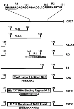
The strong NLS of ICP27 has been mapped to residues 110 to 137 of ICP27, and the NuLS has been mapped to residues 110 to 153 (45). The two arginine-rich sequences are found between residues 141 and 171. We termed these regions R1 and R2 (Fig. 1). R1 consists of an 11-amino-acid sequence, RRGRRRGRGRG, encompassing residues 141 to 151 and is part of the NuLS of ICP27. R2, which is 10 amino acids down-stream of R1, is composed of the sequence PRRRAPRTNR, from residues 162 to 171. A 75-amino-acid deletion from res-idues 103 to 178 which included the strong NLS of ICP27 as well as R1 and R2 was constructed as described in Materials
and Methods. This mutant was termed D2DS5 (Fig. 1). Into
this construct, we inserted a synthetic oligonucleotide encoding the R1 sequence. This mutant, called R1, consisted of a
dele-tion of amino acids 103 to 140, including the strong NLS of ICP27, and 152 to 178, the R2 region, but contained the first arginine-rich region, R1 (Fig. 1). The mutants S5 and S13 (23) were also used in these studies. S5 contains a deletion of R2, residues 153 to 178, maintaining both the NLS and NuLS (Fig. 1). S13 is a four-amino-acid insertion between residues 262 and 263 at the start of what we have termed the activator region of ICP27 (23). This mutant was used as a control to compare the activity of a previously characterized ICP27 mutation as ex-pressed from a stably transformed cell line.
Cell lines containing each of these mutants were generated by cotransfection of plasmid pFeLTR-neo (67) and a plasmid
containing an ICP27 mutation, pD2DS5, pRG, pS5, or pS13.
G418-resistant colonies were selected, expanded, and screened for ICP27 expression as described in Materials and Methods. To verify expression of the full-length mutant proteins from each of the stably transformed cell lines, parental Vero cells
and the cell lines D2DS5, R1, containing the RG mutation,
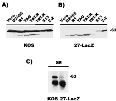
S13, and 2-2, which carries the wild-type ICP27 gene (70), were infected with HSV-1 KOS or 27-LacZ and then analyzed by Western blot analysis. The wild-type 63-kDa ICP27 protein was detected in all lanes containing lysates from KOS-infected cells, as were the mutant ICP27 proteins (Fig. 2A). The ICP27 mutant proteins were the only ICP27 proteins detected in lysates from 27-LacZ-infected cells (Fig. 2B). The apparent molecular masses of the ICP27 mutant proteins were as
[image:3.612.360.511.68.289.2]fol-lows: D2DS5 encodes a 441-amino-acid polypeptide of about
FIG. 1. Schematic representation of the amino-terminal half of the ICP27 polypeptide showing the locations of the deletion mutations and the amino acid sequences and positions of the insertion mutations. The positions of the previ-ously defined strong NLS, from residues 110 to 137, and NuLS, from residues 110 to 153, are shown (45). The amino acid sequences of the two arginine-rich regions succeeding the NLS, termed R1, from residues 141 to 151, and R2, from amino acids 162 to 171, are also depicted. The mutation D2DS5 is a deletion of amino acids 103 to 178 removing the NLS and both arginine-rich regions of ICP27. The mutation RG is a deletion of residues 103 to 140, including the NLS, and residues 152 to 178, which includes the R2 region of ICP27. The mutation S5 is a deletion of residues 153 to 178 of ICP27, including R2 (23). The mutation TAG is an insertion of the minimal NLS of SV40 large T antigen, PKKKRKV (20, 34), between residues 103 and 178 of the ICP27 polypeptide. The mutation TAT.R is an insertion of the NLS of the HIV Tat protein, YRKKRRQRRRA (15, 25, 62), between residues 103 and 178 of the ICP27 polypeptide. The mutation TAT.K is an insertion of the sequence YKKKRKKKKKA, in which lysine residues were substituted for the arginine residues in the TAT.R inserted sequence in all but the arginine residue at position 5.
on November 9, 2019 by guest
http://jvi.asm.org/
54 kDa, R1 encodes a 454-amino-acid polypeptide of about 56 kDa, and S13 encodes a 516-amino-acid polypeptide of about 63 kDa which was indistinguishable from the wild-type protein (Fig. 2A and B). This finding confirmed that each of the stably transformed cell lines expressed a mutant of ICP27 of the appropriate predicted molecular mass and allowed us to ob-serve the expression level of each of the ICP27 mutants com-pared with the wild-type level in KOS infection or in 27-LacZ infection of 2-2 cells. In all cases, expression was apparent but protein levels were lower in the 27-LacZ-infected cells than in KOS-infected cells.
The mutant S5 localized primarily to the nucleoli of infected cells, and Western blot analysis using whole cell lysates was not sufficiently sensitive to detect this protein. To detect expression of the S5 mutant protein, it was necessary to isolate the nuclei of KOS- and 27-LacZ-infected S5 cells and immunoprecipitate the mutant protein with monoclonal antibody H1119 prior to Western blot analysis. S5 encodes a 495-amino-acid polypep-tide of about 61 kDa. Thus, the expression of the full-length S5 mutant protein from 27-LacZ-infected cells was also confirmed (Fig. 2C).
To examine the localization of each of the ICP27 mutants, the cell lines were infected with 27-LacZ to activate expression of the mutant ICP27 protein, and immunofluorescence stain-ing was performed on cells fixed 2, 4, 6, and 8 h after infection. Staining was carried out with both monoclonal antibody H1119 and a polyclonal antibody to the carboxy-terminal portion of ICP27 (23) to address the possibility of differential reactivities of antibodies to mutant forms of the same protein (9). Detec-tion with the polyclonal antibody was not exclusively specific for ICP27, and thus there was some nuclear background stain-ing even in uninfected cells (Fig. 3B, mock). Also, stainstain-ing of wild-type-infected cells fixed at 6 and 8 h with the polyclonal antibody resulted in the majority of fluorescence localized to
cytoplasmic structures. It has been suggested that this pattern is due to the binding of rabbit immunoglobulin G to the HSV-1 late Fc receptor proteins (Fig. 3B, KOS) (9). This binding does not affect staining of cells expressing mutant forms of ICP27, as all of these mutants were deficient in late gene expression. Staining of cells fixed 2 h after infection with the lower-affinity polyclonal antibody was very faint (data not shown). The
re-sults showed that mutant D2DS5 was significantly cytoplasmic
at all times after infection compared with wild-type ICP27 as seen in KOS-infected Vero cells or 27-LacZ-infected 2-2 cells. This was the case with both the monoclonal antibody (Fig. 3A) and the polyclonal antibody (Fig. 3B), although slightly greater cytoplasmic fluorescence was seen with the polyclonal anti-body. It was also apparent that there was bright nuclear stain-ing with either antibody. Also, the mutant R1, which contains only the R1 arginine-rich region, showed significant cytoplas-mic fluorescence at late times after infection with the mono-clonal antibody (Fig. 3A) but was almost exclusively nuclear as detected by the polyclonal antibody. Finally, the mutant S5, which has a deletion of only the R2 region, localized prefer-entially to the nucleolus and could be detected in the nucleus only at late times (Fig. 3A). The immunofluorescence pattern seen with the polyclonal antibody was the same as that ob-served with the monoclonal antibody (data not shown). Al-though none of these deletion mutants was exclusively cyto-plasmic, presumably because of a number of weak NLSs in the carboxy-terminal portion of ICP27 (45), the localization of each of these mutants was defective compared with wild-type localization. Similar results were obtained by Mears et al. (45) for mutants lacking the major NLS.
To quantify the difference between nuclear and cytoplasmic
localization of the D2DS5 protein and the R1 protein relative
to wild-type ICP27, nuclear and cytoplasmic fractionation was performed on the mutant cell lines infected with 27-LacZ for 6 h and on Vero cells infected with KOS for 6 h. ICP27 was immunoprecipitated from each fraction prior to Western blot analysis as described in the legend to Fig. 2 for the S5 protein. Following enhanced chemiluminescence detection of ICP27, the images were scanned and the relative amounts of ICP27 in the nuclear and cytoplasmic fractions were calculated. It was found that approximately five times more wild-type ICP27 was detected in the nuclear fraction than in the cytoplasmic frac-tion in KOS-infected Vero cells. In contrast, about 0.5 times
the amount of the D2DS5 protein was found in the nuclear
fraction compared with the cytoplasmic fraction, and there were nearly equal amounts of the R1 protein in the two frac-tions. These results showed that deletion of both the NLS and arginine-rich regions of ICP27 resulted in a mutant protein that was the most defective in its ability to localize to the
nucleus (D2DS5). The first arginine-rich region, R1, acted as a
partial NLS but could not fully restore this defect. In addition, the NLS and NuLS including R1 were insufficient to properly localize ICP27 to the nucleus in the absence of the second arginine-rich region, R2, as seen in mutant S5.
Nuclear localization defects in mutants of the
amino-termi-nal half of ICP27 can be overcome by heterologous NLSs.As
deletion of the region of ICP27 including the strong NLS, R1, and R2 created a mutant which was compromised in its ability to localize to the nuclei of infected cells, we wanted to examine whether ICP27 could be directed to the nuclei of infected cells by defined NLSs from nonherpesvirus proteins. We inserted the well-characterized minimal NLS of the SV40 large T anti-gen (33, 34) into the region of the deletion in the mutant
D2DS5. The resulting mutant, TAG, contained the lysine-rich
SV40 large T-antigen minimal NLS, PKKKRKV, in frame between residues 103 and 178 of the ICP27 polypeptide,
re-FIG. 2. Western blot analysis to detect expression of full-length mutant ICP27 proteins from stably transformed cell lines. Vero cells, 2-2 cells, which contain the wild-type ICP27 gene (70), and the cell line D2DS5, R1, TAG, TAT.R, TAT.K, and S13 were infected with HSV-1 KOS (A) to determine the relative mobilities and levels of expression of the mutant proteins compared with those of wild-type ICP27. Parallel cultures were infected with 27-LacZ (B) to activate expression of the resident ICP27 gene and thus verify expression of a full-length mutant protein from each of the stably transformed cell lines. Cul-tures were harvested 6 h after infection, and whole cell lysates were fractionated on a sodium dodecyl sulfate–10% polyacrylamide gel. The proteins were trans-ferred to nitrocellulose by electroblotting. The blot was treated with a monoclo-nal antibody to ICP27, H1119. (C) S5 cells were infected, and nuclear extracts were prepared (12). Immunoprecipitation was performed with antibody H1119, and precipitated antigen-antibody complexes were fractionated by polyacryl-amide gel electrophoresis followed by Western blot analysis. Sizes are indicated in kilodaltons.
on November 9, 2019 by guest
http://jvi.asm.org/
[image:4.612.83.269.69.231.2]placing both the NLS and arginine-rich regions of wild-type ICP27 (Fig. 1). We also constructed a mutant in which the NLS of the HIV Tat protein (25, 62, 69) was inserted in frame in the same position as in the mutant TAG. The resulting mutant, TAT.R, had the arginine-rich HIV Tat NLS, YRKKRRQR RRA, inserted between residues 103 and 178 of ICP27 (Fig. 1). As this sequence was also a defined arginine-rich RNA-binding domain, this mutant would also provide a means of determin-ing whether the arginine-rich region of ICP27 is involved in a similar activity. Finally, we created a mutant in which we in-serted the amino acid sequence YKKKRKKKKKA in frame into the same region of ICP27. This sequence was an arginine-to-lysine mutation of all but one of the arginine residues in the TAT.R inserted sequence. The resulting mutant, TAT.K (Fig. 1), allowed us to determine the effect of a highly basic se-quence, which was not a defined NLS but which still main-tained the ability to bind RNA, on the localization and func-tion of ICP27.
Cell lines containing these ICP27 mutants were generated, and expression of the full-length mutant protein was verified by Western blot analysis (Fig. 2A and B). TAG encodes a 450-amino-acid polypeptide of about 55 kDa, while both TAT.R and TAT.K encode 452-amino-acid polypeptides of about 56 kDa. The expression of the appropriate-size mutant protein was confirmed in lysates from 27-LacZ-infected cells (Fig. 2B), although the relative level of expression was lower than that of the wild-type protein in KOS infections (Fig. 2A). To
deter-mine whether these mutant forms of ICP27 were localized to the nucleus, immunofluorescence staining of 27-LacZ-infected cells fixed 2, 4, 6, and 8 h after infection was performed. The TAG mutant localized to the nucleus of infected cells as effi-ciently as wild-type ICP27, as detected by either the monoclo-nal (Fig. 3A) or polyclomonoclo-nal (Fig. 3B) antibody. While the mu-tants TAT.R and TAT.K were predominantly nuclear as detected by either antibody, a slight cytoplasmic staining was observed for both mutants with the monoclonal antibody (Fig. 3A). This cytoplasmic fluorescence was also detected at early times (4 h) for the mutant TAT.K when the polyclonal anti-body was used (Fig. 3B). Thus, insertion of the SV40 large T-antigen minimal NLS directed ICP27 to the nuclei of in-fected cells as efficiently as the wild-type sequence. In addition, both the HIV Tat protein NLS and a lysine-rich mutated ver-sion of this sequence restored the nuclear localization of the ICP27 deletion mutant.
ICP27 mutants with deletions in the amino-terminal NLS or arginine-rich region cannot complement infection with an
ICP27 null mutant virus.The arginine-rich region of ICP27
and the preceding sequences have not been implicated in any of the functions of ICP27 other than nuclear/nucleolar local-ization, yet viral mutants of the NLS and NuLS regions were deficient in viral replication (45). To determine whether any of the ICP27 mutants of this region could complement an ICP27 null mutant virus in infection, the ICP27 null mutant virus 27-LacZ was plated on Vero cells, complementing 2-2 cells,
FIG. 3. Immunofluorescence analysis of the cellular location of nuclear localization mutants of ICP27 during infection. (A) Vero cells were infected with HSV-1 KOS, and cell lines carrying wild-type ICP27 (2-2) or the mutant D2DS5, RG (R1), TAG, TAT.R, TAT.K, S5, or S13 were infected with 27-LacZ. Cells were fixed 2, 4, 6, and 8 h after infection and treated with monoclonal antibody H1119. (B) Uninfected Vero cells (mock), Vero cells infected with HSV-1 KOS, and the cell lines D2DS5, R1, TAG, TAT.R, and TAT.K infected with 27-LacZ were treated with a polyclonal antibody specific for the carboxy-terminal portion of ICP27 (23).
on November 9, 2019 by guest
http://jvi.asm.org/
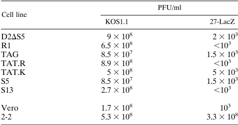
and the cell lines D2DS5, R1, TAG, TAT.R, TAT.K, S5, and S13. The efficiency of plating was compared with that of HSV-1 KOS. This comparison revealed 4- to 5-log-units-lower titers for the 27-LacZ-infected cells than for the KOS-infected cells in all of the cell lines harboring mutants (Table 1). The titer of 27-LacZ on 2-2 cells was comparable to wild-type titers (Table 1). These data indicate that ICP27 mutants of the
amino-terminal NLS and arginine-rich regions were unable to support infection in the absence of wild-type ICP27. This deficiency was not due solely to the improper localization of the mutant proteins because the TAG mutant, containing the NLS of the SV40 large T antigen, localized to the nucleus as efficiently as the wild-type protein yet was still unable to complement 27-LacZ infection (Table 1). In addition, replacement of this region with a defined RNA-binding region was also insufficient to restore its function, as evidenced by the inability of the TAT.R and TAT.K mutants to support 27-LacZ infection (Ta-ble 1).
The amino-terminal localization regions of ICP27 are
re-quired for late gene expression.ICP27 is required for late gene
expression. To determine the nature of the deficiency of mu-tants of the NLS and arginine-rich regions of ICP27 to com-plement infection of an ICP27 null mutant virus, we examined the steady-state mRNA levels of HSV-1 genes from the differ-ent kinetic classes. Northern blot hybridization was performed on total RNA extracted from Vero cells, 2-2 cells, or the stably
transformed cell line D2DS5, R1, TAG, TAT.R, TAT.K, S5, or
S13 at 2, 4, 6, and 8 h after infection with 27-LacZ (Fig. 4). Control infections were performed in Vero cells with KOS. For each cell line, one blot was sequentially hybridized with probes specific for transcripts of each kinetic class, with subsequent stripping of the blots between hybridizations with different probes as described previously (72). The blots were then hy-bridized with a 28S rRNA probe, and the autoradiographs were densitometrically scanned to ensure equivalent loading of
FIG. 4. Northern blot analysis of steady-state levels of HSV-1 mRNAs from each of the temporal classes in cells expressing ICP27 nuclear localization mutants. Vero cells were infected with HSV-1 KOS or 27-LacZ as indicated. The cell lines 2-2, D2DS5, R1, TAG, TAT.R, TAT.K, S5, and S13 were infected with 27-LacZ at a multiplicity of infection of 5. Total RNA was extracted 2, 4, 6, and 8 h after infection (3). Equivalent amounts of RNA for each sample, as determined by ethidium bromide staining of fractions electrophoresed in agarose, were denatured in glyoxal, fractionated in 1% agarose, and transferred to GeneScreen. Each blot was hybridized and then stripped of the hybridization probe after autoradiography. The blots were subsequently rehybridized with succeeding32
[image:6.612.57.297.92.218.2]P-labeled RNA probes specific for each mRNA as indicated. (A) Hybridization with probes specific for the immediate-early mRNAs ICP27 (1.8 kb) and ICP4 (4.8 kb) and both the pre-mRNA (4.0 kb) and the fully spliced mRNA (2.8 kb) of ICP0. (B) Hybridization with the probes specific for early mRNA ICP8 (4.2 kb) and the leaky late mRNAs gD (2.6 kb) and gB (3.3 kb). (C) Hybridization with probes specific for the strict late mRNAs gC (2.7 kb) and UL38 (1.8 kb). The antisense RNA probes are described in Materials and Methods.
TABLE 1. Comparisons of efficiencies of plating of HSV-1 KOS and 27-LacZ on cell lines containing ICP27 mutantsa
Cell line
PFU/ml
KOS1.1 27-LacZ
D2DS5 93108 23103
R1 6.53108 ,103
TAG 8.53107 1.53103
TAT.R 8.93108 ,103
TAT.K 53108 53103
S5 8.53107 1.53103
S13 2.73108 ,103
Vero 1.73108 103
2-2 5.33108 3.33108
a
A dilution of HSV-1 KOS or 27-LacZ, the ICP27 null mutant virus, was plated on the parental cell line Vero and on cell lines containing wild-type ICP27, 2-2, or the ICP27 mutant D2DS5, RG (R1), TAG, TAT.R, TAT.K, S5, or S13. Monolayers were overlaid with methylcellulose, and plates were incubated at 378C for 6 days, at which time plaques were stained and counted.
on November 9, 2019 by guest
http://jvi.asm.org/
total RNA. Total RNA from each mutant infection differed in concentration by less than 1.5-fold (data not shown).
To examine the effects of these mutations in ICP27 on ex-pression of the immediate-early genes, the levels of ICP27, ICP4, and both the pre-mRNA and fully spliced mRNA of ICP0 were analyzed (Fig. 4A). ICP27 mRNA expression from each of the stably transformed cell lines was confirmed, al-though the levels were lower, even in 2-2 cells, than wild-type levels. As expected, ICP27 mRNA was not detected in 27-LacZ-infected Vero cells. ICP4 RNA levels were equivalent or somewhat higher than levels detected in KOS-infected cells. An overexpression of ICP4 has been reported previously for some ICP27 mutants (63, 70). Expression levels of the fully spliced ICP0 mRNA were similar in all of the cell lines; how-ever, detection of ICP0 pre-mRNA by an RNA probe which spanned the junction of intron 1 and exon 2 varied. During wild-type infection of Vero cells, there was an accumulation of ICP0 pre-mRNA as described previously (22, 24). This accu-mulation also occurred during 27-LacZ infection of the wild-type ICP27 carrying 2-2 cells and to various degrees in the cell lines R1, TAG, TAT.R, S5, and S13, with the most pronounced effect seen in R1 and S13 (Fig. 4A). Little or no accumulation of ICP0 pre-mRNA was observed during infection of the
D2DS5 and TAT.K cell lines, similar to what is observed during
infection in the absence of ICP27 (Fig. 4A, 27-LacZ Vero). Thus, altering the NLS and arginine-rich regions of ICP27 appears to have some effect on the ability of ICP27 to interfere with splicing. However, the steady-state levels of viral imme-diate-early mRNA expressed during infection in the presence of the ICP27 mutants of the NLS and arginine-rich regions were not significantly different from wild-type levels.
We next examined representatives of the early and leaky late gene classes to determine what effects, if any, the amino-ter-minal ICP27 mutants had on their expression. RNA levels from the early gene encoding ICP8 and the leaky late genes encoding gD and gB were monitored by hybridization with
32P-labeled transcript-specific RNA probes (Fig. 4B). For
some of the cell lines (R1, TAG, TAT.K, and S13), the levels of expression of ICP8 were comparable to those seen during wild-type (KOS) and 27-LacZ infection of 2-2 cells. However, the maximum accumulation of ICP8 mRNA appeared to be
lower during 27-LacZ infection of the cell lines D2DS5,
TAT.R, S5, and Vero and was somewhat delayed in all of the ICP27 mutant cell lines compared with wild-type infection of Vero cells. The steady-state mRNA levels of gD and gB were very similar in the ICP27 amino-terminal mutant cell lines, with the levels being somewhat lower than the levels seen in KOS-infected Vero cells (Fig. 4B). There was a greater de-crease in the RNA levels of these two leaky late genes in the presence of the S13 activator mutant during infection, which correlates with the activation defective phenotype of this mu-tant as defined in transfection studies (23). Overall, these re-sults indicated that the steady-state mRNA levels of the early ICP8 and the leaky late gD and gB genes were only slightly affected in the amount or the time of maximal expression during infection in the presence of the ICP27 amino-terminal mutants. These differences would not be sufficient to account for the profound differences in plating efficiency reported in Table 1.
Finally, we examined steady-state mRNA levels of two late gene products to determine whether ICP27 mutants of the amino-terminal NLS or arginine-rich region had any effect on the expression of these genes. The gC and UL38 late gene mRNAs were analyzed (Fig. 4C). In KOS-infected Vero cells and in 27-LacZ-infected 2-2 cells, expression of both gC and UL38 RNA was detected as early as 4 h after infection. In
contrast, we were unable to detect any expression of these mRNAs during infection of the cell lines containing the ICP27 amino-terminal mutants or the activator mutant. Thus, the mutants of the amino-terminal localization regions of ICP27 were unable to support late gene expression during viral infec-tion.
As the levels of ICP27 RNA (Fig. 4A) and mutant protein (Fig. 2) expressed from the stably transformed cell lines during infection were lower than wild-type levels, we wanted to con-firm that low levels of ICP27 expression were not responsible for the lack of late gene expression. To do so, we infected Vero cells with wild-type KOS virus at multiplicities of infection of 0.1, 0.5, 1, 5, and 10 and performed Western blot analysis using monoclonal antibody H1119 (Fig. 5A). The multiplicity of in-fection at which the level of wild-type ICP27 protein most similarly approximated the levels of expression seen in the ICP27 mutant cell lines was determined to be between 0.1 and 0.5. Therefore, Vero cells were infected at multiplicities of infection of 0.1 and 0.5, and total RNA was extracted from these cells at 6 and 8 h and at 2, 4, 6, and 8 h, respectively. Northern blot hybridization analysis was then performed with antisense RNA probes for ICP27 and gC (Fig. 5B). Steady-state mRNA levels of ICP27 seen at a multiplicity of 0.1 were most similar to the levels detected in each of the ICP27 amino-terminal mutants (Fig. 4A), yet gC mRNA was clearly detected during infection at multiplicities of both the 0.1 and 0.5. Thus, low levels of ICP27 were not responsible for the lack of late gene expression observed in the presence of ICP27 amino-terminal mutants.
[image:7.612.358.510.73.192.2]These results indicated that the steady-state levels of mRNA for two immediate-early genes (Fig. 4A) and for one early gene and two leaky late genes (Fig. 4B) were not significantly altered during infection in the presence of ICP27 mutants of the ami-no-terminal localization region, although there was some effect on the accumulation of pre-mRNA from the ICP0 gene (Fig. 4A) and on the overall accumulation of ICP8, gB, and gD mRNAs (Fig. 4B). In contrast, mRNA expression from two late gene products, gC and UL38, was undetectable during infection in the presence of any of these mutants (Fig. 4C), and low-level ICP27 expression could not account for this defect (Fig. 5B). These results also suggest that aberrant localization of ICP27 was not responsible for the defect in late gene ex-pression because several of these mutants were localized prop-erly (Fig. 3). Thus, we conclude that the region of ICP27 from
FIG. 5. Expression levels of wild-type ICP27 over a range of multiplicities of infection and mRNA levels of ICP27 and gC at low multiplicities of KOS infection. (A) Cells were infected with KOS at multiplicities of infection of 10, 5, 1, 0.5, and 0.1. Cultures were harvested 6 h after infection, and Western blot analysis was performed with monoclonal antibody H1119. Size on the right is indicated in kilodaltons. (B) Cells were infected with KOS at multiplicities of infection of 0.1 and 0.5. Total RNA was extracted 2, 4, 6, or 8 h after infection as indicated. Northern blot hybridization analysis was performed with32 P-la-beled antisense RNA probes specific for ICP27 (1.8 kb) and gC (2.7 kb) mRNAs.
on November 9, 2019 by guest
http://jvi.asm.org/
residues 103 to 178 is required for late gene expression during infection.
Mutations in the amino-terminal localization region of
ICP27 do not significantly affect viral DNA replication.ICP27
appears to have a role in viral DNA replication because ICP27 deletion mutants and some nonsense mutants display substan-tial reductions in viral DNA replication (41, 55, 63). As late gene expression in HSV-1 infection is dependent on viral DNA replication (28), we wanted to determine whether the absence of detectable late gene expression during infection in the pres-ence of ICP27 mutants of the NLS and arginine-rich regions was due directly to an effect on late expression or indirectly to a reduction in DNA replication. Stably transformed cell lines
D2DS5, R1, TAG, TAT.R, TAT.K, S5, and S13, as well as
Vero cells and 2-2 cells, were infected with 27-LacZ at a mul-tiplicity of infection of 1. DNA was isolated from the infected cultures at 1 and 12 h after infection, and parallel infected cells were incubated for 24 h to monitor virus yields. Equivalent DNA concentrations were applied to nitrocellulose filters by using a slot blot apparatus, and the filters were hybridized with a radiolabeled antisense RNA probe specific to the gC gene as described in the legend to Fig. 6A. Appropriate exposures of the autoradiographs were densitometrically scanned, and the relative efficiencies of DNA synthesis were calculated on the basis of an efficiency of 100 for 27-LacZ infection of 2-2 cells, the wild-type control (Fig. 6B). The extent to which infected cell lines expressing different ICP27 mutants were able to syn-thesize viral DNA varied from 33 to 43% of the wild-type level
for D2DS5, R1, TAT.K, S5, and the activator mutant S13 to
56% for TAT.R and 74% for TAG. 27-LacZ-infected Vero cells showed replication of viral DNA at 5% of wild-type levels. Virus yields were 2 to 3 log units lower than wild-type yields for the Vero cell control and for each of the mutants except the activator mutant S13, in which the yield was only 20-fold less than the wild-type yield. All of the mutants were able to sup-port a moderate level of viral DNA replication, from 33% up to 74% of the wild-type level, yet virus yields were substantially lower than wild-type yields. Thus, the reductions in viral DNA
synthesis did not correlate with the severely reduced virus yields. In addition, these results did not correlate with the total lack of late gene expression observed during infection in the presence of each of the ICP27 amino-terminal mutants. There-fore, the effects of mutations in the amino-terminal localiza-tion region of ICP27 on viral DNA replicalocaliza-tion were separate from the effects of these mutations on late gene expression.
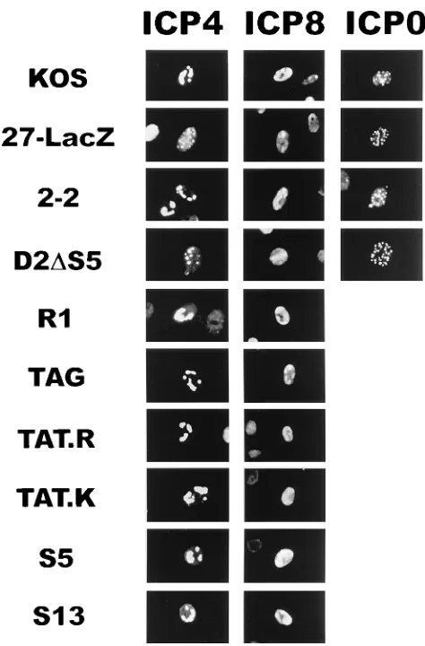
Effects of nuclear localization mutants of ICP27 on the localization of other viral proteins involved in late gene
ex-pression and DNA replication.ICP4 (11, 14, 52, 80) and ICP0
(16, 18, 50, 75) are known to function in transcriptional acti-vation of early and late genes, and ICP27 has been implicated in the regulation of the localization of both of these immedi-ate-early proteins (48, 85, 86). In addition, ICP8 is essential for viral DNA replication (4, 7, 81) and is also required for the formation of replication compartments or nuclear structures in which DNA replication (10) and also transcription (36) have been suggested to occur. The assembly of replication proteins into these active sites was found to be defective in the majority of cells infected with various ICP27 mutants (9). To examine the intracellular localization of ICP4, ICP0, and ICP8 during infection in the presence of ICP27 mutants of the NLS and
arginine-rich regions, the cell lines D2DS5, R1, TAG, TAT.R,
TAT.K, S5, and S13 were infected with 27-LacZ and examined by immunofluorescence staining. As controls, Vero cells and 2-2 cells were similarly infected with 27-LacZ or KOS. The cells shown in Fig. 7 were fixed 8 h after infection, and immu-nofluorescence staining was performed. Wild-type (KOS) in-fection of Vero cells showed the typical pattern of ICP4 intra-cellular localization in which ICP4 moved from a diffuse intranuclear localization at 2 h after infection (data not shown) to globular compartments within the nucleus (Fig. 7). These compartments are suggested sites for viral replication and late gene transcription (36, 53). During infection with 27-LacZ, ICP4 remained in the diffuse intracellular pattern, with no movement into globular compartments (Fig. 7). This defect was overcome during 27-LacZ infection of 2-2 cells which express wild-type ICP27 protein (Fig. 7). During infection in
the presence of the ICP27 mutant, D2DS5, ICP4 remained
diffusely localized throughout the nucleus at early times after infection (data not shown), with small punctate structures seen forming 8 h after infection (Fig. 7). By contrast, during infec-tion of the other mutants of the amino-terminal region of ICP27, R1, TAG, TAT.R, TAT.K, and S5, and the activator mutant S13, ICP4 was found in globular compartments of the
nucleus (Fig. 7). Aside from the mutant D2DS5, in which the
formation of punctate structures was delayed, there were no defects in the localization of ICP4 protein during infection in the presence of the ICP27 mutants.
ICP0 localizes primarily to the nuclei of infected cells in a diffuse and punctate pattern in wild-type infection (17, 47). To determine whether the mutants of the amino-terminal local-ization regions of ICP27 altered the locallocal-ization of ICP0, the staining pattern of ICP0 was monitored in Vero cells, 2-2 cells,
and the cell line D2DS5 (Fig. 7). The intranuclear localization
pattern of ICP0 was identical during infection in the absence of ICP27 (27-LacZ) or when ICP27 was improperly localized
(D2DS5) to the pattern seen during wild-type infection (KOS
and 2-2). Similar results were seen during infections of the cell lines R1, TAG, TAT.R, TAT.K, S5, and S13 (data not shown). Therefore, the intranuclear localization of ICP0 was not al-tered during infection in the presence of ICP27 mutants of the NLS or arginine-rich regions.
[image:8.612.58.301.69.230.2]ICP8 staining showed the formation of globular replication compartments as early as 4 h (data not shown) and more completely at 8 h after infection with KOS (Fig. 7). ICP8 could
FIG. 6. Measurement of DNA replication during infection in the presence of ICP27 nuclear localization mutants. Vero cells, 2-2 cells, and the cell lines D2DS5, R1, TAG, TAT.R, TAT.K, S5, and S13 were infected with 27-LacZ at a multiplicity of infection of 1. (A) DNA was purified from infected cultures 1 and 12 h after infection. Equivalent amounts of total DNA for all samples over a 100-fold concentration range were loaded onto nitrocellulose filters by using a slot blot apparatus. Blots were hybridized with a32
P-labeled, 960-nt antisense RNA probe specific for the gC gene. Autoradiographs were scanned by using Sigma Scan/Image for Windows (Jandel Scientific) to determine relative DNA synthesis efficiency. (B) Parallel cultures were incubated for 24 h, at which time virus yields were measured by plaque assay on 2-2 cells.
on November 9, 2019 by guest
http://jvi.asm.org/
be found only in smaller prereplicative sites during infection with the null mutant, 27-LacZ. Infection of the cell lines
D2DS5, R1, TAG, TAT.R, TAT.K, S5, and S13 resulted in
ICP8 localization to globular replication compartments in the nucleus like those seen in wild-type virus infection. 27-LacZ
infection in the presence of D2DS5 showed a delay in the
localization of ICP8 to globular complexes, but by 8 h, repli-cation complex formation was evident (Fig. 7). Thus, infection in the presence of the ICP27 mutants that were analyzed showed no alterations in the pattern of the localization of ICP8. These data suggest that the defect in late gene expres-sion seen in the presence of these ICP27 mutants is not due to the improper intranuclear localization of ICP4, ICP0, or ICP8.
DISCUSSION
A single consensus has not been determined to specify nu-clear localization of proteins, but two types of signals, both of which are highly basic, have become the prototypes for com-parison (49, 74). The first is defined by a short contiguous stretch of basic amino acids, consisting primarily of arginine
and lysine residues, and was originally identified in SV40 large T antigen (34). The other, which was first described for
Xeno-pus laevis nucleoplasmin (59), has a bipartite structure
consist-ing of two short stretches of basic residues separated by a spacer of nonconserved residues (13, 39, 73, 84). More specif-ically, this type of NLS consists of two consecutive basic amino acids followed by a region containing three of five basic resi-dues which are separated by a minimum of 10 amino acids (59). The arginine-rich region of ICP27 shows similarity to both of these types of NLS. The first of the arginine-rich regions (amino acids 141 to 151) of ICP27 resembles the first type of NLS and together with part of the second arginine-rich region from amino acids 145 to 166 closely resembles the second type of NLS. However, the strong NLS of ICP27 was mapped by Mears et al. (45) to the sequence just preceding these arginine-rich regions and comprises the sequence ARR PSCSPEQHGGKVARLQPPPTKAQPA, which does not re-semble either the highly basic or the bipartite signal. The first arginine-rich region is included only in the NuLS of ICP27 as defined by Mears et al. (45).
The position of an NLS and the context of its surrounding sequences have been shown to affect the function of a number of NLSs (60), including that of the HSV-1 major DNA-binding protein, ICP8 (19). Our studies suggest that the strong NLS of ICP27 functions in a context-dependent manner and that both of the succeeding arginine-rich regions are required for the appropriate localization of ICP27. The strong NLS, residues 110 to 137 of ICP27, was defined by the ability of this minimal sequence to cause efficient nuclear localization of a pyruvate kinase fusion protein and was confirmed by the partial cyto-plasmic localization of viral recombinants with a deletion of this region (45). The ICP27 NLS-pyruvate kinase fusion con-struct also showed some cytoplasmic staining. Only upon in-clusion of the region of ICP27 from residues 110 to 172 in a similar fusion construct did the expressed protein become completely nuclear (45). This finding suggested that the suc-ceeding arginine-rich regions of ICP27, residues 151 to 171, were required for the efficient localization of ICP27 in the context of the fusion protein. We have presented evidence that in the context of the ICP27 protein, the first arginine-rich region (R1) can act as a partial NLS in the absence of the NLS and second arginine-rich region (R2) (Fig. 3). Yet in combi-nation with the strong NLS, the R1 region leads to the pref-erential nucleolar localization of the mutant protein when the second arginine-rich region is not present (Fig. 3A, S5). The nuclear staining observed during infection in the presence of this mutant could best be explained by an overflowing of the nucleolar protein. This notion is consistent with the diffuse nuclear localization of this same mutant in transient transfec-tions (data not shown). As there is an uncontrolled template copy number per cell in transient transfections, it has been suggested that the excessive levels of expression result in the frequently observed aberrant localization of the plasmid-en-coded protein in these cells (45). These data suggest that the NLS, in the context of the ICP27 polypeptide, requires the presence of both of the arginine-rich regions to bring about the appropriate localization of ICP27 in the nucleus.
[image:9.612.58.295.67.427.2]Although the arginine-rich regions of ICP27 appear to be necessary to ensure efficient nuclear localization as directed by the native NLS of ICP27, there is not a requirement for the specific sequence of this region in the nuclear localization of ICP27. We have shown that an ICP27 mutant in which the NLS and arginine-rich regions are deleted can be directed to the nuclei of infected cells as efficiently as the wild-type polypep-tide by the lysine-rich NLS of SV40 T antigen (Fig. 3A, TAG). In addition, mutants which have this region replaced with the
FIG. 7. Immunofluorescence analysis of the cellular location of ICP4, ICP0, and ICP8 during infection in the presence of ICP27 nuclear localization mutants. Vero cells were infected with HSV-1 KOS or 27-LacZ as indicated. Cell lines expressing wild-type ICP27 (2-2) and the ICP27 mutants D2DS5, RG (R1), TAG, TAT.R, TAT.K, S5, and S13 were infected with 27-LacZ at a multiplicity of infection of 10. Cells were fixed 8 h after infection and stained with monoclonal antibody H1114 to ICP4 (A), monoclonal antibody H1115 to ICP8 (B), or monoclonal antibody H1112 to ICP0 (C).
on November 9, 2019 by guest
http://jvi.asm.org/
arginine-rich HIV Tat protein NLS or a lysine-rich sequence, for which no role in localization has been determined, are also localized to the nucleus more efficiently than the mutant form of ICP27 with the NLS and arginine-rich regions deleted (Fig. 3A and B, TAT.R and TAT.K). These results suggest that the nuclear localization of ICP27 does not require the specific sequences of the NLS or arginine-rich regions per se but re-quires only a strong NLS to be efficiently localized to the nucleus.
The indication that the arginine-rich regions of ICP27 do not function as the major NLS suggests that these regions may have an alternate role in the function of ICP27. Arginine-rich regions can be found in a number of RNA-binding domains. To date, three sequence motifs which are common to many RNA-binding proteins have been determined: the RNP (ribo-nucleoprotein) motif (27), the arginine-rich motif, and the RGG motif, of which the latter two require arginine residues. The arginine-rich motif consists of a short region of basic amino acids, approximately 8 to 20 amino acids in length, which is especially rich in arginine. This motif has been found in the HIV Tat and Rev proteins, in which it is involved in transcriptional activation (8, 26, 37, 46, 51, 54, 83), as well as in bacterial antiterminators and ribosomal proteins (38). The RGG motif differs from the arginine-rich motif in that it con-tains several repeats of arginine-glycine-glycine and has been found in nucleolar proteins and in heterogeneous nuclear RNPs, in which it is thought to be involved in mRNA process-ing (2, 35). Both of the arginine-rich regions of ICP27 bear resemblance to the arginine-rich binding motif. Each is be-tween 10 and 15 amino acids in length and consists of at least 50% arginine. The first arginine-rich region, R1, also includes a high percentage of glycine residues, making it similar to the RGG motif. ICP27 is involved in mRNA processing at the level of both splicing and polyadenylation (24, 43, 66), and one possibility for the means by which it does this is through a direct interaction with pre-mRNA or small nuclear RNA, as the RGG region of ICP27 closely matches the RGG sequence from the U small nuclear RNP constituent, the SmD protein (45, 61). In this regard, the mutant R1 in which the RGG-like region was inserted regained the ability to accumulate ICP0 pre-mRNA.
To determine more specifically what role these arginine-rich sequences play in the role of ICP27 during infection, we in-vestigated the abilities of mutants in this region to support infection in the absence of wild-type ICP27. It was previously observed that viral recombinants lacking either the NLS or a portion of the NuLS of ICP27 were defective for growth on Vero cells; however, it was suggested that this may be a prop-erty of the reduced levels of nucleus-localized ICP27 (45). Here, we have shown that the NLS and/or arginine-rich regions of ICP27 are required for more than the appropriate localiza-tion of ICP27. Mutants lacking the NLS and arginine-rich regions of ICP27 were efficiently localized to the nucleus dur-ing infection when heterologous NLSs were added, yet these mutants were unable to support infection of an ICP27 null mutant virus (Table 1). Infection in the presence of any of the mutants with deletions of the NLS and/or arginine-rich regions led to a deficiency in late gene expression, regardless of the ability of the mutants to localize to the nucleus (Fig. 4C). The similarities between the arginine-rich region of ICP27 and the HIV Tat protein RNA-binding/nuclear localization region led us to consider the possibility that introducing this region of the
Tat protein into the D2DS5 mutant might restore the
activa-tion of late gene expression. However, replacement with the Tat protein sequence was not sufficient to overcome the defect in late gene expression. These results do not rule out the
possibility that ICP27 is involved in RNA binding or that the arginine-rich region is required for this binding but instead demonstrate that the specific sequence of this region of ICP27 is important to its function. There is also the consideration that the context of the functional domain or the conformation which the mutant proteins assume could alter the ability of this domain to function. This question could be addressed in future studies by constructing deletions adjacent to the arginine-rich regions to ascertain what effect conformational changes may have on the functional activity of this region.
As yet, we have not examined the prospect that the R1 region may function like an RNA-binding domain of the RGG box motif as a result of its high content of glycine residues. For example, it is possible that the nucleolar localization of ICP27 seen with the S5 mutant is a consequence of this putative RNA-binding function in that ICP27 may be able to bind sequences in rRNA by a mechanism similar to that proposed for the Tat and Rev proteins of HIV. Accumulation of these proteins in the nucleolus is thought to result from the ability of the RNA-binding domains of these proteins to recognize se-quences in pre-rRNA or rRNA that may be similar to the viral pre-mRNA to which these proteins normally bind (6, 69). This would suggest that the R1 region of ICP27, which encodes the putative RGG box, might bind RNA since this region is re-tained in S5.
Although we are not able to define a mechanism for this arginine-rich region in the activation of late gene expression, we did show that the manner in which late gene expression is affected is independent of the effect of ICP27 on viral DNA replication, does not involve altered expression of genes of the other temporal classes, and does not stem from the aberrant localization of other viral proteins required for transcriptional activation or DNA replication. The relative levels of DNA synthesis for each of the mutants ranged from 33 to 76% of the wild-type level, although virus yields were significantly reduced for all mutants (Fig. 6B). In addition, the intranuclear local-ization of two viral proteins involved in the activation of late gene expression, ICP4 and ICP0, and one which is required for DNA replication, ICP8, each of whose localization has been shown to be influenced by ICP27 (9, 48, 85, 86), was unaltered in the presence of the ICP27 mutants of the NLS and/or argi-nine-rich regions. Although there are many possibilities for the mechanism by which ICP27 could affect late gene expression, these results allude to the direct involvement of this region of ICP27 in this function. Further studies will be directed toward determining whether the arginine-rich region of ICP27 is in-volved in RNA binding.
ACKNOWLEDGMENTS
This work was funded by Public Health Service grant AI21515 from the National Institute of Allergy and Infectious Diseases to R.M.S.-G. M.K.H. was supported by an NIAID training grant in virology.
REFERENCES
1. Ackerman, M., D. K. Braun, L. Pereira, and B. Roizman. 1984. Character-ization of herpes simplex virus 1 alpha proteins 0, 4, and 27 with monoclonal antibodies. J. Virol. 52:108–118.
2. Birney, E., S. Kumar, and A. R. Krainer. 1993. Analysis of the RNA-recognition motif and RS and RGG domains: conservation in metazoan pre-mRNA splicing factors. Nucleic Acids Res. 21:5803–5816.
3. Cathala, G., J. F. Savouret, B. Mendez, B. L. West, M. Karin, J. A. Martial, and J. D. Baxter.1983. A method for the isolation of intact, translationally active ribonucleic acid. DNA 2:329–335.
4. Challberg, M. D. 1986. A method for identifying the viral genes required for herpesvirus DNA replication. Proc. Natl. Acad. Sci. USA 83:9094–9098. 5. Chapman, C. J., J. D. Harris, M. A. Hardwicke, R. M. Sandri-Goldin,
M. K. L. Collins, and D. S. Latchman.1992. Promoter independent