0022-538X/95/$04.0010
Copyrightq1995, American Society for Microbiology
Isolation and Characterization of a Syncytium-Inducing,
Macrophage/T-Cell Line-Tropic Human Immunodeficiency
Virus Type 1 Isolate That Readily Infects Chimpanzee
Cells In Vitro and In Vivo
RIRI SHIBATA,
1M. DAVID HOGGAN,
1CHRISTINE BROSCIUS,
1GEORGE ENGLUND,
1THEODORE S. THEODORE,
1ALICIA BUCKLER-WHITE,
2LARRY O. ARTHUR,
3ZIMRA ISRAEL,
4ALAN SCHULTZ,
5H. CLIFFORD LANE,
6ANDMALCOLM A. MARTIN
1*
Laboratory of Molecular Microbiology,
1Vaccine Research and Development Branch, Division of AIDS,
5and
Laboratory of Immunoregulation,
6National Institute of Allergy and Infectious Diseases, Bethesda, AIDS
Vaccine Program, National Cancer Institute-Frederick Cancer Research and Development Center,
PRI/Dyncorp, Frederick,
3and Division of Molecular Virology and Immunology,
Georgetown University, Rockville,
2Maryland, and Department of Pathology,
New York University Medical Center, New York, New York
4Received 29 December 1994/Accepted 3 April 1995
Fresh human immunodeficiency virus type 1 (HIV-1) isolates from patients with AIDS were screened for
infectivity in chimpanzee peripheral blood mononuclear cells (PBMC) to identify strains potentially able to
generate high virus loads in an inoculated animal. Only 3 of 23 isolates obtained were infectious in chimpanzee
cells. Of these three, only one (HIV-1
DH12) was able to initiate a productive infection in PBMC samples from
all 25 chimpanzees tested. HIV-1
DH12tissue culture infections were characterized by extremely rapid
replica-tion kinetics, profound cytopathicity, and tropism for chimp and human PBMC, primary human macrophage,
and several human T-cell lines. An infection was established within 1 week of inoculating a chimpanzee with
50 50% tissue culture infective doses of HIV-1
DH12; cell-free virus was recovered from the plasma at weeks 1,
2, and 4 and was associated with the development of lymphadenopathy. Virus loads during the primary
infection and at 6 months postinoculation were comparable to those reported in HIV-1-seropositive
individ-uals.
There are many unanswered questions about human
immu-nodeficiency virus type 1 (HIV-1) infections in a seropositive
individual including (i) the mechanism(s) responsible for the
transient depletion of CD4-positive cells that occurs
immedi-ately following exposure to the virus (for a review, see
refer-ence 48); (ii) the pathogenic process(es) leading to the gradual
destruction or aberrant functioning of the immune system
de-spite the rapid and nearly complete elimination of the virus
from the blood following the acute infection (for a review, see
reference 65); (iii) the irreversible loss of CD4-positive T
lym-phocytes that accompanies clinical progression to disease
on-set; (iv) the contribution, if any, of specific HIV-1 phenotypes
(syncytium inducing versus non-syncytium inducing; T-cell line
tropic versus macrophage tropic) to virus transmission and
disease progression; and (v) the biological role of auxiliary viral
genes such as vpr, vif, and nef during the establishment of the
initial infection and its subsequent spread to other sites in the
body. Because a tractable HIV-1 animal model that progresses
to immunodeficiency does not exist, virtually all of our present
knowledge of HIV-1 biology comes from studies of tissue
cul-ture infections or analyses of clinical specimens obtained from
seropositive individuals.
The chimpanzee (Pan troglodytes) can be reliably infected
with relatively low doses of HIV-1 (4), but no long-standing
impairment to the immune system occurs (for a review, see
reference 23). As with human patients, a primary HIV-1
in-fection of chimpanzees is usually characterized by a brief
pe-riod of virus production that lasts for several weeks. Compared
with human patients, however, a primary infection in
chimpan-zees seemed to be much milder, as suggested by an infrequent
detection of plasma viremia (25, 26, 43, 46, 47). The acute
infection is followed by the appearance of antibodies directed
against several of the viral proteins and a rapid decline of the
virus load, typically measured by the number of peripheral
blood mononuclear cells (PBMC) needed for HIV-1 isolation
or containing viral DNA, as determined by PCR. In many
instances, virus isolation from persistently infected
chimpan-zees can become intermittent following resolution of the
pri-mary infection (references 4, 12, 32, 36, and 43 and our
un-published observations).
One explanation for the failure of the nearly 100
chimpan-zees to progress to clinical disease following an HIV-1
chal-lenge is that, in contrast to humans, they possess an immune
system uniquely able to mount a long-lasting and protective
response to HIV-1. This would be somewhat analogous to the
case of sooty mangabeys (Cercocebus atys), the natural host of
the smm strain of the simian immunodeficiency virus
(SIV
smm), which rarely develop immunodeficiency following
an SIV challenge but can experience an acute disease
syn-drome when inoculated with the macaque-passaged and highly
pathogenic SIV
smmPBj14(14, 24).
It is also possible that the unsuccessful attempts to develop
a chimpanzee–HIV-1 disease model might be related to the
intrinsic biological properties of the virus strains selected for
inoculation. In the SIV-macaque system, symptoms and
incu-bation period vary greatly among different SIV strains; several
* Corresponding author. Mailing address: Laboratory of MolecularMicrobiology, NIAID, NIH, Bethesda, MD 20892-0460.
4453
on November 9, 2019 by guest
http://jvi.asm.org/
are not pathogenic (for a review, see reference 34). In some
earlier HIV-1–chimpanzee studies, clinical specimens from
pa-tients with AIDS or primary virus isolates were used as inocula
(3, 28, 29, 32, 43). It is now appreciated that most primary
HIV-1 isolates do not infect chimpanzee PBMC as efficiently
as human PBMC (reference 55 and this study). Among various
well-characterized HIV-1 strains, only two (HIV-1
IIIB/LAV[5]
or HIV-1
SF2[13]) and their derivatives have been used for a
majority of chimpanzee inoculations (4, 22, 26–28, 43, 47, 61);
in a few instances, naive chimpanzees have been challenged
with other HIV-1 strains (28, 31, 43).
In an attempt to generate an HIV-1 chimpanzee disease
model with a potentially more pathogenic virus strain, primary
isolates from patients with AIDS were screened for their
ca-pacity to spread with rapid kinetics and release high levels of
progeny virions from chimpanzee PBMC. One of the isolates
obtained, HIV-1
DH12, exhibited rapid infection kinetics and
induced syncytia in human and chimpanzee PBMC, replicated
in both human monocyte-derived macrophage (MDM) and
human T-cell lines, and established an infection in a naive
chimpanzee within 1 week of inoculation of 50 50% tissue
culture infective doses (TCID
50) of virus. Molecular clones of
HIV-1
DH12which directed the synthesis of virus progeny
ex-hibiting growth properties in tissue culture infections
indistin-guishable from those of the parental virus were obtained.
MATERIALS AND METHODS
Cell culture.PBMC were prepared from heparinized whole blood (human and chimpanzee) or from a leukapheresis sample (human) by Ficoll-Hypaque (Phar-macia) density-gradient centrifugation. The PBMC were used immediately or preserved in cryoprotective medium (Whittaker), supplemented with 50% fetal bovine serum (FBS, HyClone) in liquid nitrogen. Fresh or frozen-thawed PBMC were stimulated with 1mg of phytohemagglutinin (PHA, Wellcome) per ml for 3 days and then maintained in RPMI 1640 (Whittaker) medium supplemented with 10% FBS–2 mML-glutamine (Gibco-BRL)–20 U of human recombinant interleukin 2 (Boehringer Mannheim) per ml–50mg of gentamicin (Whittaker) per ml–100 U of penicillin-streptomycin (Gibco-BRL) per ml.
Human blood-derived monocytes were prepared as previously described (38). Briefly, elutriated monocytes were cultured in bacteriological-grade petri dishes for 2 weeks, replated in 96-well plates (Nunc no. 1-67008), and used for infection. The MDM cultures were maintained in Dulbecco’s minimal essential medium (Whittaker, high-glucose, 4.5 g/liter), supplemented with 10% fresh human se-rum, glutamine, and antibiotics.
Human T-cell lines MT-4 (33), H9 (50), C8166 (15), and CEM-12D7 (53) were maintained in RPMI 1640 supplemented with 10% FBS, glutamine, and antibi-otics. HeLa cells were maintained in Dulbecco’s minimal essential medium (Whittaker) supplemented with 10% FBS, glutamine, and antibiotics.
Virus isolation from patients with AIDS.PHA-stimulated normal human PBMC (33106) were cocultured with an equal number of freshly isolated PBMC from patients with AIDS. Culture supernatants were collected during a 3-week period and monitored for reverse transcriptase (RT) activity. RT-posi-tive, cryopreserved tissue culture supernatants were used as inocula for subse-quent infections.
Clinical data on the patient source of the HIV-1DH12isolate.The patient was
a 35-year-old male who first tested positive for HIV in July 1989 when his CD4 count was 157 cells per mm3. From July 1990 until his last visit to the Outpatient Clinic in October 1991, he was treated with several drugs including zidovudine, didanosine, zalcitabine, and decadron. At the time of phlebotomy in October 1991, his CD4 level was 19 cells per mm3. He died approximately 1 month later because of multiple complications of HIV infection including Kaposi’s sarcoma, histoplasmosis, and toxoplasmosis.
In vitro infections.Human T-cell lines, activated human and chimpanzee PBMC (days 4 to 8 after stimulation), and human MDMs were used for in vitro virus infections. Virus inocula were normalized for32
P-RT activity and used as described in the different figure legends. Culture supernatants were collected and monitored for RT activity during a 2- to 4-week period. Virion-associated RT activity was measured in the presence of [32
P]TTP (Amersham,.400 Ci/mmol), as previously described (62). The RT activity was reported as counts of [32
P]TTP per minute incorporated in 10ml (containing 1.67ml of infected culture super-natant) of the reaction mixture.
Virus infection of a chimpanzee.One chimpanzee (no. 1206) was pretested as negative for HIV-1 antibody (enzyme-linked immunosorbent assay [ELISA]) and HIV-1 DNA (PCR). The animal was housed in a biosafety level 2 facility, and biosafety level 3 procedures were followed. A total of 50 TCID50of the
HIV-1DH12isolate (passaged twice in human PBMC and five times in chimpan-zee PBMC) was used for intravenous inoculation. The TCID50of HIV-1DH12 was determined by infecting chimpanzee PBMC in quadruplicate with serial fivefold dilutions of HIV-1DH12and then assaying for RT on day 14. The animal was physically examined by veterinarians every 2 weeks, at which time blood samples were taken.
Virus isolation from blood of the infected chimpanzee.Plasma was prepared from heparinized chimpanzee blood by centrifugation (4003g) and filtration (0.45-mm-pore-size filter). PHA-stimulated chimpanzee PBMC (106in 1 ml) were incubated with fivefold dilutions of plasma (starting at 1 ml of undiluted plasma) and monitored for RT production for 4 weeks. Stimulated fresh chim-panzee PBMC were added to the cultures on day 14.
PBMC, isolated from the heparinized chimpanzee blood described above, were depleted of CD8-positive cells, which can suppress virus replication in vitro and cause false-negative results for virus isolation (59). CD8 depletion was done by panning, with a CD8 antibody-coated flask (Applied Immune Science). Five-fold dilutions of CD8-depleted PBMC (starting from 53106
of CD82PBMC) were costimulated with human PBMC and monitored for RT production for 4 weeks. Stimulated human PBMC were added to the cocultures on day 14 and day 22.
Antibody ELISAs and lymphocyte immunophenotyping.Antibody in chimpan-zee plasma, elicited against HIV-1 proteins, was measured by ELISA with a commercial HIV-1 micro-ELISA kit (Organon Teknika). Heparinized blood was incubated with fluorochrome-conjugated monoclonal antibodies (CD8-fluores-cein isothiocyanate, CD4-phycoerythrin, CD3-peridinin chlorophyll protein [Becton Dickinson]) and then treated with fluorescence-activated cell sorter (FACS) lysing buffer (Becton Dickinson) to remove erythrocytes. Following fixation with paraformaldehyde, lymphocyte samples were analyzed by FACsort flow cytometer (Becton Dickinson).
Viral DNA copy number estimation by PCR.The copy number of viral DNA in PBMC was estimated by DNA PCR following an end-point dilution of a chimpanzee PBMC lysate. Uncultured PBMC (23106) from the infected chim-panzee were lysed in 200ml of PCR lysis buffer (100 mM KCl, 10 mM Tris-HCl [pH 8.0], 2.5 mM MgCl2, 0.5% Tween 20, 0.5% Nonidet P-40, 150mg of pro-teinase K per ml) for 3 h at 658C, incubated at 1008C for 15 min, and subjected to 30 cycles of PCR. This initial PCR was carried out in duplicate with cell lysate equivalent to 105
cells and in quadruplicate with further fivefold dilutions of the PBMC lysate (equivalent to 23104, 4,000, 800, and 160 cells). These PCR products were subsequently amplified by 30 cycles of nested PCR. Under these conditions, any amplifiable molecule would be visualized in an ethidium bro-mide-stained agarose gel. The distribution of amplifiable molecules in each PCR should follow the Poisson equation, P512e(2m)
, where P is the positive fraction and m is the average copy number. Since when m equals 1, P will equal 0.6321, the dilution of PBMC (starting at 105cells) at which 63.21% of the samples are PCR positive would correspond to a DNA copy number of 1 per sample. The percent positive values in the PCR analyses were calculated as described by Reed and Muench (52) for determining TCID50.
Both sets of PCR primer pairs map to the gp120 coding region of the HIV-1 env gene. For the first PCR, CTAAAGCCATGTGTAAAATTAACCCCACTC (located upstream of V1) and TATAGAATTCACTTCTCCAATTGTCCCT CAT (located downstream of V5) primer pairs were used. For the second amplification, TTGAAGAATGGTACTAATTTGAAGAATGG (located in the V1 region) and CCTTCAGTACCATTCCAAGTACTAT (located in the V4 region) were used. Ampli-Taq DNA polymerase (Perkin-Elmer) was used for all PCRs which were performed as follows: an initial denaturation at 948C for 5 min; 30 cycles of 948C for 45 s, 558C for 45 s, and 728C for 120 s; and a final extension at 728C for 7 min.
Molecular cloning, plasmid construction, and sequencing.The strategy fol-lowed was to clone unintegrated, circular, viral DNA molecules following their conversion to full-length linear DNA by digestion with a single cutter restriction enzyme. Earlier Southern blot analyses of HIV-1DH12-infected cellular DNA revealed that EcoRI cleaved the viral DNA a single time within vpr gene se-quences (data not shown).
PHA-stimulated normal chimpanzee PBMC (107) were infected with HIV-1DH12stock at a multiplicity of infection of approximately 1021. The inoculum used had been passaged twice in human PBMC (including the primary isolation) and then three times in chimpanzee PBMC. On day 3 of infection, low-molec-ular-weight DNA was prepared by Hirt’s method (35), digested with EcoRI, ligated with EcoRI-digested arms of Lambda Wes-B phage vector (39), and packaged in Gigapack XL (Stratagene). PCR-amplified HIV-1DH12long termi-nal repeat (LTR), env, and pol gene fragments were labeled with [32
P]dCTP and used as probes for the screening of lambda phage plaques. The following oligo-nucleotides were used for PCR primers: GATTGGCAGAACTACACACC and TGAGGCTTAAGCAGTGGGTTC (LTR); GAACTTAATAAGAGAACTCA AG and TACAGTCTACTTGTCCATGCTA (pol), and TATGAATTCATATG CTGTTAAATGGCAGCAGTCTAGCAGAA and ATTTATAGAATTCTCT TCTCCATTGTCCCTCAT (env). Phage DNAs were purified from 7 positive plaques (out of 25,000), digested with EcoRI, and recloned into the T7T3-18U (Pharmacia) plasmid vector to generate a clone containing a circularly permuted HIV genome (40). Escherichia coli HB101 (Gibco-BRL) was used to prepare large amounts of plasmid DNA.
on November 9, 2019 by guest
http://jvi.asm.org/
A plasmid carrying the two-LTR linear form of HIV-1DH127was reconstructed from the original circularly permuted plasmid clone, as follows: (i) the XhoI (blunt ended; proviral map position 8881)-plus-EcoRI (position 5734) 5.9-kb cleavage fragment (containing nef-LTR-gag-pol-vif and the 59 half of the vpr gene) was subcloned into the T7T3-18U vector between the NaeI (blunt ended) and the EcoRI sites; (ii) the EcoRI (position 5734)-plus-NarI (blunt ended; proviral position 635) 4.0-kb fragment (containing the 39half of the vpr gene plus tat-rev-vpu-env-nef and LTR sequences) was subcloned into the pUC19 vector between the EcoRI and the HindIII (blunt-ended) sites; and (iii) the EcoRI-plus-BglI (located in the ampicillin-resistant gene of T7T3 and pUC19) cleavage products from the two subclones were ligated to one another.
Three PCR clones (DH12A, DH12B, and DH12C), containing vpr-tat-rev-vpu-env and nef sequences, were amplified from the low-molecular-weight DNA fraction of HIV-1DH12-infected chimpanzee PBMC with the primer pair GCAG GAGTGGAAGCCATAA (located in vpr) and AAAGTCCCCAGCGGAA AGT (located in the LTR).
The nucleotide sequence of different HIV-1 clones was determined by the dideoxy-chain termination method. The complete sequence of the full-length HIV-1DH123clone and the vpu-tat-rev-env-nef-LTR region of the HIV-1DH125, HIV-1DH126, and HIV-1DH127clones and the vpu-env sequences from three PCR clones (DH12A, DH12B, and DH12C), amplified from cells infected with the uncloned HIV-1DH12, were determined.
Electroporation and transfection of cloned HIV-1DH12DNAs.The biological
activity of the circularly permuted clones (HIV-1DH123, HIV-1DH125, HIV-1DH126, and HIV-1DH127) was evaluated by digestion of the plasmid DNA with EcoRI to release HIV sequences from the vector, DNA ligation to regenerate the two-LTR configuration, and electroporation of the concatemerized DNA into MT-4 cells (40). The MT-4 cells (43106
) were resuspended in 0.4 ml of fresh medium containing 5mg of EcoRI-digested-ligated HIV-1DH12DNA, elec-troporated at 960mF-0.3 kV (Bio-Rad gene pulser), resuspended in 10 ml of medium containing 106fresh MT-4 cells, and monitored for virus production. Samples of the infected culture supernatants were collected prior to the peak of RT activity (day 7 to day 8), filtered (0.45-mm-pore-size filter), and then used for infectivity assays.
Virus stocks were also prepared by transfecting HeLa cells with the plasmid clone carrying HIV-1DH127in the two-LTR configuration (reconstructed from the circularly permuted clone as described in the previous section), HIV-1NL43 (1), HIV-1MAL(2, 49), HIV-1ELI(2, 49), HIV-1LAI(49, 58), HIV-1AD8(a molecular clone derived from HIV-1AD-87[51]), and HIV-1SG3.1(31) (obtained from the National Institutes of Health AIDS Research and Reference Reagent Program). HeLa cells were transfected with 30mg of plasmid DNA by the calcium-phosphate precipitation method. The culture supernatants were col-lected 48 h after transfection, filtered (0.45-mm-pore-size filter), and, following normalization for RT activity, used in infectivity assays.
RESULTS
Isolation and biological characterization of primary HIV-1
isolates capable of infecting chimpanzee PBMC.
In this study,
we decided to screen fresh viral isolates from patients with
AIDS for their ability to induce a rapid cytopathic infection in
chimpanzee PBMC, a possible predictor of pathogenicity in
vivo. Prior to initiating such a search, PBMC from several
uninfected chimpanzees were pretested for susceptibility to an
HIV-1 isolate, previously used for chimpanzee inoculations
(HIV-1
IIIB040[4]); the cells from one animal (ISIS no. 810)
reproducibly yielded high titers of virus (approximately 10
5TCID
50/ml). A large quantity of PBMC were prepared from
this chimpanzee, frozen, and used, following mitogen
activa-tion, to identify potentially useful primary HIV-1 isolates.
Attempts were made to isolate HIV-1 from 30 patients with
AIDS, seen at the NIAID Outpatient Clinic, Warren
Magnu-son Clinical Center, National Institutes of Health, by
coculti-vating their PBMC with PHA-stimulated human PBMC from
a seronegative individual. Virus was isolated from 23 of the 30
specimens as monitored by the release of
32P-RT activity into
the medium during a 21-day observation period. All 23 primary
isolates produced progeny virions within 2 weeks following
cell-free infection of fresh, activated human PBMC. In
con-trast, only 3 (HIV-1
DH12, HIV-1
DH20, and HIV-1
DH29) of the
23 HIV-1 isolates exhibited cell-free infectivity for chimpanzee
PBMC. The other 20 samples failed to infect cells from the
ISIS no. 810 animal or other chimpanzee donors.
The experiment depicted in Fig. 1 compares the infectivities
of HIV-1
DH12, HIV-1
DH20, and HIV-1
DH29for human or
chimpanzee PBMC (ISIS no. 810) with those of several other
HIV-1 isolates. Three previously characterized viruses
(HIV-1
MAL[2, 49], HIV-1
Eli[2, 49], and the macrophage-tropic
HIV-1
AD8[molecularly cloned from the HIV-1
AD-87derivative
of HIV-1
Ada] [30, 51]), as well as another representative
(HIV-1
DH32) from our cohort of 30 patients with AIDS, produced no
detectable progeny virus in chimpanzee PBMC. Inocula
pre-pared from the molecularly cloned and highly tissue
culture-adapted HIV-1
Lai(2, 49) and HIV-1
NL43(1) replicated to low
levels in chimpanzee PBMC. The HIV-1 chimpanzee challenge
stock (HIV-1
IIIB-040[4]) and an isolate (HIV-1
SG3.1[31])
pre-viously shown to be tropic for chimpanzee lymphocytes readily
infected chimpanzee PBMC, although their replication
kinet-ics were slower than those of HIV-1
DH12and
syncytium-form-ing activity in chimpanzee PBMC was difficult to detect (see
below).
Although the data presented in Fig. 1 indicate that three of
the primary HIV-1 isolates were capable of initiating infections
in PBMC prepared from the ISIS no. 810 animal, the amounts
of progeny virus produced differed greatly. Differences in
in-fectivity were also observed when PBMC from other
chimpan-zees were monitored for susceptibility to infection by
HIV-1
DH12, HIV-1
DH20, and HIV-1
DH29. As shown in Fig. 2, PBMC
from the ISIS no. 1157 donor supported the replication of all
three strains but the infection kinetics for HIV-1
DH20and
HIV-1
DH29were markedly delayed compared with those for
HIV-1
DH12. Very low levels of HIV-1
DH20and HIV-1
DH29were produced in the cells of the ISIS no. 1135 chimpanzee
compared with HIV-1
DH12. No progeny virus was detected
from the ISIS no. 1206 animal following infection of PBMC
with HIV-1
DH20and HIV-1
DH29. The screening of PBMC
from 21 additional chimpanzees indicated that all (25 total
donors) were susceptible to HIV-1
DH12(Table 1), and in most
cases, rapid and highly cytopathic (see below) infections were
observed.
These data indicate that rapid and cytopathic infection of
chimpanzee PBMC is not a common property of most HIV-1
strains, perhaps explaining the failure to detect high virus
lev-els and clinical disease in chimpanzees inoculated with clinical
specimens from patients with AIDS. No correlation between
the syncytium-inducing phenotype and chimpanzee-PBMC
tropism (55) was observed among HIV-1 strains in our cohort
(HIV-1
DH29exhibited the non-syncytium-inducing phenotype,
and most of our primary syncytium-inducing isolates fail to
replicate in chimpanzee PBMC). Furthermore, nucleotide
se-quence analyses indicate that HIV-1
DH12, HIV-1
DH20, and
HIV-1
DH29are genetically distinct clade B isolates (data not
shown).
Because it was highly infectious for PBMC from all
chim-panzees tested, we concentrated our efforts exclusively on the
HIV-1
DH12isolate and initially evaluated its capacity to infect
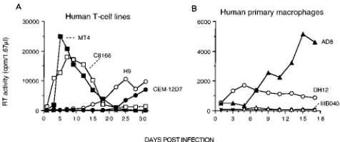
a variety of CD4-positive cell types. As shown in Fig. 3A,
HIV-1
DH12was able to infect several different human T-cell
leukemia lines although its replication kinetics in H9 and, in
particular, CEM-12D7 cells were markedly delayed. It should
be noted that MT-4 cells supported the production of
high-titered stocks of HIV-1
DH12possessing the same biological
properties as virus generated in PBMC. The HIV-1
DH12isolate
also successfully infected human MDM cultures (Fig. 3B);
although it exhibited characteristic rapid infection kinetics
even in MDM, HIV-1
DH12directed the production of lower
levels of progeny particles compared with a molecularly cloned
virus (HIV-1
AD8) derived from the macrophage-tropic isolate,
HIV-1
AD-87(51). The ability of HIV-1
DH12to infect both
MDM and human T-cell lines was somewhat unusual but was
thought to possibly reflect its recent isolation from a
on November 9, 2019 by guest
http://jvi.asm.org/
atic HIV-infected individual and the presence of different
pop-ulations of virions with diverse tropic properties within the
virus stock.
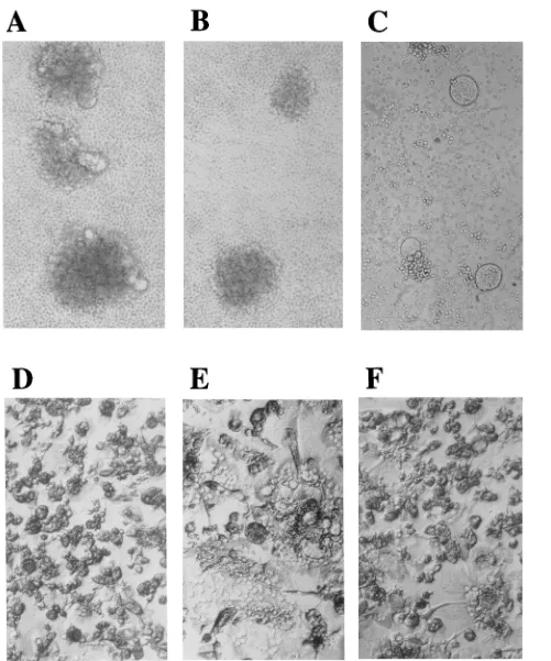
Another striking property of the HIV-1
DH12isolate was its
capacity to rapidly induce cell fusion in PBMC, MDM, and
some T-cell lines. For example, HIV-1
DH12induced syncytium
formation in both human and chimpanzee PBMC at 3 days
postinfection (chimpanzee cells are shown in Fig. 4A), whereas
only aggregated cells were visualized in the two cultures
fol-lowing exposure to HIV-1
IIIB-040(chimpanzee cells are shown
in Fig. 4B) or HIV-1
SG3.1(data not shown). Similarly, infection
of human MDM by HIV-1
DH12was associated with extensive
cell fusion on day 5 of the infection shown in Fig. 3B; a
companion HIV-1
AD8-infected MDM culture contained only a
few multinucleated cells on day 5. Two days later, both cultures
exhibited similar cytopathicity (Fig. 4E and F). Syncytium
for-mation was also detected following HIV-1
DH12infection of
CEM-12D7 (Fig. 4C), C8166, and H9 cells but not MT-4 cells.
Infectivity of HIV-1
DH12in vivo.
Since the ultimate goal of
this work was to obtain a fresh HIV-1 isolate capable of rapidly
initiating a de novo infection in a chimpanzee, care was taken
to minimize the introduction of changes into the HIV-1
DH12genome during the generation of a high-titered virus inoculum.
Consequently, the HIV-1
DH12stock used for chimpanzee
in-oculation was prepared by two passages in human PBMC
(in-cluding its initial isolation) followed by five successive passages
in chimpanzee PBMC, as described in Materials and Methods.
In several independent 2-week assays, the infectivity of this
inoculum was determined to be approximately 1.7
3
10
5TCID
50/ml in both human and chimpanzee PBMC and about
[image:4.612.128.486.70.293.2]FIG. 1. (A) Infectivity of primary patient isolates and laboratory-adapted HIV-1 strains in human and chimpanzee PBMC. A total of 2.53106PHA-stimulated chimpanzee PBMC (closed circles) or human PBMC (open squares) were infected with HIV-1DH12, HIV-1DH20, HIV-1DH29, HIV-1DH32, HIV-1NL43(derived from an infectious molecular clone [1]), and HIV-1IIIB040(4). Virus inocula were made in chimpanzee PBMC (HIV-1DH12, HIV-1DH20, HIV-1DH29, HIV-1IIIB040) or in human PBMC (HIV-1DH32, HIV-1NL43) and normalized for RT activity (approximately 106 32P cpm). Symbols3and1represent mock infections of human and chimpanzee PBMC, respectively. (B) Infectivity of virus derived from infectious molecular clones. A total of 106PHA-stimulated chimpanzee PBMC (closed circles) or human PBMC (open squares) were infected with virus prepared from the following HIV-1 molecular clones: HIV-1DH127, one of the four HIV-1DH12molecular clones; HIV-1SG3.1, a chimpanzee PBMC tropic strain (31); HIV-1AD8, a macrophage-tropic, laboratory-adapted strain derived from HIV-1AD-87(51); HIV-1MAL, an African isolate (2, 49); HIV-1ELI, an African isolate (2, 49); and HIV-1LAI(49, 58), a T-cell-line-tropic laboratory strain. Virus inocula were prepared by transfecting HeLa cells and used to infect PBMC following normalization for RT activity (approximately 105 32P cpm). The mock infections of human (3) and chimpanzee (1) PBMC, respectively, are indicated.
FIG. 2. Infectivity of three of the primary HIV-1 isolates in the PBMC of three different chimpanzee donors (no. 1157, no. 1135, and no. 1206). HIV-1DH12(circles), HIV-1DH20(squares), and HIV-1DH29(triangles) inocula were prepared in chimpanzee PBMC (no. 810), normalized for RT activity (approximately 4310
4 32 P cpm), and used to infect PHA-stimulated chimpanzee PBMC (0.53106
cells). Symbol3represents mock infection. The production of progeny virus was monitored by RT assay.
on November 9, 2019 by guest
http://jvi.asm.org/
[image:4.612.128.489.581.689.2]10 times higher when measured in MT-4 cells. In the animal
experiment to be described, the amount of HIV-1
DH12inocu-lated is expressed as TCID
50determined in chimpanzee
PBMC.
A naive adult male chimpanzee was inoculated with 50
TCID
50of virus by the intravenous route. As shown in Fig. 5,
virus could be isolated directly from fresh plasma samples
collected at weeks 1, 2, and 4 postinfection; viral RNA was also
detected in the plasma at weeks 2, 3, 4, and 17 by the branched
DNA procedure (reference 21 and data not shown). Although
previously reported (25), the development of plasma viremia is
a rarely observed phenomenon in HIV-1-inoculated
chimpan-zees. The establishment of an HIV-1
DH12infection within 1
week of inoculation was also verified by the recovery of virus
following cocultivation of chimpanzee PBMC with PBMC
from a seronegative human volunteer. At week 1, 5
3
10
6chimpanzee cells were required for virus isolation, whereas
between weeks 2 and 8, fewer (2
3
10
5) cells were needed,
indicating the presence of increasing numbers of infected cells.
Nested-DNA PCR analysis of uncultured chimpanzee PBMC,
collected at various times following inoculation, was also
car-ried out to ascertain the viral DNA copy number. HIV DNA
was initially amplified (30 cycles) from serially diluted PBMC
lysates, assayed in quadruplicate, with a primer pair
comple-mentary to conserved sequences in the C1 and C5 regions of
gp120. These primers are able to amplify env gene segments
from a variety of HIV-1 isolates tested including HIV-1
Lai,
HIV-1
NL4-3, HIV-1
SF2, HIV-1
DH12, HIV-1
DH20, HIV-1
DH29,
and four recent isolates from seropositive, asymptomatic
indi-viduals (data not shown). The second amplification (30 cycles)
utilized a primer pair specific for HIV-1
DH12that was
comple-mentary to the V1 and V4 regions of gp120. The latter primers
failed to amplify the analogous region of HIV-1
IIIB040,
HIV-1
NL4-3, HIV-1
SF2, HIV-1
DH20, and HIV-1
DH29(data not
shown). Figure 5 indicates that the viral DNA load in PBMC
peaked at weeks 3 and 8 and then fell to the 10- to
30-copy-per-10
5PBMC range by week 15.
The infected chimpanzee developed anti-HIV-1 antibody
between weeks 3 and 4 as measured by ELISA, and a sample
of plasma collected at week 15 was positive at a 1:250 dilution
for anti-HIV-1
DH12neutralizing antibody, monitored by
inhi-bition of infectivity for human PBMC (41a). Interestingly, the
infected animal also developed intermittent lymphadenopathy
(weeks 3 and 18, lasting 3 to 4 weeks each time). HIV-1
DH12was isolated from lymphocytes prepared from the lymph node
biopsied at week 3, following cocultivation with human PBMC.
Although the virus load in the peripheral blood and lymph
node at week 3, measured by the number of lymphocytes from
each source required for virus isolation, was not significantly
different, the DNA copy number per cell was approximately
five times higher in the lymph node (data not shown), a result
similar to that reported previously (54). The preinoculation
percent CD4-positive cells (46%) fell to 33% at week 3 but by
week 21 had returned to the preinfection level; no significant
changes were observed in the percent CD8 cells (data not
shown).
Molecular cloning of HIV-1
DH12DNA and characterization
of virus derivatives.
We noted earlier that some of the unusual
biological and tropic properties of the HIV-1
DH12isolate
might be explained by the presence of genetically diverse
vi-ruses within the uncloned HIV-1
DH12stock. On the other
hand, these could be unique properties of the HIV-1
DH12isolate and not represent the contribution of discrete
subpopu-lations of the virus stock. One way to resolve this issue would
be to obtain individual molecular clones of HIV-1
DH12DNA
and ascertain whether any directed the synthesis of virus with
the tropic and syncytium-inducing properties described earlier.
Unintegrated circular viral DNA, purified from HIV-1
DH12-infected chimpanzee PBMC, was cleaved with EcoRI, cloned
into the lambda phage Wes-B vector (39), and then transferred
to the plasmid vector, pT7T3-18U. Of the seven clones
ob-tained, four (DH123, DH125, DH126, and DH127) were
full-length (approximately 9.2 kb in size) and circularly permuted.
Their infectivity was assessed following digestion of the
plas-mid DNA with EcoRI; concatemerization in vitro using DNA
ligase to regenerate the linear, two-LTR configuration; and
electroporation into MT-4 cells. All four clones were able to
initiate a spreading infection in MT-4 cells, releasing RT
ac-tivity into the medium within a week of transfection (data not
shown).
Virus stocks were prepared from the electroporated MT-4
cells and assayed for infectivity in a variety of CD4-positive
cells. The virus obtained from all four infectious clones
repli-cated in chimpanzee PBMC, human PBMC, human MDM,
and human T-cell lines. Representative infections for two of
the HIV-1
DH12molecularly cloned viruses (designated
HIV-1
DH123and HIV-1
DH127) in MT-4, H9, and human primary
MDM are presented in Fig. 6. Like its uncloned parent,
HIV-1
DH127also replicated to high levels in chimpanzee PBMC as
well as in human PBMC (lower left panel, Fig. 1). The cloned
viruses also induced syncytia in human and chimpanzee
PBMC, human MDM, and T-cell lines (HIV-1
DH123-infected
H9 and CEM-12D7 cells are shown in Fig. 6B). Taken
to-gether, these data indicate that the unusual biological and
tropic features of infections initiated by the original
HIV-1
DH12are intrinsic properties of this isolate since they were
also exhibited by molecularly cloned derivatives.
[image:5.612.57.299.91.164.2]The nucleotide sequence of the entire HIV-1
DH123genome
FIG. 3. HIV-1DH12infects human T-cell lines (A) and MDM (B). The hu-man T-cell lines (0.53106
cells) MT-4 (33), C8166 (15), H9 (50), and CEM-12D7 (53) were inoculated with HIV-1DH12(approximately 4310
4 32 P cpm), prepared in chimpanzee PBMC. Human primary macrophage cultures (105
cells in a 96-well plate) were inoculated with HIV-1DH12, HIV-1IIIB040, and the mac-rophage-tropic HIV-1AD8(approximately 7310
4 32
[image:5.612.59.297.567.667.2]P cpm). Mock infections (3) are also indicated.
TABLE 1. Susceptibility of PBMC from different chimpanzee donors to primary HIV-1 isolatesa
Virus
No. of chimpanzee donors with:
Susceptible PBMC PBMC not susceptible
HIV-1DH12 25 0
HIV-1DH20 7 6
HIV-1DH29 3 7
HIV-1IIIB 9 1
a
A total of 53105
of PHA-stimulated chimpanzee PBMC were infected with cell-free virus (approximately 43104 32
P cpm) prepared in chimpanzee (ISIS no. 810) PBMC. Cultures were kept for at least 3 weeks, and virus replication was monitored by RT assay.
on November 9, 2019 by guest
http://jvi.asm.org/
FIG. 4. Syncytium induction by HIV-1DH12in various cell types. Chimpanzee PBMC, from the infection presented in Fig. 1A, are shown on day 3 following exposure to HIV-1DH12(A) or HIV-1IIIB040 (B). Syncytia were also observed in CEM-12D7 cells (0.53106) 5 days following infection with an HIV-1DH12inoculum (approximately 33105 32
P cpm of RT activity), produced in MT-4 cells (C). Uninfected (D) or HIV-1DH12(E)- or HIV-1AD8(F)-infected (day 7) primary human macrophage cultures are shown. The kinetics of RT production for these macrophage infections is presented in Fig. 3.
on November 9, 2019 by guest
http://jvi.asm.org/
as well as the 3
9
-terminal 4.0 kb (encompassing the tat, rev, vpu,
env, and nef genes and 3
9
LTR) of the HIV-1
DH125,
HIV-1
DH126, and HIV-1
DH127proviral DNAs was determined. The
sequence obtained placed the parental HIV-1
DH12strain
within the clade B of HIV-1 isolates (44). As depicted
dia-grammatically in Fig. 7A, little heterogeneity was evident
within the env gene sequences of the four full-length circularly
permuted HIV-1
DH12-derived clones. Two substitutions in
gp41 (both in HIV-1
DH125), and a valine-to-isoleucine change
in the V3 loop of HIV-1
DH127, were the only alterations
ob-served. No changes were present in the deduced amino acid
sequences of Tat and Rev. Three additional DNA PCR clones
(DH12A, DH12B, and DH12C) were obtained by amplifying
the low-molecular-weight DNA from chimpanzee PBMC
in-fected in vitro with HIV-1
DH12. Sequencing of the V3 loop of
these latter clones was consistent with the previous analysis of
the three circularly permuted full-length clones (Fig. 7B). Of
particular interest, however, was the disruption of the vpu gene
in all seven of the clones (Fig. 7B), reflecting the lack of
initiation codons (DH123, DH125, DH12B), in-frame stop
codons (DH126, DH12C), or frameshift mutations (DH127,
DH12A).
DISCUSSION
There is still no explanation for the failure of chimpanzees to
develop disease following infection with HIV-1. It is well
known that the status of an animal’s immune system can play
a critical role in the induction of disease by retroviruses. For
example, some inbred mouse strains will develop disease only
if they are infected with murine leukemia viruses during the
neonatal period, presumably reflecting the inability of an
im-mature immune system to mount a protective response (6, 8,
19). Because differences have been observed during infections
of human and chimpanzee cells both in vitro and in vivo, a
number of immunopathological mechanisms have been
pro-posed to explain the asymptomatic nature of HIV-1
chimpan-zee infections, including (i) the reported inability of the viral
gp120-chimpanzee CD4 interaction to progress to syncytium
formation (9), subsequently shown to be incorrect (reference 7
and the present work); (ii) a failure of HIV-1-infected
chim-panzee peripheral blood lymphocytes to undergo apoptosis
following cross-linking with the CD3 monoclonal antibody
(55); and (iii) the suppression of HIV-1 tissue culture
infec-tions by CD8-positive T lymphocytes, obtained from either
infected or uninfected chimpanzees (11).
One could also argue that the HIV-1 strains used thus far for
chimpanzee inoculations are deficient in some unknown
prop-erty necessary to generate high virus loads and/or for targeting
specific tissues in the infected animal. As a consequence, no
disease is induced. In contrast to humans who develop high
levels of both cell-free virus and infected PBMC during a
primary HIV-1 infection (16), inoculated chimpanzees rarely
develop a plasma viremia although infected PBMC can be
detected (25, 46).
In planning a strategy for obtaining HIV-1 strains capable of
replicating to high levels in a chimpanzee, we believed that the
source of the isolate (a patient with AIDS) and the cell type
used (chimpanzee PBMC) for preparation of the virus stock
would be of critical importance. A primary isolate would more
likely to contain a variety of virus subpopulations, including
macrophage-tropic strains, than an isolate that had been
ex-tensively passaged in human T-cell lines. In this regard, some
recent data suggest that tropism for macrophage may be of
critical importance during the establishment of an HIV-1
in-fection in humans (42, 63, 64). Patients with AIDS were
se-lected as the source of HIV-1 for chimpanzee inoculation in
view of reports suggesting that they harbor virus strains with
greater pathogenic potential than those of asymptomatic
indi-viduals (57, 60).
In addition to preserving the macrophage-tropic
constitu-ents that would be eliminated by passaging through
T-leuke-mia cell lines, the screening of primary virus isolates for
infec-tivity in chimpanzee PBMC as a predictor of replication
potential in an inoculated animal has precedents in both the
SIV and HIV systems. This approach has been useful for
identifying SIV
macisolates that successfully infect and induce
disease in rhesus monkeys (45) or HIV-2 isolates that replicate
to high titers in rhesus monkeys and baboons (10). For HIV-1,
the markedly disparate amounts of virus released into the
tissue culture medium from HIV-1
IIIB(high)- or HIV-1
SF2(low)-infected chimpanzee PBMC correlate with the levels of
virus detected in chimpanzees subsequently inoculated with
the two different virus strains (43a). Neither strain of virus
induced disease, however.
Of the 23 primary isolates from patients with AIDS, only one
(HIV-1
DH12) was able to initiate infections in all chimpanzee
PBMC tested (Table 1). The prominent features of HIV-1
DH12infections in tissue culture include extremely rapid replication
kinetics, marked cytopathicity, and tropism for a variety of
CD4
1cell types. The cytopathic effects associated with
HIV-1
DH12infections may profoundly perturb cellular metabolism
and, as was observed in MDM (Fig. 3B), actually reduce the
amount of virus progeny released compared with the HIV-1
Adaisolate, which exhibits a more slowly replicating and
dimin-ished cell-killing phenotype. The dual tropism of HIV-1
DH12for both T-leukemia cell lines and MDM and its capacity to
readily induce syncytia in PBMC were also evident in
infec-tions initiated by virus derived from molecular clones, verifying
that these characteristics were, in fact, intrinsic properties of
the parental uncloned HIV-1
DH12. Although somewhat
un-usual, another cloned isolate (HIV-1
89.6) with a similar
[image:7.612.63.297.74.254.2]ex-tended host range has also been reported (17). No obvious
FIG. 5. Infection of a chimpanzee with HIV-1DH12. Chimpanzee no. 1206was inoculated with 50 TCID50of HIV-1DH12at week 0. The percentage of CD3-CD4 double-positive cells in the total lymphocyte population (determined by FACS analysis), ELISA antibody levels (against HIV-1 virion proteins), and the viral copy number (estimated by quantitative DNA PCR) per 105CD3-CD4 double-positive cells are indicated on the ordinates. The temporal course of virus isolation from CD8-depleted PBMC or from 1 ml of plasma is presented at the bottom (filled circles represent positive virus isolations). Two episodes of lymph-adenopathy are also indicated.
on November 9, 2019 by guest
http://jvi.asm.org/
amino acid homology exists between the gp120 V3 regions of
HIV-1
DH12and HIV-1
89.6.
An interesting result of the nucleotide sequence analysis of
proviral and PCR clones was the absence of a functional vpu
gene in any HIV-1
DH12derivatives examined. The Vpu reading
frame was altered in a number of different ways, all of which
precluded the synthesis of an active protein. This same type of
genetic change has been previously reported for brain-derived
HIV-1 strains HIV-1
YU-2(41) and HIV-1
SG3.1(31) and a
de-rivative of the macrophage-tropic strain HIV-1
Ada(56a). It is
of interest that the Vpu
2phenotype is characterized by the
accumulation of cell-associated viral proteins, impaired release
of progeny virions, and increased cytopathicity during a
pro-ductive virus infection (37). How and if these properties relate
to the unique biological phenotype of HIV-1
DH12in vitro and
in vivo infections are presently under investigation.
In the absence of any induced disease, comparisons of the
HIV-1
DH12infection in inoculated chimpanzees with infection
in animals with other HIV-1 isolates are somewhat
problem-atic. The development of antibody to viral proteins and, in
some instances, intermittent virus isolations have been the
principal parameters monitored in HIV-1-infected
chimpan-zees. PCR analyses, currently used for measurements of
cell-free or cell-associated virus loads in infected individuals, have
been only rarely applied to HIV-1-infected chimpanzees.
Nonetheless, several encouraging signs have already emerged
from our HIV-1
DH12chimpanzee inoculations even though it
is probably too early to draw any conclusions about the
patho-genic potential of this isolate. A robust in vivo infection was
rapidly established by inoculating chimpanzee no. 1206 with 50
TCID
50of HIV-1
DH12and was associated with the presence of
cell-free, cultivatable virus in the plasma at weeks 1, 2, and 4
(Fig. 5). This viremia was independently confirmed by a
branched DNA signal amplification procedure. Two other
na-ive chimpanzees have recently been inoculated with 300 and 30
TCID
50of a high-titered stock of HIV-1
DH12, available for
[image:8.612.93.521.73.457.2]chimpanzee challenge experiments. Both became infected
within 1 week, and each developed lymphadenopathy by week
3 (17a). Significantly, the two animals also developed a rash
between weeks 4 and 5, which, in one chimpanzee, was widely
FIG. 6. Infectivity of virus derived from HIV-1DH12molecular clones in a variety of CD4-positive cells. (A) The human T-cell lines (0.53106cells) MT-4 and H9 were infected with virus prepared from the HIV-1DH12-derived clones DH123 and DH127 (approximately 33105 32P cpm of RT activity used as inoculum). The human macrophage cultures were infected with HIV-1DH123and HIV-1DH127(approximately 73104 32
P cpm of RT activity used as inoculum). In these experiments, the virus inocula were prepared by transfecting MT-4 cells with the indicated molecular clones. (B) Syncytium formation in H9 (left) cells and 12D7 (right) cells, 5 days following infection with HIV-1DH123.
on November 9, 2019 by guest
http://jvi.asm.org/
disseminated on the trunk, extremities, and face. Another
en-couraging sign is that the peak load of HIV-1
DH12in
chimpan-zee no. 1206, as measured by DNA PCR (one DNA copy per
10
3CD4
1cells), is in the same range as that reported during
primary HIV-1 infections of humans (7 to 13 DNA copies per
10
3PBMC [20]). The level present in the chimpanzee at 6
months (two copies per 10
4CD4
1cells) is also comparable to
that seen in asymptomatic seropositive human patients (one
copy per 6,000 to 80,000 PBMC [18, 56]). All of the HIV-1
DH12-infected animals are currently being monitored for additional
virologic and immunologic signs suggestive of clinical progression.
ACKNOWLEDGMENTS
We thank P. A. Frost (White Sands Research Center), M. A. Java-dian (WSRC), J. M. Marr (WSRC), S. M. Nigida, Jr. (PRI/Dyncorp), and L. Sawyer (NIAID) for assistance in the animal study; J. A. Metcalf (NIAID) for assistance in collection of the human clinical specimens; G. Myers (Los Alamos National Laboratory) for amino acid sequence analysis; K. K. Murthy (Southwest Foundation for Bio-medical Research), A. J. Conley (Merck Research Laboratories), W. C. Satterfield (M. D. Anderson Cancer Center), and T. Rizvi (M. D. Anderson Cancer Center), for providing advice regarding chim-panzee inoculation; T. Matthews (Duke University Medical Center)
for conducting neutralization assays; and E. Freed and R. Willey for critical reading of the manuscript.
REFERENCES
1. Adachi, A., H. E. Gendelman, S. Koenig, T. Folks, R. Willey, A. Rabson, and M. A. Martin.1986. Production of acquired immunodeficiency syndrome-associated retrovirus in human and nonhuman cells transfected with an infectious molecular clone. J. Virol. 59:284–291.
2. Alizon, M., S. Wain-Hobson, L. Montagnier, and P. Sonigo. 1986. Genetic variability of the AIDS virus: nucleotide sequence analysis of two isolates from African patients. Cell 46:63–74.
3. Alter, H. J., J. W. Eichberg, H. Masur, W. C. Saxinger, R. Gallo, A. M. Macher, H. C. Lane, and A. S. Fauci.1984. Transmission of HTLV-III infection from human plasma to chimpanzees: an animal model for AIDS. Science 226:549–552.
4. Arthur, L. O., J. W. Bess, Jr., D. J. Waters, S. W. Pyle, J. C. Kelliher, P. L. Nara, K. Krohn, W. G. Robey, A. J. Langlois, R. C. Gallo, and P. J. Fisch-inger.1989. Challenge of chimpanzees (Pan troglodytes) immunized with human immunodeficiency virus envelope glycoprotein gp120. J. Virol. 63: 5046–5053.
5. Barre-Sinoussi, F., J. C. Chermann, F. Rey, M. T. Nugeyre, S. Chamaret, J. Gruest, C. Dauguet, C. Axler-Blin, F. Vezinet-Brun, F. Rouzioux, W. Rozen-baum, and L. Montagnier.1983. Isolation of a T-lymphotropic retrovirus from a patient at risk for acquired immune deficiency syndrome (AIDS). Science 220:868–871.
6. Bear, S. E., P. N. Tsichlis, and R. S. Schwartz. 1980. H-2-mediated resistance to an ecotropic type C retrovirus: localization of controlling genes and ontogenic studies of resistance. Immunogenetics 11:451–465.
7. Broder, C. C., and E. A. Berger. 1993. CD4 molecules with a diversity of mutations encompassing the CDR3 region efficiently support human immu-nodeficiency virus type 1 envelope glycoprotein-mediated cell fusion. J. Vi-rol. 67:913–926.
8. Buller, R. S., K. Wehrly, J. L. Portis, and B. Chesebro. 1990. Host genes conferring resistance to a central nervous system disease induced by a poly-tropic recombinant Friend murine retrovirus. J. Virol. 64:493–498. 9. Camerini, D., and B. Seed. 1990. A CD4 domain important for
HIV-medi-ated syncytium formation lies outside the virus binding site. Cell 60:747–754. 10. Castro, B. A., M. Nepomuceno, N. W. Lerche, J. W. Eichberg, and J. A. Levy. 1991. Persistent infection of baboons and rhesus monkeys with different strains of HIV-2. Virology 184:219–226.
11. Castro, B. A., C. M. Walker, J. W. Eichberg, and J. A. Levy. 1991. Suppres-sion of human immunodeficiency virus replication by CD81cells from infected and uninfected chimpanzees. Cell. Immunol. 132:246–255. 12. Castro, B. A., C. M. Walker, M. Tateno, C. Cheng-Mayer, R. Heberling, J. W.
Eichberg, and J. A. Levy.1989. Human immunodeficiency virus (HIV) from experimentally infected chimpanzees: isolation and characterization. J. Med. Primatol. 18:337–342.
13. Cheng-Mayer, C., D. Seto, M. Tateno, and J. A. Levy. 1988. Biologic features of HIV-1 that correlate with virulence in the host. Science 240:80–82. 14. Cichutek, K., and S. Norley. 1993. Lack of immune suppression in
SIV-infected natural hosts. AIDS 7:S25–S35.
15. Clapham, P. R., R. A. Weiss, A. G. Dalgleish, M. Exley, D. Whitby, and N. Hogg.1987. Human immunodeficiency virus infection of monocytic and T-lymphocytic cells: receptor modulation and differentiation induced by phorbol ester. Virology 158:44–51.
16. Clark, S. J., M. S. Saag, W. D. Decker, S. Campbell-Hill, J. L. Roberson, P. J. Veldkamp, J. C. Kappes, B. H. Hahn, and G. M. Shaw.1991. High titers of cytopathic virus in plasma of patients with symptomatic primary HIV-1 infection. N. Engl. J. Med. 324:954–960.
17. Collman, R., J. W. Balliet, S. A. Gregory, H. Friedman, D. L. Kolson, N. Nathanson, and A. Srivivasan.1992. An infectious molecular clone of an unusual macrophage-tropic and highly cytopathic strain of human immuno-deficiency virus type 1. J. Virol. 66:7517–7521.
17a.Conley, A. J. (Merck Research Laboratory). Personal communication. 18. Connor, R. I., H. Mohri, Y. Cao, and D. D. Ho. 1993. Increased viral burden
and cytopathicity correlate temporally with CD41T-lymphocyte decline and clinical progression in human immunodeficiency virus type 1-infected indi-viduals. J. Virol. 67:1772–1777.
19. Corbin, A., and M. Sitbon. 1993. Protection against retroviral diseases after vaccination is conferred by interference to superinfection with attenuated murine leukemia viruses. J. Virol. 67:5146–5152.
20. Daar, E. S., T. Moudgil, R. D. Meyer, and D. D. Ho. 1991. Transient high levels of viremia in patients with primary human immunodeficiency virus type 1 infection. N. Engl. J. Med. 324:961–964.
21. Dewar, R. L., H. C. Highbarger, M. D. Sarmiento, J. A. Todd, M. B. Va-sudevachari, R. T. Davey, Jr., J. A. Kovacs, N. P. Salzman, H. C. Lane, and M. S. Urdea.1994. Application of branched DNA signal amplification to monitor human immunodeficiency virus type 1 burden in human plasma. J. Infect. Dis. 170:1172–1179.
[image:9.612.57.292.69.379.2]22. Francis, D. P., P. M. Feorino, J. R. Broderson, H. M. McClure, J. P. Getchell, C. R. McGrath, B. Swenson, J. S. McDougal, E. L. Palmer, and A. Harrison.1984. Infection of chimpanzees with lymphadenopathy-associated FIG. 7. Genome structure of HIV-1DH12-derived clones. (A) The LTRs and
open reading frames, represented by the open rectangles, of the full-length, molecularly derived, HIV-1DH123isolate are shown at the top. The tat-rev-vpu-env-nef-LTR regions of the four infectious clones are also shown with disrupted vpu and/or nef genes, indicated by the stippled boxes. Vertical lines denote the locations of amino acid substitutions in the open reading frames. The black square in clone DH126 indicates a duplication of the first 140 bp of the U3 region. (B) A comparison of amino acid sequences comprising the gp120 V3 neutralization loop and Vpu present in HIV-1DH12-derived infectious clones (DH123 and -125 to -127) or PCR clones (DH12A to -C) are shown. Dots represent amino acid identity with HIV-1DH123, and asterisks indicate a termi-nation codon. Lowercase letters signify amino acids altered because of frameshift mutations.
on November 9, 2019 by guest
http://jvi.asm.org/
virus. Lancet ii:1276–1277.
23. Fultz, P. N. 1993. Nonhuman primate models for AIDS. Clin. Infect. Dis. 17:S230–S235.
24. Fultz, P. N., H. M. McClure, D. C. Anderson, and W. M. Switzer. 1989. Identification and biologic characterization of an acutely lethal variant of simian immunodeficiency virus from sooty mangabeys (SIV/SMM). AIDS Res. Hum. Retroviruses 5:397–409.
25. Fultz, P. N., H. M. McClure, R. B. Swenson, and D. C. Anderson. 1989. HIV infection of chimpanzees as a model for testing chemotherapeutics. Intervi-rology 30(Suppl. 1):51–58.
26. Fultz, P. N., H. M. McClure, R. B. Swenson, C. R. McGrath, A. Brodie, J. P. Getchell, F. C. Jensen, D. C. Anderson, J. R. Broderson, and D. P. Francis. 1986. Persistent infection of chimpanzees with human T-lymphotropic virus type III/lymphadenopathy-associated virus: a potential model for acquired immunodeficiency syndrome. J. Virol. 58:116–124.
27. Fultz, P. N., A. Srinivasan, C. R. Greene, D. Butler, R. B. Swenson, and H. M. McClure.1987. Superinfection of a chimpanzee with a second strain of human immunodeficiency virus. J. Virol. 61:4026–4029.
28. Gajdusek, D. C., H. L. Amyx, C. J. Gibbs, Jr., D. M. Asher, P. Rodgers-Johnson, L. G. Epstein, P. S. Sarin, R. C. Gallo, A. Maluish, L. O. Arthur, L. Montagnier, and D. Mildvan.1985. Infection of chimpanzees by human T-lymphotropic retroviruses in brain and other tissues from AIDS patients. Lancet i:55–56.
29. Gajdusek, D. C., H. L. Amyx, C. J. Gibbs, Jr., D. M. Asher, R. T. Yanagihara, P. Rodgers-Johnson, P. W. Brown, P. S. Sarin, R. C. Gallo, A. Maluish, L. O. Arthur, R. V. Gilden, L. Montagnier, J. C. Chermann, F. B. Barre-Sinoussi, D. Mildvan, U. Mathur, and R. Leavitt.1984. Transmission experiments with human T-lymphotropic retroviruses and human AIDS tissue. Lancet i:1415– 1416.
30. Gendelman, H. E., J. M. Orenstein, M. A. Martin, C. Ferrua, R. Mitra, T. Phipps, L. A. Wahl, H. C. Lane, A. S. Fauci, D. S. Burke, D. Skillman, and M. S. Meltzer.1988. Efficient isolation and propagation of human immuno-deficiency virus on recombinant colony-stimulating factor 1-related mono-cytes. J. Exp. Med. 167:1428–1441.
31. Ghosh, S. K., P. N. Fultz, E. Keddie, M. S. Saag, P. M. Sharp, B. H. Hahn, and G. M. Shaw.1993. A molecular clone of HIV-1 tropic and cytopathic for human and chimpanzee lymphocytes. Virology 194:858–864.
32. Gibbs, C. J., Jr., R. Peters, M. Gravell, B. K. Johnson, F. C. Jensen, D. J. Carlo, and J. Salk.1991. Observations after human immunodeficiency virus immunization and challenge of human immunodeficiency virus seropositive and seronegative chimpanzees. Proc. Natl. Acad. Sci. USA 88:3348–3352. 33. Harada, S., Y. Koyanagi, and N. Yamamoto. 1985. Infection of HTLV-III/
LAV in HTLV-1-carrying cells MT-2 and MT-4 and application in a plaque assay. Science 229:563–566.
34. Hirsch, V. M., and P. R. Johnson. 1994. Pathogenic diversity of simian immunodeficiency viruses. Virus Res. 32:183–203.
35. Hirt, B. 1967. Selective extraction of polyoma DNA from infected mouse cell cultures. J. Mol. Biol. 26:365–369.
36. Johnson, B. K., G. A. Stone, M. S. Godec, D. M. Asher, D. C. Gajdusek, and C. J. Gibbs, Jr.1993. Long-term observations of human immunodeficiency virus-infected chimpanzees. AIDS Res. Hum. Retroviruses 9:375–378. 37. Klimkait, T., K. Strebel, M. D. Hoggan, M. A. Martin, and J. M. Orenstein.
1990. The human immunodeficiency virus type 1-specific protein vpu is required for efficient virus maturation and release. J. Virol. 64:621–629. 38. Lazdins, J. K., K. Woods-Cook, M. Walker, and E. Alteri. 1990. The
li-pophilic muramyl peptide MTP-PE is a potent inhibitor of HIV replication in macrophages. AIDS Res. Hum. Retroviruses 6:1157–1161.
39. Leder, P., D. Tiemeier, and L. Enquist. 1977. EK2 derivatives of bacterio-phage lambda useful in the cloning of DNA from higher organisms: the Lambda gtWES system. Science 196:175–177.
40. Leonard, J., C. Parrott, A. J. Buckler-White, W. Turner, E. K. Ross, M. A. Martin, and A. B. Rabson.1989. The NF-kB binding sites in the human immunodeficiency virus type 1 long terminal repeat are not required for virus infectivity. J. Virol. 63:4919–4924.
41. Li, Y., H. Hui, C. J. Burgess, R. W. Price, P. M. Sharp, B. H. Hahn, and G. M. Shaw.1992. Complete nucleotide sequence, genome organization, and biological properties of human immunodeficiency virus type 1 in vivo: evi-dence for limited defectiveness and complementation. J. Virol. 66:6587– 6600.
41a.Matthews, T. Unpublished data.
42. McNearney, T., Z. Hornickova, R. Markham, A. Birdwell, M. Arens, A. Saah, and L. Ratner.1992. Relationship of human immunodeficiency virus type 1 sequence heterogeneity to stage of disease. Proc. Natl. Acad. Sci. USA 89:10247–10251.
43. Morrow, W. J., J. Homsy, J. W. Eichberg, J. Krowka, L. Z. Pan, I. Gaston, H. Legg, N. Lerche, J. Thomas, and J. A. Levy.1989. Long-term observation of baboons, rhesus monkeys, and chimpanzees inoculated with HIV and given periodic immunosuppressive treatment. AIDS Res. Hum. Retroviruses 5:233–245.
43a.Murthy, K. (Southwest Foundation for Biomedical Research). Personal communication.
44. Myers, G. 1993. HIV-1 sequence subtypes and phylogenic trees, p. 2–3. In G. Myers, B. Korber, J. A. Berzofsky, and R. F. Smith (ed.), Human retrovi-ruses and AIDS. Los Alamos National Laboratory, Los Alamos, N.Mex. 45. Naidu, Y. M., H. W. Kestler, III, Y. Li, C. V. Butler, D. P. Silva, D. K.
Schmidt, C. D. Troup, P. K. Sehgal, P. Sonigo, M. D. Daniel, and R. C. Derosiers.1988. Characterization of infectious molecular clones of simian immunodeficiency virus (SIVmac) and human immunodeficiency virus type 2: persistent infection of rhesus monkeys with molecularly cloned SIVmac. J. Virol. 62:4691–4696.
46. Nara, P. L., W. Hatch, J. Kessler, J. Kelliher, and S. Carter. 1989. The biology of human immunodeficiency virus-1 IIIB infection in the chimpan-zee: in vivo and in vitro correlations. J. Med. Primatol. 18:343–355. 47. Nara, P. L., W. G. Robey, L. O. Arthur, D. M. Asher, A. V. Wolff, C. J. Gibbs,
Jr., D. C. Gajdusek, and P. J. Fischinger.1987. Persistent infection of chimpanzees with human immunodeficiency virus: serological responses and properties of reisolated viruses. J. Virol. 61:3173–3180.
48. Niu, M. T., D. S. Stein, and S. M. Schnittman. 1993. Primary human immu-nodeficiency virus type 1 infection: review of pathogenesis and early treat-ment intervention in humans and animal retrovirus infections. J. Infect. Dis. 168:1490–1501.
49. Peden, K., M. Emerman, and L. Montagnier. 1991. Changes in growth properties on passage in tissue culture of viruses derived from infectious molecular clones of HIV-1LAI, HIV-1MAL, and HIV-1ELI. Virology 185: 661–672.
50. Popovic, M., M. G. Sarngadharan, E. Read, and R. C. Gallo. 1984. Detec-tion, isolaDetec-tion, and continuous production of cytopathic retroviruses (HTLV-III) from patients with AIDS and pre-AIDS. Science 224:497–500. 51. Potts, B. J., W. Maury, and M. A. Martin. 1990. Replication of HIV-1 in
primary monocyte cultures. Virology 175:465–476.
52. Reed, L. J., and H. Muench. 1938. A simple method of estimating fifty per cent endpoints. Am. J. Hyg. 27:493–497.
53. Ross, E. K., A. J. Buckler-White, A. B. Rabson, G. Englund, and M. A. Martin.1991. Contribution of NF-kB and Sp1 binding motifs to the repli-cative capacity of human immunodeficiency virus type 1: distinct patterns of viral growth are determined by T-cell types. J. Virol. 65:4350–4358. 54. Saksela, K., E. Muchmore, M. Girald, P. Fultz, and D. Baltimore. 1993. High
viral load in lymph nodes and latent human immunodeficiency virus (HIV) in peripheral blood cells of HIV-1-infected chimpanzees. J. Virol. 67:7423– 7427.
55. Schuitemaker, H., L. Meyaard, N. A. Kootstra, R. Dubbes, S. A. Otto, M. Tersmette, J. L. Heeney, and F. Miedema.1993. Lack of T cell dysfunction and programmed cell death in human immunodeficiency virus type 1-in-fected chimpanzees correlates with absence of monocytotropic variants. J. Infect. Dis. 168:1140–1147.
56. Simmonds, P., P. Balfe, J. F. Peutherer, C. A. Ludlam, J. O. Bishop, and A. J. Brown.1990. Human immunodeficiency virus-infected individuals contain provirus in small numbers of peripheral mononuclear cells and at low copy numbers. J. Virol. 64:864–872.
56a.Theodore, T. S., et al. Unpublished data.
57. van Griensven, G. J., E. M. de Vroome, F. de Wolf, J. Goudsmit, M. Roos, and R. A. Coutinho.1990. Risk factors for progression of human immuno-deficiency virus (HIV) infection among seroconverted and seropositive ho-mosexual men. Am. J. Epidemiol. 132:203–210.
58. Wain-Hobson, S., P. Sonigo, O. Danos, S. Cole, and M. Alizon. 1985. Nu-cleotide sequence of the AIDS virus, LAV. Cell 40:9–17.
59. Walker, C. M., D. J. Moody, D. P. Stites, and J. A. Levy. 1986. CD81 lymphocytes can control HIV infection in vitro by suppressing virus replica-tion. Science 234:1563–1566.
60. Ward, J. W., T. J. Bush, H. A. Perkins, L. E. Lieb, J. R. Allen, D. Goldfinger, S. M. Samson, S. H. Pepkowitz, L. P. Fernando, P. V. Holland, S. H. Kleinman, A. J. Grindon, J. L. Garner, G. W. Rutherford, and S. D. Holm-berg.1989. The natural history of transfusion-associated infection with hu-man immunodeficiency virus. Factors influencing the rate of progression to disease. N. Engl. J. Med. 321:947–952.
61. Watanabe, M., D. J. Ringler, P. N. Fultz, J. J. MacKey, J. E. Boyson, C. G. Levine, and N. L. Letvin.1991. A chimpanzee-passaged human immunode-ficiency virus isolate is cytopathic for chimpanzee cells but does not induce disease. J. Virol. 65:3344–3348.
62. Willey, R. L., D. Smith, L. Lasky, T. S. Theodore, P. L. Earl, B. Moss, D. J. Capon, and M. A. Martin.1988. In vitro mutagenesis identifies a region within the envelope gene of the human immunodeficiency virus that is critical for infectivity. J. Virol. 62:139–147.
63. Zhang, L. Q., P. MacKenzie, A. Cleland, E. C. Holmes, A. J. Brown, and P. Simmonds.1993. Selection for specific sequences in the external envelope protein of human immunodeficiency virus type 1 upon primary infection. J. Virol. 67:3345–3356.
64. Zhu, T., H. Mo, N. Wang, D. S. Nam, Y. Cao, R. A. Koup, and D. D. Ho. 1993. Genotypic and phenotypic characterization of HIV-1 patients with primary infection. Science 261:1179–1181.
65. Zunich, K. M., and H. C. Lane. 1991. Immunologic abnormalities in HIV infection. Hematol. Oncol. Clin. N. Am. 5:215–228.