0022-538X/96/$04.0010
Copyrightq1996, American Society for Microbiology
Interactions between PE2, E1, and 6K Required for Assembly
of Alphaviruses Studied with Chimeric Viruses
JIAN SHENG YAO,† ELLEN G. STRAUSS,ANDJAMES H. STRAUSS*
Division of Biology, California Institute of Technology, Pasadena, California 91125
Received 16 January 1996/Accepted 21 July 1996
During the assembly of alphaviruses, a preassembled nucleocapsid buds through the cell plasma membrane to acquire an envelope containing two virally encoded glycoproteins, E2 and E1. Using two chimeric viruses, we have studied interactions between E1, E2, and a viral peptide called 6K, which are required for budding. A chimeric Sindbis virus (SIN) in which the 6K gene had been replaced with that from Ross River virus (RR) pro-duced wild-type levels of nucleocapsids and abundant PE2/E1 heterodimers that were processed and trans-ported to the cell surface. However, only about 10% as much chimeric virus as wild-type virus was assembled, demonstrating that there is a sequence-specific interaction between 6K and the glycoproteins required for efficient virus assembly. In addition, the conformation of E1 in the E2/E1 heterodimer on the cell surface was different for the chimeric virus from that for the wild type, suggesting that one function of 6K is to promote proper folding of E1 in the heterodimer. A second chimeric SIN, in which both the 6K and E1 genes, as well as the 3*nontranslated region, were replaced with the corresponding regions of RR also resulted in the pro-duction of large numbers of intracellular nucleocapsids and of PE2/E1 heterodimers that were cleaved and transported to the cell surface. Budding of this chimera was severely impaired, however, and the yield of the chimera was only ;1027
of the SIN yield in a parallel infection. The conformation of the SIN E2/RR E1 heterodimer on the cell surface was different from that of the SIN E2/SIN E1 heterodimer, and no interaction between viral glycoproteins and nucleocapsids at the cell plasma membrane could be detected in the electron microscope. We suggest that proper folding of the E2/E1 heterodimer must occur before the E2 tail is posi-tioned properly in the cytoplasm for budding and before heterodimer trimerization can occur to drive virus budding.
Alphaviruses mature by the budding of a nucleocapsid through the cell plasma membrane to acquire an envelope containing two virus-encoded glycoproteins, E2 and E1 (re-viewed in reference 28). The nucleocapsid is preassembled in the cytoplasm from 240 copies of a basic nucleocapsid protein of 30 kDa and the viral genomic RNA of 11.7 kb; it is a regular
T54 icosahedral structure with a diameter of 40 nm (4, 24).
During budding, each nucleocapsid subunit interacts with the cytoplasmic domain of an E1/E2 heterodimer present in the plasma membrane. This specific interaction provides much or most of the free energy required for budding and leads to the
assembly of a T54 icosahedral lattice of glycoprotein
het-erodimers. The resulting alphavirus is a precise structure with
T54 icosahedral symmetry (reviewed in reference 29), in
con-trast to many enveloped viruses, in which the assembled par-ticles are heterogeneous in structure and composition. In ad-dition to the glycoprotein-tail-nucleocapsid interactions that occur during assembly of alphaviruses, there are specific inter-actions between the glycoproteins that contribute to the free energy of budding. Glycoproteins E1 and PE2, a precursor to E2, form a specific heterodimer in the endoplasmic reticulum shortly after synthesis; this heterodimer is the functional gly-coprotein subunit (reviewed in reference 28). At some time during transport of the PE2/E1 heterodimer to the cell surface, PE2 is cleaved to E2, with the result that the PE2/E1 het-erodimer is converted to an E2/E1 hethet-erodimer. Shortly before or during budding, three glycoprotein heterodimers interact
with one another to form a trimeric spike, and trimerization has been postulated to contribute to the free energy of budding (5). There are also believed to be longer-range interactions among the 80 trimeric spikes on the surface of the virus (4, 30). The envelope glycoproteins are produced as part of a struc-tural polyprotein that is processed by cellular and viral pro-teases to the final products. During processing, a small hydro-phobic peptide called 6K (from its molecular mass), located in the polyprotein sequence between PE2 and E1, is produced. 6K is membrane associated and is believed to span the bilayer twice, such that both the N terminus and C terminus are external. It is known that 6K is required for efficient budding, and it is found in the virus in submolar amounts (6, 7, 9, 13, 17); however, it is unknown how 6K affects budding.
We have previously used chimeras between Sindbis virus (SIN) and Ross River virus (RR) to study interactions that occur between alphavirus proteins and between alphavirus proteins and sequence elements in the RNA that are required for efficient replication and assembly (10, 12, 18). In one study on virus assembly, we showed that an RR E1/RR E2 het-erodimer would interact with the SIN nucleocapsid if most of the residues which differ between SIN and RR in the cytoplas-mic tail of E2 were changed from the RR sequence to the SIN sequence (18). In the present study, we have constructed chi-meric alphaviruses to study the interactions between glycopro-teins E2 and E1 and between the glycoproglycopro-teins and the 6K protein that are required for assembly of progeny virions.
MATERIALS AND METHODS
Cell lines and viruses.BHK 21 (clone 15) was grown in Eagle’s minimal essential medium (Gibco Laboratories, Grand Island, N.Y.) containing 10% fetal bovine serum and nonessential amino acids. Parental viruses were recovered from full-length cDNA clones, SIN clone pToto54 (pSIN), and RR clone pRR64,
* Corresponding author. Phone: (818) 395-4903. Fax: (818) 449-0756. Electronic mail address: [email protected].
† Present address: B. C. Research Institute for Child and Family Health, Vancouver, B.C. V5Z 4H4, Canada.
7910
on November 9, 2019 by guest
http://jvi.asm.org/
as previously described (11, 12, 26). Chimeric virus cDNA clones were con-structed from these two clones as described below, and chimeric viruses were recovered from these clones as described previously (11, 12, 18, 26).
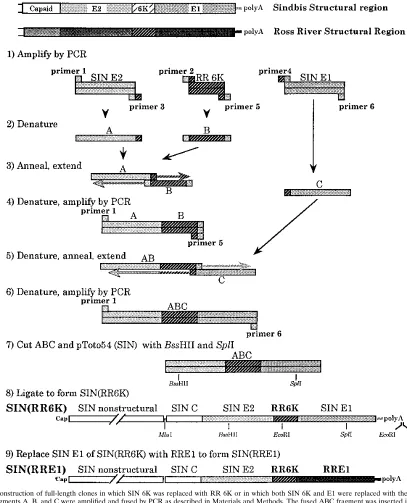
Construction of chimeric clones.The chimeric clone pSIN(RR6K), containing the SIN genome with the gene for RR 6K substituted for the SIN 6K gene, was produced by PCR with six primers, as illustrated in Fig. 1. Primer 1 is a sense primer, 59-CGGAACCAACCACTGAAT-39(SIN residues 9541 to 9558, found in the SIN E2 protein region). Primer 2 is a sense chimeric primer, 59-AGGTC GGCCAATGCTGCATCATTCGCTG-39, which consists of the last 14 nucleo-tides in the sequence of SIN E2 (residues 9885 to 9899 in the SIN genome) and the first 14 nucleotides in the sequence of RR 6K (residues 9834 to 9846 of the
RR genome). Primer 3 is the complement of primer 2 and is an antisense chimeric primer. Primer 4, 59 -CCTCCGCAAAAGCTTACGAACATGCGAC-39, is a sense chimeric primer consisting of the last 14 nucleotides of the sequence of RR 6K (RR residues 10000 to 10013) and the first 14 nucleotides of the sequence of SIN E1 (SIN residues 10065 to 10078). Primer 5 is the complement of primer 4. Primer 6 is an antisense primer, 59-AGTTGTTTTTCCACATCTC -39, located in SIN E1 (SIN residues 10803 to 10786).
[image:2.612.98.505.76.579.2]Primers 1 and 3 were used to prime a PCR starting from SIN clone pToto54 to produce a fragment containing 359 nucleotides (nt) of SIN E2 joined to 13 nt of RR 6K (A in Fig. 1). A second fragment, B, containing the entire RR 6K sequence (180 nt) flanked on each end by SIN sequence, was produced by FIG. 1. Construction of full-length clones in which SIN 6K was replaced with RR 6K or in which both SIN 6K and E1 were replaced with the comparable RR proteins. Fragments A, B, and C were amplified and fused by PCR as described in Materials and Methods. The fused ABC fragment was inserted into SIN pToto54 as shown to produce the full-length clone pSIN(RR6K). To construct pSIN(RR6KE1), a shuttle vector was first constructed by inserting the SspI-SspI fragment from pSIN(RR6K), containing the structural protein region and 39NTR, into the EcoRI site in pGEM3Z. This shuttle vector contains two EcoRI sites, one within the 6K protein and the other following the poly(A) tract. The EcoRI-EcoRI fragment in the shuttle vector was replaced with the comparable fragment from pRR64, which contains RR E1 and the RR 39NTR. Then the MluI-XhoI fragment containing SIN capsid, SIN E3, and SIN E2 plus RR6K, RR E1, and the RR 39NTR was excised from the shuttle vector and ligated into SIN pToto54 cut with same enzymes to produce the full-length clone pSIN(RR6KE1) [shortened to SIN(RRE1)].
on November 9, 2019 by guest
http://jvi.asm.org/
primers 2 and 5 with RR clone pRR64. Primers 4 and 6 were used with pToto54 to produce a third fragment called C, consisting of 739 nt from SIN E1 joined to 13 nt of RR 6K. In each case, the reaction mixture contained 100 ng of template, 0.2 mM deoxynucleoside triphosphates, and each primer at a final concentration of 1 pM. PCRs were carried out at 958C for 2 min followed by 25 cycles of 948C for 10 s, 558C for 30 s, and 728C for 2 min and finally by an extension at 728C for 7 min. After purification of each fragment on a low-melting temperature agarose gel, fragment A was fused to fragment B as follows: 100 ng of fragment A was mixed with 100 ng of fragment B, and the DNA was denatured and annealed at 558C for 2 min. These partially double-stranded molecules were made fully double stranded by extension at 728C for 3 min under the same conditions as for the PCR. The AB fusion DNA was then amplified with primers 1 and 5 for 25 cycles of PCR consisting of treatment at 948C for 10 s, 558C for 30 s, and 728C for 2 min, followed by a final extension at 728C for 7 min. The amplified product fragment AB was purified and fused to fragment C by using the same procedure and primers 1 and 6. The final fused fragment ABC consisted of the entire RR 6K protein fused exactly in frame to SIN E2 on the upstream side and to SIN E1 on the downstream side.
Fragment ABC was purified and cut with restriction enzymes BssHII and SplI and ligated to pToto54 SIN cDNA that had been cut with the same enzymes. After construction, the region between BssHII and SplI was sequenced to ensure that no error had been introduced during PCR amplification.
The construct pSIN(RR6KE1), which consists of pToto54 with the 6K, E1, and 39nontranslated region (NTR) of SIN replaced by those from RR, was con-structed by replacing the EcoRI (RR nt 9894, in 6K)-EcoRI [pToto54 nt 11742, just downstream of the poly(A) tract] fragment of pSIN(RR6K) with that from pRR64, using a multistage procedure that is described in the legend to Fig. 1. Throughout the text, the name of this chimeric construct has been shortened to pSIN(RRE1) and that of the virus recovered from it has been shortened to SIN(RRE1). Ten independent clones of pSIN(RRE1) were isolated and tested to ensure that the properties observed for the chimera did not result from exogenous changes introduced during the construction process.
Transfection of cells with RNA.BHK cells were harvested when slightly sub-confluent, washed with ice-cold phosphate-buffered saline (PBS) lacking Ca21 and Mg21, and resuspended at a concentration of 107/ml. To 0.45 ml of cells was
added about 10mg of RNA transcribed in vitro from the different cDNA clones by SP6 RNA polymerase, and the mixture was transferred to a 2-mm cuvette. Electroporation was performed at room temperature with two consecutive 1.5-kV, 25-mF pulses from a Gene Pulser apparatus (Bio-Rad). The cells were then diluted into 10 ml of medium and seeded into 10-cm culture plates.
Assay of released virus and nucleocapsids.After 8 h of incubation at 378C following electroporation, the medium was replaced with medium containing 5% dialyzed fetal calf serum and 1mg of dactinomycin per ml. At 4 h later, the medium was changed to medium lacking dactinomycin but containing [3
H]uri-dine (20mCi/ml), and the transfected cells were labeled for 10 to 12 h. Virus released into the medium and intracellular nucleocapsids were assayed from the same cell culture. Released virus was assayed by precipitating the virus from the culture fluid with polyethylene glycol (25) and resuspending it in 1 ml of TNA (50 mM Tris [pH 7.5], 200 mM NaCl, 1 mM EDTA) and sedimenting it in a linear 15 to 30% sucrose gradient in the same buffer at 34,000 rpm at 58C for 1.5 h in an SW41 rotor. Intracellular nucleocapsids were prepared as described previ-ously (18) and sedimented for 2.5 h at 58C at 34,000 rpm in a linear 10 to 40% sucrose gradient in TNA buffer containing 0.1% Triton X-100 in an SW41 rotor. Fractions of 0.4 ml from these gradients were assayed for radioactivity by liquid scintillation counting.
Metabolic labeling and immunoprecipitation.Following incubation at 378C for 22 to 24 h after electroporation, transfected cells were washed with Eagle’s medium and overlaid with methionine-free medium for 30 min at 378C. The medium was replaced for 10 min at 378C with medium containing [35
S]methi-onine (100mCi/ml), and the cells were washed with medium containing 1003
methionine and chased for various times in medium containing 103methionine. At each chase point, the plate was put on ice and the cells were washed with ice-cold PBS and lysed with 200ml of lysis buffer (1% Triton X-100, 50 mM Tris-HCl [pH 7.5], 150 mM NaCl, 2 mM EDTA, 100mg of phenylmethylsulfonyl fluoride per ml, 25 mM iodoacetamide). The lysate was centrifuged to remove nuclei and immunoprecipitated with monoclonal antibodies or polyclonal anti-bodies. For this, 20ml of lysate was diluted to 200ml with lysis buffer and 2ml of monoclonal antibody or 5ml of polyclonal antibody was added. After incubation at 48C for 1 h, the reaction mixture was treated with 50ml of 10% Staphylococcus aureus in lysis buffer and incubated for a further 30 min at room temperature. The S. aureus cells were then pelleted, washed three times with lysis buffer at room temperature, resuspended in 40ml of nonreducing sample buffer (100 mM Tris [pH 8.8], 500 mM sucrose, 5 mM EDTA [pH 8.0], 4% sodium dodecyl sulfate [SDS], 10 mg of methionine per ml, 0.02% bromophenol blue), and heated to 858C for 10 min. The released protein was analyzed by polyacrylamide gel electrophoresis in the presence of SDS.
Biotinylation of proteins at the cell surface.Transfected cells were pulse-labeled with [35S]methionine and chased for various times as described above.
The cells were put on ice, washed with ice-cold PBS, and treated with 1 mg of sulfosuccinimidyl 6-(biotinamido)hexonate (NHS-LC-biotin; Pierce Chemical Co.) per ml for 30 min at 48C. The reaction was quenched by washing twice with ice-cold PBS containing 50 mM NH4Cl followed by washing with PBS alone, and
the cells were lysed with 200ml of lysis buffer. A 40-ml sample of the lysate was diluted to 200ml with lysis buffer, and the biotinylated proteins were captured by adding 40ml of 50% (vol/vol) streptavidin-agarose (Sigma) and incubating at 48C for 2 h. The agarose pellet was washed three times with lysis buffer, once with 10 mM Tris (pH 7.5), and once with 1% SDS. Finally, the sample was heated in sample buffer to 958C for 10 min and analyzed by gel electrophoresis.
Low-pH-mediated cell fusion from within.At 12 h posttransfection, the cells were washed once with PBS and treated with low-pH buffer [Eagle’s medium containing 10 mM 2-(N-morpholino)ethanesulfonic acid (MES) and 10 mM N-2-hydroxyethylpiperazine-N9-2-ethanesulfonic acid (HEPES) (pH 5.3)] for 3 min at 378C. The control medium was pH 7.2. After removal of the low-pH buffer, the cells were incubated at 378C for 1 h, and fusion was observed by light microscopy after fixation and staining with Giemsa.
Immunofluorescence assay.At 12 h posttransfection, the cells were washed with PBS and fixed with 4% paraformaldehyde at room temperature for 10 min. After being washed twice with PBS, the cells were treated at room temperature for 15 min with either 0.2% Triton X-100 in PBS or PBS lacking Triton X-100. The cells were washed twice with PBS and treated with rabbit anti-SIN E2 polyclonal antiserum at 378C for 1 h. The cells were washed twice with PBS, goat anti-rabbit immunoglobulin G conjugated to fluorescein isothiocyanate was added, and the mixture was incubated at 378C for 40 min. The cells were washed and observed by fluorescence microscopy.
Electron microscopy.At 12 or 24 h posttransfection, the cells were treated with 2% glutaraldehyde in 0.1 M cacodylate buffer (pH 7.2), immediately scraped off the plate, and kept at 48C for 15 min. After being pelleted, the cells were resuspended in 0.1 M cacodylate buffer (pH 7.2) containing 0.2% sucrose. The cells were postfixed, dehydrated, and infiltrated with Epon. The section was cut, stained, and observed by electron microscopy.
RESULTS
Construction of chimeric viruses.We constructed two chi-meric viruses starting from a full-length cDNA clone of SIN, pToto54 (10), and a full-length cDNA clone of RR, pRR64 (12). The first chimera, called SIN(RR6K), consisted of SIN in which the 6K protein of SIN had been replaced by that of RR, and the second chimera, called SIN(RRE1), consisted of SIN
in which the 6K protein, E1, and the 39NTR of SIN had been
replaced by the corresponding regions from RR (Fig. 1). In a preliminary characterization of the chimeric viruses, RNA transcribed in vitro from the chimeric or parental cDNA clones was used to transfect BHK cells by using Lipofectin transfec-tion, and the cells were overlaid with liquid medium to assay the rate of virus growth (Table 1). Lipofectin transfection re-sults in only a small percentage of the cells being initially infected, and the rise in titers at later times results from mul-tiple rounds of infection, amplifying differences in growth rates. SIN(RR6K) is impaired in virus production; at later times, the titers were only about 2% those of SIN. SIN(RRE1)
is severely impaired and produced only about 1027the amount
of virus as did wild-type SIN (Table 1).
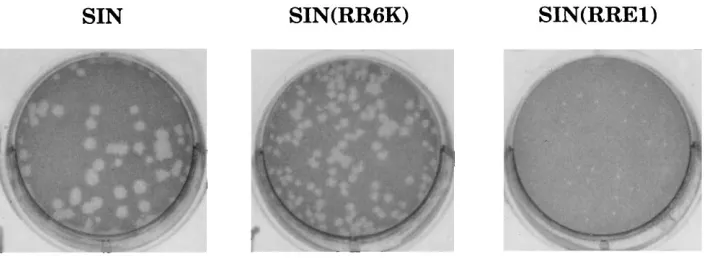
In a second experiment, transfected cells were overlaid with agarose to determine the specific infectivities of the RNAs (Table 1). The specific infectivities of the two chimeric RNAs and SIN RNA were the same within experimental error, i.e., 10 to 20 plaques per ng of RNA. Thus, although there is a great difference in growth rates, a cell transfected by chimeric RNA produces sufficient virus to propagate a plaque. However, the differences in growth rates are reflected in the size of the plaques (Table 1), which is not always true (11, 22, 23). In particular, the plaques formed by SIN(RRE1) are minute and at the limit of detection (Fig. 2). Because the specific infectiv-ities of the RNAs are the same, it is probable that the limited growth of the chimeras that is observed does not result from the early appearance of variants.
RR RNA transcribed from pRR64 is only about 1/10 as infectious as SIN RNA, and RR grows less rapidly than does SIN in BHK cells (Table 1), as has been previously described (12). Note that RR produces plaques the same size as those produced by SIN despite the difference in growth rates (Table 1).
on November 9, 2019 by guest
http://jvi.asm.org/
SIN(RR6K) and SIN(RRE1) are impaired in budding.To determine where in the infection cycle virus production was blocked, BHK cells were transfected by electroporation, so
that most of the cells were infected, and labeled with [3
H]uri-dine. The amount of radioactivity in nucleocapsids present in the infected cells at 24 h after transfection and the amount of radioactivity in virus released into the medium at this time were determined by sucrose gradient fractionation. Some of the results are shown in Fig. 3, and the results of two such experiments are quantitated in Table 2. Nucleocapsids were assembled in large amounts in cells transfected with the chi-meric RNAs and SIN RNA. The radioactivity incorporated into SIN capsids and SIN(RR6K) capsids was similar, but there was about half as much label in SIN(RRE1) capsids (Table 2). However, the amount of radiolabel in virus released from the transfected cells was very different. The radioactivity in re-leased SIN(RR6K) virions was about 10% of that in rere-leased SIN virions, consistent with the results reported in Table 1. Furthermore, the specific infectivities (PFU/cpm) of SIN and SIN(RR6K) virions in the peak fractions from the sucrose gradients were identical (data not shown), indicating that the reduced yield of infectious virus was the result of reduced virus assembly rather than low infectivity of assembled virus. For cells transfected with SIN(RRE1) RNA, no detectable radio-labeled virus was released (Fig. 3) and the small amount of infectious virus produced is thus due to a failure to assemble virions rather than to the assembly of defective virions. The total amount of radioactivity in encapsidated RNA of SIN (RRE1) (the sum of that in intracellular capsids plus capsids in virions) was about 20% of that in SIN capsids, suggesting that this virus may be slightly impaired in RNA replication, which may result from an effect of structural proteins on the rate of
RNA synthesis (10). However, it is clear that the major im-pairment in the production of SIN(RRE1) virus is in the in-corporation of capsids into mature virions.
Production of E1/E2 heterodimers.To examine the synthesis and heterodimerization of glycoproteins in cells transfected with the chimeric viruses, the cells were transfected by
elec-troporation and pulse-labeled with [35S]methionine. After
dif-ferent chase periods, the cells were lysed by treatment with Triton X-100 and the radiolabeled proteins were displayed on SDS-containing polyacrylamide gels (Fig. 4). Nonreducing (but denaturing) gels were used for all experiments here because the resolution of SIN E2 from SIN E1 or RR E1 was clear in nonreducing gels but poor in reducing gels (results not shown). After a short chase (10 or 20 min), PE2, E1, and C were present in cells transfected with any of the three viral RNAs. Also present were small amounts of higher-molecular-weight proteins, which represent the untranslocated polyprotein PE2-6K-E1 or different nonstructural protein precursors. After a longer chase (40 or 80 min), PE2 was cleaved to E2 and the efficiency of PE2 cleavage appeared to be similar. Since cleav-age of PE2 to E2 occurs during transport of PE2 to the cell surface (reviewed in reference 28), we conclude that the gly-coproteins are translated as polyproteins, cleaved, glycosy-lated, and transported normally (other experiments that ad-dress transport are also described below).
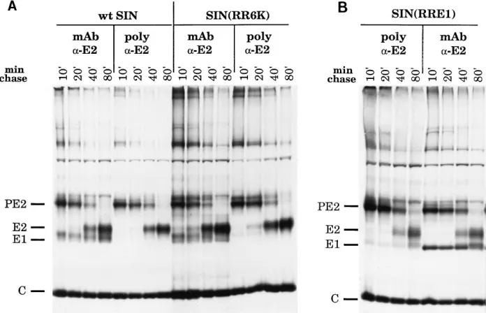
[image:4.612.60.556.81.163.2]We next examined whether PE2/E1 heterodimers were formed by immunoprecipitating proteins from Triton X-100 lysates of transfected cells with an anti-SIN-E2 monoclonal antibody. After a short chase (10 or 20 min), SIN E1 (Fig. 5A) or RR E1 (Fig. 5B), depending on the virus, coprecipitated with PE2, showing that heterodimers were formed between SIN PE2 and SIN E1 in wild-type SIN or the SIN(RR6K)
[image:4.612.131.483.577.706.2]FIG. 2. Plaques formed by chimeric viruses. Stocks of SIN, SIN(RR6K), and SIN(RRE1) from RNA transfection were subjected to titer determination on BHK cells. The cells were stained with neutral red after 48 h and photographed 12 h later.
TABLE 1. Virus growth after RNA transfection
RNA
Virus yield (PFU/ml)aat:
Plaque size Infectivity
(PFU/ng of RNA)b
6 h 12 h 24 h 30 h 48 h
SIN Toto 54 43102 63104 23108 93108 531011 1.5–2.0 19
RR64 ,50 ,50 13103 43104 53107 1.5–2.0 1
SIN(RR6K) 43102 13104 33106 13107 83109 1.0–1.5 16
SIN(RRE1) ,50 ,50 50 33102 73103 ,0.5 11
a
BHK cells were transfected with RNA transcribed in vitro from the various cDNA clones, using Lipofectin (12), and the transfected cells were overlaid with liquid medium. At the indicated times, the medium was assayed for infectious virus by plaque assay on BHK cells.
b
BHK cells were transfected with RNA as in footnote a but overlaid with medium containing agarose so that individual plaques formed. The cells were stained with neutral red after 48 h, and the plaques were measured and counted 12 h later. The size of the plaques (in millimeters) formed in this primary transfection assay and the specific infectivity of the RNA (in PFU per nanogram of RNA) determined from the number of plaques formed are given.
on November 9, 2019 by guest
http://jvi.asm.org/
chimera and between SIN PE2 and RR E1 in the SIN(RRE1) chimera. Since less E1 than PE2 was precipitated, there may be partial dissociation of the PE2/E1 complexes during sample preparation or immunoprecipitation, but the results with the chimeras and with wild-type SIN appeared to be similar; if anything, the SIN PE2/RR E1 complex appeared to be more stable than the SIN PE2/SIN E1 complex. After the longer chase periods (40 or 80 min), during which time PE2 was converted to E2, the RR E1 or SIN E1 coprecipitated with SIN E2, showing that both the SIN E2/SIN E1 and SIN E2/RR E1 complexes are stable during the course of the experiments.
It is perhaps significant that the coprecipitated E1 in SIN(RR6K) infection appears to be a doublet (Fig. 5A). SIN E1 is known to undergo a series of conformational changes during maturation that involve the formation of disulfide bonds with concomitant changes in migration if the protein is not reduced (19). Furthermore, it is known that PE2 and E1 are not complexed when first synthesized but rapidly associate to form a heterodimer with a half-time of 4 min or less (2, 20). Before association, E1 begins to fold in association with the molecular chaperone BiP, but folding continues after het-erodimerization with PE2 (20). The doublet seen in the case of SIN(RR6K) might result from a lower rate of maturation of E1
or from a failure of E1 to mature completely when RR 6K rather than SIN 6K is present, suggesting that the 6K protein might interact with the PE2/E1 complex in a way that aids the folding of E1.
[image:5.612.63.296.67.391.2]As a control to rule out the possibility that nonspecific ag-gregation was responsible for coprecipitation, we also immu-noprecipitated the Triton X-100 extracts with a rabbit anti-E2 polyclonal antibody. The strong interaction of the polyclonal antibody with PE2 or E2 led to the dissociation of the SIN PE2(E2)/SIN E1 heterodimer, and when this antibody was
FIG. 3. Nucleocapsids and virions formed following transfection of cells with chimeric RNAs. BHK cells were transfected with RNA transcribed in vitro from pSIN, pSIN(RR6K), and pSIN(RRE1) by electroporation and labeled with [3
H]uridine from 12 to 24 h after transfection. Labeled virions released into the cell culture medium and labeled intracellular nucleocapsids were assayed by sedimentation in appropriate sucrose gradients. In the nucleocapsid gradients, the faster-sedimenting peak contains 140S nucleocapsids and the more slowly sedimenting peak contains free RNA or RNA complexed with cellular proteins. Sedimentation is from right to left.
FIG. 4. Synthesis of structural proteins following transfection by chimeric RNAs. BHK cells were transfected by electroporation with RNA transcribed in vitro from pSIN, pSIN(RR6K), or pSIN(RRE1). The transfected cells were pulse-labeled for 10 min with [35S]methionine at 378C at 22 to 24 h after
[image:5.612.316.557.90.234.2]electroporation and chased with medium containing unlabeled methionine for various times as indicated. The labeled cells were lysed with buffer containing 1% Triton X-100, and the whole lysate was analyzed on 10% nonreducing polyacryl-amide gels containing SDS. wt, wild type.
TABLE 2. Radioactivity in nucleocapsids and virions 24 h after transfection of BHK cells with RNAs
RNAa
Nucleocapsidsb Virionsc
Radiolabel
(105cpm) Ratiod
Radiolabel
(105cpm) Ratiod
Expt 1e
SIN 12.0 1.0 11.0 1.0
SIN(RR6K) 8.4 0.7 0.9 0.1
SIN(RRE1) 4.4 0.4 ,0.002 ,0.0002
RR NDf ND
Expt 2e
SIN 3.5 1.0 1.4 1.0
SIN(RR6K) 4.4 1.2 0.2 0.1
SIN(RRE1) 1.9 0.5 ,0.003 ,0.002
RR 0.2 0.1 0.2 0.1
aRNA was transcribed in vitro from the various cDNA clones and used to
transfect BHK cells by electroporation. The transfected cells were labeled with [3H]uridine from 12 to 24 h after electroporation.
bNucleocapsids were isolated from the transfected cells and sedimented on
sucrose gradients as in Fig. 3. The radiolabel in the nucleocapsid peak was summed.
cVirions were isolated from the cell supernatant by sucrose gradient
sedimen-tation as in Fig. 3. The radiolabel in the virion peak was summed.
dRatio of radiolabel to that in SIN Toto54.
eExperiments 1 and 2 are independent experiments performed several months
apart.
fND, not done.
on November 9, 2019 by guest
http://jvi.asm.org/
[image:5.612.342.533.480.654.2]used, no detectable SIN E1 protein coprecipitated (Fig. 5). However, small amounts of RR E1 protein did coprecipitate with PE2 or E2 (Fig. 5), again suggesting that the SIN PE2(E2)/RR E1 interaction is more stable than the SIN PE2(E2)/SIN E1 interaction. These results confirm the identity of SIN E2 (and, by extension, of E1) in the gels. The nucleo-capsid protein C is present in the gels because it precipitates nonspecifically under these conditions (see, for example, ref-erence 27).
Transport of heterodimers to the cell surface.Cleavage of PE2 to E2 is a late event in the transport of the glycoproteins to the cell surface (reviewed in reference 28), and the fact that this cleavage occurred quantitatively in cells transfected by the chimeric viruses is evidence that the PE2/E1 heterodimer is transported normally in cells infected by the chimeras. To directly examine the presence of the E2/E1 heterodimer at the cell surface, we used three different methods. In one set of experiments, the transfected cells were exposed briefly to pH 5.3 and the cells were examined for fusion. Representative results are shown in Fig. 6. Cells transfected with RNA from SIN or from either of the chimeras showed strong fusion when exposed to pH 5.3 but not when exposed to pH 7.2. Control BHK cells did not exhibit fusion when exposed to pH 5.3. Fusion is believed to be a property of E1 (reviewed in refer-ence 28), and we conclude that E1 is present on the surface of the infected cell following infection by the chimeras and is functional in fusing cells when exposed to low pH.
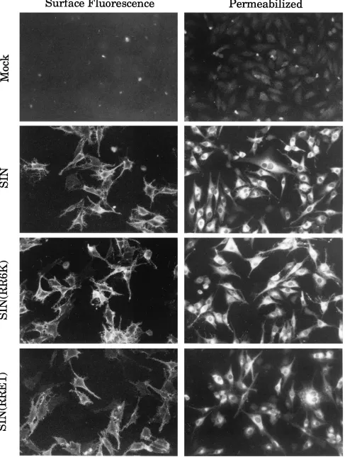
In a second set of experiments, we examined the surface of transfected cells for the appearance of E2 by using an immu-nofluorescence assay with an anti-E2 monoclonal antibody (Fig. 7). The cell surface showed strong staining by anti-E2 antibodies whether infection was by SIN or by either of the chimeras. Thus, E2 has appeared at the surface of the cells infected by the chimeras.
In a third experiment, transfected cells were pulse-labeled
with [35S]methionine and chased for 10 or 80 min. Intact cells
were then reacted with activated biotin and lysed, and the biotinylated proteins were isolated (Fig. 8). After 10 min of
chase, no 35S-labeled glycoprotein was detectable at the cell
surface. After 80 min of chase, however,35S-labeled viral
gly-coproteins at the cell surface were reactive with biotin. In cells transfected with SIN RNA, both E2 and E1 reacted efficiently with biotin. However, in cells transfected with SIN(RR6K) RNA, only the E2 protein was detected following reaction with biotin; the SIN E1 in the heterodimer does not react with biotin when 6K comes from RR rather than SIN. In contrast, in cells transfected with SIN(RRE1) RNA, only the RR E1 protein was reactive with biotin; thus, in the SIN E2/RR E1 heterodimer, the SIN E2 does not react with biotin. We con-clude that the PE2/E1 heterodimers are formed and trans-ported to the cell surface in all three cases but that the con-formation of the heterodimers is different in the three cases, resulting in a difference in the accessibility to biotinylation of E2 and E1. It is unknown whether the primary amines that are biotinylated are present in the N-terminal residue or in inter-nal residues or both.
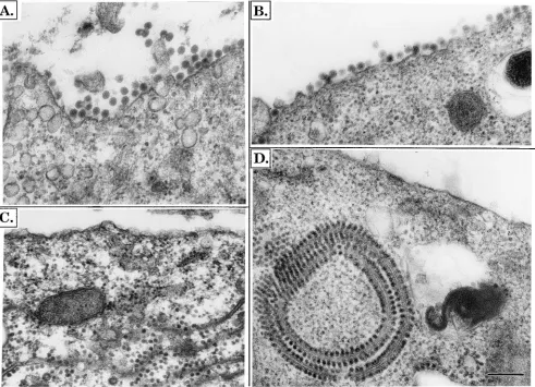
[image:6.612.130.478.71.295.2]Electron microscopy of infected cells.To examine whether the chimeric heterodimers present on the cell surface can in-teract with the viral nucleocapsids, we examined transfected cells by electron microscopy. At 12 or 24 h posttransfection, the cells were fixed, dehydrated, and embedded, and thin sec-tions were cut and examined. Representative micrographs are shown in Fig. 9. After 12 h of infection by SIN (Fig. 9A) or SIN(RR6K) (Fig. 9B), large numbers of virus are seen budding from the plasma membrane. Subjectively, it appears that more virus is budding from SIN-transfected cells than from SIN(RR6K)-transfected cells, but accurate quantitation is not possible. In contrast, in cells infected with SIN(RRE1), no budding virus is seen and no nucleocapsids are aligned along the plasma membrane, although large numbers of nucleocap-sids are present within the cell (Fig. 9C). After 24 h, budding virus continues to be seen in cells infected with SIN or
FIG. 5. Formation of PE2/E1 and E2/E1 heterodimers by chimeric viruses. BHK cells were transfected, pulse-labeled, chased, and lysed as in Fig. 4. The lysates were immunoprecipitated with either an anti-SIN-E2 monoclonal antibody (lanes labeled mAb) or an anti-SIN-E2 polyclonal antibody (lanes labeled poly). The immunoprecipitates were collected with S. aureus and analyzed on 10% nonreducing polyacrylamide gels containing SDS. (A) Wild-type (wt) SIN and SIN(RR6K); (B) SIN(RRE1).
on November 9, 2019 by guest
http://jvi.asm.org/
SIN(RR6K), and the cytoplasm contains tubular or vacuolar structures that have many nucleocapsids attached to the cyto-plasmic face but do not appear to be sites of budding (data not shown, but see Fig. 9D and reference 21); these structures have been called cytopathic vacuoles (21). In cells infected with SIN(RRE1), budding virus or nucleocapsids aligned at the plasma membrane are still not seen after 24 h but cytopathic vacuoles with attached nucleocapsids are readily visible within the transfected cell (Fig. 9D). It thus appears that the interac-tion of the nucleocapsids with the glycoproteins in the cell
surface is defective in the SIN E2/RR E1 chimera, since nu-cleocapsids are not attracted to the plasma membrane of in-fected cells and do not bud. The presence of nucleocapsids aligned along cytopathic vacuoles, however, may indicate that the interaction is not totally defective.
DISCUSSION
Role of the 6K protein in virus assembly.The 6K protein is a 55-amino-acid (in SIN) hydrophobic peptide found in the
FIG. 6. Cell fusion assays to detect the appearance of E1 on the cell surface. BHK cells were transfected by electroporation with RNA transcribed from pSIN, pSIN(RR6K), or pSIN(RRE1). At 12 h after electroporation, the transfected cells were treated with low-pH buffer (pH 5.3) for 3 min at 378C or with control medium (pH 7.2). After removal of the low-pH buffer, the cells were incubated for 1 h at 378C in normal medium, fixed, and stained.
on November 9, 2019 by guest
http://jvi.asm.org/
FIG. 7. Immunofluorescence assay for the appearance of E2 at the cell surface. BHK cells were transfected by electroporation with RNA transcribed from pSIN, pSIN(RR6K), or pSIN(RRE1). At 12 h after electroporation, the transfected cells were fixed and half of the samples were permeabilized with 0.2% Triton X-100. Both nonpermeabilized and permeabilized cells were then reacted with rabbit anti-SIN-E2 polyclonal antibody, followed by goat anti-rabbit immunoglobulin G conjugated to fluorescein isothiocyanate and examined by fluorescence microscopy.
7917
on November 9, 2019 by guest
structural polyprotein precursor between PE2 and E1 (28, 29). It is believed that cleavages by signalase form both its N and C termini, so that the final protein is membrane associated and spans the membrane twice, with both the N and C termini in the lumen. The N-terminal membrane-spanning region is thought to act as a stop transfer signal, and the C-terminal spanning domain is thought to act as an internal signal se-quence which transfers the N terminus of E1 into the lumen. In addition to its function as a signal sequence, the 6K protein is known to be required for the correct and efficient assembly of virus, and submolar amounts of 6K are normally present in SIN virions (7); however, its exact role in virus assem-bly is unknown. In Semliki Forest virus, deletion of 6K re-sults in the production of only very small amounts of virus, but the virus that is produced is normal (13). In SIN, mutations in 6K can lead to defective assembly of virus, resulting in particles containing more than one nucleocapsid (6, 9). It has been postulated that one function of the 6K protein in budding might be to allow lipids in the membrane to flip from one side of the bilayer to the other (6); the virus mem-brane is very sharply curved, and the cross-sectional area of the outer leaflet is 50% greater than the cross-sectional area of the inner leaflet, requiring some reorientation of lipids during bud-ding.
The use of chimeric viruses to study protein interactions has a number of advantages over approaches previously taken to study the functions of 6K. The substitution in SIN of the RR 6K for its SIN counterpart is equivalent to introducing numer-ous mutations into the 6K protein; of the 55 residues in SIN 6K, the RR 6K differs at 39 positions, and in addition, the RR protein has a 5-amino-acid insertion relative to the SIN se-quence, so that it is 60 residues in length. However, the final product (RR 6K) is known to be fully functional in the correct
context, unlike the insertion of random mutations into the SIN 6K sequence, which might affect its folding or its activities in unknown ways. Furthermore, when the resulting chimera is viable but attenuated, it is often possible to isolate variants with compensating mutations that adapt the disparate proteins to one another, making it possible to map interacting domains (8, 18).
Our results with SIN(RR6K) indicate that there is a se-quence-specific interaction between 6K and PE2(E2) or be-tween 6K and E1 that is required for efficient virus assembly, since the 6K protein of RR cannot substitute for that of SIN in the chimera SIN(RR6K) to give wild-type virus yields. The results of the immunoprecipitation experiments, in which dif-ferences in the conformation of E1 were found, as well as the results of the experiments to assess the biotinylation of E2 and E1 at the cell surface, in which E1 could be biotinylated in one case but not the other, clearly show that the conformation of E1 in the SIN E2/SIN E1 heterodimer is different when it is formed in the presence of RR 6K from when it is formed in the presence of SIN 6K. These results suggest that 6K may en-hance assembly by promoting proper folding and maturation of E1 within the heterodimer. In addition to this role and its function as a signal sequence, still other roles for 6K, such as promotion of lipid exchange during budding, are possible.
The E1/E2 heterodimer.PE2 and E1 are separate when first formed but associate within minutes to form a heterodimer (2, 20). Using SIN(RRE1), we have shown that SIN PE2 will form a heterodimer with RR E1 and that this heterodimer is trans-ported to the cell surface and cleaved to form an E2/E1 het-erodimer, even though SIN E1 and RR E1 share only 51% amino acid sequence identity. Chimeric as well as wild-type PE2/E1 and E2/E1 heterodimers are sufficiently stable to be coprecipitated with anti-E2 monoclonal antibodies, but the conformation of the chimeric heterodimer at the cell surface is different from that of a SIN E2/SIN E1 heterodimer. First, SIN E2 could be labeled with biotin at the cell surface when present in a SIN E2/SIN E1 heterodimer, regardless of the source of 6K, but not when present in a chimeric SIN E2/RR E1 het-erodimer. Second, we could find no budding figures or specific association of nucleocapsids with chimeric heterodimers in the plasma membrane, whereas nucleocapsids were readily seen at the plasma membrane when cells were infected by SIN or by the SIN(RR6K) chimera. Related results have been seen with a temperature-sensitive mutant of SIN, ts23, which has two mutations in the ectodomain of E1 (14). In ts23-infected cells at the nonpermissive temperature, nucleocapsids are found scattered throughout the cytoplasm; in contrast, in cells in-fected with ts20, a mutant having a mutation in E2, nucleocap-sids were found aligned underneath the plasma membrane at the nonpermissive temperature, presumably interacting with the glycoproteins, although budding did not take place in ei-ther case (3).
It is well accepted that there is a sequence-specific interac-tion between the cytoplasmic domain of glycoprotein E2 and the nucleocapsid protein that is important for virus budding (reviewed in references 28 and 29). There are two models, based upon this interaction, to explain our results with the chimeras. In one model, E2 and E1 must interact with one another in a favorable way for the tail of E2 to be properly positioned for interaction with the nucleocapsid. In this model, the E1-E2 interactions are not favorable in the chimeras; in SIN(RR6K), the weakened interaction results in a lower rate of budding, but in SIN(RRE1), the interaction is so defective that there is an almost total lack of glycoprotein-nucleocapsid interaction at the plasma membrane. Biochemical studies have shown that in the absence of proper orientation of the E2 tail,
FIG. 8. Labeling of E1 and E2 at the cell surface with biotin. BHK cells were transfected, pulsed with [35S]methionine, and chased as in Fig. 4, and cell surface
proteins were biotinylated. The biotinylated proteins were captured with strepta-vidin-agarose and analyzed on 10% denaturing (but nonreducing) polyacryl-amide gels. Lanes labeled lysate display samples of whole-cell lysate. Lanes labeled avidin display the proteins captured with streptavidin.
on November 9, 2019 by guest
http://jvi.asm.org/
no E2/nucleocapsid interaction is observed by electron micros-copy (15, 16), but there is no direct evidence currently that E1-E2 interactions can affect the orientation of the tail. In a second model, heterodimer trimerization, which has been pos-tulated to involve E1-E1 interactions (1), is blocked by subop-timal conformations of the chimeric heterodimers. It is un-known whether the interaction of a single E2 tail with a nucleocapsid is sufficient to hold the nucleocapsid at the cell surface or whether trimerization of heterodimers is required before a stable interaction can occur. It is possible that for SIN(RR6K), a lower rate of heterotrimerization results in a lower rate of budding, and that for SIN(RRE1), a very low or nonexistent rate of heterotrimerization results in almost total
failure of the capsid-E2 interaction. Eckstro¨m et al. (5) have
produced evidence that trimerization may provide a significant fraction of the free energy required for budding and have postulated that the formation of a trimeric spike increases the affinity between the glycoproteins and the nucleocapsid be-cause of the multivalent nature of the interaction. It is un-known if heterodimer trimerization is absolutely required for budding, however. We are continuing our investigation of these interactions in the chimeras by isolating variants in which SIN E2 and RR E1 are adapted to each other such that an increased rate of budding is obtained.
ACKNOWLEDGMENTS
We are grateful to J. E. Edens for assistance and training in electron microscopy, to Y. Shirako for stimulating discussions during the course of these experiments, and to E. Lenches for expert technical assistance. This work has been supported by grants AI 20612 and AI 10793 from NIH.
REFERENCES
1. Anthony, R. P., and D. T. Brown. 1991. Protein-protein interactions in an alphavirus membrane. J. Virol. 65:1187–1194.
2. Barth, B.-U., J. M. Wahlberg, and H. Garoff. 1995. The oligomerization reaction of the Semliki Forest virus membrane protein subunits. J. Cell Biol.
128:283–291.
3. Brown, D. T., and J. F. Smith. 1975. Morphology of BHK-21 cells infected with Sindbis virus temperature sensitive mutants in complementation groups D and E. J. Virol. 15:1262–1266.
4. Cheng, R. H., R. J. Kuhn, N. H. Olson, M. G. Rossmann, H.-K. Choi, T. J.
Smith, and T. S. Baker.1995. Nucleocapsid and glycoprotein organization in an enveloped virus. Cell 80:621–630.
5. Ekstro¨m, M., P. Liljestro¨m, and H. Garoff.1994. Membrane protein lateral interactions control Semliki Forest virus budding. EMBO J. 13:1058–1064. 6. Gaedigk-Nitschko, K., M. Ding, and M. J. Schlesinger. 1990. Site-directed
mutations in the Sindbis virus 6K protein reveal sites for fatty acylation and the underacylated protein affects virus release and virion structure. Virology
175:282–291.
7. Gaedigk-Nitschko, K., and M. J. Schlesinger. 1990. The Sindbis virus 6K protein can be detected in virions and is acylated with fatty acid. Virology
[image:10.612.64.555.72.427.2]175:274–281.
FIG. 9. Electron microscopy of cells infected by chimeric viruses. BHK cells were transfected with RNA as in Fig. 4. At either 12 or 24 h after electroporation, the transfected cells were fixed, sectioned, stained, and examined by electron microscopy. (A) SIN, 12 h after transfection; (B) SIN(RR6K), 12 h after transfection; (C) SIN(RRE1), 12 h after transfection; (D) SIN(RRE1), 24 h after transfection.
on November 9, 2019 by guest
http://jvi.asm.org/
8. Hahn, C. S., S. Lustig, E. G. Strauss, and J. H. Strauss. 1988. Western equine encephalitis virus is a recombinant virus. Proc. Natl. Acad. Sci. USA
85:5997–6001.
9. Ivanova, L., S. Lustig, and M. J. Schlesinger. 1995. A pseudo-revertant of a Sindbis virus 6K protein mutant, which corrects for aberrant particle forma-tion, contains two new mutations that map to the ectodomain of the E2 glycoprotein. Virology 206:1027–1034.
10. Kuhn, R. J., D. E. Griffin, K. E. Owen, H. G. M. Niesters, and J. H. Strauss. 1996. Chimeric Sindbis-Ross River viruses to study interactions between alphavirus nonstructural and structural regions. J. Virol. 70:7900–7909. 11. Kuhn, R. J., Z. Hong, and J. H. Strauss. 1990. Mutagenesis of the 39
nontranslated region of Sindbis virus RNA. J. Virol. 64:1465–1476. 12. Kuhn, R. J., H. G. M. Niesters, H. Zhang, and J. H. Strauss. 1991. Infectious
RNA transcripts from Ross River virus cDNA clones and the construction and characterization of defined chimeras with Sindbis virus. Virology 182: 430–441.
13. Liljestro¨m, P., S. Lusa, D. Huylebroeck, and H. Garoff.1991. In vitro mu-tagenesis of a full-length cDNA clone of Semliki Forest virus: the small 6,000-molecular-weight membrane protein modulates virus release. J. Virol.
65:4107–4113.
14. Lindqvist, B. H., J. DiSalvo, C. M. Rice, J. H. Strauss, and E. G. Strauss. 1986. Sindbis virus mutant ts20 of complementation group E contains a lesion in glycoprotein E2. Virology 151:10–20.
15. Liu, N., and D. T. Brown. 1993. Phosphorylation and dephosphorylation events play critical roles in Sindbis virus maturation. Virology 196:703–711. 16. Liu, N., and D. T. Brown. 1993. Transient translocation of the cytoplasmic (endo) domain of a type I membrane glycoprotein into cellular membranes. J. Cell Biol. 120:877–883.
17. Loewy, A., J. Smyth, C.-H. von Bonsdorff, P. Liljestro¨m, and M. J. Schlesinger.1995. The 6-kilodalton membrane protein of Semliki Forest virus is involved in the budding process. J. Virol. 69:469–475.
18. Lopez, S., J.-S. Yao, R. J. Kuhn, E. G. Strauss, and J. H. Strauss. 1994.
Nucleocapsid-glycoprotein interactions required for alphavirus assembly. J. Virol. 68:1316–1323.
19. Mulvey, M., and D. T. Brown. 1994. Formation and rearrangement of disul-fide bonds during maturation of the Sindbis virus E1 glycoprotein. J. Virol.
68:805–812.
20. Mulvey, M., and D. T. Brown. 1995. Involvement of the molecular chaperone BiP in maturation of Sindbis virus envelope glycoproteins. J. Virol. 69:1621– 1627.
21. Murphy, F. A. 1980. Togavirus morphology and morphogenesis, p. 241–316. In R. W. Schlesinger (ed.), The togaviruses: biology, structure, replication. Academic Press, Inc., New York.
22. Niesters, H. G. M., and J. H. Strauss. 1990. Defined mutations in the 59
nontranslated sequence of Sindbis virus RNA. J. Virol. 64:4162–4168. 23. Niesters, H. G. M., and J. H. Strauss. 1990. Mutagenesis of the conserved 51
nucleotide region of Sindbis virus. J. Virol. 64:1639–1647.
24. Paredes, A. M., D. T. Brown, R. B. Rothnagel, W. Chiu, R. J. Schoepp, R. E.
Johnston, and B. V. V. Prasad.1993. Three-dimensional structure of a membrane-containing virus. Proc. Natl. Acad. Sci. USA 90:9095–9099. 25. Pierce, J. S., E. G. Strauss, and J. H. Strauss. 1974. Effect of ionic strength
on the binding of Sindbis virus to chick cells. J. Virol. 13:1030–1036. 26. Rice, C. M., R. Levis, J. H. Strauss, and H. V. Huang. 1987. Production of
infectious RNA transcripts from Sindbis virus cDNA clones: mapping of lethal mutations, rescue of a temperature-sensitive marker, and in vitro mutagenesis to generate defined mutants. J. Virol. 61:3809–3819. 27. Rice, C. M., and J. H. Strauss. 1982. Association of Sindbis virion
glycopro-teins and their precursors. J. Mol. Biol. 154:325–348.
28. Strauss, J. H., and E. G. Strauss. 1994. The alphaviruses: gene expression, replication, and evolution. Microbiol. Rev. 58:491–562.
29. Strauss, J. H., E. G. Strauss, and R. J. Kuhn. 1995. Budding of alphaviruses. Trends Microbiol. 3:346–350.
30. von Bonsdorff, C.-H., and S. C. Harrison. 1978. Hexagonal glycoprotein arrays from Sindbis virus membranes. J. Virol. 28:578–583.