To compare the effect of ‘lactate-based’
and ‘acetate-based’ intravenous fluids on
the electrolytes and the acid-base status
A DISSERTATION SUBMITTED AS PART FULFILMENT
FOR
M.D DEGREE BRANCH X (ANAESTHESIA)
EXAMINATION
CERTIFICATE
Department of Anesthesia
Christian Medical College and Hospital Vellore-632004
Tamil Nadu- India
This is to certify that the dissertation entitled “To Compare the effect of lactate-based and acetate-based intravenous fluids on the electrolytes and the acid-base status” is a bonafide work of Dr. Vanjare Pankaj Arvind in partial fulfillment of the requirement for the M.D degree (Branch X) Anesthesiology Examination of The Tamil Nadu Dr. M.G.R Medical University, Chennai, to be held in February / March 2009.
Dr. Verghese T Cherian Dr. Sarah Ninan.
Professor and Guide, Professor and Head,
Department of Anesthesiology, Department of Anesthesiology, Christian Medical College & Christian Medical College & Hospital, Vellore. Hospital, Vellore.
ACKNOWLEDGEMENT
This thesis was an eye opener for me into the world of “clinical research”
and I could not have done it without the able guidance and assistance
of Dr. Verghese T Cherian, Professor in Anesthesiology, Christian
Medical College and Hospital, Vellore.
I am grateful to Dr. Sarah Ninan, Head of the Department of Anesthesia,
Christian Medical College and Hospital, Vellore for his valuable support and
guidance throughout this endeavor.
I thank my colleagues in Anesthesia, nursing staff of our general surgical
ward for their co-operation and encouragement.
I would like to thank my statistician Mr. Prasanna. S who helped me in my
statistical analysis.
Last but not least I thank God for his grace and mercy to complete this
INDEX
Page
AIM
-5
INTRODUCTION
-6
REVIEW OF LITERATURE
-7
METHODOLOGY
-28
RESULTS
-33
DISCUSSION
-40
CONCLUSION
-44
BIBLIOGRAPHY
-46
APPENDICES
-52
Patient information & Consent form
Performa
Data
AIM
To compare the effect of lactate-based and acetate-based intravenous fluids
on the electrolytes and the acid-base status, in patients undergoing major
Introduction
The choice of the intravenous fluid during surgery is often assumed to be of
little significance. Isotonic crystalloid solutions are used for volume
repletion during surgery, but various crystalloids can impact electrolyte and
acid-base balance. Although, 0.9% (normal) saline has been used for over
half a century, administration of large volumes of this fluid can lead to
hyperchloremic acidosis [4,5,6,8,14,19]. This is not seen with the
administration of balanced salt solutions such as Ringer lactate [3,4] or
Plasmalyte 148 [4] despite having similar pH. However, intravenous
administration of large amounts of Ringer lactate can increase serum lactate
levels [8]. Plasmalyte A, an acetate-based balanced salt solution with a pH
of 7.4, is currently available. This study is an effort to compare the effects of
Plasmalyte A and Ringer lactate, on the electrolyte and acid-base status in
patients undergoing major abdominal surgery.
Review of literature
Intravenous fluid administration is an important part of anesthetic
management as adequate plasma volume is essential in maintaining cardiac
output and tissue perfusion. Intraoperative fluid has always been a topic of
debate with regard to requirement, composition and type of solution.
Fluid management strategies have undergone several swings over the past 50
years. Prior to the 60’s fluid restriction was practiced during the
intraoperative period. In the early 1960s it was demonstrated that major
surgery was associated with significant fluid loss and these patients need
large volume of peri-operative fluid. However, this was questioned by
Moore and Shires [1] who suggested that trauma and surgery were
associated with obligatory metabolic and endocrine response leading to
retention of fluid. In the late 80s and early 90s the concept of achieving
supernormal oxygen delivery ushered in the era of administering large
volumes of fluid intraoperatively. However more recently the goal-directed
Similarly, there is much debate about the choice of crystalloid during
surgery, some of which is not based on scientific evidence. The
compositions of the various available crystalloids vary significantly and this
has its implication on the homeostasis, especially after infusion of large
volumes. Ringer lactate, 0.9% saline and dextrose saline are the commonly
used crystalloids for peri-operative fluid maintenance and replacement. The
table compares the composition of these fluids with that of Plasmalyte, a
newer balanced salt solution.
Plasma 0.9% saline Dextrose saline Ringer lactate Plasmalyte 148 Plasmalyte A Na
mEq.L-1 134-146 154 77 131 140 140
Cl
mEq.L-1 98-108 154 77 109 98 98
K
mEq.L-1 3.4-5 4 5 5
Ca
mEq.L-1 2.25-2.65 2.7
Mg
mEq.L-1 0.7-1.1 3
Glucose
mmol.L-1 5-6 50
Lactate
mEq.L-1 0-1 28
Gluconate
mEq.L-1 23 23
Acetate
mEq.L-1 27 27
pH
7.3-7.4 5.6 4.0 5.5 5.5 7.4 Osmolarity
0.9% Saline
Over the last 50 years, 0.9% (‘normal’) saline has been used for
intra-operative maintenance and replacement and during other clinical conditions
such as trauma and diabetic ketoacidosis. However this solution is neither
‘normal’ nor physiological (3)
0.9% saline has 9gm of NaCl dissolved in 1 liter of water. The mass of 1 mL
of this solution is 1.009 gm. The molecular weight of sodium chloride is 58
g.mole-1. Therefore, this solution contains 0.154 mole.L-1 of NaCl which is
equal to 154mE q.L-1. Since NaCl dissociates into two ions (Na+ and Cl-)
each is present in a concentration of 154mEq.L-1. Since, osmolarity is equal
to the number of osmotically active particles per litre, the osmolarity of
0.9% saline is 308 mosmol.L-1.
Saline-based intravenous fluids are non-physiological in three ways. Firstly,
the level of chloride (154mmol.L-1) is significantly above that of plasma
(98-102 mmol.L-1). Secondly they lack electrolytes present in the plasma
bicarbonate or bicarbonate-precursor buffer necessary to maintain plasma
pH within normal
limits. These factors may be responsible for the homeostatic disruption and
the development of metabolic acidosis. [3]
The intra-operative development of metabolic acidosis is frequently
attributed to hypovolemia, tissue hypo perfusion, and lactic acidosis. In a
prospective, observational study, Waters et al evaluated dilutional acidosis
as a possible mechanism for the routine development of intraoperative
acidosis in patients undergoing non-cardiovascular surgery. Twelve patients
scheduled to undergo surgical procedures expected to last more than 4 hrs
were enrolled in the study. Peri-operative management was based on the
judgment of the attending anesthesiologist without knowledge of the study's
intent. Arterial blood gas parameters, serum electrolytes and urine
electrolytes were measured pre- and post-operatively. Pulmonary artery
catheters were placed for hemodynamic measurement and oxygen delivery
calculations. Plasma volume was measured both pre- and postoperatively,
using the Evans blue dye dilution technique. Although significant changes in
to explain the change in base excess (0.8±2.3 to -2.7 ±2.9). Chloride levels
significantly increased (106 ±3 to 110 ±5) with a significant correlation
between the degree of change in chloride to that in base excess. There was a
strong correlation between the change in base excess and the volume of
normal saline administered and more so the total chloride administered.
Classically, ‘dilutional acidosis’ would explain the predominance of this
acidotic change; however, since there was no increase in plasma volume,
chloride administration was thought to be the cause of the increase in base
deficit. The absence of plasma volume change would suggest that the
mechanism postulated to result in dilutional acidosis is incomplete. The
common treatment of administering more fluid for intra-operative acidosis
may be inappropriate and contribute to the acidosis. The authors suggested
that the chloride levels should be assessed whenever a metabolic acidosis is
seen peri-operatively. [6]
Potassium containing solutions such as Ringer lactate have been avoided in
patient with renal failure with the aim to avoid hyperkalemia. In a
prospective randomized double blinded trial O’Malley et al compared the
effects of intraoperative administration of normal saline versus ringer lactate
6 liters, while the Ringer lactate group received 5.6 liters intraoperatively.
Significantly higher number of patients developed hyperkalemia (p=0.05)
and metabolic acidosis (p=0.004) in Normal saline group which had to be
treated as compared to Ringer lactate group. The study concluded that large
volumes of Ringer lactate in patients undergoing renal transplant may be
safe and better than normal saline for intravenous fluid therapy. [11]
Correcting the fluid status of the surgical patient is an integral part of good
anesthetic practice. Controversies still exist as to what the best solution to
give is, whether it be a colloid or a crystalloid, and how and when to give it.
There has been an increased awareness about the different properties of
colloids and their carrier solution, essentially a choice between saline or
Ringer's lactate (compound sodium lactate or Hartmann's solution). Stephens
et al have reviewed recent studies involving crystalloids, the 'new colloids'
and on the amount and timing of fluid therapy. [24] Saline based fluids
(including most colloids) are associated with a hyperchloremic metabolic
acidosis and a hypocoagulable state. Saline may have deleterious effects on
renal function. Colloids in solutions similar to Ringer's lactate ('balanced
solutions') may avoid these effects. The study concluded that compared with
hyperchloremic metabolic acidosis and a hypocoagulable state but it may not
be associated with adverse patient outcomes. Increasing awareness of the
'Stewart hypothesis' has led to new ways of managing hyperchloremic
metabolic acidosis.
Saline-based fluids can have effects on coagulation and urine output in
addition to their effects on acid-base balance. [24, 25] Hyperchloremia
produces a progressive renal vasoconstriction and fall in glomerular
filtration rate that is independent of the renal nerves, is potentiated by prior
salt depletion and is related to tubular Cl- reabsorption. Chloride-induced
vasoconstriction appears specific for the renal vessels. [25]
Ringer lactate
The electrolyte content of Ringer lactate solution resembles that of plasma,
more closely than 0.9% saline. It is moderately hypotonic, the chloride
content is higher and sodium content is lower. The calcium chloride in water
dissociates to provide calcium and chloride ions. Approximately 80% of
body calcium is excreted in the feces as insoluble salts; urinary excretion
provide potassium and chloride ions. Potassium is found in low
concentration in plasma and the extra-cellular fluids (3.5-5.0mEq.L-1)
compared to the intracellular concentration (160mEq.L-1). Sodium chloride
in water dissociates to provide Na+ and Cl-. Sodium is the principal cation of
the extra cellular fluid and plays a large part in the therapy of fluid and
electrolyte disturbances. Chloride has an integral role in buffering action
when oxygen and carbon dioxide exchange occurs in the red blood cells.
Sodium lactate provides sodium and lactate ions. The lactate anion is in
equilibrium with pyruvate and has an alkalizing effect resulting from
simultaneous removal by the liver of lactate and hydrogen ions. In the liver,
lactate is metabolized to glycogen which is ultimately converted to carbon
dioxide and water by oxidative metabolism. The Na+ combines with HCO 3
-produced from carbon dioxide of the body and thus retains bicarbonate to
combat metabolic acidosis.
In a double-blind, randomized control trial, Waters et al compared 0.9%
saline and Ringer lactate as intraoperative fluid. Thirty-three patients
undergoing aortic reconstructive surgery were enrolled and were randomly
assigned to receive Ringer lactate or 0.9% saline. Patients receiving 0.9%
therapy (30± 62 ml versus 4 ± 16 ml) which was given if the base deficit
was greater than -5 mEq.L-1. The normal saline group was associated with
significantly more acidosis and received significantly more blood products.
They concluded that predominant use of 0.9% saline in major surgery has
little impact on outcome as assessed by duration of mechanical ventilation,
hospital stay and post-operative complications, but it appear to be associated
with increased blood loss during the surgery. [5]
Scheingraber et al, in a dose-response study, randomly assigned 24 patients
undergoing major abdominal gynaecological surgery to receive either 0.9%
saline or Ringer lactate solution in a dose of 30ml.kg-1.hr-1. The pH, PaCO 2
and serum concentrations of sodium, potassium, chloride, lactate, and total
protein were measured in 30 minute intervals. The serum HCO3
concentration was calculated using the Henderson-Hasselbalch equation and
also using the Stewart approach. The strong ion difference was calculated as
the difference between the positive (serum Na+ and K+) and the negative
(serum Cl-1 and lactate). The amount of weak plasma acid was calculated as
2.43 times the serum total protein concentration (g.dL-1). They found that
rapid infusion of 0.9% saline caused a metabolic acidosis with
compared to those who received Ringer lactate. Calculating the serum HCO3
concentration using the Henderson-Hasselbalch equation or the Stewart
approach produced equivalent results. They concluded that infusion of
30ml.kg-1.hr-1 of 0.9% saline leads to metabolic acidosis, which is not
observed with administration of Ringer lactate and the acidosis is associated
with hyperchloremia [13]
Takil et al compared the effects of 20ml.kg-1.hr-1 of 0.9% saline or Ringer
lactate on electrolytes and acid-base balance during major spine surgery.
Thirty patients aged 18-70 yr were randomly grouped to receive either
Ringer lactate or 0.9% saline. General anesthesia was standardized. The
electrolytes (Na+, K+, Cl-) and the arterial blood gases were measured
pre-operatively, every hour intra-operatively and at the 1st, 2nd, 4th, 6th and 12th
hour post-operatively. In the 0.9% saline group, the pH, HCO3 and base
excess decreased and the Cl- values increased significantly at the 2nd hour
and the Na+ values increased at the 4th hour intra-operatively (P < 0.001).
The values returned to normal ranges at the 12th hour post-operatively. In the
Ringer lactate group, the blood gas and the electrolyte values did not show
any significant difference intra-operatively, but the increase in PaCO2, the
post-operatively. The study concluded that administration of 0.9% saline leads to
hyperchloremic acidosis which was not seen in those who receive Ringer
lactate. However, infusion of Ringer lactate leads to post-operative
respiratory acidosis and mild hyponatremia.[18]
Plasmalyte 148 / Plasmalyte A
Plasmalyte A is a balanced salt solution with a pH of 7.4. The contents of
Plasmalyte A are (in 100ml) 526mg of NaCl, 502 mg of Na gluconate,
368mg of Na acetate trihydrate, 37mg of KCl and 30mg of MgCl2. The pH
is adjusted with NaOH. Plasmalyte A produces metabolic alkalinizing effect.
The acetate (bicarbonate precursor) and the gluconate ions are metabolized
in the liver to CO2 and water and this reaction requires H+ ions for its
formation. The caloric content is 21 kcal.L-1. Plasmalyte A produces a
metabolic alkalinizing effect. Plasmalyte 148 differs from Plasmalyte A by
having a pH of 5.5.
Lee et al compared the effect of infusing 15 ml.kg-1.hr-1 of either 0.9% saline
or Plasmalyte 148 on 30 patients undergoing hepato-biliary or pancreatic
lactate) were measured at the beginning, the end and at 24 hrs after the
surgery. The patients who received 0.9% saline had a significant increase in
chloride levels (p < 0.01), decreased standard bicarbonate concentration (p <
0.01) and increased base excess (p < 0.01) compared to those who received
Plasmalyte 148. It was concluded that exclusive use of 0.9% saline intra-
operatively can produce temporary hyperchloremic acidosis. It may also
exacerbate acidosis resulting from an actual pathological condition. The use
of a balanced salt solution such as Plasmalyte 148 may avoid these
complications. [4]
Hadimioglu et al in a double-blind study tried to quantify the changes in the
acid-base balance, and the levels of potassium and lactate levels caused by
the administration of different crystalloid solutions, during kidney
transplantation. Patients were randomized to three groups of 30 each, to
receive 0.9% saline, Ringer lactate or Plasmalyte, at 20-30 mL.kg-1.hr-1. The
arterial blood analyses were done before induction of anesthesia and at 30
minute intervals during the surgery. The urine output, the serum creatinine,
the BUN and the creatinine clearance were recorded on the 1st, 2nd, 3rd and 7th
post-operative days. There was a statistically significant decrease in the pH
significant increase in serum Cl- (104 ± 2 vs 125 ± 3 mEq.L-1) in patients
who received 0.9% saline. The lactate levels increased significantly in
patients who received Ringer lactate (0.48 ± 0.29 vs 1.95 ± 0.48). There was
no significant change in the acid-base measures or the lactate levels in
patients who received Plasmalyte. The potassium levels did not show any
significant change in any group. They concluded that all three crystalloid
solutions can be safely used during uncomplicated, short-duration renal
transplants; however, the best metabolic profile is maintained in patients
who receive Plasmalyte [7].
Restricted Vs Liberal fluid regime
Branstrup et al in a randomized assessor-blinded multicentric trial studied
the effects of intravenous fluid restriction on post operative complications
after colorectal surgery. One hundred and seventy two patients were
allocated to receive either a restricted or a standard intra-operative and
postoperative fluid regime. The ‘standard’ fluid regime consisted of
preloading with colloid, replacing third space loss and loss during fasting
with crystalloids. In the ‘restricted’ regime there was no preloading or
1500 ml was replaced with crystalloids/colloid and above that with blood.
The restricted regime aimed at maintaining preoperative body weight. The
primary out-come measured were complications like anastomotic leak,
peritonitis, wound dehiscence, bleeding, and intestinal obstruction. The
secondary outcome measured death and adverse effects. The restricted fluid
regimen significantly reduced the postoperative complications both by
intention-to-treat (33% versus 51%, p- 0.013) and per-protocol (30% versus
56%, p- 0.003) analyses. The numbers of both cardiopulmonary (7% versus
24%, p- 0.007) and tissue-healing complications (16% versus 31%, p- 0.04)
were significantly reduced. No patients died in the restricted group
compared with 4 deaths in the standard group (0% versus 4.7%, p- 0.12).
The study concluded that the restrictive peri-operative fluid regime reduces
complications significantly. [26]
Kita T et al. studied 112 patients, undergoing trans-thoracic esophagectomy
for carcinoma. They observed the incidence of post-operative pulmonary
complications in these patients, when a restricted fluid regime was given and
compared it to a historical group in whom intra-operative fluids were
administered liberally. The fluid was administered at 4-5 ml.kg-1.hr-1 and the
crystalloids or colloid until the fall in hematocrit was less than or equal to 25
%. Blood was transfused if hematocrit fell below 25%. The intra-operative
volume balance decreased more so in the late period compared with early
period (p< 0.0001). The need for tracheostomy, bronchoscopic suctioning,
and extubation failure on 1st post-operative day were significantly more
frequent in the early period than in the late period (p- 0.0083, p- 0.0319, and
p- 0.0024, respectively). The hospital recovery period after surgery was
shortened during the late period (p- 0.032). Intra-operative volume balance
affected the need for tracheostomy and frequent bronchoscopy
post-operatively. The author concluded that restricted intra-operative fluid
administration reduces post-operative pulmonary complication and shortens
recovery time. [9]
Determining adequate fluid volume resuscitation is a major clinical
challenge. It is not uncommon for patients undergoing major surgical
procedures to gain 5-10 kg in weight as a result of positive fluid balance.
[10] Positive salt and water balance, sufficient to cause a 3 kg weight gain
after surgery delays return of gastrointestinal function and prolongs stay in
Nisanevich et al studied the impact of two intraoperative fluid regimes on
post-operative outcome in 152, ASA grade I-III, patients who underwent
elective intra-abdominal surgery. Patients were randomly assigned to receive
either liberal (liberal protocol group [LPG], n = 75); bolus of 10 ml.kg-1
followed by 12 ml.kg.-1hr-1, or restrictive fluid regime (restrictive protocol
group [RPG], n = 77), 4 ml.kg.-1hr-1 of Ringer lactate, intra-operatively. The
primary endpoint was the number of patients who died or experienced
complications. The secondary endpoints included time to initial passage of
flatus and feces, duration of hospital stay, and changes in body weight,
hematocrit, and albumin serum concentration in the first 3 postoperative
days. Analysis showed that number of patients with complications were
lower in the RPG (P = 0.046). Patients in the LPG passed flatus and feces
significantly later (flatus, median (range): 4 (3-7) days in the LPG vs. 3 (2-7)
days in the RPG; p< 0.001; feces: 6 (4-9) days in the LPG vs. 4 (3-9) days in
the RPG; p< 0.001), and their post-operative hospital stay was significantly
longer (9 (7-24) days in the LPG vs. 8 (6-21) days in the RPG; p- 0.01).
Significantly larger increases in body weight were observed in the LPG
compared with the RPG (p< 0.01). In the first 3 postoperative days,
hematocrit and albumin concentrations were significantly higher in the RPG
restrictive fluid management may be advantageous in patients undergoing
elective intra-abdominal surgery, because it reduces postoperative morbidity
and shortens hospital stay. [15]
‘Goal-directed’ intra-operative fluid
It is suggested that goal-directed fluid therapy may allow individualized
fluid therapy that adapts to changing patient needs during the perioperative
period and prevents subtle hypovolemia or hypervolemia. Goal-directed
therapy uses the concept of fluid challenge to optimize the predetermined
goal. Individualized goal-directed fluid optimization facilitates early
recovery and reduces hospital stay. [21]
Fasting, anesthesia and surgery affect the body's physiological capacity to
control its external fluid and electrolyte balance. Abnormalities of fluid and
electrolyte balance may adversely affect organ function and surgical
outcome. Perioperative fluid therapy has a direct bearing on outcome of the
patient’s condition. The goal of fluid therapy in the elective setting is to
maintain the effective circulatory volume while avoiding interstitial fluid
be minimized in an attempt to achieve a 'zero fluid balance status. Patients
should arrive in the theatre room in a state of normal fluid and electrolyte
balance so as to avoid the need to resuscitate fluid-depleted patients.
Optimal fluid delivery should involve preoperative fasting, adequate fluid
administration and analgesia, avoidance of nasogastric tubes, early
mobilization, and early return to oral feeding, as exemplified by the
enhanced recovery after surgery programme. [23]
In a prospective, randomized study the effect of goal-directed intra-operative
fluid administration was studied on duration of post-operative hospital stay.
One hundred patients who were scheduled to undergo major elective surgery
with an anticipated blood loss greater than 500 ml were randomly assigned
to a control group (n= 50) that received standard intra-operative care or to a
protocol group (n= 50) that, in addition, received intra-operative plasma
volume expansion guided by the esophageal Doppler monitor to maintain
maximal stroke volume. Length of operative hospital stay and
post-operative surgical morbidity were assessed. The protocol group had a
significantly higher stroke volume and cardiac output at the end of surgery
compared with the control group. Patients in the protocol group had a shorter
days, with a median of 6 versus 7 days, respectively (p- 0.03). These patients
also tolerated oral intake of solid food earlier than the control group: 3 ± 0.5
versus 4.7 ± 0.5 days, with a median of 3 versus 5 days, respectively (p-
0.01). Therefore, ‘goal-directed’ intra-operative fluid administration results
in earlier return to bowel function, lower incidence of postoperative nausea
and vomiting, and decrease in length of postoperative hospital stay.[16]
Acid-Base - Stewart approach
Stewart theory rests on two important physicochemical principles:
1. The law of electro-neutrality – In a solution, the number of positively
charged ions must be equal to the number of negatively charged ions.
2. The law of conservation of mass - The total amount of a substance
remains constant, unless it is added to or generated, or removed or
destroyed.[3]
Stewart concluded that one might model acid-base disturbances, based on
the three conceptual contributors,
1- Strong Ion difference (SID)
3- PCO2.
According to the law of electrical neutrality:
[Na+] + [K+] + [H+] = [Cl-] + [lactate-] + [HCO
3-] + [A-] + [CO32-]
Ignoring the minimal contribution of [H+], [HCO
3-] and [CO32-]:
[SID] = [HCO3-] + [A-]
Stewart puts the three variables together in the Stewart Equation described in
the equation box. If contribution of albumin is ignored in this equation, it
simplifies to the Henderson-Hasselbalch equation. Thus, albumin is the
major variable that Stewart has added in, left out by Siggaard-Andersson for
reasons of simplicity
Stewart originally described the equation as follows:
Lactate
One of the best indicators currently available to assess the adequacy of
oxygen delivery in the critically ill patient is the blood lactate concentration.
During tissue hypoperfusion and hypoxia, anaerobic metabolism occurs and
pyruvate generated by glycolysis is converted to lactate and is often
measurable as an increase in the circulating lactate concentration. [20]. The
severity of lactic acidosis in critically ill patients correlates with overall
oxygen debt and survival. Lactate determinations may be useful as an
ongoing monitor of perfusion as resuscitation proceeds. Therapy of critically
ill patients with lactic acidosis is designed to maximize oxygen delivery in
order to reduce tissue hypoxia by increasing cardiac index, while
maintaining hemoglobin concentration. Buffering agents have not been
shown to materially affect outcome from lactic acidosis caused by shock.
Methodology
The study protocol was approved by the institutional review board, prior to
the commencement of the clinical trial. The study was explained to all the
patients and an informed consent was obtained from each patient who
volunteered. Sixty, ASA (American Society of Anesthesiologists) grade I or
II, patients scheduled for upper abdominal surgery were enrolled and
randomly allocated to one of the two groups.
Group A - Plasmalyte A
Group B - Ringer lactate
Anesthetic Technique
The anesthetic technique was standardized in all the patients. General
anesthesia was induced with thiopentone-vecuronium-fentanyl and
maintained with O2-air-isoflurane. The peri-operative monitoring of the
patient included electrocardiogram (ECG), Pulse oximetry (SpO2),
non-invasive blood pressure (NIBP), central venous pressure (CVP), end-tidal
Study intervention
When the patient arrived inside the operating room, the randomization
envelope, as per the serial number, was opened and the patient was allotted
to either Group I (Plasmalyte A) or Group II (Ringer lactate). The allotted
bag of fluid was wrapped within an opaque cover, so as to blind the
anesthesiologist of the type of fluid. The study fluid was infused at 5-7
ml.kg-1.hr-1. The aim was to maintain the CVP at 5 mm Hg. If it was not
achieved with the use of the study fluid a bolus of 100ml of a colloid,
tetra-starch (Voluven ®) was administered. An arterial blood sample was taken
after induction of anesthesia and at the end of the surgery to measure the
arterial blood gas and the serum electrolytes (sodium, potassium, chloride
and lactate).
Sample size calculation
The sample size was calculated based on two similar studies [4, 5] using the
Lee et al [4] compared the effect of Plasmalyte 148 with 0.9% saline,
administered at the rate of 15ml.kg-1.hr-1 as intraoperative maintenance fluid.
The post-operative base excess was significantly lower in patients who
received 0.9% saline (-5.0 ±2.1) as compared to those who received
Plasmalyte 148 (-1.2 ±1.1).
Similarly, Waters et al [5] compared Ringer lactate and 0.9% saline and
demonstrated that the post-operative base excess was significantly lower in
those who received 0.9% saline (-3.8 ±3.9) compared to those who received
Ringer lactate (-2.2 ± 2.0).
The sample size was calculated using the formula:
n = (Z α/2 + Z 1- β) ² x 2 x S²
D²
S = Average of the two standard deviations
D = Difference in the means
Z α /2 = 1.96 (5% LEVEL OF SIGNIFICANCE)
Since this study was to compare Plasmalyte A and Ringer lactate, the
post-operative base excess values (mean and standard deviation) of Plasmalyte
148 and Ringer lactate from the two studies were taken. However, since
Plasmalyte A has a pH of 7.4 compared to 5.5 of Plasmalyte 148, the
difference in mean was taken as 1.2 instead of 1 and the average standard
deviation as 1.6. Substituting these values into the formula gave a sample
size of 27 in each arm. It was decided to study 30 patients in each group.
Randomization and Blinding
A computer-generated randomization table was drawn up in blocks of ten.
The name of the study fluid to be used (Plasmalyte A or Ringer lactate) was
written on a piece of paper and enclosed in serially numbered envelopes.
When the enrolled patient arrived in the operating room the envelope, as per
the serial number, was opened by a person not involved in the study. The
correct fluid was then connected to the administration set and the bag was
wrapped with an opaque cover so as to blind the investigating
anesthesiologist.
The basic structure of the analysis was a comparison of two groups of
patients. The primary aim was to detect any post-operative change in the
acid-base status of these patients. The analysis was done using ‘SPSS for
Windows’ version11 software. The mean and the standard deviation of the
pre- and the post- operative measured parameters were calculated and the
test of significance was applied. The post-operative values were
Results
Sixty, ASA grade I or II, patients scheduled for major abdominal surgery,
were randomly allocated to either Group 1 (Plasmalyte A) or Group 2
(Ringer lactate). The demographic characteristics of the patients were
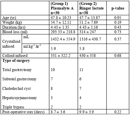
comparable between the two groups (Table 1). The type of surgery and the
duration of surgery were also similar. There was no statistically significant
difference in the volume of study crystalloid administered between the two
groups. None of the patients had any significant complications during or
Table 1 Patient characteristics and operative details. (Group 1) Plasmalyte A n=30 (Group 2) Ringer lactate n=30 p-value
Age (yr) 47.8 ± 10.23 47.7 ± 13.87 0.95
Weight (kg) 54.7 ± 12.11 51.2 ± 7.89 0.19
Duration (hrs) 4.43 ± 1.35 4.43 ± 1.16 0.43
Blood loss (ml) 293.33 ± 218.8 314 ± 247 0.73
Crystalloid infused
mL 1432.4 ± 554.9 1316 ± 430.7 0.37
ml.kg-1.hr-1
5.9 5.8
Colloid infused 331 ± 322.2 430 ± 358 0.68
Type of surgery
Total gastrectomy Subtotal gastrectomy Choledochol cyst Hepaticojejunostomy Triple bypass 10 7 8 3 2 11 6 7 4 2
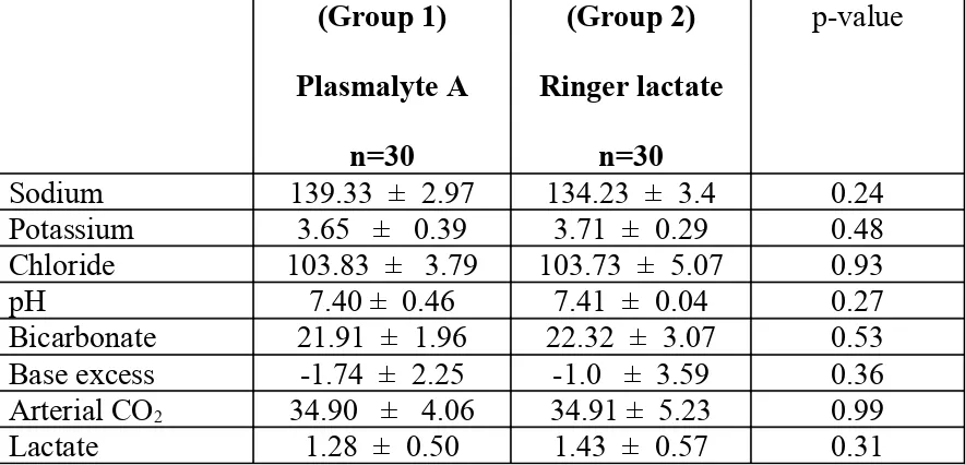
The serum electrolytes and the arterial blood gas analyses were done in all
the patients before and after the surgery. There was not statistically
significant difference between the two groups with regard to the
pre-operative and the post-pre-operative values of serum electrolyte, arterial
[image:35.612.86.528.267.480.2]acid-base status. (Table 2 & 3)
Table 2: Pre-operative values of serum electrolytes and arterial blood gases
(Group 1) Plasmalyte A n=30 (Group 2) Ringer lactate n=30 p-value
Sodium 139.33 ± 2.97 134.23 ± 3.4 0.24
Potassium 3.65 ± 0.39 3.71 ± 0.29 0.48
Chloride 103.83 ± 3.79 103.73 ± 5.07 0.93
pH 7.40 ± 0.46 7.41 ± 0.04 0.27
Bicarbonate 21.91 ± 1.96 22.32 ± 3.07 0.53 Base excess -1.74 ± 2.25 -1.0 ± 3.59 0.36 Arterial CO2 34.90 ± 4.06 34.91 ± 5.23 0.99
Lactate 1.28 ± 0.50 1.43 ± 0.57 0.31
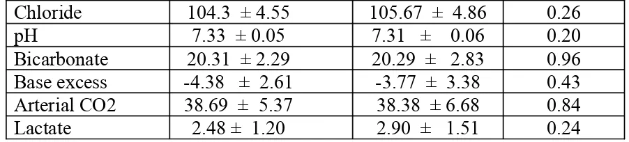
Table 3. Post-operative values of Serum electrolyte and arterial blood gas
(Group 1) Plasmalyte A n=30 (Group 2) Ringer lactate n=30 p-value
[image:35.612.85.528.605.723.2]Chloride 104.3 ± 4.55 105.67 ± 4.86 0.26
pH 7.33 ± 0.05 7.31 ± 0.06 0.20
Bicarbonate 20.31 ± 2.29 20.29 ± 2.83 0.96 Base excess -4.38 ± 2.61 -3.77 ± 3.38 0.43 Arterial CO2 38.69 ± 5.37 38.38 ± 6.68 0.84
Lactate 2.48 ± 1.20 2.90 ± 1.51 0.24
The groups were analyzed to assess if the administration of either
Plasmalyte A or Ringer lactate resulted in any change in the measured value,
within the group. (Table 4 & 5)
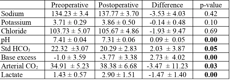
In both the groups, there was no significant difference between the
pre-operative and the post-pre-operative electrolyte values. However, there was
significant decrease in the pH in the post-operative blood gases compared to
the pre-operative values, in both the groups. This acidosis was not clinically
significant. The development of acidosis seems to be contributed by increase
in arterial CO2 levels as well as reduction in serum bicarbonate levels. The
[image:36.612.80.531.72.174.2]level of lactate was also significantly increased in both the groups.
Table 4: Group A- Plasmalyte A (n=30)
Preoperative Postoperative Difference p-value Sodium 139.33 ± 2.97 138.43 ± 3.26 0.90 ± 2.60 0.68 Potassium 3.65 ± 0.39 3.71 ± 0.28 -0.05 ± 0.51 0.54 Chloride 103.83 ± 3.79 104.3 ± 4.55 -0.47 ± 4.71 0.59
Bicarbonate 21.91 ± 1.96 20.31 ± 2.29 -1.60 ± 2.36 0.001
Base excess -1.74 ±2.25 -4.38 ± 2.61 2.63 ± 2.97 0.000
Arterial CO2 34.90 ± 4.06 38.69 ±5.37 -3.79 ± 5.94 0.002
Table 5: Group B- Ringer lactate (n=30)
Preoperative Postoperative Difference p-value Sodium 134.23 ± 3.4 137.77 ± 3.70 -3.53 ± 4.03 0.42 Potassium 3.71 ± 0.29 3.86 ± 0.50 -0.14 ± 0.48 0.10 Chloride 103.73 ± 5.07 105.67 ± 4.86 -1.93 ± 9.47 0.69
pH 7.41 ± 0.04 7.31 ± 0.06 0.09 ± 0.05 0.00
Std HCO3 22.32 ±3.07 20.29 ± 2.83 2.03 ± 3.87 0.05
Base excess -1.0 ± 3.59 -3.77 ± 3.38 2.73 ± 4.02 0.00
Arterial CO2 34.91 ± 5.23 38.38 ± 6.68 -3.47 ± 11.23 0.03
Lactate 1.43 ± 0.57 2.90 ± 1.51 -1.47 ± 1.40 0.00
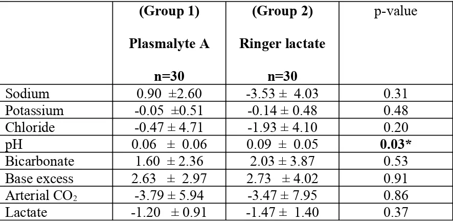
The changes caused in each group were compared as shown in Table 6. The
decrease in pH was significantly more among those who received Ringer
lactate as compared to that seen among those who received Plasmalyte A.
Table 6: Student t – test for change
(Group 1)
Plasmalyte A
n=30
(Group 2)
Ringer lactate
n=30
p-value
Sodium 0.90 ±2.60 -3.53 ± 4.03 0.31
Potassium -0.05 ±0.51 -0.14 ± 0.48 0.48
Chloride -0.47 ± 4.71 -1.93 ± 4.10 0.20
pH 0.06 ± 0.06 0.09 ± 0.05 0.03*
Bicarbonate 1.60 ± 2.36 2.03 ± 3.87 0.53
Base excess 2.63 ± 2.97 2.73 ± 4.02 0.91
Arterial CO2 -3.79 ± 5.94 -3.47 ± 7.95 0.86
Discussion
The aim of this study was to compare the electrolyte levels and the acid-base
status in patients receiving either Plasmalyte A or Ringer lactate as
maintenance fluid, during major abdominal surgery. This double-blind,
randomized trial has demonstrated that administration of 5-7 ml.kg-1.hr-1 of
either of these fluids does not change the electrolytes significantly.
However, this volume of fluid reduces the pH which, although not clinically
significant, is statistically more with the use of Ringer lactate. The decrease
in pH seems to be contributed by increase in arterial CO2 as well as decrease
in serum bicarbonate.
0.9% ‘normal’ saline has been used as intra-operative fluid therapy and for a
multitude of clinical conditions including trauma and diabetic ketoacidosis.
Yet, it is neither ‘normal’ nor physiological [3]. Hyperchloremic acidosis is
now a well accepted entity observed after infusion of large volumes of 0.9%
saline. [4, 5, 6, 8, 14, 11, 19] This is attributed to dilution of plasma by large
volume of fluid with high chloride content and deprived of bicarbonate or its
precursor. However, this explanation is not convincing since studies have
to the Stewart hypothesis, infusion of large quantity of Cl- reduces the strong
ion difference leading to acidosis. This has lead to the use of balanced salt
solutions such as Ringer lactate and Plasmalyte. Several studies have shown
that the use of 0.9% saline significantly increases the acidosis and chloride
level as compared to when Ringer lactate or Plasmalyte is used [4, 5, 6, 11,
19]. Although, these studies have shown that the effect of the balanced salt
solutions on acidosis is minimal, there are none that have compared the two
balanced salt solutions in patients undergoing major abdominal surgery.
Most studies have looked into the effect of transfusing large volumes of
crystalloids (15-30 ml.kg-1.hr-1) and demonstrated significant effect on the
electrolytes and the acid-base status. However, it is well accepted now that
fluid restriction reduces the incidence of peri-operative complications such
as cardiopulmonary events and impaired bowel motility, while improving
wound and anastomotic healing. In this study, the volume of fluid infused
was 5-7 ml.kg-1.hr-1, based on the various studies which recommend
‘restricted’ fluid regime [9,10,15,26]. The central venous pressure was
measured and maintained at 5 mmHg, with boluses of tetrastarch.
within normal limits. Although, infusion of large volume of Ringer lactate is
known to increase lactate levels, [7] rising serum lactate is generally
associated with tissue hypoperfusion. During tissue hypoperfusion and
hypoxia, the pyruvate generated by glycolysis is converted to lactate by
anaerobic metabolism. [20]. It could be argued that it may be that the
volume of fluid infused (5-7ml.kg-1.hr-1) was relatively low as evidenced by
the fact that 1-2 ml.kg-1.hr-1 of colloid was needed to maintain the CVP at 5
mmHg. However, the volume of colloid infused matched the calculated
volume of blood loss.
The acidosis caused by 0.9% saline is attributed to the dilution of
bicarbonate and the infusion of high concentration of Cl-. According to
Stewart’s hypothesis, the reduction in strong ion difference (SID = Sodium +
Potassium – Chloride – lactate) is associated with rise in H+ leading to
acidosis. SID can also be affected by the presence of weak organic acids
such as lactate and possibly (?) acetate. Since these negatively charged
molecules reduce the SID they lead to acidosis. In this study, although, the
pH was decreased marginally more in the Ringer lactate group the reduction
in SID was slightly more in group A (37.9 to 35.4) as compared to no
change in group B (32.8). Could this have been due to the unmeasured
In the liver, lactate is metabolized to glycogen which is ultimately converted
to carbon dioxide and water by oxidative metabolism. The CO2 and water
produces bicarbonate. Acetate is also metabolized to produce bicarbonate.
The acetate-containing solutions have at least a theoretical advantage over
lactated solutions, since the capacity to metabolize lactate is mainly in the
liver and kidney, while acetate can be metabolized in all tissues, which could
be beneficial in patients who are hypovolemic or in shock. [27]
The shortcomings of this study include some unanswered questions:
1. Does 0.9% saline infused in the dose of 5-7 ml.kg-1.hr-1 cause any
electrolyte and acid-base abnormality?
If a third ‘control’ group, that could be randomized to receive the same
volume of 0.9% saline, was included in the study, it would have answered
this query and also how it compares to that seen with Ringer lactate or
Plasmalyte A.
2. Could infusion of a larger volume have prevented the rise in lactate
levels?
The administration of the saline-based colloid to maintain the CVP at 5
avoided if the study fluids were administered at about 10 ml.kg-1.hr-1.
Conclusion
The choice of the intravenous fluid during surgery is often assumed to be of
little significance. However, the widely used 0.9% saline has been shown to
cause significant acidosis when compared to balanced salt solutions such as
Ringer lactate or Plasmalyte. This study was an effort to compare the effect
of administering lactate-based and acetate-based intravenous fluids on the
electrolytes and the acid-base status.
This double-blind randomized control study was approved by the
Institutional Review Board and all the patients enrolled signed an informed
consent. Sixty, ASA grade I or II, patients scheduled for major abdominal
surgery, were randomly assigned to receive either Ringer lactate or
Plasmalyte A at the rate of 5-7 ml.kg-1.hr-1. The arterial blood gas analysis
and the electrolytes were measured before and at the end of surgery.
pre-operative and the post-operative electrolyte levels showed no significant
difference in either group. However, when the pre-operative and
post-operative values were compared, there was statistically significant decrease
in pH in both the groups, with it being marginally more among those who
received Ringer lactate. There was both a slight rise in PaCO2 as well as a
slight decrease in bicarbonate. However, the post-operative pH was within
normal limits and was not clinically significant.
This study concludes that in patients undergoing major abdominal surgery,
the administration of 5-7 ml.kg-1.hr-1 of either Plasmalyte A or Ringer lactate
leads to statistically significant decrease in pH, but this change is not
clinically significant. However, when the two groups are compared, there is
no statistically significant difference in the electrolyte and the acid-base
BIBLIOGRAPHY
1. Moore FD, Shires T; Editorial; Annals of Surgery 1967; 166:300-1
2. Shires T, Williams J, Brown F - Acute change in extra-cellular fluids
associated with major surgical procedures. Annals of Surgery 1961 154
no 5
3. Edward B, Antony M, Michael G, Mythen MG:Hyperchloremic acidosis:
Pathophysiology and Clinical Impact ; Transfusion Alternatives in
Transfusion Medicine2003; 5:424-430
4. McFarlane A. Lee: A comparison of Plasmalyte 148 and 0.9% saline for intra-operative fluid replacement; Anesthesia 1994;49: 779–781.
5. Waters, J.H., Gottlieb, A., Schoenwald, P., Popovich, M.J., Sprung, J.
and Nelson: Normal saline vs Ringer lactate solutions for fluid
management in patients undergoing abdominal aortic aneurysm repair;
6. Waters JH, Miller, LR. Clack S and Kim JV: Cause of metabolic acidosis
in prolonged surgeries; Critical Care Medicine 1999; 27; 2142-46
7. Hadimioglu N, Saadawy I, Saglam T, Ertug Z, Dinckan A: The effect of
different crystalloid solutions on acid-base balance and early kidney
function after kidney transplantation; Anesthesia 2008; 107:264-9.
8. Joshi G: Intraoperative fluid restriction improves outcome after major
elective gastrointestinal surgery; Anesthesia-Analgesia: 2005:101; 601-5
9. Kita T, MammotoT, Yoshihiko Kishi: Fluid management and post
operative respiratory disturbances in patients with transthoracic
esophagectomy for carcinoma; Journal of Clinical Anesthesia 2002;
14;252-6.
10.Lobo DN. Bostock KA: Effect of salt and water balance on recovery of
gastrointestinal function after elective colonic resection; Lancet 2002;
11.O’Malley CMN, Robert JF, Mark AH: A randomized double blind
comparision of lactate ringer solution and normal saline during renal
transplantation; anesthesia- analgesia 2005;100.1518-24
12.Skellett S, Mayer A, Durward A, Tibby S M, Murdoch A: Chasing the
base deficit: Hyperchloraemic acidosis following 0.9% saline fluid
resuscitation; Archive Dis Child 2000; 83:514-516
13.Scheingraber, Stefan , Rehm, Markus , Sehmisch, Christiane; Finsterer,
Udilo: Rapid Saline Infusion Produces Hyperchloremic Acidosis in
Patients Undergoing Gynecologic Surgery; Anesthesiology: 1999;90:
1265-1270
14.Holte K, Foss N B, Andersen J, L Valentiner, C Lund, P Bie and H
Kehlet: Pathophysiology and clinical implications of perioperative fluids
excess: British Journal of Anesthesia 2002; 89; 622-3
15.Vadim N, Itamar F: Effects of intraoperative fluid management on
16.Gan TJ, Soppitt A, Maroof M, Robertson K, Moretti E, Dwane P, Glass
P: Goal directed Intraoperative Fluid Administration Reduces Length of
Hospital Stay after Major Surgery; Anesthesiology 97(4), 2002, 820-826
17.Handy JM and Soni N: Physiological effects of hyperchloremia and
acidosis; British Journal of Anesthesia 2008(2):101;141–50.
18.Takil A, Eti Z, Irmak P, Yilmaz Göğüş F: Early postoperative respiratory
acidosis after large intravascular volume infusion of lactated ringer's
solution during major spine surgery; Anesthesia Analgesia. 2002 ;
95(2):294-8
19.Didwania A, Miller J, Kassel D, Jackson EV Jr, Chernow B: Effect of intravenous lactated Ringer's solution infusion on the circulating lactate
concentration; Critical Care Medicine.25(11):1851-4;997
20.Mizock, Barry A, Falk, Jay L: Lactic acidosis in critical illness; Critical
21.Ana M, Girish J: Perioperative Fluid Management: Minimization Versus
Goal-Directed Therapy; American Society of Anesthesiologist news
letter72(4) 2008
22.Lobo DN, MacAfee DA, Allison SP: How perioperative fluid balance
influences postoperative outcomes; Best Pract Res Clin Anaesthesiol.
2006 Sep;20(3):439-55
23.Stephens R, Mythen M: Optimizing intraoperative fluid therapy; Current
Opinion in Anaesthesiology 2003 Aug;16(4):385-92
24.Stephens R, Mythen M: Saline-based fluids can cause a significant
acidosis that may be clinically relevant; Critical Care Medicine. 2000
Sep;28(9):3375-7.
25.Christopher S. Wilcox ;Regulation of Renal Blood Flow by Plasma
26.Brandsturp B, Ttonnesen H et al: effects of intravenous fluid restriction
on postoperative complication: Comparision of Two perioperative fluid
regimes. Annals of Surgery 238(5)2003
27.Prough DS, Svensen CH. Crystalloid solutions. In Hahn RG, Prough DS,
Svensen CH (eds) Perioperative Fluid Therapy. New York. Informa
INFORMED CONSENT DOCUMENT
PATIENT INFORMATION:
0.9% saline and Ringer lactate are the most widely used intravenous fluids used in our hospital. It has been shown that both these fluid can contribute to metabolic acidosis when used in large amounts, with it being more with 0.9% saline. A new balanced solution, Plasmalyte A is now available which has a pH similar to that of blood, and is thought to contribute very minimal to acidosis with its use.
In this study we want to compare the effect of either Ringer lactate of Plasmalyte A on the acidosis and the electrolytes.
If you volunteer for this study, you will be randomly assigned to receive either Plasmalyte A or Ringer lactate. Two samples of blood (6 ml each) will be drawn, while you are asleep, before the start and at the end of surgery.
The participation in this study is entirely voluntary, and the care you receive will be the same whether you volunteer or not.
CONSENT
I have been explained the study protocol, by Dr. ________________________in detail, in the language I understand. I am willing to volunteer for this study out of my own free will and am aware that I can withdraw from the study at any time, without it in anyway affecting the treatment I receive in this hospital.
Patient’s Signature--____________________
Performa S.No-Name Age Weight Hospital no
Diagnosis Procedure –
preoperative postoperative ---pH ---PaCO2 ---PaO2 ---HCO3 ---ABE ---Na ---K ---Cl ---Lactate ---Duration- blood loss- urine
output-Fluid
administeredCrystalloid colloid blood blood products -Patient transferred to - ward/shdu/sicu.