EVALUATION OF MARGINAL FIT OF METAL
CERAMIC AND METAL FREE CERAMIC
CROWNS- AN INVITRO STUDY
A Dissertation Submitted
to the Tamil Nadu Dr. M.G.R. Medical University
In partial fulfillment of the requirement for the degree
of
MASTER OF DENTAL SURGERY
(BRANCH VI-PROSTHODONTICS)
CERTIFICATE
This is to certify that the dissertation titled “Evaluation Of
Marginal Fit Of Metal Ceramic And Metal Free Ceramic Crowns- An
Invitro Study” is a bonafide record of work carried out by Dr.V.PARIMALA , during the period of 2006-2009. This dissertation is
submitted in partial fulfillment, for the degree of Master of Dental Surgery awarded by Tamil Nadu Dr. MGR Medical University, Chennai in the branch of Prosthodontics. It has not been submitted partially or fully for the award of any other degree or diploma.
Guided By HEAD OF THE DEPARTMENT,
Dr. C.THULASINGAM, MDS,
Professor and Head of the Dept, Dept. of Prosthodontics,
Tamil Nadu Govt Dental College & Hospital
Chennai- 3
PRINCIPAL
Dr.K.S.G.A NASSER, M.D.S
Tamil Nadu Govt Dental College &Hospital Chennai-3
Dr A.Meenakshi,M.D.S
Additional Professor Dept. Of Prosthodontics,
Tamil Nadu Govt Dental College & Hospital
DECLARATION
I Dr.V.PARIMALA, do hereby declare that the dissertation titled “Evaluation
of Marginal Fit of Metal Ceramic and Metal Free Ceramic Crowns- An
Invitro Study” was done in the Department of Prosthodontics, Tamil Nadu
Government Dental College & Hospital, Chennai 600 003. I have utilized the facilities
provided in the Government dental college for the study in partial fulfillment of the
requirements for the degree of Master of Dental Surgery in the speciality of
Prosthodontics (Branch VI) during the course period 2006-2009 under the
conceptualization and guidance of my dissertation guide, Dr.A.Meenakshi, MDS.
I declare that no part of the dissertation will be utilized for gaining financial
assistance for research or other promotions without obtaining prior permission from the
Tamil Nadu Government Dental College & Hospital.
I also declare that no part of this work will be published either in the print or
electronic media except with those who have been actively involved in this dissertation
work and I firmly affirm that the right to preserve or publish this work rests solely with
the prior permission of the Principal, Tamil Nadu Government Dental College &
Hospital, Chennai 600 003, but with the vested right that I shall be cited as the author(s).
Signature of the PG student Signature of the HOD
ACKNOWLEDGEMENT
I am extremely thankful to Dr.C.THULASINGAM, MDS. Professor and Head of the Department, Department of Prosthodontics, Tamil Nadu Government Dental College and Hospital for his constant guidance,
encouragement, and monitoring during this study. I also thank him for the valuable guidance he has given throughout my post graduation.
My sincere thanks to Dr.K.S.G.A.NASSER, MDS Principal, Tamil
Nadu Government Dental College and Hospital, for his kind help, and permitting me to use the facilities in the institution
I consider it my utmost privilege to express my sincere and heartful
gratitude to my dissertation guide Dr.A.MEENAKSHI, MDS. Additional Professor, Department of Prosthodontics, Tamil Nadu Government Dental College and Hospital for her able guidance and support she has rendered at various stages of the dissertation.
I am extremely thankful to Dr.C.SABARIGIRINATHAN, MDS,
Assistant Professor/ senior civil surgeon, Department of Prosthodontics, Tamil Nadu Government Dental College and Hospital, for his instant help,
I am thankful to Dr. T.JEYANTHI KUMARI MDS Assistant Professor, for guiding and helping me throughout this study.
I also thank, Dr. K.VINAYAGAVEL,, MDS, Dr.V.BALAJI, MDS, Dr.G.GOMATHI M.D.S, Dr K.RAM KUMAR, MDS, Dr M.KANMANI, MDS, Dr V.HARISHNATH, MDS, Assistant Professors for helping me at different stages of this study.
. I thank BALA DENTAI LAB and GANI DENTAL LAB helping for this study. I also thank CHENNAI METCO PVT LTD AND KOSACA
CALIBERATION LAB for their help in sectioning, and measuring of samples
for the study.
I thank Dr.RAVANAN, Professor, Dept. of Statistics, Presidency College, and Chennai for helping me to carry out statistical analysis of the
various test results. I am highly indebted to my family members, all my friends and my fellow post graduates who have helped me in several ways during the course of the study.
CONTENTS
S.NO
.
CONTENTS
PAGE NO.
1
INTRODUCTION
1-7
2
REVIEW OF LITERATURE
8-21
3
MATERIALS AND METHODS
22-34
4
RESULTS
35-42
5
DISCUSSION
43-52
6
SUMMARY AND CONCLUSION
53-55
7
APPENDIX
56-62
INTRODUCTION
T
he smile is one of the most appealing aspects of the human face and is considered to be the very image of the soul. An esthetically pleasing appearance of teeth is the best asset for a good smile.Ceramic restorations because of their natural translucency and tooth like color provide excellent esthetics and are virtually indistinguishable from the adjacent natural dentition. Full veneer ceramic crowns are very successful in this aspect because by covering the entire
tooth they can totally mask the previous condition to create a new appearance.
Dr. Charles Land 37 introduced one of the earliest forms of
ceramic crowns in 1903. Then the first metal-ceramic crown was described by Brecker 37 in 1956. Since then various types of metal ceramic restorations
have been developed with advancements being made in both the type of the
metal & the porcelain for an effective metal ceramic bond.
used for both single crowns and fixed partial dentures. But the esthetic limitations like lack of metal translucency, exposure of metal collar in the
anterior region, paved way for all ceramic metal free restorations.
The absence of metal layer in the all ceramic restorations helps in transmission of light through the full depth of the restoration, thereby enhancing the translucency creating a life like appearance.
Mcleaan and Hughes 44 in 1965 introduced the first all ceramic
porcelain jacket crowns made of high strength alumina core.
Heat pressed ceramics were introduced by Ivoclar in the 1990s 44.
Here a ceramic substructure (ceramic core) is made by pressing the ceramic ingots into a refractory mold made by the lost wax technique. These ceramic copings thus obtained can be finished to the final form either by a
characterization technique, involving surface stain only, or by layering technique involving application of veneering porcelain.
Depending upon the clinical demand, both all ceramic and porcelain fused to metal can be used for full veneer crowns. The clinical success of a
tooth, exhibit a minimum cement margin, be adequately retained and restore function & esthetics.
Of all these marginal fit is considered to be a primary and significant factor in the prevention of secondary caries, and is an important indicator of the overall acceptability and longevity of the restoration.
Clinically distortion of the margins creates a potential space between
the crown and the tooth preparation. This space promotes leakage & dissolution of cement, encourages plaque deposition, initiates secondary caries, leads to gingival inflammation, periodontal disease, & finally results
in deterioration of the restoration and the supporting tooth. So, the intimacy of fit of the crown margins is critical to ensure a smooth surface that will not promote accumulation of plaque.
Mclean & Von Fraunhofer 36 after examining more than 1000 crowns
clinically over a period of 5 years showed that the clinically acceptable crown marginal discrepancies could range up to 120 µm after cementation.
The marginal fit of any restoration depends upon the inherent physical
without a metal substructure, most of the marginal discrepancies are induced in the fabrication stage because of multiple high temperature firing cycles
used for porcelain application.
Ando et al 36 in 1972 reported that when a metal casting is subjected
to degassing, there is a significant marginal discrepancy of about 100 - 150µm.
Other studies by various authors have identified many factors such as the type of the alloy, metal oxide formation on the fitting surface of the alloy, the thickness of the coping, mismatch of the porcelain-metal thermal
contraction, porcelain firing shrinkage, release of casting induced stresses and several other factors as contributing to the distortion of metal ceramic crowns during fabrication. Regardless of the specific factor involved, the
common denominator is the elevated temperature which definitely causes loss of the initial fit of the metal coping.3, 7,14,15,21.
Similarly in the fabrication of all-ceramic crowns made by application of veneering ceramic over a ceramic coping (layering technique),
there is deformation of the ceramic substructure at repeated exposure to high temperatures. This has been proved in a study by Mehmet et al 48 on the
Earlier studies have reported the marginal fit of all ceramic crowns to be inferior to that of the traditional metal ceramic crowns.
But of late, the heat pressed ceramic restorations provide a better marginal fit because of the leucite reinforcement in them which imparts more strength to the core ceramic, and also because of the technology of heat pressing the ceramic ingot which is more accurate than the earlier
methods.
Edward et al 17 in 2005 studied the marginal fit of leucite glass
pressable ceramic restorations and concluded that a pressed ceramic
restoration has better marginal adaptation than a traditional metal ceramic restoration.
Hence this study was performed to analyze the marginal fit of heat
The Aims Of The Study
are
1. To evaluate the initial marginal fit of the metal copings made by induction casting with that of the ceramic copings made by heat pressing.
2. To evaluate the discrepancy in the marginal fit that has occurred after porcelain application in both the groups.
3. To compare the ultimate marginal fit of conventional metal
ceramic crowns with that of heat pressed all ceramic crowns.
.
REVIEW OF LITERATURE
Ideally cemented crown margins meet prepared tooth margins in perfect non detectable junctions. The definition of fit in the dental literature has been imprecise and various terms
have been used to describe essentially identical measurements. The bulk of literature and research on complete crown margins falls into one of the 3 broad categories
1. Location of the margin
2. Configuration
3. Fit
Improper marginal fit clinically seen as an increased space between the
tooth and the crown margin can be attributed to at least 2 separate sets of factors.
1. Distortion of margins of the restoration either during fabrication or during clinical use.
spite of having structurally good defect free margins.
The marginal fit of ceramic crowns is affected by both these factors.
Shillingburg et al. (1973)36 published the results of facial margin
opening changes of four different coping designs that resulted during porcelain firing & concluded that the greatest marginal opening occurred
during those cycles in which porcelain was fired to Au-Pd-Ag copings. They also said that the shoulder margins had less marginal distortion than the chamfer margins
Feichtinger et al( 1973 )36 - marginal distortion changes of upto
64µm were observed to occur during the metal oxidation treatment but not during the porcelain firing procedure
Iwashita et al( 1977)36 – studied the effect of full coverage porcelain
& the partial veneer designs on fit of a metal ceramic crown. They found that the full coverage specimens showed the largest gap change of 187µm . Most of the marginal discrepancy occurred after the degassing procedure.
The effect of thermal contraction differential between the metal & the porcelain was also found to be significant.
Kulmer et al. (1978)36 demonstrated that gold alloy crowns which
less than 19 ulm. This finding suggests that porcelain-metal incompatibility stresses were not capable of producing metal distortion effects.
Robert R.Faucher et al (1980 )58investigated the distortion in relation
to the margin design in porcelain fused to metal restorations. They concluded that shoulder & shoulder with bevel designs exhibit significantly less marginal distortion than the chamfer margins and also said that the
placing of additional metal at the gingival margin reinforces this margin and inhibits marginal distortion
Faucher and Nichols (1981)36 monitored the change in the fit during
a series of firing procedures. They said that after the porcelain firing cycles there was an increase in the mesiodistal dimensions and a decrease in the faciolingual dimensions. As reported by the previous authors they also
concluded that most of the distortion occurred during the degassing cycle.
Buchanan et al( 1981)36 reported greater marginal discrepancy of the
base metal alloys(70µm) when compared to that of the precious metals. The larger discrepancy of the base metal alloy was explained on the basis of the
formation of oxide on the internal surface of these copings.
VanNortwick &gettleman et al( 1981 )66 found statistically significant
improvements in the fit of the castings by applying die-spacer. The amount
K.J.Anusavice et al (1984)54 studied the effect of metal design on the
marginal distortion of metal ceramic crowns and concluded that the
calculated marginal distortions due to metal-porcelain thermal contraction mismatch depend primarily on the metal-porcelain combination and are insensitive to the coping design. They also confirmed that if the coping is kept in a minimal thickness of 0.1mm or less then they are more susceptible
to localized or generalized distortion than a coping of higher thickness.
In yet another study by Anusavice et al in( 1987)41 it was conclusively
demonstrated that incompatibility stress induced by a positive contraction
mismatch is not a primary cause of marginal or generalized distortion of metal-ceramic crowns and suggests that external grinding and internal abrasive blasting of crowns are more likely to cause this effect.
Richter- snapp et al in (1988)42 – studied the effect of alloy type,
margin design and porcelain proximity on the marginal fit of metal ceramic crowns and concluded that none of these factors or a combination of these affected the final fit of the restoration. Rather the only factor which could
influence was the exposure to increased temperature.
It was Homes et al in (1989 )30 who comprehensively discussed the
description of crown fit. He established measurements of both internal fit &
absolute marginal discrepancy. This was simply described as the distance from the margin of the casting to the cavosurface angles of the tooth
preparation. Homes et al stated that this distance would always be the largest measurement of error at the margin and reflect the total crown misfit at that point both vertical & horizontal.
As mentioned earlier apart from structurally good margins the
complete seating of the crown is essential for a good fit. The methods for measuring the marginal fidelity includes four different categories
1. Direct view
2. Cross-sectional method
3. Impression technique
4. Explorer &visual examination.
John A.Sorensen etal (1990)35 compared & scrutinized the various
methods used to measure the crown margin fidelity and claimed that the cross-sectional method of evaluation of margins allows greater precision in determination of the measuring points and also in measuring the absolute
actual surface of the marginal gap and in addition provides consistent reproducible &standardized points of measurement.
Farhad et al (1991)22 compared the marginal fit of all ceramic crowns
with metal ceramic crowns [renaissance crowns vs metal ceramic & dicor crowns]. This study concluded that Dicor & metal ceramic crowns fit better than the renaissance crowns.
Tam S.Hager et al (1993)69 studied the effect of selective die spacer
placement techniques on the seatability of the castings. The results of this study pointed out the need for providing additional relief at the axial walls
when casting base metal alloys. This was because of the casting shrinkage resulting in tight fitting crowns binding along the axial walls more frequently than in other areas. So an additional layer of die spacer has to be
applied to these areas for better seating of the crown.
Stephen D .Campbell et al (1995)68 studied the effects of firing cycle
and surface finishing on the distortion of metal ceramic castings and reported that thermocycling of metal ceramic restorations resulted in
ceramic restoration was observed when the initial thermal cycle was completed before the specimens were finished.
Deniz Gemalmaz et al in (1995)15 analysed the marginal fit changes
that occur during the porcelain firing cycles of palladium – copper and nickel chromium copings and concluded that a greater change in the marginal fit occurred after the degassing stage and after the firing of body
porcelain. Palladium copper copings showed more marginal discrepancy (19.39µm) than nickel chromium copings(8.65µm).
Christian Lehner et al (1997)10 did a short term [ 2 years] clinical
evaluation of the heat pressed IPS-Empress crowns and reported that 79.5% of the crowns were rated excellent from the clinical & functional standpoint. The marginal adaptation of the crowns was good with high degree of patient
satisfaction
Kenneth et al (1998)43 measured of fit of the Procera all ceram
crowns fabricated with Procera CAD/CAM technology and concluded that the marginal fit of these crowns were below 70µm which is conductive to
clinical success.
is because of the unique physical characteristics of titanium alloys such as biocompatibility, high modulus of elasticity, low mass, high mechanical
strength & resistance to corrosion.But there are some difficulties because of the casting difficulties of titanium and also because the thick oxide layer interferes with the ceramic bonding
Morakot Piemjai et al (2001)51 evaluated the effect of seating force ,
margin design, cement on marginal seal, and retention of complete metal crowns. These authors said that there was a significant difference in the marginal seating (P<.05) between the different seating forces , for marginal
design, with 2 different luting cements. Increasing the seating force from25 to 300N significantly improved the seating of the complete metal crowns with chamfer, shoulder &shoulder with bevel margins.
Efstratios Papazoglou et al (2001)18 evaluated the distortion of high
palladium metal ceramic crowns during ceramic firing at 4 stages- the as cast fit , after oxidation , after 2 simulated opaque porcelain firings, and after 2 simulated dentin porcelain firings. The results of this study suggested that
M.J.Cattell et al in (2001)45 analysed the flexural strength
optimization of a leucite reinforced glass ceramic.. The study showed
concluded that the fine leucite crystal size and the uniformity of microstructure caused by the dispersion of the leucite crystals during heat pressing is associated with significant increase in the biaxial flexural strength and reliability of the core ceramic. Such leucite reinforced ceramics
have distinct advantages & are very useful in the fabrication of durable & esthetic heat pressed dental restorations.
Michael behr et al (2003)49 did an in-vitro study to investigate the
marginal adaptation and fracture resistance of heat pressed glass ceramic and fiber reinforced composite molar crowns with 3 types of cements. They found that both the restorations had highest fracture resistance & better
marginal adaptation if they were luted with resin cement.
Stefan Wolfart et al (2003)67 did a clinical evaluation of the marginal
fit of heat pressed all ceramic crowns before and after cementation and found that adhesive cementation caused a significant increase of the
outflow of the cement as the crown is seated. All these factors increase the hydraulic pressure on the resin cement thus preventing its complete seating.
Xin –Hua Gu et al (2003)72 evaluated the marginal discrepancies and
leakage of all-ceramic crowns cemented with different luting agents after fatigue tests. They reported that the marginal discrepancies of the all-ceramic crowns cemented with 3 different luting agents were smaller than
that of the metal ceramic crowns. This result is mainly attributed to the excellent adaptation properties of the heat pressed ceramics. They also concluded that the adhesive composite resin luting system Panavia
minimized the marginal leakage of all ceramic crowns and was far superior to zinc phosphate and compomer luting cements.
Massimiliano Guazzato et al (2004)47 evaluated the strength
,reliability and mode of fracture of bilayered porcelain/core ceramics and concluded that in bilayered ceramics the strength and reliability were improved by the core material that possessed better mechanical properties.
Mehmet et al (2005)48 studied the influence of firing cycles on the
because the milled crowns are subjected to glaze firing only. Whereas in-ceram crowns and copy milled in-in-ceram crowns showed significant marginal
distortion (57±24µm, 57±32µmrespectively) because of the repeated high temperature firing cycles during porcelain build up.
Edward B. Goldin et al ( 2005 )17 compared the marginal fit of
leucite –glass pressable ceramic restorations and ceramic pressed to metal
restorations with a traditional metal ceramic crown. They concluded that a pressed ceramic restoration [81±25µm] with or without a metal has equal or better marginal adaptation than a traditional metal ceramic
restoration[94±41µm].
Giuseppe Isgro et al (2005)24 evaluated the influence of multiple
firing on the thermal contraction of ceramic materials used for the
fabrication of layered all- ceramic restorations. It was concluded that glass-ceramics and aluminous porcelain exhibited more stable thermal behavior after repeated firing. So the fabrication of a layered all- ceramic restoration , using these materials , could be more reliable.
Niek De Jager et al (2005)53 studied the influence of the stresses
sufficiently strong to produce long lasting restorations, the stresses in the veneering porcelain can reduce the longevity. At the core veneer interface,
the maximum tensile stress is higher for crowns with ceramic cores. So they suggested that the bonding between veneering porcelain and these strong ceramic cores should be improved to exploit fully the strength of these materials.
Jianxiang Tao et al (2006)33 analyzed the fit of metal ceramic crowns
cast in Au-1.6wt%Ti alloy for different finish line curvature and concluded that these titanium alloys had accuracy of marginal fit as good as that of gold
alloy. After porcelain application, there was a larger labial marginal gap corresponding to the finish line curvature.
Carla Castiglia et al (2008)9 did an elaborate study to analyze the
mechanical properties & porosity of 2 commercial glass ceramics – [ leucite reinforced & lithium di silicate reinforced] after hot pressing them at different temperatures. They concluded that the microstructure of leucite reinforced ceramic was not affected by the different amounts of energy
provided during the hot press technique, leading to similar mechanical properties in different temperatures. This is because these ingots were already subjected to heat treatment between 920 - 1200°c by the
the nucleating centers in the base glass. So when the ingots are heated again during the firing procedure, the leucite crystals undergo a maturing process
resulting in a more homogeneous distribution throughout the glassy matrix. Because of this the leucite reinforced glass ceramics have good mechanical properties, low porosity & good marginal fit .
.
MATERIALS AND METHODS
This invitro study was performed to evaluate the accurate marginal
fit of the metal ceramic and pressed ceramic jacket crowns over the prepared tooth. This study also involved to evaluate the distortion of metal and ceramic copings due to repeated heating during the processing
procedure.
A typhodont maxillary right central incisor was prepared for a jacket crown. A uniform labial nd axial reduction of 1.5 mm was made
along with a 2mm incisal reduction and .an uniform shoulder of 1.5mm. The height of the preparation was 7mm with a convergence angle of 6°.
Before starting the preparation the index of the unprepared tooth was made with putty silicone to facilitate the accurate reduction of the
tooth. The occlusal and axial reductions of the teeth were carried out using this putty index.
20 impressions of the prepared tooth were made in light body poiy
vinyl siloxane impression material by using individualized custom made impression trays. The impressions were cast on die stone.
Group I - consists of 10 stone dies for the fabrication of metal ceramic crowns
Group II- consists of 10 stone dies for the fabrication of Pressable ceramic crowns
FLOW CHART
20 stone dies
Group - I
10 Stone dies for making metal ceramic crowns
Group-II
10 stone dies for making pressable ceramic crowns
Sub-Group-IA Metal Coping before veneering of porcelain
Sub-Group-IIA Heat pressed all Ceramic Coping
Before porcelain veneering
Sub-Group-IB Metal Crown after veneering of porcelain
Sub-Group-IIB Heat pressed all ceramic
Crown After porcelain
MATERIALS USED IN THIS STUDY
Sl no Material Manfacture name
1. Die stone zerhmark
2. Bellavest Beggo, germany
3. Addition silicone Ivocloar,
4. Inlay wax GC coporation
5. Ceramic ingots &
veneering ceramic
Cergo dentsply
EQUIPMENTS USED IN THIS STUDY
Sl no Equipments used Manfacture name
1. Casting machine Fornax, beggo Germany
2. Ceramic furnance Mulitmat two touch and press,
Dentsply
3. Vaccum Mixer Beggo, Germany
I. FABRICATION OF METAL COPINGS
Application of die spacer
First the preparation margin on the die was defined and dies
hardener applied. 3 layers of paint on die spacer producing a thickness of
approximately 33-40µm was applied 1mm short of the margins.
Preparation of wax pattern
The wax patterns were prepared using inlay casting wax to an
uniform thickness of 0.5mm. The dimensions were confirmed by measuring at multiple points with a wax thickness caliper.25,58 [± 0.1 mm]
Investing
The wax patterns were coated with debubblizer and invested in
phosphate - bonded investment, strictly adhering to the manufacturer’s instructions.
Burn out
The invested rings were placed in the preheating furnace and heated
Casting
Casting was done in an induction casting machine and the nickel chromium alloy was used for this purpose. A total of 10 metal copings
were cast in the similar fashion. The copings vere then retrieved cleaned and inspected for any visible flaws. Those with gross defects were rejected and new ones made. Small nodules that might prevent complete
seating of the coping were removed with a round bur at slow speed. After final trimming, the thickness of the coping was confirmed with a metal thickness caliper at multiple locations, the copings were then seated on their respective dies.
II. FABRICATION OF PRESSABLE CERAMIC COPINGS
Application of die spacer
First the preparation margin o the die is defined and die hardener
applied, two layers of die spacer is applied 1mm short of the margins to
get a thickness of approximately 30-40µm.
Wax pattern
The wax patterns were made by using inlay casting wax intended for pressable ceramic work, to a uniform thickness of 0.7 mm as
Investing
The wax patterns were sprued in such a way that both the pattern and the sprue lie on the same line vertically, to facilitate same direction of
flow of ceramic.
The patterns were immediately invested in phosphate bonded investment, strictly adhering to the manufacturers instructions.
Burn out
The mould is heated from room temperature to 800°C.
PRESSING
It is then taken from the furnace and a pellet of pressable ceramic
[cergo, dentsply] is placed in the mould. The aluminium oxide plunger is placed over the ceramic pellet. The mould with the pellet and the plunger is immediately placed in the press furnace [MULTIMAT TOUCH AND
PRESS] and the press progame is started. It takes 45 minutes for the press procedures to be complete and the temperature reaches a maximum of 960°C. The ingot is pressed into the mould at 960°C with a holding time of 20 minutes. Then the mold is left to cool at room temperature. The
diamond discs using water cooling without applying pressure. A total of 10 ceramic copings were made in the similar fashion.
III. MEASUREMENT OF INITIAL MARGINAL FIT OF THE COPINGS:
The copings were then seated on their respective dies with finger
pressure and the marginal fit was measured using the video measuring system. This is to ensure that the copings had a good initial fit before application of porcelain.The measurementa were made at the following 6
points around the circumference of the crown.
1 point- mid facially 1 point — midpalatally
The mean of all the 6 values measured is the initial marginal fit of the coping. The measurement sites were maked for reproducible
identification by scoring the die with a sharp blade. Care was taken not to contact the marginal areas.48
The copings are now ready for porcelain build up.
IV. PORCELAIN BUILD UP ON THE METAL COPING BY LAYERING
The following schedule was followed for application of porcelain by layering technique.
Degassing -by placing the coping directly at 1200° F (650°C) and the
elevating the temperature at the rate of 15°F (31oC) per minute. Final
temperature of 1925°F (1050°C) is reached- for 15 minutes. After degassing the copings were cooled in open air.
Opaque porcelain-applied to a thickness of 0.5mm and condensed by
vibration
Dried by placing them on firing tray on a hot plate 700°F (370°C) 20
minutes
→
Transferred to the furnace already preheated to 1200°F (650°C)
→
Maximum temperature 1750°FA correction bake using identical procedures was performed to attain a baked thickness of 0.3mm
Body and incisal porcelain — added to make contours of all specimens as uniform as possible
Dried by placing them on firing tray on a hot plate 700°F (370°C) 20
minutes
→
Transferred to the furnace already preheated to 1200°F (650°C)
→
Maximum temperature 1750°F(950°C) in partial vaccum (720mm/Hg.
After reaching 1750°F the vaccum was released and the copings were allowed to air fire an additional 1 minute at 1750°F (950°C). A patch bake of either body or incisal porcelain or both was accomplished
using the same procedures.
Before glazing the margins were carefully finished to the proper contours with a rubber wheel.
Glazing
Preheated to 700°F (3
70°C)
→
Transferred to the furnace already preheated to 1200°F
(650°C)
→
Maximum temperature 1800°F (980°C) atmospheric pressure
for 3 mm.
PORCELAIN BUILD UP - ON THE CERAMIC COPING BY LAYERING TECHNIQUE
Firing programmes
Pre heating
oC Vaccumhpa Firingtemp Timemin
1st Dentine firing 450oC 50 800oC 1 min
2nd Dentine firing 450oC 50 800oC 1 min
Glaze firing 450oC – 790oC 1 min
A total of 10 heat pressed all ceramic crowns of uniform contour was made.
III. SECTIONING OF THE FINAL SAMPLES
The crowns were cemented to the respective dies with ZnPO4
cement using finger pressure. The crown die complexes were then embedded in rectangular acrylic block. The samples were then sectioned
faciolingually and mesiodistally with a diamond sectioning saw, following the guide marks on the dies. There are 2 interfaces for each point of sectioning so measurements can be made at 8 points around the
Photographs
PREPARED TOOTH
STONE DIE
WAX PATTERN FOR METAL
COPING INVESTING
PRESSING OF CERAMIC INGOT
FINISHED CERAMIC
VIDEO MEASUREMENT
VIDEO MEASUREMENT
SECTIONING OF SAMPLES
BRAIN CUT-UM THIN SECTIONING MACHINE
SECTIONED PRESSED CERAMIC SAMPLE
EQUIPMENTS USED IN THE STUDY
MULTIMAT 2 TOUCH &PRESS FURNACE
INDUCTION CASTING MACHINNE
RESULTS
This in vitro study was performed to analyze the marginal fit of
metal and metal free ceramic jacket crowns and also to compare the amount of distortion that had occurred during the high temperature firing cycles during porcelain veneering.
The test samples were divided into 2 major groups based on the type of copings used for the jacket crown.
Group I - metal coping – for metal ceramic crowns
Group II - heat pressed ceramic coping – for all ceramic crowns
They were further subdivided into 2 subgroups each based on the stage of processing as , before and after veneering of porcelain.
Accordingly,
Sub group IA - metal coping before veneering of porcelain
Sub group IIA - ceramic coping before veneering of porcelain
1. The marginal fit of these copings were verified by measuring at 6
The same copings were then veneered with porcelain to the desired contour. The veneering porcelain was added incrementally in layers [layering
technique] thereby subjecting the coping to multiple high temperatures firing cycles. The completed crowns were cemented on their respective dies using ZnPO4 cement in the correct consistency. These are the final samples & they were considered as
Subgroup I B - metal ceramic crown after veneering of porcelain
Subgroup II B - all ceramic crowns after veneering of porcelain
The marginal fit of these final samples were assessed by the cross-sectional
method. The samples were mounted in acrylic blocks and then sectioned faciolingually and mesiodistally following the markings on the dies. This creates 8 points of measurements around the circumference of the crown.
The mean of all the 8 values was taken as the final marginal fit of the samples.
The means and standard deviations were calculated for all the samples and the results were statistically analyzed using analysis of variance test ]
Table .1- Mean values, standard deviations, and the results of one way ANOVA between subgroup IA and subgroup IIA
Metal coping before
veneering
[SUBGROUP-IA]
Pressed ceramic coping
before veneering [SUBGROUP-IIA]
P VALUE
MEAN ± S.D MEAN ± S.D
.00001*** 51.34 ± 3.46 45.65 ± 4.27
*** denotes significant at1 % level.
Table .2 - Mean values, standard deviations, and the results of one way ANOVA between subgroup IA and subgroup IB
*** denotes significant at1 % level Metal coping before
veneering [SUBGROUP-IA]
Metal ceramic coping after
veneering [SUBGROUP-IB]
P VALUE
MEAN ± S.D MEAN ± S.D
Table .3 - Mean values, standard deviations, and the results of one way ANOVA between subgroup IIA and subgroup IIB
Pressed ceramic coping before veneering.
[Sub group IIA]
Pressed ceramic crown after veneering
[SUBGROUP-IIB]
P VALUE
MEAN ± S.D MEAN ± S.D .0000I***
45.65 ± 4.27 67.97± 3.34
[image:50.612.105.566.463.651.2]*** denotes significant at1 % level
Table 4 - Mean values, standard deviations, and the results of one way ANOVA between subgroup IB and subgroup IIB
Metal ceramic crown after veneering [SUBGROUP-IB]
Pressed ceramic crown after veneering [SUBGROUP-IIB]
P VALUE
MEAN ± S.D MEAN ± S.D
.00002*** 79.15 ± 3.07 67.97± 3.34
INTERPRETATION OF RESULTS
Table 1.
It can be observed that the samples in subgroup II A have lesser values of marginal fit [45.6µm ] than the samples in subgroup IA [ 51.2 µm ] . This1difference is significant at 1% level.
This implies that the pressed copings had a better marginal fit than
the metal copings.
Table 2
It can be seen that the samples in subgroup IB [79.32 µm] have
marginal fit values higher than that of the samples in subgroup IA [51.2 µm] .The difference is significant by 1% level.
This implies that there was an increase in the marginal opening of the
metal ceramic copings after veneering with porcelain.
Table 3
It can be observed that the samples in subgroup IIB have higher values of marginal fit [68.21 µm] than that of the samples in
This indicates that there was an increase in the marginal fit of the pressed ceramic copings after veneering of porcelain. But the amount
of distortion was less than that observed for the metal copings.
Table 4
The values indicate that the samples of subgroup II B had lower values [68.21µm] of marginal fit than the samples of subgroup I B
[79.32. µm]. The difference is significant by 1% level
This implies that the pressed ceramic crowns had a better marginal fit than that of the metal ceramic crowns.
Table 5
P value is insignificant, when the marginal fit values were compared among the different locations between the groups. This means that all
DISCUSSION
Porcelain jacket crowns have historically been
considered the most esthetic restorations. They are made either with or without a metal sub structure. Although the metal ceramic systems have proved to be successful, the increasing demand for esthetic materials in
dentistry has resulted in the development of many new metal free ceramic systems with better physical properties.
Ceramic core materials with new chemical composition have
been developed, for use with processing methods, combining pressure and high temperature. These are nothing but the heat pressed ceramics which are Leucite-reinforced feldspathic porcelains strengthened by incorporating leucite K2OAL2O34 SiO2 crystals approximately 45 volume percent , in
the glass matrix.44
. Heat treatment of Leucite enhances the formation of a highly crystallized microstructure that imparts strength and resists the crack
propagation and distortion thus increasing the strength of the ceramic substructure [coping] of the pressed ceramic crown.61, 34 These core
porcelains have also been designed to be compatible for veneering with
making the most esthetic restorations. Thus a 2 layered all ceramic crown fabricated by heat pressing combines the strength of the core ceramic and
the esthetics of the veneering porcelain.
This study analyses the marginal fit of porcelain jacket crowns made with a Leucite reinforced pressable glass ceramic with that of a traditional metal ceramic crown . Moreover this study also analyses the distortion of
the coping margins in relation to marginal fit of both the types of crowns induced by the repeated high temperature firing cycles used for veneering of porcelain.
The methodology involved preparation of a typhodont maxillary right central incisor for a porcelain jacket crown. A standardized tooth preparation was done with a uniform shoulder finish line. 20 stone dies
were made from individual impressions. This method of making stone dies for study purposes was described by many authors.
Siegbert Witkowski et al 2006 used stone die duplicates of a human maxillary central incisor prepared for a metal ceramic crown, in his study
on the marginal fit of titanium copings. James .D. Weaver et al32 1991,
John .A. Sorensen et al35 1992 had also followed the same method of
evaluation. Shillin burg et al36 1973 found that shoulderfinish lines with or
without a bevel exhibited less marginal distortion in a porcelain fused to
metal restoration.This finding was again re inforced by Faucher et al 36
who studied the distortion related to margin design in PFM restorations and concluded that shoulder & shoulder bevel designs exhibit less marginal distortion than chamfer designs. Hence in this study, the tooth preparation
was done with a shoulder finish line.
All the metal copings in this study were in a uniform thickness of 0.5 mm. This thickness is as recommended by Strating et al 25in 1981—who
evaluated the marginal accuracy of ceramo metal restorations based on the type of the metal and the coping design. They recommended that the metal copings should have a minimum thickness of at least 0.4 mm to resist the
distortion in high temperatures. They also showed that nickel chromium alloys can be cast as accurately as semiprecious or precious ceramo metal alloys.
Silver et al 1960 58observed that if the marginal metal was thinned
ceramic copings were made in a uniform thickness of 0.7 mm as recommended by the manufacturer.
All the copings were seated on their respective dies and the initial marginal fit was verified by using the video measuring system at a magnification of 100 x. Copings with visibly increased marginal openings or defects in the margins were rejected and new ones were made .The initial
marginal fit values were obtained for both the group of copings. This served as the control and was compared with the final values obtained after veneering of porcelain. . This method has been described by Mehmet et al 48
2005.
The copings were then veneered with their respective veneering porcelains strictly adhering to the manufacturer’s instructions and subjected
to the firing cycles. Ten metal ceramic and ten pressed ceramic crowns of uniform thickness were made
The final marginal fit of the samples after veneering with porcelain was analyzed by the cross sectional method.
John A.Sorensen et al 35 did a review of all the available methods of
determination of the measuring points and also in measuring the absolute marginal fit.
They also added that the marginal fit should be measured after cementing the crowns on the test dies. This is because, even in the presense of structurally good margins, complete seating of the crown over the prepared tooth is equally important for obtaining an accurate
marginal fit. Increased cement thickness or inadequate space for the cement will prevent the crown from seating completely thus resulting in increased marginal opening. Also, cementation avoids damage to the master die
because of repeated usage.
So, in this study the completed crowns were cemented on the dies , mounted in acrylic blocks and then sectioned faciolingually and
mesiodistally and the marginal fit was measured at 8 points around the circumference of the crown.
Homes et al 30 in 1989 established measurements of both internal fit &
marginal fit and concluded that the best method was to determine the
absolute marginal discrepancy by measuring the distance from the margin of the casting to the cavosurface of the tooth preparation. He stated that this distance would always be the largest measurement of error at the margin and
In this study also, the measurements of the marginal fit was made from the margin of the casting to the cavosurface of the tooth preparation.
The results of this present study shows that the initial marginal fit of the pressed ceramic copings were better than that of the metal copings.
This is consistent with the findings of M .J Cattell et al 45 in 1999
who reported that the restorations made of pressable ceramic have a good
marginal fit He said that it is mainly because of the accuracy of the heat pressing technology. In this system an accurate wax pattern is invested into a refractory mould which is burnt out to allow a ceramic ingot to be pressed
into it under high pressure &high temperature. Since the wax fabrication techniques are used in conjunction with this technique it is possible to achieve good marginal adaptation and occlusal relationships with ease45. As
Giuseppe Isgro et al 24 in 2003 said that avoiding large pore formation is the
major advantage of the hot pressing technique. This method also allows
better distribution of the crystalline phase within the glass matrix there by producing restorations of better strength and accuracy.
Mehmet et al 48 2005 studied the influence of firing cycles on the marginal
distortion of 3 ceramic systems and concluded that the multiple high
temperature firing cycles during the addition of porcelain to the copings of all ceramic systems caused distortion of the ceramic core resulting in misfit.
Comparison of the results of this study shows that the marginal fit of the
pressed ceramic copings had decreased after veneering with porcelain. So it is possible that the repeated firing cycles used for veneering the copings has caused distortion of the core ceramic resulting in increased marginal opening
when compared to that of the initial fit.
But the magnitude of distortion is less in pressed ceramics when compared to the metal copings as shown by the results of this study.The reduced distortion of the pressed copings may be because of the
The thickness of the core ceramic coping [0.7mm] is more than that of the metal coping [0.5mm]. This is because the metal copings have to be
made only at the minimum possible thickness in order to provide enough space to accommodate the veneering porcelain.
Whereas in a pressed ceramic system ,the ceramic ingots which are used to form the core , are available in different shades.So the thickness of the
veneering porcelain added to improve the esthetics is less when compared to that of the metal ceramic crowns . Therefore the ceramic copings are subjected to less number of firing cycles than that of the metal copings.
Like wise metal copings are subjected to degassing before application of porcelain. It has been proved by many authors that most of the distortion of the margins occurs during degassing. This step is not needed for the heat
. According to the literature 21, 25 the factors responsible for the marginal
distortion of porcelain fused to metal crown include –
1. Contraction of the veneering porcelain
2. Contamination of the casting that reduces the melting temperature
3. Release of casting induced stresses
4. Type of the alloy used [noble vs base metal alloy]
5. Grain growth of the alloy
6. Progressive reduction in the resilience of the metal because of repeated firing
7. Design of the margin
8. Inadequate support of the metal frame work during firing
9. Thermal contraction mismatch between the metal and the porcelain.
However the common denominator is the repeated exposure to high temperature which definitely causes a misfit of the PFM crowns.
pressed ceramic crowns had a better marginal fit than the metal ceramic crowns.
The success of the pressable ceramic system is attributed to both the strength of the core material and also the accuracy of the heat pressing technology.
However this study has also got some limitations. The marginal fit of
the copings were not measured after each firing cycle. So it was not possible to assess the amount of distortion during the various stages of porcelain veneering. Further longitudinal studies may be helpful to prove the accuracy
SUMMARY AND CONCLUSION
Recent days the ceramic crowns are the material of choice for
esthetic restorations because of their excellent translucency and life like appearance. However just as metal ceramic crowns these crowns also can be distorted during the fabrication procedure especially during the
porcelain firing steps thus causing a negative effect on the marginal fit and hence the success of the restorations.
This invitro study was performed to evaluate the accurate marginal fit of the metal ceramic and pressed ceramic jacket crowns over the
prepared tooth and also to analyze the distortion of metal and ceramic copings due to repeated firing during the processing procedure.
The test samples were divided into 2 major groups based on the
type of copings used for the jacket crown and further divided into 2 subgroups each based on the stage of processing as, before and after veneering of porcelain. So a total number of 20 samples were prepared
consisting of 10 samples in one subgroup.
Within the limitations of the present study and from the results
obtained the following conclusions were drawn.
1. The pressed ceramic copings have better initial marginal fit than the metal copings.
2. Both metal copings and pressed ceramic copings show a decrease in
the marginal fit after veneering of porcelain. But the amount of distortion in the pressed ceramic copings is less than that observed in the metal copings.
3. The pressed ceramic crowns have a better adaptation than the metal ceramic crowns even after exposure to multiple high temperature firing cycles.
The results of the study show that the pressed ceramic jacket crowns can be reliably used as an alternative to the traditional metal ceramic crowns because of their better marginal fit and superior esthetic value. However further longitudinal studies under conditions simulating
APPENDIX
Table 6 . Marginal fit of metal coping before veneering
Sub group IA
No F-1 P-1 M-1 M-2 D-1 D-2 Mean
value
MC1 52 49 51 39 50 48 49
MC2 48 54 49 52 57 42 49.5
MC3 50 54 51 55 56 53 54.5
MC4 53 59 52 48 51 49 50
MC5 48 52 55 50 49 46 47.5
MC6 54 52 56 54 53 51 52
MC7 47 54 48 52 56 51 53.5
MC8 50 53 47 48 53 51 52
MC9 52 54 48 52 53 55 54
MC10 46 50 55 52 51 53 52
Table 7.Marginal fit of metal ceramic crown after porcelain veneering
Sub group IIB
NO Facial side Palatal side Mesial side Distal side Mean
value
F-1 F-2 P-1 P-2 M-1 M-2 D-1 D-2
MC1 73 76 78 81 80 75 75 78 77.000
MC2 79 81 84 82 76 80 78 82 80.250
MC3 76 74 77 74 79 81 83 80 78.000
MC4 79 83 80 82 75 78 74 73 78.000
MC5 81 84 82 85 80 83 79 82 82.000
MC6 80 76 82 84 75 81 79 75 79.000
MC7 78 80 81 85 79 81 80 84 81.000
MC8 74 77 78 76 72 78 75 78 76.000
MC9 77 81 80 76 78 81 77 82 79.000
Table .8 Marginal fit of pressed ceramic coping before veneering with porcelain
Sub group IIA
NO Facial side
Palatal side
Mesial side Distal side Mean value
F- P- M-1 M-2 D-1 D-2
PC1 40 46 56 38 51 40 45.17
PC2 43 45 47 49 46 48 46.33
PC3 40 48 43 49 38 45 43.83
PC4 43 52 44 40 51 46 46.00
PC5 39 43 41 40 46 44 42.17
PC6 52 54 45 43 46 42 47.00
PC7 51 53 49 38 40 45 46.00
PC8 41 48 43 39 52 48 45.17
PC9 47 54 48 52 43 50 49.00
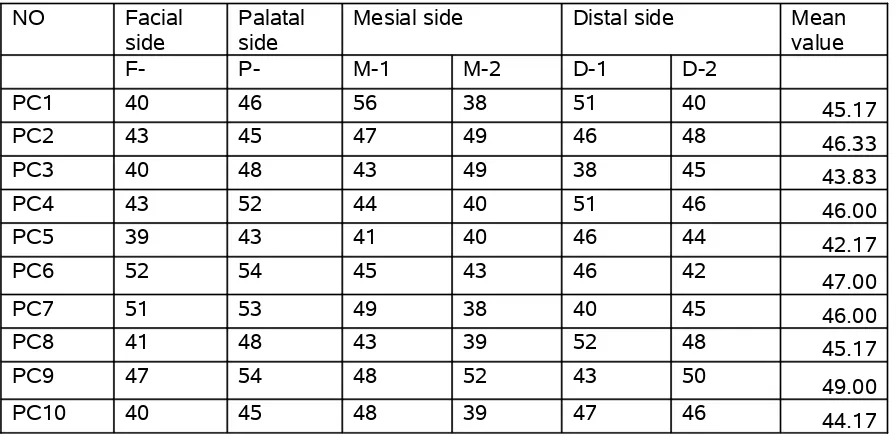
[image:69.612.102.550.146.364.2]PC10 40 45 48 39 47 46 44.17
Table 9 .Marginal fit of pressed ceramic crowns after application of porcelain
Sub group IIB
NO Facial side Palatal side Mesial side Distal side Mean value F-1 F-2 P-1 P-2 M-1 M-2 D-1 D-2
PC1 67 64 69 67 60 65 71 69 66.50
PC2 71 72 74 73 72 70 73 72 72.13
PC3 66 69 70 64 65 70 68 72 68.00
PC4 68 71 72 69 67 73 68 72 70.00
PC5 66 64 67 65 64 65 66 65 65.25
PC6 66 69 70 68 64 67 65 67 67.00
PC7 64 69 68 71 62 65 63 66 66.00
PC8 71 73 70 72 71 74 71 73 71.88
PC9 62 66 68 66 64 67 63 65
65.13
Table. 10 .Comparision of initial marginal fit of metal coping with pressed ceramic coping
Surfaces
Metal coping before veneering
Pressed ceramic coping before veneering
SUB GR- IA SUB GR-IIA
MEAN of all the 10 samples
S.D MEAN of all
the 10 samples
S.D
FACIAL F-1 50 2.71 43.6 4.77
MEAN 50 2.71 43.6 4.77
PALATAL P-1 53.1 2.73 48.8 4.13
MEAN 53.1 2.73 48.8 4.13
MESIAL M-1 51.2 3.26 46.4 4.27
M-2 50.2 4.54 42.7 5.29
MEAN 50.7 3.9 44.55 4.78
DISTAL D-1 52.9 2.73 46 4.67
D-2 49.9 3.81 45.4 2.95
Table 11 .Comparision of marginal fit of metal coping before and after veneering of porcelain.
SURFACES SUB GR- IA SUB GR-IB
MEAN S.D MEAN SD
FACIAL F-1 50 2.71 77.9 2.92
79.6 3.63
MEAN 50 2.71 78.75 3.275
PALATAL P-1 53.1 2.73 80.6 2.46
81 4.19
MEAN 53.1 2.73 80.8 3.325
MESIAL
M-1 51.2 3.26 77.7 3.2
M-2 50.2 4.54 80 2.36
MEAN 50.7 3.9 78.85 2.78
DISTAL D-1 52.9 2.73 78 2.79
D-2 49.9 3.81 79.8 3.74
Table 12. Comparision of marginal fit of pressed ceramic coping before and after veneering of porcelain.
SURFACES
I SUB GR-IIA SUB GR-IIBMEAN S.D MEAN S.D
FACIAL F-1 43.6 4.77 66.9 2.81
69 3.4
MEAN 43.6 4.77 67.95 3.105
PALATAL P-1 48.8 4.13 70 2.16
68.9 3.48
MEAN 48.8 4.13 69.45 2.82
MESIAL M-1 46.4 4.27 65.6 3.75
M-2 42.7 5.29 68.5 3.27
MEAN 44.55 4.78 67.05 3.51
DISTAL D-1 46 4.67 67.5 3.41
D-2 45.4 2.95 69.3 3.27
Table 13. Comparisons of final marginal fit of pressed ceramic crowns with metal ceramic crowns
SURFACES SUB GR-IB SUB GR-IIB
MEAN SD MEAN S.D
FACIAL F-1 77.9 2.92 66.9 2.81
F-2 79.6 3.63 69 3.4
MEAN 78.75 3.275 67.95 3.105
PALATAL P-1 80.6 2.46 70 2.16
P-2 81 4.19 68.9 3.48
MEAN 80.8 3.325 69.45 2.82
MESIAL M-1 77.7 3.2 65.6 3.75
M-2 80 2.36 68.5 3.27
MEAN 78.85 2.78 67.05 3.51
DISTAL D-1 78 2.79 67.5 3.41
D-2 79.8 3.74 69.3 3.27
BIBLIOGRAPHY
1.A.J.Hunter And A.R.Hunter -Gingival Crown Margins Confirgurations :
A Review And Discussion .Part 1:Terminology And Width. The Journal Of Prosthetic Denstistry Vol64, Issue1, Nov1990
2. Adriana Ferreira Quaintas, Dds, Phd Fabiano Oliveira,Dds - Vertical
Marginal Discrepancy Of Ceramic Materials,Finish Lines,And Luting Agents:An In Vitro Evaluation The Journal Of Prosthetic Denstistry Vol92, Issue3, Sep2004.
3. Aldo Lomanto Dmd And Sual Weiner Dds - A Comparative Study Of
Ceramic Crown Margins Constructed Using Different Technique The Journal Of Prosthetic Denstistry Vol67, Num2, June1992
4. Alfredo Julio Fernandes Neto , Heitor Panzers - Bond Strength Of
Three Dental Porcelains To Ni-Cr And Co-Cr-Ti Alloys Braz Dent J (1)2006
5. Allyn J. Coleman, Dmd, Ms1/Haig Rickerby, Dmd2/Lisa –
Macromolecular Leakage Beneath Complete Cast Crowns A 6-Month In
6. Anelise R.Ferreira, Dds,Msc Gelson L.Adabo,Dds,Msc,Phd -
Evaluation Of The Thermal Shrinkage Of Titanium And The Setting And
Thermal Expansion Of Phosphate Bonded Investments The Journal Of Prosthetic Denstistry Vol98, July2007.
7. Anthony H.L.Tjan Dds, Tao Li,Bds - Marginal Accuracy Of Complete Crowns Made From Alternative Casting Alloys The Journal Of Prosthetic
Denstistry Vol66, Num2, Aug1991.
8. Anthony H.L.Tjan, Dr.Dent ,Dds, - Effect Of Preparation Finish On Retention And Fit Of Complete Crowns The Journal Of Prosthetic
Denstistry Vol56, Issue3, Sep1986
9. Carla Castiglia Gonzaga , Paulo Francisco Cesar -Mechanical Properties Of Dental Glass Ceramics Hot Pressed At Different
Temperatures. Materials Research Vol 1. No3, 301 – 306 , 2008.
10. Christian Lehner ,Stephen Studer, - Short Term Results Of
Ips-Empress Full Porcelain Crowns. Journal Of Prosthodontics 1997;6;20 – 30.
11. Christina A.Mitchell Bds,Phd Mariar.Pintado,Mph -Non Destructive
12. Constaninos Yfantis Dimitrios Fantis-Analytical And Electrochemical Evaluation Of The In Vitro Corrosion Behavior Of Nickel –Chrome And
Cobalt-Chrome Casting Alloys For Metal Ceramic Restorations
13. D.J.Wanserski Dds,Ms K.P.Sobczak Dds -An Analysis Of Margin Adaptation Of All Porcelain Facial Margin Ceramometal Crown. The Journal Of Prosthetic Denstistry Vol56, Issue3, Sep1986
14. D.N.Dederich C.W.Svare - The Effect Of Repeated Firings On The Margins Of Non Precious Ceramometals The Journal Of Prosthetic Denstistry Vol51, Issue5, May1984
15. Denis Gemalmaz Dds,Phd Hasan Needet Alkumuru,Dds,Phd - Marginal Fit Changes During Porcelain Firing Cycles The Journal Of Prosthetic Denstistry Vol73, Num1, Jan1995.
16. Denis Gemalmaz Dds,Phd Hasan Needet Alkumuru,Dds,Phd -
Thermal Cycling Distortion Of Porcelain Fused To Metal Fixed Partial Dentures The Journal Of Prosthetic Denstistry Vol80 Num 6 Dec1998
17. Edward B.Goldin,Dds,Norman W.Boydiii,Dds - Marginal Fit Of
Leucite-Glass Pressable Ceramic Restorations And Ceramic-Pressed To Metal Restorations-The Journal Of Prosthetic Denstitry
18. Efstration Papazoglou Dds,Ms,Phd William M.Johnston - Evaluation Of High Temperature Distortion Of High Palladium Metal
Ceramic Crowns The Journal Of Prosthetic Denstistry Vol85, Num2, Feb2001
19. Elaine R.Schilling Dds,Ms, Barbara H.Miller Dds,Ms - Marginal Gap Of Crowns Made With A Phosphate Bonded Investment And Accelerated
Casting Method The Journal Of Prosthetic Denstistry Vol81, Num2, Feb1999
20. F.Michael Gardner D.D.S - Margins Of Complete Crowns-Literature
Review The Journal Of Prosthetic Denstistry Vol48, Num4, Oct1982.
21. F.Van Rensburg H.Strating - Evaluation Of The Marginal Integrity Of Ceramometal Restorations The Journal Of Prosthetic Denstistry Vol52,
Issue2, Aug1984
22. Farhad Vahidi Dmd,Msd, Erwin T.Egloff Dds,Ms - The Journal Of Prosthetic Denstistry Vol66, Num4, Oct1991.
23. George Salem Dmd - Margin Design For Esthetic Posterior Metal
24. Giuseppe Isgro , Cornelis J. Kleverlaan , - The Influence Of Multiple Firing On Thermal Contraction Of Ceramic Materials Used For The
Fabrication Of Layered All-Ceramic Dental Restorations. Dental Materials Vol 11 , 557 564, 2005
25. H.Strating,B.Ch.D,M.Sc.D C.H.Pameijer,Dmd,Msc.D,Dsc.D -
Evaluation Of The Marginal Integrity Of Ceramometal Restorations The
Journal Of Prosthetic Denstistry Vol56, Issue1, July1981
26. Hajime Hamaguchi, D.D.S, Anelo Cacciatore,D.D.S – Marginal Distoration Of The Porcelain Bonded To The Metal Complete Crown The
Journal Of Prosthetic Denstistry Vol47, Num2, Feb1982.
27. Harold F.Morris ,Dds,Ms-Department Of Veterans Affairs Cooperative Studies Project No:242 Quantitative Evaluation Of The Marginal Fit Of The
Cast Ceramic Porcelain Shoulder And Cast Metal Full Crown Margins The Journal Of Prosthetic Denstistry Vol67, Issue2, Feb1992
28. Ivy S.Schwartz Dds,Ms.Ed -A Review Of Methods And Techniques To Improve The Fit Of Cast Restorations The Journal Of Prosthetic Denstistry
29. J.Fischer P.W.Fleetwood - Improving The Processing Of High Gold Metal Ceramic Frame Works By A Pre-Friring Heat Treatment Dental
Materials 16(2000)109-113.
30. J.Robert Homles.Considerations In The Measurement Of Marginal Fit.
The Journal Of Prosthetic Dentistry 1989;62;405-408
31. J.Robert Kelly Dds, Ms, Dmedsc Ichiro Nishimura Dds
Dmedsc,Dmd-Ceramics In Dentistry:Historical Roots And Current
Perspectives The Journal Of Prosthetic Denstistry Vol75, Num1, Jan1996
32. James D.Weaver,Dds, Glen H.Johnson Dds,Ms -Marginal Adaptation
Of Castable Ceramic Crowns The Journal Of Prosthetic Denstistry Vol66, Num6, Dec1991.
33. Jianxiang Tao, Masanobu Yoda - Fit Of Metal Ceramic Crowns Cast
In Au-1.6 Wt% Ti Alloy For Different Abutment Finish Line Curvature Dental Materials (2006) 22,397-404.
34. John .M. Powers Craig’s Restorative Dental Materials
35. John A.Sorensen D.M.D - A Standardized Method For Determination
36. John W Mclean- Dental Ceramics Proceedings Of The First International Symposium On Ceramicsquintessence Publishing
37. John W Mclean - The Science And Art Of Dental Ceramics Vol I & Vol II
38. John W.Mclean, O.B.E, D.Sc, M.D.S, L.D.S,- Dental Ceramic Proceedings Of The First International Symposium On Ceramics
39,. John W.Mclean, O.B.E, D.Sc, M.D.S, L.D.S,Edmund
E.Jeansonne,D.D.S,-A New Metal-Ceramic Crownthe Journal Of Prosthetic Dentistry Sep 1978 Vol 40 Number 3 Page(273-287)
40. Johnson Campideli Fonseca, Guilherme Elias Pessanha Henriques - Stress Relieving And Porcelain Firing Cycle Influence On Marginal Fit Of Commercially Pure Titanium And Titanium Aluminum Vanadium Copings
Dental Materials19(2003) 686-691
41. K. J. Anusavice. , J.E.Carroll. – Effect Of Incompatibility Stress On The Fit Of Metal Ceramic Crowns. J. Dent.Res.66(8) ; 1341-1345.Aug 1987
42. K.Richter Snapp A.Aquilino - Change In Marginal Fit As Related To