NATIONAL INSTITUTE OF SIDDHA
Tambaram sanatorium, chennai – 47.AFFILIATED TO THE TAMIL NADU DR. M.G.R. MEDICAL UNIVERSITY, CHENNAI - 600 032.
A STUDY ON
UTHIRAVATHA SURONITHAM
(DISSERTATION SUBJECT)
For the partial fulfillment of the requirements to the Degree of
DOCTOR OF MEDICINE (SIDDHA)
BRANCH I – POTHU MARUTHUVAM DEPARTMENT
MARCH– 2009
ACKNOWLEDGEMENT
I feel immense awe and colossal gratitiute in my heart of hearts to God
Almighty for making this dissertation have its present form.
I take this opportunity to express my gratitude and acknowledge to the, The
Tamil Nadu Dr.M.G.R. Medical University, Chennai.
I express my deep sense of gratitude to our Director, Prof.Dr.S. Boopathiraj,
M.D(s), National Institute of Siddha, Chennai.
I express my sincere thanks to Prof. Dr.R.S. Ramasamy, M.D(s). Dean,
National Institute of Siddha, Chennai.
I would like to express my profound sense of gratitude to our H.O.D.
Dr.K.Manickavasagam, M.D(S). Head of the Department, Pothu maruthuvam,
National Institute of Siddha, Chennai for his valuable guidance to complete my project.
I express my sincere thanks to Dr.M.Logamanian, M.D(s), Phd, Head of the
Department, Noi nadal, Hospital Superintendent, National Institute of siddha, Chennai.
My deep sense of gratitude to Dr. Subburayalu M.D.Gen.Med. Madras
Medical College, Chennai.
I express my grateful thanks to Dr.R.Lakshmi kantham M.D(s), Department
of Pothu maruthuvam, National Institute of Siddha, for his moral support and
encouragement
I express my sincere thanks to Dr.H.Vethamerline kumari M.D(s),
Department of Pothu maruthuvam, National Institute of Siddha, for his memorable
I express my sincere thanks to Dr.Selvashanmugam M.D. (S), Department of
Pothu maruthuvam, Medical officer, for his valuable support during this work.
I express my sincere gratitude to Mr. M. Subramanian M.Sc. SRO, National
Institute of Siddha, for his guidance in statistical analysis.
I acknowledge my thanks to Prof. Dr. C.Venkatraman, The Director, C.L.Beid
Metha Pharmacy College, Thorappakkam, Chennai-96, for his support in
Pharmacological and Toxicological studies.
I wish to thank my beloved family members and friends for whose selfless
help for this work.
I wish to thank all the faculties especially Librarians and Lab technicians in
CONTENTS
S.NO
TITLE
PAGE.
NO
1.
2.
3.
4.
5.
6.
7.
8.
9.
10.
INTRODUCTION
AIM AND OBJECTIVES
REVIEW OF LITERATURE
MATERIALS AND METHODS
OBSERVATION AND RESULTS
DISCUSSION
SUMMARY
CONCLUSION
ANNEXURES
I. DRUG REVIEW.
II. TOXICITY STUDY OF TRIAL DRUG. III. PHARMACOLOGICAL STUDY OF TRIAL
DRUG.
IV. CHEMICAL ANALYSIS OF TRIAL DRUG. V. STATISTICAL ANALYSIS.
VI. PROFORMA.
INTRODUCTION
Siddha is a holistic medical system that gives importance to mental as well as physical well beings of a patient. The word siddha means "established truth". The word siddha comes from the word siddhi that means an object to be attained such as perfection in life or heavenly bliss.
Medicine is all about preventing and treating ailments thus postponing death. As interesting aspect of siddha medicine is its view about death. Thirumoolar defines the ailment of the body and mind as diseases. In the same breath he defines death as disease and hence could be prevented.
"¯¼õÀ¡ø «Æ¢Â¢ü ¯Â¢Ã¡ ÄÆ¢Å÷
¾¢¼õÀ¼ ¦Áö»¡Éõ §ºÃ×õ Á¡ð¼¡÷
¯¼õ¨À ÅÇ÷ìÌõ ¯À¡Âõ «È¢ó§¾
¯¼õ¨À ÅÇ÷ò§¾ý ¯Â¢÷ ÅÇ÷ò§¾§É".
- ¾¢ÕãÄ÷.
Siddhars not only defines death as disease, but they also earnestly tried to prevent death by advocating life style modification and developing new drugs. Siddha drugs are derived from natural sources such as plants, animals and minerals. Plant drugs are its mainstay. The standing instruction of siddhars to medical practitioners is to use herbal drugs first in any ailment, if herbal drugs are ineffective then only advised to go herbomineral preparations.
"«ñ¼ò¾¢Öûǧ¾ À¢ñ¼õ À¢ñ¼ò¾¢Öûǧ¾ «ñ¼õ."
things including drugs and diet articles. Various tissues of the body are the combination of these five elements in different proportions.
The physical function of the body is mediated and maintained by three forces. They are vali, Azhal and iyam. In normal state they are called three forces or three thathu that sustain and nourish the body. In disease state when the three forces are vitiated they are called mukkutrams. When these three forces are in balance one is healthy. When vitiated singly or combination bring about disease.
Seven pillars or fundamental tissues called thathus supporting every living body. When the three forces are vitiated the tissues, disease will occur.
Various siddhars classified disease, one of is Yugi munivar. Yugi munivar classifies disease based on clinical signs and symptoms along with humoral pathology. Yugi classified vatha diseases in to 80 types in "Yugivaithiya chinthamani". Uthiravatha suronithan is one of them.
The disease Uthiravatha suronitham has a very close relevance to Rheumatoid arthritis. The disease Uthiravatha suronitham involving multiple system of the body especially musculoskeletal system affected first and then other systems are affected depends upon their immunology. Single and herbal drug therapies are not much useful to cure the disease. So only the author selected herbomineral drug for internal medicine.
Anupoga vaithiya navaneetham said that the Rasapralaya chenthuram is good for vatha diseases and Theraiyar thylavarukka surukkam describes the Navanatha siddhar thylam is useful to reduce inflammatory conditions of vatha diseases. The principle drugs Rasapralaya chenthuram which is useful in chronic musculoskeletal disorder and Navanatha siddhar thylam is an external application reducing inflammatory condition because of its contents has effective anti-inflammatory action.
AIM AND OBJECTIVES
The knowledge of preserving one’s health sound and thus prolonging life is said to have descended in this modern technological world. The prolonged and uncertain course of the disease “Udhira Vatha Suronitham” calls for special emphasis and intended the author to bring an ideal treatment for the disease.
Medicine, as everyone knows is not a mere science, but is an art as well. It is the only want of men with such knowledge of science and its practical practice with efficacious cures on hand.
x The principal aim of the present study is to estimate the efficacy of the siddha drugsRasapralaya chenthuram and Navanatha siddhar thylam.
x To ensue a new approach in diagnosis for the disease.
x To know whether the drug has any side- effects or not.
OBJECTIVES:
@ The main objective of the present study is to create awareness about the siddha sciences and to highlight the efficacy of siddha drugs among the public.
@ To collect various informations about “Uthiravatha Suronitham” and to expound the characteristics of etiology, premonitory symptoms, signs and pathogenesis based on both siddha and modern aspects.
@ To access the prevalence of the disease with reference to age, sex, diet habits, socio economic status, family history etc.
@ To highlight the siddha diagnostic principles in diagnosing the disease.
@ To conduct a thorough study on Uthiravatha suronitham with Rheumatoid arthritis.
@ To have a complete study of the disease Uthiravatha suronitham, under the headings of
(a) Pori Pulangal (b) Mukkutram
(c) Udal Kattugal (d) Ennvagai thervugal etc.
@ To evaluate the pharmacological study on the trial drug.
@ To study the bio-chemical and chemical analysis on the trial drug.
@ To evaluate the efficacy of the trial medicine on Antimicrobial activity by in vitro studies.
REVIEW
OF
SIDDHA ASPECT
The things exist in the universe also exist in the human beings. Any adverse changes of these two, even a minute change will be reflecting on the other.
In siddha system of medicine, the physiological function in human system is mediated by three substances viz, Vatham (vali + veli), Pitham (thee), Kabham (neer + man). These three humors maintain the upkeep of the human body through their combined functioning. When deranged, they bring about diseases peculiar to their influence. Uthiravatha suronitham the disease taken for study is one of the vatha diseases described by Yugi vaithya chinthamani.
DEFINITION OF VATHAM:
Vatham is a clinical condition characterized by pain, swelling, pricking sensation and loss of function due to vitiated vatha, which is the principle humour of the body.
‘nghw;wh kiuahd; Gidnka; auz;fhf;Fk; nghw;wh kiuahd; Gfy;tnjd; nghw;whk; tstpdpNy ahf;Fuk;ig kd;ndd;d kd;d tstpdpNy ahf;Fk; tsp.”
- Njiuah; akf ntz;gh.
Vatham is being hailed as the king, who rules the fort (Body) and enables the dwelling of the citizen (Uyir) in the fort. Hence Theraiyar lauds Vatham as the prime force in normal state.
AETIOLOGY:(Neha; tUk;top)
'vd;dNt thjk;jh ndz;gjhFk;
,fj;jpNy kdpjh;fSf; nfa;AkhW gpd;dNt nghd;jidNa NrhuQ; nra;J
nghpNahh;fs; gpuhkziu J~dpj;Jk; td;dNj tw;nrhj;jp NrhuQ; nra;J
khjh gpjh FUit kwe;j Ngh;f;Fk; fd;dNt Ntjj;ij epe;ij nra;jhy;
fhaj;jpw; fye;jpLNk thje; jhNd jhd; vd;w frg;NghL Jth;g;G iwg;G
rhjfkha; neQ;RfpDQ; rikj;j tz;zk; Mdd;w thwpdJ nghrpj;jyhYk;
Mfhaj; NjwsJ Fbj;j yhYk; ghndd;w gfYf;f kpuh tpopg;G
gl;bdpNa kpftWjy; ghunka;jy; Njndd;w nkhopahh; Nkw; rpe;ij ahjy;
rPf;fpukha; thjkJ nrdpf;Fk; jhNd."
- A+fp itj;jpa rpe;jhkzp.
Excessive sexual indulgence, over consumption of bitter, astringent and salty-tasted foods, alcoholism, and daytime sleep, night time over work, starvation and lifting over weight will aggravates Vatham.
Pararasasekaram describes the factors for vitiation of vatham:
'njhopy; ngW ifg;G fhh;j;jy; Jth;j;jy; tpQ;RfpDQ; NrhWk; gioajhk; tuF kw;iwa ige;jpid aUe;jpdhYk;
vopy;ngwg; gfYwq;fp ,utpdp Ywq;fhjjhYk; kio epfh; FoypdhNs thjk; Nfhgpf;Fk; fhNz."
Consumption of excessive bitters, astringents, salty tasted foods, rancid foods, Daytime sleep and lacking of night sleep vitiates Vatham.
According to Sabapathy Kaiyedu:
tspf;Fw;wj;ijj; J}z;Lk; czTfshfpa thiof;fha;> fpoq;F> Gsp> japh;> Nghd;wtw;iw mjpfkhf cz;gjhYk; Fsphpy; cyty;> ngz;bh; kaf;fk;
Kjypatw;whYk; thjNeha; tUk;.
thjk; th;j;jpf;Fk; fhyk;:
\thjth;j; jidfhy NkNjh ntd;dpy; kUTfpd;w Mdpfw; flf khFk;
Mjitg; grpNahL fhh;j;jpif jd;dpy; mlUNk kw;wkh jq;f ld;dpy;
NghjNt rkpf;fpd;w fhy khFk|;.
tspf;Fw;wk; Mdp> Mbapy; jd;dpiy tsh;r;rpAk;> Ig;grp fhh;;j;jpifapy; gpuNfhgKk; milAk;. kw;w khjq;fspy; jd;dpiy milAk;.
Mbahjp aha; Ig;grp <wha; mdpykjw; Nfhuurpay; fhyk.;|
~flfKjy; Jyhk; tiuapy; thjkhFk; fz;zhb iag;grpA kJNt ahFk;.|
- rjfehb.
CHARACTERISTICS OF VATHA DISEASE:
'thjtPW md;dkpwq;fhJ fLg;Gz;lhk; tz;zKz;lhk; NkhJfl;L Nuhfk; RuKz;lh kpUkYkh Kwq;fhnjd;Wk; XJ#hpa thj kdyhF eLf;f Kz;lhk; nghUs;fsha;j; jPjdNt euk;gprpj;J re;Jfs; NjhWq; fLf;Fe; jpdKk;jhNd" - Njud;thflk.;
Loss of appetite, pain and redness, fever, cough, insomnia, shivering and pain in all over the joints.
're;jputhjKld; Fsph;e;njOe;Nj eLf;Fe; rPjtha;thk;
Ke;jpa Fj;jp rpthe; re;Jfs; NjhWk; File;J nkhspfs; tPq;Fk; te;jpa njhe;jthjk; tPf;f Kz;lh Klypw;wp uz;lhNk"
Chillness of the body, rigor, spasm, pain and tenderness over the joints and swelling of the joints.
"fhzg;gh thjkPwpy; fhy;iffs; ngUj;J NehTk; G+zg;gh Fly; Gul;Lk; kyryk; nghUkpf; fl;Lk;
Czg;gh FspUq; fha;r;ry; clk;ngy;yhk; Fj;J tha;T tPzg;gh FjkpWf;Fk; tpah;itAk; Nth;f;Fk; jhNd."
- mfj;jpah; itj;jpa fhtpak; 1500.
"Nktpa thjQ; nra;Ak; Fze;jid tpsk;gf; Nfsha; jhtpNa tapW ke;jQ; re;Jfs; nghUe;J NehthQ; rPtpa jhJ ehrQ; nrWj;Jld; rpWePh; tPOq;
fhtpaq; fz;zpdhNs kykJ fUfp tPOk;”.
- mfj;jpah; MAs; Ntjk;.
"vwpa ey;thj nkwpf;Fq; Fzq;NfS Fwpnad iffhy; Fisr;R tpyhr;re;J gwpnad nehe;Jlw; gr;irg; Gz;zhFNk Gz;zha; typf;Fk; ngUFk; FlNyhbj; jz;zh kyj;ijj; jk;gpf;Fk; Nghf;fhJ xz;zhd Mrdk; cwNt ruf;fpLk; gz;zhh; Fsph;rPjk; gUj;jpLk; thjNk”.
- jpU%yh; itj;jparhuk;.
‘thjk; te;Jw;w NghJ tapwJ nghUkpf; nfhs;Sk; jhjtpo;e;jpLg;G iffhy; re;Jfs; fLg;Gj; Njhd;Wk; rPnjhU kyK ePUe; rpWj;jJld; fLj;J tpo khjtkiu Nky; te;j thjj;jpd; FzkpjhNk.”
- A+fp Kdpth; ngUE}y; itj;jpa fhtpak; 1000.
From the above poetic versions, it is clear that the major characters of vatha diseases are joint pain, swelling present over the joints, difficulty in walking, constipation, burning micturition, oliguria, dyspnoea, flatulence, fever, fatigue, giddiness and nerve weakness etc.
RNuhzpj Neha; vd;gjw;F T.V.rhk;grptk;gps;is mth;fs; khjtplha; rk;ge;jkhf Vw;gLk; Neha; vd;W $wpAs;shh;.
Suronitha Vatham is a disorder of menstruation in women characterized by affection in chest and limbs, extreme sensibility to pain, dryness in the skin, pain in nerves accompanied by intense bodily pain.
VATHA SURONITHAM:
Yugi Vaidhya Chinthamani classified Vatha suronitham into 7 types. They are: 1. Vatha Suronitham
2. Uthiravatha Suronitham 3. Sithuvatha Suronitham 4. Vaikithavatha Suronitham 5. Paithiyavatha Suronitham 6. Slethuma vatha Suronitham 7. Udharavatha Suronitham
1. VATHA SURONITHAM:
'mwpe;jpl;l mq;fnky;yh nkypT khfp mirthd jt;tplq;fs; tPf;fkhfp ewpe;jplp;l eilnfhlh jhdpUj;jy; typahfp nkhop nkhopa tPf;fkhfr; nrhwpe;jpl;l Njfnkq;F kirT fhzy; Nrhw;wpd;Nky; dpidtpd;wpj; J}f;fkhjy; twpe;jpl;l thajdp dPh;jh D}wy; thj RNuhzpjj; jhYk; tFj;j thNu"
Emaciation, swelling of joints, restricted movement, pain, tenderness, discomfort, loss of appetite and excessive salivation.
2. UTHIRAVATHA SURONITHAM:
nra;fpjkha; rpWtpuy;fs; kpfTk; nehe;J rpe;ij jLkhwpNa rypg;Gz;lhFk;
igfpjkhk; gapj;jpaj; jpy;yhj kpQ;rpg; ghukha; cw;gtpj;J moYz;lhFk; ca;fpjkhk; mrdkJ jhDk; Ntz;lh cjputhjr; RNuhzpjj;jp Dzh;r;rpahNk"
Pain and swelling in ankle joints, knee joints and all small joints of the hands and toes.
3. PAITHIYAVATHA SURONITHAM:
'czh;r;rpaha;r; RNuhzpje;jhd; kpfnt Jk;gp Cf;fkha;j; Njfnkq;F kpfNt nehe;J Kzh;r;rpaha; Koq;fhy;fs; Koq;if nahf;f Kidahd rpWtpuy;fs; fd;dk; new;wp jzh;r;rpaha;r; re;JrU thq;f nkq;Fk; jhl;bf kha;f;File;J RuK Kz;lhk; gzhr;rpaha;g; ghz;lJNghd; Nkdp ahFk; gapj;jpath jRNuhzpjj;jpd; gz;G jhNd"
Pain in all over the body, pain in elbow joints, knee joints, fingers, cheek, forehead, hyper pyrexia, and anemia.
4. SLETHUMAVATHA SURONITHAM:
'ghz;ghf Tly;Fsph;e;J VW tPq;fpg; gijg;ghd tple;njhl;lhw; ghu Nehthk; jpz;ghd rpuRnew;wp Nehf;fh Lz;lhk; rpNyl;Lkkha;f; NfhionahL Rthr khFk; kz;ghf kaf;fnkhL fdT Kz;lhk; tha;tuz;L Urpapy;yh tUj;j khFk; ez;ghf ehbANk glg lf;Fk;
ew;Nrl;g RNuhzpjkhk; ehLq; fhNy"
5. UTHARAVATHA SURONITHAM:
'ehLNk Ruk;te;J eLf;f Yz;lhk; ehtuz;L jiynehe;J clk; gOe;jp thLNk Njfnky;yh kdpr;rk; g+g;Nghy; kfhtUj;j Kz;lhfp kaf;f khFk; rhLNk abf;fbjhd; Ngjp jhDk; jtpf;FNk jz;zPh;jh dhl;l khfpj; NjLNk Nrhw;wpd;Nky; epidT jhDk; nra;Tju thjRNuh zpje;jh ndd;Nd."
Fever with rigor, dryness of the mouth, headache and analgia, giddiness, passing loose stools, thirst and excessive appetite.
6. SITHUVATHA SURONITHAM:
'thwhd rhPunky;yh jpiwe;J Cjy; khrw;w Njhy;jhDk; jpiue;J NghFk; ehwhd ehWNghy; euk;G Rf;Fk;
ehf;Fj;jhd; totoj;Jf; Nfhio ahFk; J}whd neUg;Gj;jhd; gl;lhw;Nghy nehe;JNk rlnky;yhq; nfhg;g spf;Fk; tPwhd Thpe;Jgpd;id ntJk; gPq;Fk; kpf;frpj;J thjRNuh zpj khNk"
Wrinkled skin, accumulation of phlegm in the throat, vesicles presents all over the body, exfoliation and anasarca.
7. VAIKITHA VATHAM:
'Mnkd;w tPq;fpdNjhh; tplj;jpy; uj;jk; mOj;jkha;j; jpuz;LNk vq;Fk; gha;e;J Xnkd;W xl;bNa jpuz;bUf;Fk;
Swelling, hematomas, cough, hyperpyrexia, numbness in soles and pain in all over the body.
DHANVANTHRI VAITHYAM QUOTES THE CLINICAL FEATURE OF
SONITHA VATHA ROGAM AS:
'fhZNk vyptplk; Nghy; fdg;Gld; jbg;Gkhfp g+Zlk; GisAk; Fj;Jk; nrhwp fdg;Gld; NjhzpUe; Jbg;Gj; Njfk; fps;spdhw; NrhjpaJ khdpy Kiyaha; RNuhzpj thjkhNk"
'ifapdpw; fwz;il jz;zPh; fl;LQ; rijNjhy; tPq;F ikaW fUik nrk;ik ngaHj; njwpe;joYk; tPf;fk; nka;apidg; gpsf;Fk; thjk; tpahgpf;Fk; clk;gjhf;F ikayh; clk;G jk;gpf;Fk; thjRnuhzpj kpnjd;Nd."
Pyrexia and swelling of the body as in rat poison intoxication, pain and tenderness, twitching of muscles, loss of sensation, swelling of wrist and phalanges, black and redness of swelling due to vascular failure and hyperaemia.
PARARASA SEKARAM CLASSIFIES VATHA SURONITHAM INTO FIVE
HEADINGS.
Pararasa Sekaram describes Suronitham as,
'tpo;ngW RNuhzpje;jhd; kpfTld; nkypTkhfpj; jho;tpy; re;JfNs tPq;fpj; jif ngw eil nfhlhky; tho;TW ifAq;fhYk; trkpd;wp aod;W Nehthk; gho;ngW kzq;fpdhNs gaDwg; gfh;e;Jl;NlhNk"
Debility in raktha thathu (anaemia), swelling of peripheral joints, deformed movement of joints, pain in upper and lower limbs.
UTHIRA VATHAM IN PARARASA SEKARAM:
kpf;FNk cjputhjk; vd;wpJ tpsk;gyhNk"
Pain and tenderness of chest and axilla, emaciation, pain and swelling of upper and lower limbs.
CLINICAL FEATURES OF VATHASURONITHAM:
'gfh;e;jpLk; thje; jd;dpw; RNuhzpjk; gfUk; fhiyg; GFe;Jlnyq;Fk; nehe;J NghjNt Njhy; jpiue;J Gife;jpL neUg;G gl;lhw; NghyNt nfhg;gspj;J kpFe;jpl nthpe;J gpd;id ntJk;gpNa tPf;fkhFk;."
Pain and tenderness, exfoliation, eruption as in burns and swelling in joints.
'tPq;fpa tplj;jpw;jhNd kpfj;jpuz;Nlfp Kl;b
Mq;fij tpuyhw; njhl;Nl aOj;jpw; nkj;njd;wpUf;Fk; Xq;fpa typg;G Kz;lh Klypdpw; fLg;G Kz;lhk; Nfhq;nfY KiyapdhNs $wpa Fzq; fNlNu."
Soft swelling on touch and pricking nature of pain.
CLINICAL FEATURES OF SEETHAVATHA SURONITHAM:
'nrhw;rPj Tjuthj RNuhzpj Koq;fhy; jhDk; nghw;fidf; fhYk; re;Jk; Gwtb jhDk; tPq;fp ew;NfhZ tpuy;fs; nehe;J eLigj;jpa thjj;jpy; cw;gt FzKz;lh %W E}ypy; nrhd;djhNk."
Swelling of the knee joint, ankle joint and feet, pain and swelling over the phalanges.
CLINICAL FEATURES OF PAITHIYAVATHA SURONITHAM:
Pain and swelling of the metacarpo phalangeal joints and proximal interphalangeal joints and headache.
CLINICAL FEATURES OF SILETHUMAVATHA SURONITHAM:
'Nehw;wpa Nrw;gthj RNuhzpj Kly; Fsph;e;Nj Vw;wkha; tPq;fpae;j tple;njhl;lhd; kpfr; rpf;nfd;Wk; khw;Wwg; nghUe;Jr; rpf;Fk; tphptpop kley;yhNs rhw;wpa ey;Nyhh; Ntjj;jpdpDiuj; jpl;lthNu."
Chillness of the body, tenderness and swelling of the joints.
CLINICAL FEATURES OF UTHARAVATHA SURONITHAM:
'ciungW cjuthj RNuhzpj Kiwf;Fk; fhiyj; jiungW thjj;J}w;Nw RNuhzpj FzKk;jf;f tphpTW gypj;Jthj RNuhzpjf; FzKkpf;f RiungW cjuthj RNuhzpj FzK Kz;lhk;."
Vitiation of Vatha aggravates the signs and symptoms of Vatha Suronitham.
The term suronitha vatha is also mentioned in Aathma Ratchamirtham, Anuboga Vaithya deva ragasiyam handled the term Uthiravatha surothinam as Sonitha vatha Rogam.
Our text book Siddha maruthuvam handle the term Uthiravatha Suronitham as Vali Azhal Keelvayu as per Literature Sabhabathi Kaiyedu.
MUKKUTRA VERUPADUGAL (SIDDHA PATHOLOGY)
‘gpzpapDw; gj;jpia NgRtd; gpzpKjy; thjgpj; jq;fg kd; ke;jphp je;jphp
tPjkh Alyuz; nka;k;Gu tuR nra; Kiw nrAkhjyhd;.”
- Njiuah; fhg;gpak;.
Which highlights that the main factor in the causation of the disease are vatham, pitham and kabham.
VATHAM:
Vatham is the prime force that impacts movement to every living cell in the body. Its dwelling place lies in the bones, muscles, nerves, joints etc. Hence it is responsible for the movement of parts involved in locomotor system. When vatham is affected, the other two pitham and kabham also gets deranged and in turn, they vitiates the other structural and functional elements of the living body called seven Udal thathus.
¾ Viyaanan which is responsible for the voluntary and involuntary movements
and nutrition of the tissue gets affected leading to restriction of movements and lassitude.
¾ Samaanan which neutralizes other vitiated vayus gets affected. Further it is
needed for normal digestion. So derangement of this vayu produces loss of appetite and indigestion.
¾ Involvement of Abaana Vayu also plays a main role in the manifestation of signs and symptoms. Abaanan which is responsible for distribution and assimilation of nutritional factors gets affected leading to symptoms like constipation.
¾ Kirukaran and Thevathathan are also affected because of loss of appetite and
sleeplessness respectively.
PITHAM:
¾ Ranjaga pitham which gives colour to blood.
¾ Saathaga pitham which is needed to carry out normal activities.
¾ In few, Anal pitham which is needed for digestion gets affected leading to anorexia.
¾ Prasaga pitham which gives complexion to skin gets affected leading to pallor
of skin.
KABAM:
The detoriation of the two main kuttram accompany the Kabha kutram whose structure is Earth + Water and is concerned with the maintenance of smooth working of joints, integration of structural elements of the body into stable structures etc.
¾ Santhiga kabham which is needed for normal maintenance of synovial fluid
gets affected.
¾ Avalambagam which forms the basis for all the other four types of Kabham
gets affected.
¾ In few, Kilethagam gets affected leading to loss of appetite.
Thus disturbance in Mukkutram produces,
¾ Pain, swelling of joints, joint stiffness, restriction of movements, loss of appetite and sleeplessness and constipation due to vatham.
¾ Inflammatory changes in joints like redness, warmth, loss of appetite and anemia due to pitham.
¾ Erosion of bony margin, osteoporotic changes, increases in the synovial fluid are due to disturbed kabham.
UDALTHATHUKKAL:
1. Saaram - Strengthens the body and mind.
2. Senneer - Gives power, knowledge and boldness to the mankind.
3. Oon - It gives structure and shape to the body and is responsible for movements of the body.
4. Kozhuppu - It lubricates the joints and organs and facilitates their functions.
5. Enbu - It protects all the internal organs and forms structural framework of the body.
6. Moolai - Resides inside the core of bones. It strengthens and maintains the normal condition of bones. 7. Sukkilam/ - Meant for reproduction (Male and Female Suronitham respectively).
In Uthiravatha suronitham, the affected Udal thathus are,
¾ Saaram - Loss of appetite, lassitude. ¾ Senneer - Anaemia, presence of RA factor). ¾ Oon - Muscle wasting, swelling.
¾ Kozhuppu - Emaciation, restriction of joint movements.
¾ Enbu - Vague pain and swelling of joints and deformity of joints.
DIFFERENTIAL DIAGNOSIS
:
Yugi Munivar in his “Yugi Vaidhya Cinthamani” mentioned about 80 types of Vatha diseases. Among them, the following diseases have joint pain as main clinical feature.
1. Oorusthamba vatham. 2. Malaithakamba vatham. 3. Santhu vatham.
5. Vatha suronitham.
1
. CU];jk;g thjk;
(Oorusthamba vatham):
‘Mnkd;w thjkJ cs;s lq;fp
mbj;Jiljhd; Fwq;fpuz;L kstha;g; gw;wpf; fhnkd;w iffhypy; tpuYQ; Rw;wpf;
fdj;JNk rhzpaJ nghjpe;jhw; Nghyj; Njnkd;w rpue;jdpNy ghu Kz;lha;j; Njfnkq;F %jpNa jpkpUz;lhFk; ehnkd;w elf;nfhzh nthLf;f khfp eypA+Uj; jk;gkJ eZFq; fhNz.”
Pain in both the thighs, swelling of fingers and toes, numbness, generalized edema of the body and inability to walk are the symptoms of this disease.
2
. kiyj;jfk;g thjk;
(Malaithakamba vatham)
‘Fk;gkhq; ifapd;kzpf; fl;L jd;dpw;
Ftpe;jiuapy; nkhj;ijNgh Yuj;Jf; fhZe; Jk;gkh kq;fq;fs; Jbg;g jhFe;
Jtz;LNk fhy; ifA eLf;f khFk; mk;gkh kiuf;Ff;fPo; jpkpUz; lhFk; mjuNk kpff;fWj;J ntbg;Gz; lhFk; tk;gkhk; thAe;jhd; gupA ehw;wk;
tUkiyj;j fk;gj;jpd; khh;f;fkhNk.”
Congestion of wrist joint, twitching, tremors in upper and lower extremities, numbness below the hip joint, fissured lips and passing foul flatus will be seen.
3. re;J thjk;
(Santhu vatham):
ehzpNa Kd;Nghy; eil nfhlhJ ikifjhd; kaf;fnkhL tha;ePUWk; tuz;bLNk ehTjh dbf;f bf;Ff; iffhy;jhd; juzpjdpw; wupf; nfhzhJ rQ;rupf;FQ; re;Jthk; thjq; NfNs.”
Pain in joints, body pain, pilo erection, inability to walk, giddiness, dryness of the tongue, excessive salivation and unable to keep the limbs in floor are the features of this disease.
4.
gapj;jpathj RNuhzpjk;
(Paithyavatha suronitham)
‘czu;r;rpaha;r; RNuhzpje;jhd; kpfnt Jk;gp Cf;fkha;j; Njfnkq;F kpfNt nehe;J Kzu;r;rpaha; Koq;fhy;fs; Koq;if nahf;f Kidahd rpWtpuy;fs; fd;dk; new;wp jzu;r;rpaha;r; re;JrU thq;f nkq;Fk; jhl;bf kha;f;File;J RuK Kz;lhk; gzu;r;rpaha; ghz;lJNghy; Nkdp ahFk; gapj;jpa thjRNuhzpjj;jpd; gz;GjhNd.”
Generalised body pain, severe pain in the knee joint, elbow joint, minor joints, temporo mandibular joint and all other joints, fever and anemia are the features of this disease.
5
. thj RNuhzpjk;
(Vatha suronitham)
‘mwpe;jpl;l tq;fnkyh nkypTkhfp
mirthd jt;tplq;fs; tPf;fkhfp ewpe;jpl;l eilnfhlh jhdp Uj;jy;
eypahfp nkhop nkhopa tPf;fkhfr; nrwpe;jpl;L Njfnkq;F kirT fhzy; Nrhw;wpd;Nk dpidtpd;wpj; J}f;fkhjy; mwpe;jpl;l thajdp dPh;jh D}wy;
Emaciation, swelling in movable joints, inability to walk, tremors, anorexia, increased sleep and excessive salivation are the features of this disease.
DIFFERENTIAL DIAGNOSIS (NOI NITHANAM)
Uthiravatha suronitham is differentiating from other types of vatha suronitham as follows:
S.NO
DISEASES
SIGNS AND SYMPTOMS
Uthiravatha suronitham
x Swelling of ankle joints, hip joints and knee joints.
x Pain and tenderness of minor joints especially phalanges.
x Depression.
x Loss of appetite.
x Increased vatha and pitha.
DIFFERENTIAL DIAGNOSIS
1.
Vatha suronitham
x Emaciation.
x Swelling of joints.
x Restricted movements.
x Joint pain.
x Discomfort.
x Excessive salivation.
2. Sithuvatha suronitham
x Anasarca.
x Wrinkles.
x Neural pain.
x Glossy tongue.
x Sialorrhoea.
x Bullous eruption as in burn.
x Exfoliation, swelling and warmthness.
3. Vaikithavatha suronitham
x Swelling with hyperemia.
x Soft on touch.
x Cough with pyrexia.
x Irritability.
4. Paithiyavatha suronitham
x Hyperemia.
x Tenderness in knee, elbow and smaller joints.
x Poly arthralgia.
x Pyrexia.
x Anemia.
5. Slethumavatha suronitham
x Chillness with abdominal distension.
x Severe pain and headache.
x Syncope and hallucination.
x Dryness of mouth and anorexia.
x Tachycardia.
6. Utharavatha suronitham
x Fever with rigor.
x Dryness of mouth.
x Pain in all over the joints.
x Headache, Giddiness.
x Diarrhoea.
x Hungry.
NAADI PATHOLOGY:
‘jpUj;jkhk; thjj; NjhNl jPq;nfhL gpj;jQ;Nrupy;
nghUe;Jfs; NjhWk; nehe;J”.
- Fzthflk; Nehapd; rhuk;.
‘fhzg;gh thjkPwpy; fhy;iffs; nghUe;jp NehFk;.”
- fhtpa ehb.
Vitiation of vatham and pitham produces joint pain.
‘thjj;jpd; FzNk jd;dpy;
tapwJ nghUkpf; nfhs;Sk; jhjj;jpy; Nkdp iffhy;
re;JNk fLg;G Njhd;Wk.;”
- Fwpailahs ehb.
Increases vatham results abdominal distension and pain in the joints.
‘mwpe;Jghh; thjNk jdpj;jjhdhy;
md;dk; Nghy; elf;Fkg;gh ehbghU rupe;jplNt fhy;Klf;Fk; NghJ fhl;Lk;”
- mfj;jpah; uj;jpdr; RUf;fk;.
Vitiated vatham causes difficulty in walking and impaired functioning of the lower extremities.
njsptpy;iy Gj;jpnadr; nrg;G”
- fz;Zrhkpak; vd;Dk; itj;jpa Nrfuk;.
MODERNASPECTS
ANATOMY:
Articulations or Joints are specialized anatomical structures at which the ends of certain bones are joined or the borders of other bones are juxtaposed. These osseous junctions are secured by ligaments, fibrous capsule and other binding tissues, which restrict movements or permit varying degrees of movements. Joints vary widely in their structure, frequently presenting unique morphological features, adapted to specific functional requirements.
JOINT CLASSIFICATION:
Depending on the morphological characteristics of the joints, they are classified into, Fibrous Joints - Many of which are immovable and are
united by fibrous tissue (synarthroses). Cartilaginous Joints - Slightly movable, the union between
the bones occurs via cartilage (amphiarthroses). Synovial Joints - Freely movable (diarthroses).
SYNOVIAL JOINTS
:
STRUCTURES:
Articular Cartilage firmly adherent to the articular surfaces of majority of bones.
They are either innervated or supplied with blood vessels.
Ligaments are composed mainly of bundles of collagenous fibres. They are pliant and
flexible to allow perfect freedom of movement.
Articular Capsule forms a complete envelope for a freely movable joint and consists
of external fibrous layer and internal synovial layer. The fibrous layer gets attached to the periosteum along the entire circumference of the articular end of each bone. Its flexibility permits movement, yet its strength protects joint from dislocation.
Synovial Membrane covers the inner surface of the fibrous capsule, forming a closed
sac called the synovial cavity. It is composed of loose connective tissue and it has a free surface of finger like projection called the Synovial villi. The synovial cavity contains only enough synovial fluid to moisten and lubricates the synovial surface, but in an injured or inflamed joint, the fluid may accumulate in painful amounts.
PHYSIOLOGY OF JOINTS
:
the synovial membrane surface in addition to another protein structure called Lubricin and is involved in the lubrication of articular cartilage. The substance moving over the surface of joints is called Boundary Lubrication. A second mechanism of lubrication of cartilage is affected by fluid being squeezed out of the cartilage on to the surface when weight bearing occurs.
RHEUMATOID ARTHRITIS:
Rheumatoid arthritis embraces an amazing array of hereditary and acquired disorder with a wide variety of clinical features. Rheumatoid arthritis is a disease of unknown cause, and the current thinking is that interplay between genes, infectious agent contributes to initiate an autoimmune disease mechanism that results in inflammation, dominantly at limb joints, often with destructive features. The term rheumatoid arthritis was first used by sir Archibald Garrod to describe a chronic non-suppurative inflammatory arthropathy (Rheuma - flux, eidos - resemblance), a condition resembling rheumatism.
We are all familiar with the saying regarding rheumatic fever, “It licks the joints but bites the heart.” Contrarily it can be said of rheumatoid arthritis, “It bites the joints, licks all other systems of the body and barks at the treating physician.”
DEFINITION:
Rheumatoid arthritis is a highly inflammatory polyarthritis often leading to joint destruction, deformity and loss of function. Additive, symmetric swelling of peripheral joints is the hallmark of the disease. Extra-articular features and systemic symptoms can commonly occur and may antedate the onset of joint symptoms. Chronic pain, disability and excess mortality are unfortunate sequelae.
FREQUENCY:
AETIOLOGY:
The cause of rheumatoid arthritis is unknown. Genetic, environment,
immunologic, and infectious factors may play significant roles. Socioeconomic, psychological and lifestyle factors may influence disease outcome.
1. Age:
The frequency of RA increases with age and peaks in persons aged 25-50 years. Nevertheless, the disease is observed in both elderly persons and children.
2. Sex:
Women before menopause are affected 3 times more often than men.
After the menopause the frequency of onset is similar between the sexes, suggesting an etiological role for sex hormones.
3. Genetic:
¾ The disease is familial but sporadic. In occasional families it affects several generations.
¾ HLA types: There is strong association between susceptility to RA and certain
HLA heliotypes. HLA – DR4 which occurs in 50 – 75% of patients. In addition, HLA-DR1 also carries this shared epitope and confers risk in certain areas.
4. Environmental:
For many decades, numerous infectious agents have been suggested to induce
RA. Among these are Mycoplasma organisms, Ebstein-Barr and Rubella viruses and others.
This supposition is further supported indirectly by the following:
¾ Occasional reports of flulike disorders preceding the stage of arthritis.
¾ The inducibility of arthritis in experimental animals with different bacteria or bacterial products (eg, streptococcal cell walls)
¾ The presence of bacterial products including bacterial RNA in patient joints. ¾ The activity of several agents that have antimicrobial effects as
5. Immunologic:
All of the major immunologic elements play a fundamental role in the initiation, propagation and maintenance of the autoimmune process of RA. The exact orchestration of the cellular and cytokine events that lead to pathologic consequences, such as synovial proliferation and subsequent joint destruction, is complex. It involves T and B Lymphocytes, antigen-presenting cells (e.g. B cells, macrophages, dendritic cells) and numerous cytokines. Aberrant production and regulation of both pro and anti-inflammatory cytokines and cytokine pathways are found in RA.
T cells are assumed to play a pivotal role in the initiation of RA and the key player in this respect is assumed to be the Th1 CD4 cells.
These cells may subsequently activate macrophages and other cell populations, including synovial fibroblasts. The latter 2 populations are the main producers of the proinflammatory cytokines TNF-alpha and IL-1 that appears to be the major driving forces of inflammation.
B cells are important in the pathologic process because they may serve as antigen-presenting cells and activated T cells produce numerous autoantibodies (e.g. RF, Citrullinated proteins) and secrete cytokines.
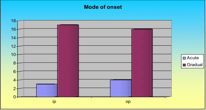
6. Onset:
Mostly onset is insidious. 75 % - insidious onset. 15 % - Acute onset. 10 % – Sub acute onset.
IMMUNOPATHOLOGY:
CLINICAL FEATURES
:Patients often present with constitutional complaints including malaise, fever,
fatigue, weight loss and myalgias. Most patients with the disease have an insidious
onset. It may begin with systemic features, such as fever, malaise, arthralgias and weakness, before the appearance of joint inflammation and swelling.
JOINT FEATURES
:
RA is typically a distal, symmetrical, small joint polyarthritis involving
proximal interphalangeal and metacarpophalangeal joints of the hands, wrist,
metatarsophalangeal joints, ankles, knees and cervical spine. The shoulders,
elbows and hips are less frequently involved, but can be a major source of morbidity.
Any synovial joint in the body may be affected. In addition periarticular synovial structures such as bursae and tendon sheaths are commonly inflammed.
Most common symptoms described by patients are pain and pronounced
stiffness. The later frequently exhibits a diurnal rhythm, worse on rising in the morning
and then recurring towards the evening, perhaps reflecting the diurnal variation in plasma cortisol levels. The affected joints are frequently tender, swollen and warm
and there may be limitation of both active and passive movements. Progressive
UPPER LIMBS
:
Hands and wrists:
Early in the disease there may be soft tissue swelling around the affected joints. Involvement of the proximal interphalangeal joints gives a spindle shaped appearance
to the fingers and soft tissue swelling can be observed over the ulnar styloid and in the
2nd and 3rd metacarpophalangeal joints (MCP). Tenosynovitis of the long flexor tendons in the palm of the hand may exacerbate stiffness of the fingers and cause “Trigger finger”. Similar synovitis of the wrist within the flexor retinaculam may cause
compression of the median nerve with the typical features of Carpal Tunnel
Syndrome. Persistent synovitis with erosion of the articular surfaces, weakening of the
joint capsules and muscle weakness, with or without tendon rupture will inevitably lead to deformities.
Ulnar deviation and subluxation of the fingers:
Occurs as a result of instability of the metacarpophalangeal joints. The fingers may tend to drift in an ulnar direction because of the ulnar vector of the action of both flexor and extensor finger tendons.
Swan Neck deformity:
Develops from hyper extension of the proximal interphalangeal joints in conjunction with flexion of distal interphalangeal joints with subsequent contracture of the intrinsic muscles which become extensors rather than flexors of the proximal interphalangeal joints.
Boutonniere (Button-Hole) deformity:
Results from flexion contractures of proximal interphalangeal joints
associated with hyper extension of distal interphalangeal joints. A similar process
at the carpometacarpal joint of the thumb may give rise to the Z-thumb deformity.
Can be detected when weakening of the distal radio ulnar ligament by synovitis allows the distal ulna to migrate dorsally so that it overrides the radius. The ulna can be depressed by pressure like a piano key.
Elbows and Shoulders:
Involvement of the elbows is less common than of the wrist but severe
destruction may occur, leading to pronounced deformity and disability. There may be inflammation of the subacromial bursae or supraspinatous tendon in addition to glenohumeral joint synovitis, producing a typical painful arc syndrome.
LOWER LIMBS:
Feet and Ankles:
Active synovitis of the metatarso phalangeal joints leads to spreading of fore foot. Subluxation of metatarsal heads into the soles results in cockup and valgus
deformities causing painful walking and difficulty with foot wear. Pain arises in the ball of foot (metatarsalgia). .
Knee :
Involvement of knee is an important cause of disability from an early stage of disease. Synovial proliferation is usually most obvious in the supra patella pouch and there may be pronounced wasting of the quadriceps as a result of reflex muscle
inhibition. Synovial effusion typically produces posterior knee pain in the early stages by stretching the posterior capsule of the joint. This may lead to the development of a popliteal cyst (Baker’s cyst). Valgus deformity of the knee is usual consequence of loading.
Hip:
Involvement of hip is uncommon. Pain is usually present in the groin; buttock and abduction of hip are reduced ultimately leading to fixed flexion deformity of the joint.
Spinal involvement is limited to upper cervical articulation. Neckpain and stiffness are common and leads to erosion of bones and ligaments in cervical spine. Vertebral arteries may also be compressed resulting in vertebro basilar insufficiency with vertigo or syncope especially on downward gaze. The risk of cord compression is greatest in those with a subluxation exceeding 8mm and there is also vertical subluxation of the atlanto axial joint.
Symptoms suggestive of atlantoaxial disease include high cervical pain radiating to the occiput and temporal regions, exacerbated by neck movements.
x Brisk tendon jerk.
x Positive Hoffman sign.
x Upgoing plantar response.
x Loss of proprioception.
x Vibration sense indicates damage to posterior column.
OTHER JOINTS:
Hoarseness of the voice may occasionally be caused by effusion within the cricoarytenoid joints.
EXTRA ARTICULAR FEATURES
:
These tend to be more numerous and severe in those with high titers of
rheumatoid factor in blood. Three major pathological phenomena dominate the disease. ¾ Inflammation of membranes
¾ Nodule formation ¾ Vasculitis.
A. RHEUMATOID NODULES:
Subcutaneous and intracutaneous nodules are the hall mark of the disease in ¼ of the patients. They are firm, non-tender swellings that occur on the extensor surface of the fore arm and olecranon sites, where repeated minor trauma could initiate their formation. They may also develop in many other tissues including eye (Scleromalacia), pleura, pericardium, parenchyma of lungs and heart.
B. HAEMATOLOGICAL MANIFESTATION:
Anaemia:
Moderate normochromic normocytic anaemia is a finding in active RA. Factors that are related to the inflammatory process probably contribute to this anaemia. There may be ineffective erythropoiesis and red blood cell survival is reduced. Iron binding capacity is typically reduced in active rheumatoid arthritis.
Thrombocytosis, Leukopenia is a finding in patients with Felty’s Syndrome.
Vasculitis:
Intimal hyperplasia of the small terminal digital vessels causes very limited cutaneous lesions (Nail fold infarcts, rashes, splinter haemorrhages). In contrast severe life threatening tissue infection may develop when there is involvement of large blood vessels by leucocytoclastic or necrotizing vasculitis.
C. LUNG INVOLVEMENT:
The fluid has more protein, low glucose; low complement levels and is typically positive for RA factor.
2. Nodules (Caplan syndrome):
More in upper zone than lower zones. Cavitation may occasionally lead to haemoptysis.
3. Pulmonary fibrosis:
It causes progressive dyspnoea, clubbing of fingers, fine late inspiratory crepitations.
4. Obliterative Bronchiolitis:
Acute onset of breathlessness. Many patients have evidence of airway obstruction. Bronchiectasis also appears to be more common.
D. CARDIAC INVOLVEMENT:
Pericardial effusion can be found by ultra-sonography in patients with seropositive nodular disease. Constrictive pericarditis is more common and presents with dyspnoea, right side heart failure and peripheral oedema.
Valvulitis: Granulomatous thickening of the cusps of the aortic valve occurs
more frequently than in the mitral valve, rarely producing incompetence.
E. EYE INVOLVEMENT:
Common in RA and may be due to localized tissue involvement.
Episcleritis:
Appears as a raised lesion in the anterior sclera with hyperaemia of the deeper layers.
scleritis:
Is less common and may leads to progressive thinning of the sclera (Scleromalacia) and even perforation.
Keratoconjunctivitis sicca (dry eyes) due to secondary Sjogren’s syndrome. Corneal melting is a rare manifestation. Clinical features are pain, redness and
blurred vision with corneal thinning.
Peripheral neuropathies can be produced by proliferating synovium causing compression of nerves. A mild glove and stocking sensory neuropathy is relatively common in RA.
G. MUSCLE INVOLVEMENT:
Is attributed to the reflex inhibition and wasting resulting from severe joint pain.
H. BONE INVOLVEMENT:
Juxta – articular osteoporosis is an early feature. A small proportion of patients may develop osteomalacia.
I. FELTY’S SYNDROME:
Lymphadenopathy is common. It is more obvious in patients with Felty syndrome (Rheumatoid arthritis, Splenomegaly, Leucopenia). Other features include anaemia, thrombocytopenia, persistent vasculitic leg ulceration, cutaneous
pigmentation, weight loss and recurrent infection.
DIAGNOSIS:
The American College of Rheumatology (1988 revised) developed the following criteria for the classification of rheumatoid arthritis.
1. Morning Stiffness: This occurs in and around the joints and lasts at least 1 hour
before maximal improvement.
2. Arthritis of 3 or more joint areas: At least 3 joint areas simultaneously have soft
tissue swelling or fluid (not bony overgrowth) observed by a physician.
3. Arthritis of hand joints of at least one area swollen in a wrist, MCP, or PIP joint.
4. Symmetric arthritis with simultaneous involvement of the same joint areas on both
sides of the body. Bilateral involvement of PIPs, MCPs, and MTPs is acceptable without absolute symmetry.
5. Rheumatoid nodules: Subcutaneous nodules are present over bony prominences or
extensor surfaces or in juxta-articular regions.
6. Serum Rheumatoid Factor: Abnormal amounts of serum RF are demonstrated by
7. Radiographic changes typical of RA on posteroanterior hand and wrist radiographs, which must include erosions or unequivocal bony decalcification localized in or most marked adjacent to the involved joints. Osteoarthritic changes alone do not qualify.
A patient can be classified as having RA if 4 of 7 criteria are present. Criteria 1- 4 must be present for at least 6 weeks, and a physician must observe criteria 2 - 5.
COMPLICATIONS:
RA is not fatal, but complications of the disease may shorten survival by a few years in some individuals. In general, RA is progressive and cannot be cured. In some, the disease gradually becomes less aggressive and symptoms may even improve. However, if bone and ligament destruction and any deformities have occurred, the effects are permanent. According to one survey, 70% of patients with RA believe that the disease prevents them from living a fully productive life.
Lymphoma and other cancers: Alterations in the immune system associated with RA
may play a role in the higher risk for lymphoma observed in patients with RA. Aggressive treatments for RA that suppress the immune system may help preventing this cancer, but more research is needed to evaluate this possibility. Other cancers that may occur with increased frequency in patients with RA include prostate and lung cancers.
Macrophage Activation Syndrome: This is a life-threatening complication of RA and
requires immediate treatment. Patients should be aware of symptoms, which include persistent fever, weakness, drowsiness, and lethargy.
PROGNOSIS:
The following factors at presentation are associated with a poor prognosis. ¾ Higher baseline disability.
¾ Female gender.
¾ Involvement of Metatarsophalangeal joints. ¾ Positive Rheumatoid Factor.
¾ Disease that remains persistantly acting for more than a year is likely to lead to joint deformities and disability around 80%.
INVESTIGATIONS:
No pathognomonic test is available to confirm the diagnosis of RA; instead, the diagnosis is made using clinical, laboratory, and imaging features.
HAEMATOLOGICAL:
1. Normochromic normocytic anaemia is frequently present in active RA. 2. The WBC count is usually normal, but a mild leucocytosis may be present. 3. Eosinophilia when present usually reflects severe systemic disease.
4. The Erythrocyte Sedimentation Rate is increased in nearly all patients with active RA.
5. The levels of acute phase reactants including Ceruloplasmin and C-reactive protein are also elevated.
6. Increased IgG, IgM, IgA and gamma globulin.
IMMUNOLOGICAL:
1. Rheumatoid factor (RF):
The presence of rheumatoid factor does not establish the diagnosis of RA, but can be of prognostic significance. RA factor are auto antibodies reactive with the Fc position of IgG. Presence of RF can be detected by several tests such as Rose Waaler, Latex fixation test and other slide agglutination test. The test can be employed to confirm a diagnosis in individuals with suggestive clinical presentation and if present in high titer, to designate patients at risk for severe systemic disease. Other conditions associated with RA are SLE, chronic liver disease, sarcoidosis, interstitial pulmonary fibrosis, hepatitis B, tuberculosis, syphilis and malaria.
2. Antinuclear antibodies: These are present in approximately 40% of patients
3. Newer antibodies (anti-CCP): Recent studies of antibodies to cyclic
citrullinated peptide suggest a sensitivity and specificity equal to Rheumatoid factor.
SYNOVIAL FLUID ANALYSIS:
x Colour - Yellow
x Clarity - Cloudt
x Viscosity - Reduced
x Mucin clot - Poor
x WBC - > 3000 μL to 50000 μL
x Total protein - >3 gm.Microscopic feature - RA cell.
x Polymorpho neuclear leucocyte - >70.
SYNOVIAL BIOPSY:
Villus formation with thickening of synovial layer and infiltration with abnormal cells.
RADIOGRAPHIC EVALUATION:
¾ Diagnosis is supported by a characteristic pattern of abnormalities including tendency towards symmetric involvement.
¾ Soft tissue changes, juxto-articular osteoporosis may become apparent within weeks of onset.
¾ Loss of articular cartilage and bone erosion develop after months of sustained activities. Joint space changes, alignment, deformities, subluxation, bony ankylosis develops in the late stage.
ARTHROSCOPY:
In acute RA synovium is edematous, diffusely erythematous and friable. In more chronic condition it becomes thickened.
MRI: Used in patients with abnormalities of the cervical spine.
This allows recognition of effusions in joints that are not easily accessible. High resolution ultrasound images may allow visualization of tendon sheaths, changes and degree of vascularization of the synovial membrane and even erosions.
BONE SCANNING:
Findings may help to distinguish inflammatory from non-inflammatory changes in patients with minimal swelling.
DENSITOMETRY: Findings are useful to diagnose changes in bone mineral density
indicative of osteoporosis.
OTHER TESTS: HLA – DR4 may constitute a helpful marker in early
undifferentiated arthritis.
OTHER PROCEDURES: Joint aspiration and biopsies (skin, nerve, rectum and
kidney) maybe considered if vasculitis is suggested.
DIFFERENTIAL DIAGNOSIS
:
1. Acute viral arthritis (Rubella, Hepatitis B, Parvovirus) 2. Bacterial endocarditis
3. Acute Rheumatic fever 4. Sarcoidosis
5. Reactive arthritis (Reiter’s disease) 6. Psoriatic arthritis
7. Inflammatory bowel disease 8. Systemic Lupus Erythematous 9. Sjogren’s syndrome
10. Polymyositis 11. Vasculitis syndrome 12. Polyarticular gout
Origin of disease (Uthira Vatha Suronitham)
Dietary Changes
Socio economical factor
Seasonal
Changes
Genetic
Changes
Immoral
Activities
Stress like factors Immunological Changes
Reflected on
Soul – Mind – Body
Anatomical Pathology Patho physiology
Patho psychology
Affected seven
affected three humors affected Trigunam
Udal Thathukkal
Saaram Vatham
Sathuva
gunam
Senneer
Pitham Rajo
gunam
Oon
Kabham
Thamo
gunam
Kozhuppu
Enbu
Moolai
Sukkilam / Suronitham
Disease
Affect
ed Th
ree hu
mo
rs
Vatham
P
itham
Kabham
¾
Joint pain
1.Inflammatory
¾
E
ros
ion of bony margin
¾
Joint Swellin
g
C
h
anges in jo
ints
¾
Restriction of
m
ovements
¾
Joint stiffness
(Rednes
s, Warmth)
¾
C
h
an
ges in synovia
l fluid
¾
Res
triction of m
o
vements
2.
Lo
ss of app
et
ite
¾
C
o
n
stip
at
ion.
3
. Anem
ia.
Abaa
nan
Constipation.
Anal Pitham
Loss o
f ap
petite
Avalambagam
Dera
ngement of other
kabha types
Viyaanan
R
estriction of
Ranja
ga Pitham
Ana
em
ia
Kilethagam
Lo
ss o
f appeti
te
M
ovements
.
Samaanan
Vitiation of
Other
P
r
asaga P
itham
Pallor of skin
Santhigam
Joint swelling
Va
yus
a
n
d
Restriction
of
movem
ents.
Loss of ap
petite.
Kiru
k
a
ran
Lo
ss of appetite.
Affect
ed Ezhu
Ud
al Th
athu
kk
al
Saaram
Senneer
Oon
Ko
zh
uppu
Enbu
Loss of ap
petite
Decrease in Udal Van
mai
Musc
le
wasting
Restric
tion of Mov
ement
Joint s
w
ell
ing
(Anaem
ia,
increased
E
S
R
,
Join
t
sw
ell
ing
Bony
erosion
p
o
si
ti
v
e
R
A
f
ac
to
r)
D
ef
o
rm
it
y
Diagnosis (Envagai Thervugal)
Naa
Nira
m
Moz
hi Vizhi
Malam
Moothir
am
Na
adi
Sparisa
m
Coated tongue
Normal
Constipation
Vat
ha
Pitham
Pallor
of
tongue
Pitha
Vatham
Pallor of
s
k
in (A
naem
ia) 1.Pallor of C
onjunctiva
1.
Neerkur
i
- Normal
(Affected
Jo
ints)
Redness o
v
er the affected joints
2. Disturbance
in vis
ion
2.
Neik
u
ri
- Sn
ak
e
like
1.Swollen
Ring like, Pearl like
2. Tender a
n
d Warm
STAGES OF PATHOGENES IS PR OCESS Princi p a l targe t site - S y novium - Vasc uliti s (Incre as ed va sc u larity, e d em a, congesti
on, villous hy
pe rtro ph y other sites F ib ri noid deg ener atio
n / necrosis
(Rice bod ies) (Cor ne a, P leura, Pericardi u m ) Infla m m ator y
cell infiltration (Ly
m p hocy tes, m o nocy tes, m ac rop ha g es, p las m a cel ls) Fibroblast ic p roliferation (P allis ad ed hist iocy tes) Rheu m ato id granulation (“Pannus” invading con nective ti ssue by creepi n g substitu ti o n ) Capsu le and li g am en ts Tendons and aponeur osis Joints/articu lar cartilag e subcuta n eou s ti ssues/bursa e Edem a Tendon sy novia l sh eath infiltr atio n Pannus infi ltrat ion fro m sy novial Bursiti s Reflection in to subchon dral bon e Laxity Fibrosi s P
annus invasion of adjacent
te ndons Deprivati on of nutri tion Nodules (A sc hoff’s nodu le s) Subl uxation Contract ures Disl ocati on Severe deform ities Avascularity and attritio n Cellu lar deat h Att enuation or rupture Fragm
entation of artic
ular ca
rtila
ge
or erosi
on of pla
ques
of devitalized cartilage
Se co nda ry d efor m ity Tota l de struc ti on of ca rtil ag e A n ky lo si s D is o rg an iza tio n o f jo in t Sec onda ry d isloc
ation or juxta
-artic
ular pathol
o
gical fractur
AUTO ANTIBODIES IN RHEUMATOID ARTHRITIS:
Auto antibodies
Target
Possible pathogenic role % positiveRheumatoid factor Self IgG Generation of immune
complex
70
Anti nuclear
antibodies
Various nuclear
components
Reaction with dead cells 4 - 6
Antihistones Histones-I-IV Vasculitis and uveitis 10 - 30
Anti ribinuclear
protein
Ribonuclear
proteins
Polyclonal b-cell activation 30
Antikeratin Keratin Disease severity 95
Anti cardiocipin Diphosphate dyl
glycerol
Effect on PGI2 release,
platelet aggragation
10
Anti collagen Type II collagen Complement fixation joint 25
Antigliadin Intestinal mucosa Intestinal permeability to
bacterial antigen triggers
PINIYARI MURAIMAI
Piniyari muraimai is a method of diagnosing a disease.
“Pini” means = Disease
“Ari” means = Identify
“Muraimiai” means = Method
This is based upon three main principles and Envagai Thervugal. The three main principles are,
Poriyal arithal (Inspection)
Pulanal arithal ( Palpation)
Vinathal ( Interrogation)
Physicians ‘Pori’ and ‘Pulan’ are used as tools for examing the ‘Pori Pulan’ of the patient.
The above principles correspond to the methodology of
Inspection
Palpation and
Interrogation in modern medicine, in arrives a clinical diagnosis of the disease.
1. Poriyal arithal (Inspection)
Pori is considered as the five senses of perception namely,
1. Nose
2. Tongue
3. Eye
4. Skin
5. Ear
2. Pulanal arithal (Palpation)
Pulan are functions of five senses. They are,
1. Smell
2. Taste
3. Vision
5. Hearing.
Examinations of Pori and pulan of the patient by Pori and pulan of the physician.
3. Vinathal (Interrogation)
Vinathal is asking the information regarding the history of the disease, its clinical feature etc.,
from the patient or his close relatives who are taking care of him.
msitfs; : (Logics):
Alavaigal are used in clinical diagnose of a disease.
“msit fhz;ly; fUjy; ciu mghtk; nghUs; xg;ghnwd;gH
msit NkYk; xopGz;ik iajpfj; Njhbay; ngd ehd; fsit fhz;gH mitapw;wpd; NkYk; miwtH mitnay;yhk; msit fhz;ly; fUjy; ciu vd;Dk; %d;wpylq;fpLNk”
- rptrpj;jpahH msit vz;. 6
Alavai is divided into ten types, they are,
1. Observation – fhz;ly;
2. Inference – fUjy;
3. Authority, literature – ciu
4. Preception – mghtk;
5. Presumption – mUj;jg;gj;jp
6. Comparison – cgkhdk;
7. Inference by elimination – ghhpNr\k;
8. Probability – rk;gtk;
9. Tradition – IjPfk;
10. Natural Inference – ,ay;G
1. Kaandal ( Inspection by Siddha method):
Through ‘Kaandal’ the physician can directly see the patient, hear the patients all the complaints
and at length concludes a diagnosis.
2. Karuthal (Through Siddha Investigations)
Through Envagai thervu and Neerkuri as well as Neikuri, we can diagnose a disease by Karuthal.
3. Urai (Literature evidence of Siddha)
Comparative study of the signs and symptoms of the patient with the reference books and come to a
diagnosis.
Ennvagai thervuagal (Eight diagnostic tools)
Siddhars have developed a unique method of diagnosing the disease by “Ennvagai thervugal”.
“ehb ];ghprk; eh epwk; nkhop tpop
kyk; %j;jpukpit kUj;Jt uhAjk;”
- rpj;j kUj;Jt Neha;ehly; Neha;Kjy; ehly; jpul;L.
Hence the diagnosis is made by the following,
1. Naadi (Pulse)
2. Sparisam (Sensation to touch)
3. Naa (Tongue)
4. Niram (Colour)
5. Mozhi (Voice)
6. Vizhi (Eyes)
7. Malam (Faeces)
8. Moothiram (Urine)
The specialty of eight tools of diagnosis is mentioned in the following verses also,
‘njhLf;fYw;w ml;ltpjg; gupl;ir jd;id
tFf;fhpa Njfnkdj; njhl;Lg; ghU rfpf;fhpa kyj;ijg; ghh; ryj;ijg; ghU rhu;e;j tpopjidg; ghh;j;J njspthaf; fhNz.”
- mfj;jpah; itj;jpa ty;yhjp 600.
1. Naadi (Pulse):
The science of pulse forms a very important branch in the siddha system of medicine. Naadi is
the seat anchor of energy. It is the binding force between soul and body. The pulse-waves as felt on
the radial artery, one inch from the wrist by means of palpation with the tip of index, middle and ring
finger corresponds to Vatham, Pitham and Kabham. They exist in the ratio of 1:1/2:1/4 normally.
Derangement of this ratio leads to various disease entities.
In Udhiravatha Suronitham, Vatha pitha naadi, Pitha vatha naadi and Vatha kaba naadi are
commonly seen.
2. Sparisam (Skin):
Skin examination can be made by inspection and palpation (touch). It reveals about the
warmth/chillness, dry/weeping skin, rough/smooth, soft/hard, tenderness, presence of ulcers, fissures,
swelling, wrinkles etc.
In Udhiravatha Suronitham, the affected part feels warm with redness, swelling, tenderness and
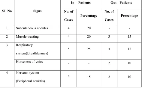
subcutaneous nodules can be noticed.
3. Naa (Tongue examination):
The colour, character and condition of tongue change according to changes in mukkutram.
In Udhiravatha Suronitham, few cases had coated tongue. In few cases that were anaemic, the
tongue was pale and some were have glossy.
4. Niram (Colour):
Signs of different complexions in Vatham, Pitham, Kabham and Thontha thegis, cyanosis,
pallor, yellowish discolouration can be studied by means of niram.
In Udhiravatha Suronitham, the patient is of mixed complexion and the affected parts get
5. Mozhi (Speech):
It constitutes high, low pitched voice, slurring and incoherent speech, nasal speech, hoarseness
of voice etc.
In Udhiravatha Suronitham, the speech is normal in most of the patients. Only two patients had
horseness of voice.
6. Vizhi (Eye):
Both motor and sensory disturbance of eye are noticed. Burning sensation, redness of eyes,
paleness, excessive lacrimation, swelling, sunken eyes, corneal ulcers, other diseased conditions
should be noted.
In Udhiravatha Suronitham, if the patient is anaemic, pallor of conjunctiva will be seen.
7. Malam (Stools):
Vatha type : Black coloured stools with constipation.
Pitha type : Loose stools with yellowish red colour.
Kabha type : White coloured stools with mucus.
Thontha type : Stools possess some of the features of
two thodams.
In Udhira Vatha Suronitham, constipation is noted in few patients
8. Moothiram (Urine):
It includes Neerkuri and Neikuri.
Neerkuri:
¾ Niram - Indicates the colour of urine voided.
¾ Manan - Indicates the smell of urine voided.
¾ Edai - Indicates the specific gravity of urine voided.
¾ Nurai - Indicates the frothy nature of urine voided.
¾ Enjal - Indicates the quantity of urine voided.
In addition, the frequency of micturation and sedimentations are noted.