A Dissertation on
"CALCULOUS DISEASE OF THE URINARY TRACT"
- A CLINICAL AND EPIDEMIOLOGICAL STUDY
Dissertation submitted to
THE TAMIL NADU DR.M.G.R.MEDICAL UNIVERSITY CHENNAI.
with fulfillment of the regulations
for the award of the degree of
M.S. (General Surgery)
Branch - I
KILPAUK MEDICAL COLLEGE,
CHENNAI.
CERTIFICATE
This is to certify that this dissertation in "CALCULOUS DISEASE OF THE URINARY TRACT" - A CLINICAL AND EPIDEMIO -LOGICAL STUDY is a work done by Dr.KUMARESH .T.S. under my guidance during the period 2003 - 2006. This has been submitted in partial
fulfillment of the award of M.S. Degree in General Surgery (Branch - I) by the
Tamil Nadu Dr.M.G.R. Medical University, Chennai.
Prof.Dr.THIRUNARAYANAN, M.S.,F.I.C.S.,
Professor and Head of Department Departmnt of Surgery
Government Royapettah Hospital, Chennai.
Prof.Dr.G.GUNASEELAN, M.S.,
Addl. Professor of Surgery Government Kilpauk Medical College & Hospital,
ACKNOWLEDGEMENT
It is my immense pleasure to thank the Dean Prof.Dr.THIAGAVALLI KIRUBAKARAN,M.D., of Kilpauk Medical College and Hospital for kindly permitting me to conduct this study in surgical department of Government Kilpauk Medical College and Hospital, Chennai.
My heartful gratitude to Prof.Dr.THIRUNARAYANAN, M.S., Head of the Department of General Surgery for his esteemed guidance and valuable suggestions. It is my privileged duty to profusely thank my teacher - guide and mentor Prof.Dr.G.GUNASEELAN, M.S., under whom I have the great honour to work as a post graduate student.
I express my sincere and heartful thanks to Prof.Dr.P.VAIRAVEL, M.S.,DGO.,M.Ch., Head of the Department or Urology, Kilpauk Medical College and Hospital for his timely suggestions and valuable guidance.
I am greatly indebted to my Unit Assistant Professors Dr.P.K.BASKARAN M.S., Dr.B.SATHYAPRIYA M.S., Dr.S.SURESH M.S., who have put in countless hours in guiding me in many aspects and also in honing my surgical skills.
My gratitude to Prof.Dr.P.KULOTHUNGAN, M.S., Prof.Dr.SHYAMALA, M.S., and Assistant Professors of all other units.
My gratitude to the urology department Assistant Professor Dr.P.LEELA KRISHNA, M.Ch., and postgraduates of urology department for immense help and guidance in every stage of the study.
CONTENTS
Sl.No. Topic Page No.
1. INTRODUCTION 1
2. AIMS OF THE STUDY 2
3. REVIEW OF LITERATURE 3
4. MATERIALS AND METHODS 28
5. PROFORMA UNDER STUDY 30
6. OBSERVATION 32
7. DISCUSSION 49
8. CONCLUSION 54
9. BIBLIOGRAPHY 56
ABBREVIATIONS
* RTA - Renal Tubular Acidosis
* IBD - Inflammatory Bowel Disease
* PUJ - Pelvi Ureteric Junction
* VUJ - Vesico Ureteric Junction
* U/3,M/3,L/3 - Upper Third, Middle Third, Lower Third
* IVU - Intra venous urography
* AUG - Ascending Urethrography
* KUB - Kidney, Ureter, Bladder
* LUTS - Lower urinary Tract Symptoms
* HUN - Hydro Uretero Nephrosis
* BPH - Benign Prostatic Hyperplasia
* GUTB- Genito Urinary Tuberclosis
* ANL - Anatrophic Nephro Lithotomy
* PCNL - Percutaneous Nephro Lithotripsy
* ESWL- Estracorporeal shock wave Lithotripsy
INTRODUCTION
Urolithiasis is one of the common clinical condition, a practising
clinician is likely to encounter in his practice. The renal calculus disease
patient may well test the diagnostic skill of a shrewed physician and
subsequent evaluation of the patient may be an acid test for his knowledge
and experience. Though the composition of the calculus and treatment
modalities might have been changed recently, archaeologic studies show the
urinary tract stone disease was an affliction of human earlier than 4800 B.C.
This is a study of 112 patients with calculous disease of the urinary
tract seen during the period of 3 years, from September 2003 to March 2006
with particular reference to epidemiological workup and clinical evaluation,
with thorough clinical examination, plain x-ray KUB, USG, IVU, Blood
Examination, Stone analysis and urine examination.
The present study deals with epidemiology of urolithiasis, their
distribution along urinary tract, various Investigations to confirm the
diagnosis and to arrive at a decision making process and finally the treatment
AIMS AND OBJECTIVES
* To evaluate all patients with calculus disease of the urinary tract with
special reference to the clinical epidemiology including :
a. Age and Sex
b. Heredity / metabolic diseases
c. Environment
d. Urinary infection
e. Dietary factors
f. Distribution of calculi within the urinary tract
* To clinically evaluate all patients with thorough clinical examination,
their common modes of presentation, associated urological problems
and by investigating them to arrive a decision making process.
* To study various modalities of treatment offered at different levels of
REVIEW OF LITERATURE
EPIDEMIOLOGY
The prevalence of urolithiasis is approximately 2.3% in the general
population and the estimated life time risk of developing a kidney stone is
about 12% for Indian males. Approximately 50% of patients with previous
urinary calculi have a recurrence within 10 years.
INTRINSIC FACTORS
Genetics
Urolithiasis requires a polygenic effect.
The hereditary diseases identified to be associated with stone disease
are cystinuria, renal tubular acidosis and familial idiopathic hypercalciuria.
Age & Sex
Peak age incidence occurs during 3rd to 5th decade. About 3 males
are affected for every female. However in cystinuria and
hyperparathyroidism, increased frequency in females have been noticed.
EXTRINSIC FACTORS
Geography
tropical areas.
Climatic
Acid stones are more common during summer and infection stones
(struvite) are more common during winter. Increased mean environmental
temperature is related to increased incidence of urinary calculi.
Water intake
Risk factors which promote crystallization of salts in a patient with
stone disease includes :
a. Low urinary volume
b. Low levels of zinc
e. Excessive water hardness.
Diet
Several studies have shown that high purine intake increases urinary
calcium, oxalate and uric acid excretion. Excess intake of vit.c produces
oxalate and increases the risk of stone production. Lack of fibre diet also
contributes for stone formation.
Occupation
Increased risk was observed in sedentary personnels, cooks and after
INHIBITORS OF CRYSTALLIZATION
Organic
Peptide, Alanine, High Molecular weight glycoproteins namely
Nephrocalcin and Tomm Horsfall protein and citrate.
Inorganic
Phosphates, zinc and magnesium.
PATHOPHYSIOLOGY
More than one or three mechanisms are likely to be active in stone
formation.
i. Possible presence of substance that promote crystallization.
ii. Possible relative lack of substance that inhibits crystallization.
iii. Possible excessive excretion or concentration of salts in the
urine, which leads to supersaturation of the crystallizing salt.
The greater the degree of supersaturation, the greater the rate of
growth of the calculi.
HYPERCALCIURIA
Accounts for 75 - 85% of urinary stones. Approximately one half of
demonstrates intermediate fragility to ESWL.
7/8th calcium stones contains calcium oxalate dihydrate. They may be
spiculated, dotted, mulberry or Jack stone appearance. The remaining 1/8th
of stones are composed of calcium phosphate (apatite) or calcium oxalate
monohydrate. These stones are denser and consequently, least responsive to
ESWL.
CAUSES
85% of calcium stones are due to idiopathic hypercalciuria and occurs
for more than half of patients with calcium oxalate stones. Hypercalciuria
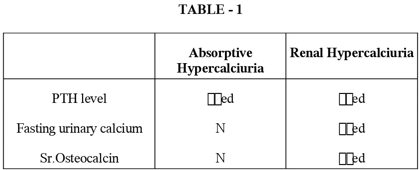
TABLE - 1
Absorptive
Hypercalciuria Renal Hypercalciuria
PTH level ed ed
Fasting urinary calcium N ed
Sr.Osteocalcin N ed
10-15% of calcium stones are due to hypercalcemic nephrolithiasis.
The causes include :
i. Hyperparathyroidism (5-10%)
ii. Renal tubular acidosis
iii. Malignancy associated hypercalcaemia
iv. Sarcoidosis (ed production of vit-D)
v. Immobilization (#, CVA)
vi. Glucocorticoid induced
vii. Pheochromocytoma
RENAL TUBULAR ACIDOSIS : (RTA)
Type I (Distal RTA) is associated with stone disease where as Type II
(Proximal RTA) and Type IV RTA does not predispose to stone formation.
In Type I (distal) RTA, there is decreased ability to lower urinary pH levels
(pH >6) and causes alkaline urine, hypercalciuria, phosphaturia.
HYPEROXALURIA
Most commonly occurs with malabsorption due to any causes, like
small bowel resection, jejuno - illeal bypass, vit-c overdose or renal failure.
Idiopathic hyperoxaluria is a rare disease.
HYPOCITRATURIA
Citrate complexes with calcium and inhibits spontaneous nucleation
and aggregation of calcium oxalate.
Acidosis is an important risk factor for hypocitraturia and occurs in
Distal RTA, Thiazide diuretics, Inflammatory bowel disease, (IBD) Chronic
diarrhoea, etc.
HYPOMAGNESIURIA
TRIPLE PHOSPHATE (STRUVITE) STONES
Constitutes 10 - 20% of urolithiasis. They enlarge and branch into
calyces to form staghorn calculus. They are caused by urea splitting bacteria
like Proteus, Klebsiella, Pseudommas which potentially lead to renal failure.
HYPERURICOSURIA
Uric acid stones constitutes 5 - 10% of renal stones. These are smooth
radiolucent stones on conventional X-rays but opaque on CT. The causes
include small bowel resection, gout, cell lysis due to leukemia, starvation
etc.
CYSTINE STONES
Cystinuria is an autosomal recessive disorder with defect in the
transmembrane cystine transport leading to increased urinary excretion of
cystine, ornithine, arginine and lysine.
Diagnosis is by cyanide nitroprusside calorimetric test.
CLINICAL PRESENTATION
ACUTE STONE EPISODE
A urinary calculus usually announces its presence with an acute
become trapped in some segment of the upper urinary tract.
First, stones may become impacted in any calyx of the upper urinary
tract. Individual calyces may, therefore become distended and painful to
create hematuria.
Second area in which a calculus may become impacted is at the PUJ
(Pelviureteric junction). It is here that the relatively large diameter of the
renal pelvis (1 cm) abruptly decreases to that of ureter (2 - 3mm).
Third area of impaction is at or near the pelvic brim, where the ureter
begins to arch over the illiac vessels posteriorly into true pelvis.
Fourth area, especially in females, is the posterior pelvis, where the
ureter is crossed anteriorly by the pelvic blood vessels and by the broad
ligament.
Finally, the most constricted area through which the urinary calculus
must pass is the ureterovesical jn (VUJ). These normal anatomic variations
probably explains the frequency with which calculi become impacted in
certain portions of the urinary tract than in others. To become impacted, the
calculi usually must have one diameter in excess of 2mm. If the smaller
PHYSICAL SIGNS
The patient almost always have moving pain with irritation. They
rarely can find comfort in any given position. Fever is rare unless urinary
infection occurs along with the calculus. Acute hydronephrotic kidney may
be palpable.
Microscopic or gross hematuria is frequently present in patients with
acute colic. Some 80% of patients do not demonstrate hematuria especially if
the calculus has caused complete obstruction. When significant number of
pus cells are present in the urine, however, a thorough search for infection
should be made.
PATIENT EVALUATION
URINALYSIS
Urine is examined for hematuria and pyuria, urine crystals may reveal
TABLE - 2
Sl.No. Crystal Shape under optical microscope
1. Ca oxalate monohydrate
(whewellite) Dumbell or hourglass
2. Ca oxalate dihydrate
(Weddelite)
Bipyramidal
3. Ca Phosphate (apatite) Amorphous
4. Brushite Needle Shaped
5. Struvite Coffin lid
6. Cystine Benzene ring
PLAIN X-RAY (KUB)
Ninety Percent of stones in the urinary tract are radiopaque.
Calcium phosphate stones are most radiopaque, calcium oxalate are
almost opaque, magnesium - ammonium phosphate are less radiopaque.
Cystine calculi are partly radiodense because of their sulfur content. Only
calculi of relatively pure uric acid or xanthine stones are radiolucent. A small
ureteric calculus may be difficult to see due to gas and faeces or to confusion
with other opacities such as phleboliths and arterial calcification.
ULTRASOUND (KUB)
[image:17.595.116.503.96.391.2]appearances. The sensitivity of USG (KUB) in diagnosing a ureteric calculus
is only 43%.
Apart from delineating the presence, location and size of the calculus,
this modality of investigation also shows the state of renal substance and an
obstructed, hydronephrotic kidney.
INTRAVENOUS UROGRAPHY
The first indication for the presence of urolithiasis is delay in the
appearance of contrast medium in the nephrogram following its injection.
Films at 20, 30 and 60 min shows better localisation and presence of
calculus. It is essential to ensure that renal function is not significantly
impaired (by serum creatinine concentration) before doing urogram. If there
is no visualisation, retrograde pyelography may be indicated.
CT SCAN
Non-contrast Helical / spiral CT gains more importance in detecting
radiolucent stones. All stones, regardless of composition are visualised on
CT scan with the exception of small percentage of Indinavir stones from
patients under Antiretroviral treatment.
RETROGRADE PYELOGRAPHY
Where other techniques are not successful in locating a calculus, this
needed when there is no function in the affected kidney.
SPECIFIC LOCATION OF CALCULI
Renal calculi
Stones >1 cm donot pass spontaneously and they usually occupy the
renal pelvis and calyces, and gets impacted at the PUJ. When all forms of
renal stones are considered, the incidence in men and women are
approximately equal, but calcium containing stones occur in men 3 times
more often than in women.
Ureteral calculi
a. Site of Origin
Usually originates in the kidney and then passes into the ureter.
Because of the smooth mucosal lining of the ureter and is constantly bathed
with urine, primary ureteral stones are rare. They may be formed primarily in
association with ureterocele, neoplasms, ectopic ureter, saculations etc.
b. Site of impaction
Most common site is vesico ureteric junction (VUJ) followed by pelvi
ureteric junction (PUJ). Other places of impaction were already discussed.
c. Size, weight and shape
Stone that weigh more than 0.1 gm have a diameter of >1 cm and associated
with urinary infection are not as likely to pass spontaneously.
d. Laterality
Ureteral calculi are equally frequent on the left and right sides,
although in certain patients stone formation seems to be limited to one side
(ie left). Several comparitive studies shows that there is a slight
preponderance to left side.
3. VESICAL CALCULI
a. Age
An etiological factor, incidence in calculi of the bladder varies in
difference parts of the world. Previously belived that the disease was largely
limited to children, but now increased incidence is being observed in adults
also.
b. Sex
Vesical calculus is predominantly a disease of males of all ages in all
races. Increasing incidence noticed in men > 50 years. Factors that give rise
to retention of urine such as stricture urethra, prostatic hypertrophy,
diverticulum of the bladder, cystoceles and neurogenic bladder are
associated with formation of struvite stones. Other bladder stones are formed
Usually a single stone is observed in the bladder, but in the prsence of
retained urine, multiple stones, 2 or 3 to 100 may be formed. Multiple stones
usually occur when there is a diverticulum of the bladder.
URETHRAL CALCULI
Constitutes <1% of all urinary stone disease.
Majority of the urethral calculi in the male consist of stones expelled
from the bladder into the urethra. Rarely primary urethral stones are formed
when stricture or diverticulum is present.
A stone the progresses through the normal urethra may get arrested in
prostatic urethra, the bulb, fossa navicularis or the external meatus.
DIAGNOSTIC AND TREATMENT DECISION PROCESS
After diagnosing a urolith, first assessment is of the degree of
seriousness of the disease process.
INDICATIONS FOR HOSPITALISATION
* In patients with symptoms not controlled with oral medications.
* In the presence of calculus anuria, usually seen in patients with
solitary kidney.
ANALYSIS OF URINARY STONES
Following treatment of acute phase, stone recovery is of paramount
importance. Most medical treatment for stone disease is now based on
analysis of calculi and decisions about proper procedures for treatment
requires knowledge of stone composition.
METHODS OF STONE ANALYSIS
1. Chemical
* Qualitative spot test
* Quantative analysis
* Chromatographic and autoanalysed methods
2. Optical
Binocular dissection microscopy with petrography
(Polarisation)
3. Instrumental
* Radiographic crystallography
* Thermo analytic
* Scanning Electron Microscopy
Although many types of stone analysis have been proposed, the most
practical type is the chemical analysis. For the practicing surgeon without
access to large analytic laboratories, the most useful methods, are chemical
analysis and petrographic methods through the polarising microscope.
Because it is relatively simple to instruct laboratory personnels in these
techniques, almost any small hospital laboratory or large clinic have the
[image:23.595.137.483.385.558.2]ability to analyse the urinary calculi.
TABLE - 3
NORMAL 24 hrs URINE VALUE (mgm)
Biochem
Component Males Females
Calcium <300 <250
Oxalate <50 <50
Uric acid <800 <750
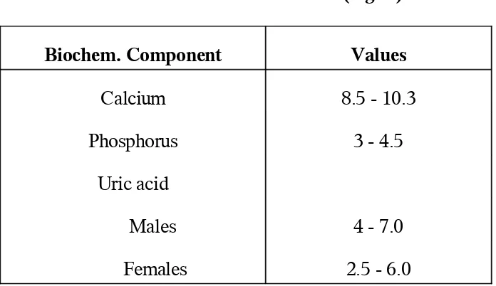
TABLE - 4
NORMAL SERUM LEVELS (mg/dl)
Biochem. Component Values
Calcium 8.5 - 10.3
Phosphorus 3 - 4.5
Uric acid
Males 4 - 7.0
TREATMENT OF UROLITHIASIS
Can be divided into
* Treatment of acute episode
* Interval treatment
* Prevention of recurrences or new stone formation.
TREATMENT OF THE ACUTE EPISODE
EXPECTANT TREATMENT
If the greatest diameter of the stone is <4 mm, spontaneous passage is
very likely, but surface characteristics of the stone may be as important as
size.
The first priority is to relieve pain, NSAID's is most commonly used
where morphine is the choice when there are containdications to NSAID's
are present like, pregnancy, Asthma and peptic ulcer.
The common measure of forced diuresis in the acute situation may be
unhelpful, as an increase in diuresis may decrease peristalsis and hinder the
Indications for Emergency intervention
1. Significant obstruction with infection
2. Intractable pain
3. Progressive renal deterioration
4. Anuria due to obstruction in a solitary kidney
5. Pyelonephritis (without significant obstruction) not responding
to antibiotic treatment.
THE OPTIONS FOR EMERGENCY TREATMENT ARE :
1. Nephrostomy
2. Stenting
3. Uterteroscopic stone extraction
4. ESWL, if on site.
INTERVAL TREATMENT
a. Surgical Treatment
Surgery forms the mainstay of treatment of calculous disease of the
urinary tract both in the acute phase and as interval treatment, after an acute
episode.
Various modalities of surgical treatment includes, percutaneous
nephrolithotomy (PCNL), extracorporeal shock wave lithotripsy (ESWL),
ESWL
Indications include,
1. Renal Calculi
Stones <2 cm diameter - 90% chances of fragmentation and
clearance.
Stones in the lower pole / calyceal diverticular region give
lower success rate.
2. Ureteral calculi
ESWL is recommended as the first line treatment for most
patients with stones 1 cm or less in the proximal ureter.
ESWL and ureterorenoscopy are equally acceptable choices for
stone of this size in the distal ureter.
3. Bladder Calculi
Can be treated with the patient prone.
CONTRAINDICATIONS TO ESWL
Absolute : Pregnancy
Uncontrolled coagulopathy
Urinary tract obstruction distal to the stone
Urinary tract infection with fever.
Relative : Urinary tract infection
Distal ureteric calculi in women of child - bearing
age.
Complications of ESWL
1. Stein strasse "stone street"
2. Bleeding
3. Gastrointestinal side-effects, like pancreatitis, elevation of
hepatic enzymes, incidental fragmentation of gall stones,
causing biliary colic.
4. Mortality (0.02%)
Ureterorenoscopy (URS)
The indications are :
* Ureteric calculi that cannot be visualised for ESWL or which
have not responded to ESWL.
* Renal calculi not responding to ESWL or residual stones after
percutaneous treatment.
* Radiolucent stones or filling defects which need to be
inspected.
Stenting with a double pigtail (JJ) stent is generally recommended
after ureteroscopic treatment.
PERCUTANEOUS NEPHROLITHOTOMY (PCNL)
PCNL was the first key hole surgery.
The absolute indication are :
* Staghorn calculi and large (> 3 cm) renal calculi
* Failed ESWL for stones < 3 cm
* Cystine stones refractory to ESWL
* An infected obstructed system - PCNL is done in 2 stages
insertion of a neophrostomy followed by nephrolithotomy after
The relative indications for PCNL
* Horse shoe kidney - where imaging is difficult.
* Morbidly obese patients where stone imaging and the weight of
the patient may be problems.
* Upper and midureteric calculi - with a dilated system above the
stone.
Complications of Access include
Haemorrhage, pneumothorax, hydrothorax and injury to the
neighbouring viscera.
The risk of fluid absorption and sepsis increases after this procedure
Open Stone surgery
In developing countries, open stone surgery still has a significant role.
The procedures performed are pyelolithotomy, anatrophic and radial
nephrolithotomy and partial nephrectomy.
Non - Surgical Treatment
* Uric acid stones can be dissolved by a high fluid intake with
alkalinization of the urine. Allopurinol, an uricosuric agent
prevents reformation of uric acid stones.
* Cystine stones are dissolved by alkalization and with cystine
competing agents, such as D-penicillamine and α -
mercaptoproionyl glycine (MPG).
* Struvite stones may undergo partial or complete dissolution
after antimicrobial therapy.
Prevention of Recurrences
50 - 75% of patients have recurrences within 10 years of the first
episode. Ideally, prevention requires analysis of the chemical composition of
DIETARY ADVICE
1. Increase in the fluid intake to produce atleast 2 lts. of urine
output per day decreases the risk of stone formation.
2. Restriction of calcium is not advised as it increases oxalate
absorption.
3. High levels of dietary protein and sodium increases the risk of
calcium oxalate and uric acid stone recurrence.
MATERIALS AND METHODS
The present study involves a total of 112 patients with calculous
disease to the urinary tract observed during the period of 3 years, from
September 2003 to March 2006.
The study was conducted both by prospective as well as by
retrospective methods, by analysing the case sheets on a random basis.
All patients of prospective study were followedup in speciality
department, where they were subjected to a detailed clinical epidemiological
workup. Complete hemogram, urinalysis, urine culture, serum biochemistry,
were performed in all cases.
Chemical analysis of stones were performed in 39 cases
postoperatively after stone retrieval by open surgeries.
Radiological investigations included plain x-ray KUB, ultrasound IVU
series, CT, depending on clinical situation. Ultrasound KUB was often
performed while endoscopic procedures were usually undertaken for
EXCLUSION CRITERIA
* Paediatric urolithiasis was excluded as they were referred
directly to childrens hospital.
* Cases directly attending to the speciality outpatient department
PROFORMA UNDER STUDY
Name : Age :
Sex : MRD No :
Occupation : Residence :
Epidemiological Factors :
1. Heredity : Yes / No
2. Metabolic : Yes / No
3. Climatic Influence : Mar to Jul
Aug to Jan
4. Dietary factors : Low fluid intake
Less fibre diet
5. Urinary infection :
+/-Organism on C/S. Distribution of Calculi :
1. Upper urinary tract / lower urinary tract 2. Site / Side of Stone impaction
Clinical Presentation :
Pain (Colic), Palpable mass, Hematuria, Fever, LUTS.
HUN / BPH / GUTB / Stricture Urethra, etc.
Method of Diagnosis :
USG, X-ray (KUB), IVU, AUG, CT Scan, Sr.Biochemistry and 24 hrs urinalysis.
Mode of Treatment :
Endoscopic / open stone surgery
OBSERVATIONS
1. AGE AND SEX INCIDENCE
a. UPPER URINARY TRACT STONE DISEASE
(Renal, Ureteral, Multiple Stones)
AGE
* Max. Age Incidence (37%) for upper urinary tract stone
disease was observed in between 30 - 40 years.
* 75% of total cases observed were found during 2nd and 5th
decade.
* Only 5 cases were found <20 years.
SEX
* Male : Female ratio observed is 1.5 :1.
Age (Yrs) Male % Female % Total %
11 - 20 2 4 3 8 5 6
21 - 30 12 24 7 21 19 22
31 - 40 18 35 13 38 31 37
41 - 50 6 12 8 24 14 16
51 - 60 9 17 - - 9 11
61 - 70 3 6 2 6 5 6
> 70 1 2 1 3 2 2
Total 51 100 34 100 85 100
b. LOWER URINARY TRACT STONE DISEASE
(Vesical, Urethral Stones)
AGE
* Max. Age Incidence (52%) for vesical and urethral stones was
observed to be within 40 - 60 years.
* 20% of cases were found in between 20 - 30 years.
SEX
* Of the 27 cases of lower urinary stones, only one female
patient was found to have bladder stone.
* 96% male predominance observed.
Age (Yrs) Male % Female % Total %
11 - 20 1 4 - - 1 4
21 - 30 5 19 0 0 5 19
31 - 40 4 15 - - 4 15
41 - 50 6 23 1 100 7 26
51 - 60 7 27 - - 7 26
61 - 70 1 4 - - 1 3
> 70 2 8 - - 2 7
2a. DISTRIBUTION OF CALCULI
(In General)
* Of the 112 cases studied, it was observed ureteral stones were
the commonest (46 cases).
* 25 cases of Renal stones and 24 cases of bladder stones
observed.
* 3 cases of urethral stones found.
* Stones at multiple sites accounts 14 cases
Site No %
Renal 25 22
Ureteral 46 41
Vesical 24 21
Urethral 3 3
Multiple sites 14 13
2b. DISTRIBUTION OF RENAL CALCULI
* Most common site of stone impaction was the renal pelvis
(63%).
* Stones on right kidney were found to be more frequent (60%)
than the left.
* B/L renal stones were found in 6 cases of 35.
Site No %
Pelvis 22 63
Lower calyx 5 15
Middle calyx 4 11
Upper calyx 4 11
Total 35 100
Laterality No %
Right 21 60
Left 8 23
Bilateral 6 17
Total 35 100
2c. DISTRIBUTION OF URETERAL CALCULUS
ureteric Jn. were more common than upper ureteric stones.
* Stones Above pelvic brim constitutes 40% and Below pelvic
brim constitutes 60%.
* Ureteral stones were found to have almost equal incidence on
right (52%) as well as on left (45%) side.
* B/L ureteral stones found in 2 cases of 56.
Site No %
Above pelvic Brim (U/3 + M/3) 22 40
Below pelvic Brim (L/3 + VUJ) 34 60
Total 56 100
Laterality No %
Right Side 29 52
Left Side 25 45
Bilateral 2 3
2d. DISTRIBUTION OF CALCULI
* 3 cases of male urethral calculi observed during the study
where the site of blockage were one at fossa nasicularis and the
other 2 at posterior urethra.
* Stones at multiple sites observed includes
either B/L renal or B/L ureteral or
multiple renal on the same side or renal with ureteral or
vesical with urethral stones.
* One case of post ESWL steinstrasse was noticed.
3. EPIDEMIOLOGY
* Only 2 cases with metabolic disorder was found. Both cases
have primary hyperoxaluria.
* Climatic influence on stone disease found in 20% cases. 23
cases presented during summer season (March - June).
* Only 8 cases showed Diet / fluid influence on stone disease.
* Urinary infection was observed in 19% of cases. Patients with
multiple stones and Bladder stones were found to have urinary
infection.
Pseudomonas was found in 2 cases.
Factor No %
Hot climate 23 20
Diet / fluid 8 7
Metabolic 2 2
Urinary infection 21 19
Organism No %
E.coli 14 68
Klebsiella 3 14
Proteus 2 9
Pseudomonas 2 9
4a. CLINICAL PRESENTATION
* Pain was the commonest symptom observed (68%)
* Upper urinary stone disease presented with colic either renal or
ureteric as the most common symptom (87%).
* Lower urinary stone disease presented with either one or more
symptoms of LUTS like dysuria, hesistancy, terminal hematuria
and dribbling.
* Hematuria noticed in 15% cases and positive urine culture
found in 21 cases (18%).
* Palpable mass was found in 5 patients. Hydronephrosis (2
cases) and pyohephnosis (3 cases).
Symptoms / Signs Upper
Tract No % Lower Tract No % TotalNo %
Colic / pain 74 87 2 5 76 68
Fever 6 7 2 5 8 7
Hematuria 9 10 8 22 17 15
Palpable Mass 5 5 - - 5 4
LUTS 4 4 16 43 20 12
4b. ASSOCIATED UROLOGICAL PROBLEM
* It was observed hydroureteronephrosis was found in 9 cases
and were more frequently found in multiple site stones.
* Pyonephrosis found in 3 cases, of which 1 had staghorn
calculus.
* BPH was the most common associated urological problem with
lower urinary stones.
* One case of genitourinary tuberculosis with stone disease
noticed.
* 6 cases of stricture urethra associated with urethral and bladder
stones observed.
Associated problem No %
HUN / PUJ Obstruction 9 38
Pyonephrosis 3 12
BPH 8 33
GUTB 1 4
5. METHOD OF DIAGNOSIS
* USG (KUB) and x-ray (KUB) were done in almost all cases
and constitutes 64% and 87.5% respectively as a diagnostic
tool for stone disease.
* CT (Scan) helped in 10 cases of doubtful diagnosis and
planning treatment.
* IVU was done in 37 cases with normal urea, creatinine levels.
* Ascending urethrogram (AUG) was done in 6 cases of stricture
associated with vesical and urethral stones.
Investigation No %
X-ray (KUB) 98 87.5%
USG (KUB) 72 64%
IVU 37 33%
CT San 10 9%
Urinalysis for crystals -
* Serum biochemistry for renal parameters showed ed levels in
9 cases (obstructive uropathy).
* Sr. Biochemistry and 24 hours urine for calcium, phosphorus
and oxalate showed 2 case of primary hyperoxaluria.
6. TREATMENT MODALITY
a. RENAL CALCULUS
* One case of renal stone was managed conservatively during
this study.
* Pyelolithotomy was the most common procedure done for renal
stone obstructing at pelvis (68%).
* 3 cases of anatrophic nephrolithotomy done and
* 4 cases of nephrectomy done for obstructed and infected
system with non functioning kidney (with opposite side normal
functioning kidney).
* ESWL / PCNL was not done in any case.
Treatment No %
Conservative 1 4
Pyelolithotomy 17 68
Nephrolithotomy
(Anatrophic) ANL 3 12
ESWL / PCNL -
6b. URETERAL STONES
* Ureterorenoscopy (URS) with lithotripsy and DJ stenting was
done in most cases for ureteral stones (51%).
* Whereas Meatotomy was done in stones at VUJ (17%).
* Open stone surgery for larger (>1cm) ureteral stones, ie.
uretero lithotomy was done in 12 cases.
* Conservative treatment was observed in 3 cases only, as OP
cases were excluded in the study.
Treatment No %
Conservative 3 6
URS / Stenting 24 51
Meatotomy 8 17
6c. MULTIPLE SITE STONE DISEASE
* Obstructive uropathy and infection were more frequently
observed in multiple stone disease.
* URS / DJ stenting was observed in 43% and ureterolithotomy
was done in 2 cases (14%).
* One case of post ESWL stein strasse was noticed, for which
URS / DJ stenting was done.
* One case of nephrectomy done for non functioning calculous
pyonephrotic kidney.
Treatment No %
Conservative 2 14
URS / Stenting 6 43
Ureterolithotomy 2 14
Pyelolithotomy 1 7
Nephrectomy 1 7
Cystoscopy / vesicolithotripsy / O.I.U 2 14
• B/L stone disease were treated according to the site, side of
6d. VESICAL AND URETHRAL CALCULUS
* Cystoscopy and vesicolithotripsy was most commonly done for
bladder stones (63%).
* Vesicolithotripsy with TURP was done in 4 cases of bladder
stones associated with BPH.
* Vesicolithotomy was done in 5 cases.
* Urethral stones at posterior and bulbar urethra were retrived by
(O.I.U) optical internal urethrotomy to relieve stricture and
lithotripsy.
* One case of fossa navicularis stone was retrieved by
meatotomy.
BLADDER STONES
Treatment No %
Vesicolithotripsy 15 63
Vesicolithotomy 5 20
Vesicolithotripsy / TURP 4 17
URETHRAL STONES
O.I.U / Lithotripsy 2
7. STONE ANALYSIS
* Of the 112 cases studied, stone analysis were performed in 39
cases after postoperative stone retrieval.
* Done in, Renal stones - 23 cases
Ureteral stones - 10 cases and
Vesical stones - 6 cases
* The most common stone was found to be calcium oxalate with
phosphate.
* Struvite stones found in 3 cases of staghorn calculus.
Sl.No. Composition No. %
1. Pure calcium oxalate 2 5
2. Calcium oxalate and phosphate 34 87
3. Struvite (Mg-ammonium
phosphate)
3 8
4. Uric acid -
-DISCUSSION
* The observed age and sex incidence and the review of literature is
tabulated below.
Upper
Urinary Urinary Lower
Observed Literature Observed Literature
Age (Yrs) - Peak
Incidence 20 - 40 yrs 3rd - 5 th decade 40 - 60 years >50 years
Sex (M.F) 1.5 : 1 3:1 96% males Male
predominance
* Maximum age incidence for urinary calculi observed were in
accordance with literature, both for upper and lower urinary stone
disease.
* Sex prepondarance was in accordance with literature for vesical
stones where as increasing female preponderance was observed for
renal / ureteral stones.
* Of the 112 cases studied, only 5 cases were <20 years of which 2
cases had primary hyperoxaluria (metabolic), as paediatric urolithiasis
was excluded.
* The incidence of calculi were definitely high during hot environment, as evidenced by their presentation during summer season (20% of
* The role of fluid intake / dietic influence on renal calculus disease could not be evaluated as history of many Patients were highly unreliable.
* 19% cases have clinical evidence of urinary infection and the most
common organism grown in culture was E.coli. Those with multiple
site stones and bladder stones mostly had urinary infection.
* Regarding distribution of calculi along the urinary tract, ureteral
calculus were most frequent (41 cases) than renal calculus. Vesical
calculus (24 cases) showed increasing incidence as renal stones and 3
cases of urethral stones were observed during the study.
* Among the renal stones, renal pelvis was the commonest site of stone
impaction. 83% had single stone and 17% had multiple stones.
* There was a slight prepondance on right side both for renal stones
(60%) as well as ureteral stones (52%). Literature says several of the
comparative study shows left sided preponderance of ureteral calculi.
* The site of stone impaction for ureteral stones was found to be below
the pelvic brim (60%) more often than above pelvic brim (40%).
* Pain was the commonest clinical presentation, although small minority
of patients presented with hematuria (12%) and fever (6%). 4 patients
had palpable mass due to hydronephrosis. LUTS were the most
* Hydroureteronephosis was commonly associated with upper urinary
stones where as BPH (3%) and stricture urethra (20%) were
commonly associated with vesical / urethral stones. One case of
GUTB was observed during the study.
* Although the sensitivity of USG (KUB) is 43%, it was observed that
USG (KUB) was the most common method of stone diagnosis and it was performed along with X-ray (KUB) in almost all cases. IVP was
done in 37 cases and AUG was done in 6 cases. It was the combined methods of investigations helped not only in diagnosing the calculus
but also to plan for treatment process.
* Regarding the treatment, pyelolithotomy was the common procedure
done for renal stones obstructing the pelvis. Nephrectomy was
performed in 4 cases for obstructed and infected system with non
function kidney.
* Patients with renal stones <1cm and symptoms not subsided by
conservative methods were referred to centres for ESWL.
* URS (Ureterorenoscopy) / lithotripsy / DJ stenting was the most
common procedure performed for ureteral stones <1cm size.
* Open stone surgery for ureteral stone (uretero lithotomy) was done is
12 cases with larger stones (> 1 cm).
* Conservative treatment for ureteric colic was observed only in 3
patients as op cases were excluded in this study.
* The treatment for multiple site stones was done depending on the side
/ site of obstruction and the clinical presentation (symptoms). It was
found that obstructive uropathy and the infection were more common
with multiple site stones.
* Cystoscopy and vesicolithotripsy was the most commonly done
procedure for vesical stones (63%). Open stone surgery for bladder
stones were done only in 5 patients with Larger stones. TURP was
combined with vesicolithotripsy in cases associated with BPH.
* Urethral stones were treated with O.I.U (Optical Internal
Urethrotomy) for relieving stricture and lithotripsy for posterior
urethral stones and stone at fossa navicularis was treated by
meatotomy.
* Of the 112 cases studied, stone analysis were performed in 39 cases
after post operative stone retrieval.
Calcium oxalate and phosphate stones were major constituents of
stones, and the comparative study also support this. 3 cases of staghorn
CONCLUSION
* Urolithiasis is predominantly a disease of males of 3 - 5 th decade.
* Increasing incidence in female has been noted with upper urinary
stones.
* 2 cases of metabolic disorder (Hyperoxaluria) were found.
* There was a definite association of stone disease with hot
environment and people with restricted fluid intake.
* Urinary infection was observed in majority of patients with multiple
stones and vesical stones.
* The most common organism in urine culture was E.Coli.
* Ureteral stones were found more frequently than renal / vesical stones
with slight predominance on right side and mostly obstructing below
the pelvic brim.
* Renal stones also show a predominance towards right side with
mostly obstructing at Renal pelvis. 83% had single stone whereas
17% had multiple stones.
* Pain was the commonest presentation although hematuria, fever and
palpable mass were found in minority of patients with upper urinary
* BPH and stricture urethrae were commonly associated with lower
urinary tract stones.
* USG (KUB) and X-ray (KUB) were performed in almost all cases
which diagnosed calculus disease in majority of patient.
* In patients with renal stones, open stone surgeries (Pyelolithotomy /
nephrolithotomy) still have a significant role than endoscopic
treatment (PCNL). Cases with smaller stones were referred out for
ESWL.
* In patients with ureteral and vesical stones, endoscopic stone retrieval
by ureterorenoscopy (URS) and cystoscopy respectively remained
promising results than with open stone surgeries.
BIBLIOGRAPHY
1. Bailey and Love's short practice of surgery: 24th Edition P-1317 to
1323; P-1347 to 1350.
2. Essential surgical practice - Sir Alfred cushieri; Robbert. J.C.Steele, Abdul Rahim Moossa: 4th Edition; Page-1269-1280.
3. Drach GW: Urinary lithiasis: chapter 96 and 99; Page - 3209 to 3301 in campell's urology.
4. Dretler, S.I.: Calculi/ureteral stone diseases options for management: Urological clinics of North America: June 1998.
5. Patrick SJ, Marthi I Reswiek; Urinary stones: Chap 16 pp. 271 in
General Urology; E.A. Tanago and J.W.M.C. Aniah in Appleton and Lange 1900.
6. Urolithiasis-Current concepts and management Protocol: Urological North American Clinics: May 2000.
7. D.E.Nurre: P.D. McIneshey, P.J. Thomas and A.R.Mundy: British
Journal of Urology; May 1998.