JOURNAL OFVIROLOGY,June 1994,p. 3761-3772 Vol.68, No. 6 0022-538X/94/$04.00+0
Copyright ©) 1994, AmericanSocietyfor Microbiology
Neuron-Specific Restriction of a Herpes Simplex Virus
Recombinant Maps to the UL5 Gene
DAVID C. BLOOMANI) JACK G. STEVENS*
Departnment
of Microbiology andImmunology,
UniversityofCalifornlia, LosAngeles,School ofMedicine, LosAngeles, Califomia 90024 Received 27October 1993/Accepted 8 March 1994
We have previously shown that, when compared with either parent, a herpes simplexvirus type 1/herpes simplex virus type 2 intertypic recombinant (R13-1) is attenuated by 10,000-fold with respect toneurovirulence inmice. Despite this, after intracranial inoculation, R13-1 replicated to titers of105PFU per brain. We present evidence that the restriction is specificfor replication in neurons and have taken a three-step approach in determining thebasis of the attenuation by (i)characterizingcellulartropismof the virus in both central and peripheralnervous systems,(ii) definingwhere in the viralreplication cycletherestriction ismanifest,and(iii) identifying thegenetic basis of the restriction through marker rescue analysis. Followinginoculationinto the animal, R13-1 viralantigens predominate in nonneuronal cells, and the block to replication in neurons was found to be beyond the level ofadsorption andpenetration. Despitetherestrictedreplicationwithin neurons, the virus established a latent infection in spinal ganglia and could be reactivated by in vitro cocultivation of theganglia. In studies carried out in cell culture, R13-1 was found to replicate normally in mouse embryo fibroblasts andprimarymouseglialcells but was restricted by1,000-foldinprimarymouseneurons andPC12 cells. R13-1 appeared toproduce normal levels of early RNA in these cells, but production of DNA and late RNA was less than that of the wild type. Marker rescue analysis localized the fragment responsible for restoringneurovirulence to UL5, a component of the origin-binding complex implicated in replication of the viral genome. Our results with this virus, with a cell-specific restriction, suggest that a neuron-specific component is involved in viral replication.
Adefinitionofhowviruses replicate preferentially in certain organs and tissues is central to an understanding of viral disease.Withrespect toherpessimplex viruses (HSVs) specif-ically, thenervous systemplaysacritical pathogenetic role,and thecentral nervous system (CNS) is asignificant targetorgan. In recent years, we and others have identified viral genes specifically involved in accessing the CNS (neuroinvasive genes) and in causing disease after the virus is in this organ (neurovirulencegenes).
Inallstudies,mutantstrains ofHSV have proveninvaluable, not only in defining differences between neuroinvasion and neurovirulence but alsoin facilitating molecular dissection of the genetic basis for these properties. For example, noninva-sive mutants have highlighted the importance of viral glyco-proteins facilitating spread through the nervous system (11, 28). Mutants in several viral genes have defined neuroviru-lence, and in general these mutants replicatewell in dividing cells but are restricted in postmitotic cells (particularly
neu-rons) of the nervous system. Viruses of this group contain mutations in the thymidine kinase (TK) (6, 8), ICP34.5 (3), dUTPase (2, 21), or US3 genes (19, 20), and all replicate poorly in the CNS.
We previously showed that, following intracranial
inocula-tion,
an intertypic recombinant of HSV(R13-1) replicates tohigh titersinthe CNSs of mice.However,whencompared with the wild type, it is attenuated by some 10,000-fold in its capacity to kill the animals (12). In this communication, we characterizepropertiesof the mutant which definethebasis for
*Correspondingauthor.Mailingaddress: Department of
Microbi-ologyandImmunology, UCLA, 43-239 CHS,LosAngeles, CA 90024-1747. Phone: (310) 825-5663. Fax:(310) 206-3865.
thislack ofconcordance between viralreplicationandcapacity to induce disease.
In initialexperiments, theviruswasshown tobe nonneuro-invasive, andthisproperty was used todefine cellular tropism invivo.Followingfootpad inoculation, the viruswasrestricted within spinalganglia, andanalysesofacutely infected ganglia revealed that although viral antigen was easily detected in supporting cells, it could be detected in only afew neurons. This result led to the hypothesis that restricted replication withinneurons was responsible forthe lack ofneurovirulence seenearlier. Studies with cellculturesindicatedthatR13-1 was restricted inprimaryneuronsbut replicated tonear-wild-type levels in primary glial cell preparations. The virus also repli-catedverypoorly in PC12 cellsandexhibited restricted DNA synthesisin thesecells.Toidentifytheviral mutationinvolved, marker rescue
experiments
with HSV type 1 (HSV-1) DNA fragments were performed, and UL5, a gene encoding a component of the primase-helicase complex, was shown to confer neurovirulence to R13-1(which
has HSV-2 sequences at this locus). The recombinants alsoreplicated
normally in PC12 cells, indicating thatthe UL5 locusin R13-1 is respon-sible forboth the restrictedreplication
and the lack ofneuro-virulence.
This mutant,with the UL5 gene of HSV-2replacingthe UL5 geneofHSV-1, definesa newtypeofneurovirulencemutantin which the property of replication in the CNS is not directly linkedtocapacity to induce disease.Since the virus is ableto
replicateinglial cells,
significant
replicationin these cells isnotcritical for survival of the animal.
Finally,
since the involvedgene is
specifically
concerned with viral DNAreplication,
it ispostulated that a host component which interacts with the helicase-primase complexisrequired forthisprocess andthat
3761
on November 9, 2019 by guest
http://jvi.asm.org/
this component is present in an altered form or quantity in neurons.
MATERIALS ANDMETHODS
Cells and virus. The viruses used in this
study
were the standard HSV-1 laboratory strain 17syn+ and therecombinant R13-1, both of which have been described previously (12). Rabbit skin (RS) cells, employed to prepare and titrate virus stocks and for transfection experiments, were prepared and passaged by standard methods(25, 26).
The RS cells were propagated in minimal essential medium supplemented with 5% calfserum,250Uofpenicillin,
250pg
ofstreptomycin
per ml,2.5pLg
ofamphotericin Bper mland 292pg
ofL-glutamine per ml. Rat-2 cells were obtained from the American Type Culture Collection and werepropagated
in minimal essential medium supplemented with 10% fetal bovine serum(FBS),
250 U of
penicillin,
250plg
ofstreptomycin
perml,
2.5 Fg ofamphotericin
B perml,
and 292pLg
ofL-glutamine
per ml.PC12
(rat pheochromocytoma)
cells were obtained from theAmerican Type Culture Collection and were propagated in RPMI 1640 supplemented with 10% horse serum, 5% FBS, 250 U of
penicillin,
250 1Lg ofstreptomycin
perml, 2.5pLg
of amphotericinBperml,and 292plg
ofL-glutamine
perml. Forexperiments
inwhich PC12 cellsweredifferentiated,
50 ngofmouse
salivary
gland nerve growth factor(Sigma,
St. Louis,Mo.)
permlwasadded tothe culture medium 4days prior
toinfection,
andtissue culture disheswere pretreated withpoly-i-lysine (10). Primary
mouseCNS neuronswereprepared from12- to
14-day
Swiss-Websterembryos(9)
andwerepropagated in serum-free medium(Dulbecco
modifiedEagle
medium-nutrient mixture F-12 Ham[1:1]
[Sigma],
3 gofglucose
per liter,2.5 mgof insulinperml,20[ig
ofprogesterone perml, 1pLg
ofnervegrowth
factorperml,
15FM
sodiumselenite,
100 Fig of transferrin perml). Briefly,
brains were removed and placed in serum-supplemented medium(Dulbecco
modified Eaglemedium-F-12;
3 g ofglucose perliter,
10% FBS). The meninges and blood vessels were removed bydissection,
and the tissue was minced into0.5-mm3 pieces
and dissociated byvigorous aspiration through
a5-mlpipette
several times. Thesuspension
wascentrifuged
at700xgfor5 minat4°C,
and thepellet
wasresuspended
in 5 ml of medium. The cells werecounted and
plated
at adensity
of 2 x 10⁵ cells per ml inserum-supplemented
medium into 12-well cluster dishes thathad been
poly-L-lysine
treated. These dishes were incubated for 30 min at37°C.
At the end ofthe incubationperiod,
the medium wasremoved,
and 2 ml of serum-free medium was added to each well. For primaryglial cells,
the above proce-dure was modifiedby
using
newborn mouseembryos,
and medium with D-valinewassubstituted forthatwith L-valine.Infections of mice. For neurovirulence
determinations,
groups of six
mice
were inoculated in the left cerebral hemi-spherewith various dilutions of virus inafinal volume of 30jIl,
and
PFU/50%
lethal dose(LD50)
were calculated by the formula of Reed and Muench(22).
For tracer studies, mice wereinfectedby scarificationof both rearfootpads,and at the indicated times groups of four mice were killed. Feet, sciatic nerve, dorsal roots,spinal ganglia, spinal cord, andbrains weredissected,
frozen in liquidnitrogen,
andtitered for infectiousvirus as previously described (26).
In order to test for the relative abilities oflatently infected dorsal root
ganglia
toreactivate,
micewereinfected with 5 x10³
PFUof either17syn4'
orR13-1 on both rearfootpads,and 21days
afterinfection,
spinal ganglia (L4andL5)from each ofeight
mice perviruswerepooled separately(four ganglia)andcultured on RS cells. Cultures were maintained in minimal
essential medium supplemented with 10% FBS and fed every other day. The cultures were examined daily and were scored positive for reactivation on the basis of the appearance of cytopathic effects of the monolayer.
Immunofluorescence. For detection of viral antigen, spinal ganglia were removed from mice following perfusion with phosphate-buffered saline(PBS;0.1 MNaPO4 [pH 7.2], 0.875 g of NaCl per liter) and 4% paraformaldehyde fixative (4% paraformaldehyde,0.1 MNaPO4[pH 7.2],0.875 gofNaCl per liter) (16). The ganglia were postfixed in 4% paraformaldehyde fixative for 2h and equilibrated with 30% sucrose (in PBS) for 4 h.The ganglia were then frozen in liquid nitrogen, and 6-pm sections were cut onto poly-L-lysine-treated slides. Sections wereblockedwith3%normal goat serum(NGS)in PBS for 30 min. The sections were then stained with fluorescein isothio-cyanate-conjugated rabbit anti-HSV antisera (Dako Corp., Carpinteria, Calif.) at a dilution of 1:250 in PBS plus 1% NGS for1 h at room temperature, washed 5 times with PBS plus 1% NGS, mounted with I mg of p-phenylenediamine per ml in 90% glycerol (13), and viewed by indirect immunofluores-cence.
Transfections and marker rescue analysis. Subcloning of the region of the genome implicated in the neurovirulence phenotype wasperformed by standard methods (1). Cotrans-fections of plasmid DNA with full-length R13-1 DNA were performed with Lipofection reagent (LifeSciences, Gaithers-burg, Md.) on 80% confluent monolayers of RS cells in 60-mm-diameter dishes.Alltransfectionswere setup in dupli-cateand typicallyinvolved I pLgof viral DNAwith an amount of plasmid DNA yielding a molar ratio of 1:10. Transfected cultures were harvested after 96 h by scrapingcells into the media, and the suspensionwas assayed for virulenceas previ-ously described (12, 26).Briefly, 0.03mlof the transfectioncell suspensionwasinoculated into thebrains oftwo mice. When the mice died, brains were removed and homogenized as a 10% (wt/vol) solution in complete media. The homogenatewas clarified by centrifugation (3,000 x gfor 5 min at 4°C), and 0.03 ml of this homogenate was inoculated into each of six mice. If two or more ofthese mice died, brainswereremoved and virus was isolated and purified by plaque purification. Plaques were picked into 96-well dishes containingRS cells. After all cells were killed, the cells were blotted to nylon membranes (Hybond-N; Amersham, Arlington Heights, Ill.) withadot blotminifold,andthe membranesweretreatedwith 1.5 MNaCl-0.1 MNaOH-0.2MTris(pH 7.5)-2x SSPE (1x SSPEis0.18 MNaCl, 10 mM NaPO4,and 1 mM EDTA [pH 7.7]). The blotswerethen hybridized withoneoftwo oligonu-cleotidesspecific for the HSV-1 UL5gene,either5' CAC GTC GAGCTG TTG TTC GTCC 3' or5' AGGCCC CCGCTC CTACAC GCCTTGCT3', bytetramethylammonium chlo-ridehybridization methodologyat55°C (1).Duplicate blots of
each
screening
were hybridized sothat recombinantshybrid-izing with both probes could be selected. HSV-1 and HSV-2 controls wereincludedoneach blot sothat thespecificities of the tetramethylammonium chloride hybridizations could be assessed.
Quantitation of the amounts of virus reaching the spinal ganglia at early times after infection. After infection of the footpad,four mice at each interval werekilledat3, 4,and 5h, timesatwhich we know that viral DNA canfirstbe detected in thespinal ganglia(24) but whicharepriortotheonsetofDNA replication. Controls included mice killed at time zero. Inall cases, ganglia (L4 and L5) were dissected from the mice, snap-frozen inliquidnitrogen, pulverized withasterilepestle, and resuspended in 200
[I
oflysis buffer (100 mMNaCl, 10 mMTris[pH
8.0], 0.5%sodiumdodecyl sulfate[SDS],
0.1 mgon November 9, 2019 by guest
http://jvi.asm.org/
HSV NEUROVIRULENCE AND UL5 3763
Feet
Sciatic
nerve
1 2 3 4 5 6 7 0 1 2 3 4 5 6 7
Days post inoculation Dayspostinoculation
Dorsal
roots
Spinal
cord
1 2 3 4 5 6
Dayspostinoculation
Spinal ganglia
2 3 4 5
Days postinoculation
Brain
0 1 2 3 4 5 6 7 0 1 2 3 4 5 6 7
Days post inoculation Days postinoculation
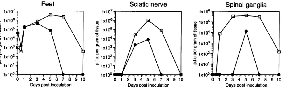
FIG. 1. Yields of 17syn' and R13-1 viruses in various neuronal tissues following footpad inoculation of mice. Six-week-old female Swiss-Webster mice were inoculatedonbothrearfootpadswith106PFU(total)ofeither17syn+orR13-1. Atthe timesindicated,groups of four micewere killed, and thefeet, sciatic nerves,spinal ganglia,dorsal roots, spinalcords, and brainsweredissected and pooled. The tissues were homogenized as 10%(wt/vol) suspensions and titrated for infectious virus. O, 17syn+;0,R13-1.
ofproteinase Kperml). Sampleswere then incubated for 3h at55°C, extractedoncewithphenol-chloroform
(1:1)
andonce with chloroform-isoamyl alcohol(24:1),
ethanol precipitated, and resuspended in Tris-EDTA.Oligonucleotide primers
spe-Feet
cific for sequences within theDNA polymerase gene of HSV wereused to amplifya92-bpfragment by PCR. The reaction mixtures included 0.5 ,ug of each sample of ganglionic DNA; a 0.5 ,uMconcentration of eachprimer
(primer
1, 5' CATCACSciatic
nerve
Spinal ganglia
0 1 2 3 4 5 6 7 8 9 10 DayspostInoculation
0)
cno
E
co
I-..
10.
0 1 2 3 4 5 6 7 8 9 10 Days post inoculation
0 1 2 3 4 5 6 7 8 9
[image:3.612.75.565.80.409.2]Dayspostinoculation FIG. 2. Yields of17syn+ andR13-1 viruses invariousneural tissuesfollowing footpadinoculation of mice withdifferingdosages.The doseof virus used to infect feet wasadjusted so thatanequivalent amount of each viruswasdelivered tospinalganglia. Thus,tocompensate for the reducedyield of R13-1 observed in feet (Fig. 1), 10-fold-less 17syn+ wasused. Viral yields from feet, spinalganglia, and spinal cord were determinedasforFig. 1. C1,17syn';
0,
R13-1.VOL.68, 1994
on November 9, 2019 by guest
http://jvi.asm.org/
[image:3.612.76.566.534.688.2]A.
B.
on November 9, 2019 by guest
http://jvi.asm.org/
HSV NEUROVIRULENCE AND UL5 3765
FIG. 3. 17syn+ and R13-1viral antigens in infected murine spinal ganglia. Micewere infectedonbothrearfootpads, and4days postinfection spinal gangliawere removed andfixedasdescribed in Materials and Methods. The gangliawere frozen and cryosectioned into6-,umsections. Thesesectionswere incubated withafluorescein isothiocyanate-conjugated anti-HSV rabbit antiserumandviewedby indirect immunofluores-cence. (A) 17syn'-infected ganglia; (B) R13-1-infected ganglia. (B) A large neuron (indicated by n) that is not expressing viral antigens is surrounded by supporting cells thatare. Bar, 100p.m.
CGA CCC GGA GAG GGA 3';primer 2,5' GGG CCA GGC GCT TGT TGG TGT 3'); 67 mM Tris (pH 8.8); 16.6 mM ammonium sulfate; 6.7 mM MgCl2; 0.17 mg of bovine serum albumin per ml; a 1.25 mM concentration of each of dGTP, dCTP, dATP, and dTTP; 1 ,uCi of
[32P]dATP
(-6,000Ci/
mmol); and 2.5 U of Taq polymerase (Perkin-Elmer). The thermal cycles used for the PCR of the samples were 94, 68, and 72°C, with thefirst cycle havingaduration of 3 min at each temperature; an additional 30 cycles were for 1 min at each temperature. Astandard curvewasincluded to provide a basis for quantitation. Following PCR, the samples were extracted with chloroform-isoamyl alcohol (24:1), ethanol precipitated,
s.
.^,_
.
.
..
g~~~~~~~~~~~~~~~~~~~~~~~~~~~~~~~~~~~~~~~....
...1 2 3 4 5 6 7 8 9 10 11 12 13 14
FIG. 4. Quantitative PCR analysis of HSV DNA reaching spinal ganglia 3, 4, and 5hafterfootpad infection. Micewereinoculated with 107 PFU of either R13-1 or 17syn'. At the times indicated below, spinal gangliawereremoved (4 mice at each time point), DNAwas
extracted, and sampleswereanalyzed by PCR asdescribed in Mate-rials and Methods. Primers specific fora 92-bp fragment within the DNA polymerase gene of HSV inconjunction with [32P]dATPwere
usedtoidentify the HSVDNA inthesamples. Lane 1, ganglia taken attimezero;lanes 2 and3, 17syn+ and R13-1at3 hpostinfection; lanes 4and5, 17syn'and R13-1 at4 hpostinfection; lanes 6 and 7, 17syn'
andR13-1 at5 hpostinfection. Lanes 8 through 14, standardcurvein which0, 1.6, 0.16, 0.016, 0.0016, 0.00016, and 0.000016pg,respectively, ofplasmidDNAcontaining thetargetsequencesfor the PCRprimers
were added to samples containing 0.5 jig of DNA from uninfected ganglia. The gelwas scanned with a PhosphorImager, and the area
under the peaks was integrated with ImageQuant software. The
number ofgenome equivalents present in each set ofganglia was
calculated after the sample values wereplotted against the standard
curve. The samples in the standard curve correspond to 0, 16,000,
1,600, 160, 16, 1.6, and 0.16 viralgenomes(lanes 8to14, respectively). Genomes in the test samples are calculated to be0, 687, 718, 750, 1,312, 562, and 1,750 in lanes 1 to7, respectively.
andresuspendedinloading buffer, and one half of each sample was resolved on a7.5% polyacrylamide gel. Following electro-phoresis for 2.5 h at 150 V, the gel was dried and exposed to film. Theextent ofhybridization wasdetermined by scanning the bloton aPhosphorlmager and then integrating the product bands with ImageQuant software (Molecular Dynamics, Sunnyvale, Calif.).
RNAand DNAanalyses. Viral DNA accumulation in cul-tured cells was determined by infection of cells in 60-mm-diameter dishes, and at various times postinfection the cells were harvested by being scraped into the media. The cell suspensionwasthencentrifugedat800xg,and thepelletwas resuspended inlysis buffer(50mM Tris [pH
8.0],
1% SDS; 1 mgof proteinaseKperml). After 3 h of incubation at 55°C, the samples were extracted once with phenol and once with phenol-chloroform. Dilutions of the resulting samples were blottedonto anylon membrane as described above. These dot blots were then hybridized with a32p
probe spanning the HindlIl A fragment of the viral genome, and the relative intensities of the hybridization signal were quantitated with aPhosphorlmager
(Molecular Dynamics).RNAwas extracted from 60-mm-diameter dishes following lysis of the monolayers with a solution of 4 M guanidinium isothiocyanate; 0.5% sodium N-lauryl sarcosine, 25 mM so-dium citrate
(pH 7.0),
0.1 M 2-mercaptoethanol, and 0.1I% antifoam A. The lysed cell suspension was then sheared by trituration with a 22-gauge needle and applied to a CsCl cushion(5.7MCsCl, 0.1M EDTA[pH 7.5]), and theRNA was pelleted by centrifugationat 44,000 rpm ina Beckman SW55 rotor for 12 h. The pellets were resuspended in Tris-EDTA and blotted tonylon membranesasdescribed above. Replicate blots were then hybridized with probes specific for the early message, TK (internalXbaI-HindIII
fragment); the late mes-sage,glycoprotein
C(internal
EcoRI-XbaIfragment),
or the immediate-early message, ICP4 (internal 3.2-kb HinclIfrag-ment).
RESULTS
R13-1 is restricted insensoryneurons.Previous studies had demonstrated that R13-1 neurovirulence isattenuated 10,000-fold compared with that in wild-type virus
(12).
In order to address the pathogenesis of R13-1 in a more manipulatable system,weassumedthat the virus would also be restricted in sensoryneurons and examined infectedsensoryganglia.Inthis system the cell typesareeasiertodistinguish
and theviruscan be introduced with no trauma to the tissues to be studied.Equivalent
amountsof R13-1 and HSV-1 strain17syn'
wereinoculated onto the rear footpads of mice, and at various intervals the amountsofvirus in relevant tissueswere deter-mined.AsFig. 1indicates,attheinitialsite of
replication
there isapproximately 10-fold-less R13-1 than17syn'
at 24h after inoculation. In spinal ganglia, differences in relativeyields
betweenthe two virusesincrease,with theyieldofR13-1being some100-fold-less thanthatof thewild type.Atthenextmajor site ofviral
replication,
thespinal cord,
therestriction isevengreater, with no R13-1 being detected until
day
5 and the difference in titers being10,000-fold. Finally,
in the case of VOL.68, 1994on November 9, 2019 by guest
http://jvi.asm.org/
[image:5.612.67.304.299.530.2]Mouse primary neurons
5 10 15 20 25 30 35 40 45 Hours post adsorption
C.
0.
CL
a)
U)-0 a)
c
50
10 20 30 40 50 60 70 Hours post inoculation
20 30 40 50 60
Hourspostadsorption 10 20
30 40 50 60 70 80 90 100
[image:6.612.64.552.80.471.2]Hourspostinoculation
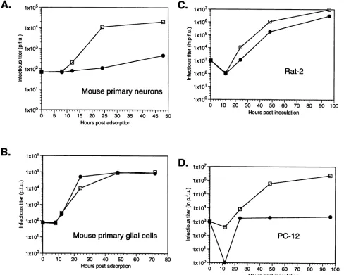
FIG. 5. Replication cyclesof17syn'andR13-1 viruses in neural cells maintained in vitro. Cultureswere infectedatamultiplicityofinfection of 0.1 PFU with17syn+ (Cl)orR13-1(-),andatthe times indicated cellswereharvested and titrated for virus.Replication cyclesinmouseprimary
neuron(A),primary glialcultures (B),Rat-2 cells (C),and PC12 cells(D)are shown.
R13-1, no virus could be detected in the brain, and the
amountsof R13-1 found in the sciatic nerveanddorsal roots
were alsogreatly reduced. From these results, it is clearthat
R13-1 isseverelyrestricted withrespect tospreadthroughthe
nervous system.
In dorsal rootganglia R13-1 antigen is detected predomi-nantly in nonneuronal cells. To determine the basis for the restriction observed in the spinal ganglia, we examined, by
immunofluorescence methods,theexpressionof viralantigens in spinal ganglia. Inorderto evaluate theantigen production resulting from similar amounts of each virus reaching spinal ganglia from the feet, the dose of R13-1 was adjusted to compensatefortheslightlyreducedyields observedinthefeet (Fig. 1). Inthismanner, an amountof R13-1 identicaltothat of17syn+ could be deliveredtothespinal ganglia just priorto the timewhen theantigen productioninthegangliawastobe observed (4 days postinfection). With this modification, viral antigen production and distribution withindifferent cell types in the ganglia could be compared. As shown in Fig. 2, the adjusted dose results in a comparable amount ofvirus being
produced in the primary infectionoffeet for the first 3 days after infection. As is also evident from thisfigure,the restric-tion of virusyieldinspinal gangliaisdramatic; comparedwith that in
17syn',
over 50-fold-less virus is detectable at day 5 postinfection,whilenovirus isseenatearlier times. It should be noted that while theadjusteddoseof virus compensates foradecrease in virus in the feet untilday3 in thesetissues,R13-1
decreasesmore rapidlythan does
17syn'
onsubsequent days.One probable contributing phenomenon to these kinetics is seeding of the foot at later times by virus produced in the
spinal ganglia, with
17syn'
supplying more virus to the feetoverthese intervals. In the immunohistochemicalexperiments
(Fig. 3), a significant number of neurons can be seen to be producingviralantigensin
17syn'-infected
ganglia. However, in the case of R13-1, only a few neurons could be seen to expressviralantigens,andmostantigensarefoundin support-ing cellssurrounding theneurons. These results suggest that thevirus is restricted inreplicationwithin dorsalrootganglion (DRG)neurons.Finally,it should be noted that whilethe virus isseentobe restricted inreplicationandantigen productioninA.
1X10⁴-1-:
Cl
1X10³-0O
1x101-:
0
B.
-O.
C')D
-O
.0
C_
on November 9, 2019 by guest
http://jvi.asm.org/
HSV NEUROVIRULENCE AND UL5 3767
-o
N
.0
n
-o
C)
0G.
U3
30 40 50 60 70
Hours postinoculation
N 1x10⁶
.0~~~~~~~E
.E 1x1
05-0
1 x10⁴
o
PC-12
1
x103-0 10 20 30 40 50 60 70 80
Hours postinoculation
FIG. 6. Accumulation of17syn4' or R13-1 viral DNA in Rat-2 and PC12 cells. Rat-2 or PC12 cells were infected at a multiplicity of infection of 0.1 with either 17syn+ (O) or R13-1
(@),
and at the indicated times the cells were harvested and DNA dot blots were prepared as described in Materials and Methods. The blots were hybridized with a probe specific for a common region of the viral genome,and the relative intensityofhybridization signalwas quanti-tated witha Phosphorlmager.neurons, it establishes latent infection in these cells. In vitro cocultivation experiments inwhich ganglia from mice latently infected with either 17syn+ or R13-1 were induced to reacti-vate demonstrate that pooledL4 and L5 (four ganglia) spinal ganglia from each of eight mice reactivated. The times in culture until virus-specific cytopathic effects was observed ranged from5 to 8 days (average, 6days) for each virus.
R13-1 infects sensory nerve termini and is transported to gangliaasefficientlyas17syn+. Inorder to determine whether therewas aninitial restriction inthe abilityof R13-1 to infect nervetermini in thefeetandthen betransportedtothe somas in thespinal ganglia,weinfected mice with
107
PFUofeither R13-1 or17syn+.Atintervals gangliawereremoved, extracted, and subjected to PCR in order to detect viral DNA, asICP4
tk
gC
17syn+
0 hr
R13-1
2 hr
4 hr
17syn+ *
R13-1
1E
l7syn+R13-1
t
1 7syn+ E * *
8
hr_
R13-1
1 7syn+ * *
16 hr
[image:7.612.354.533.77.306.2]R13-1
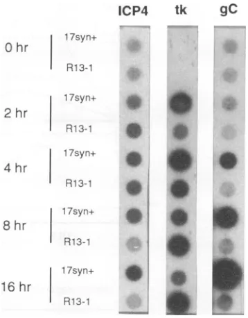
FIG. 7. Comparison ofimmediate-early,early, andlateviral mes-sage synthesis in PC12 cells infected with 17syn+ or R13-1 viruses. Cellswereinfected with 17syn andR13-1at amultiplicity of infection of 10 PFU and then harvestedatintervalspostinfection, and RNA was extracted.ThisRNAwasblotted withadot blot minifold and probed with probes to the ICP4, TK, and gC genes. Representative values determined by Phosphorlmageranalysisare asfollows. ICP4 at 2 h:
17syn', 11,898, and R13-1, 19,641; ICP4 at4 h: 17syn', 16,960, and R13-1, 19,672; TKat2h: 17syn', 45,365, and R13-1, 16,823; TK at4 h: 17syn',62,309, andR13-1, 40,463; TK at 8h: 17syn', 39,519, and R13-1,59,086; gCat8 h: 17syn+,102,392, andR13-1,6,982; and gC at 16h: 17syn',866,625, andR13-1,9,753.
described in MaterialsandMethods.AsshowninFig.4, at 3h after infection, 2,900 counts are detected in ganglia infected with 17syn+ and 3,025 counts are detected in those infected with R13-1. Theamount of viralDNAincreasesat 4and 5 h, respectively, with slightly more DNA being detected in the samples containing R13-1. In other experiments, if the levels were not equivalent, therewas always asmall increase in the amount of R13-1 DNA detected. The reason for this is not clear, but it could be related to differences in particle/PFU ratios between the two viruses. In any case, we conclude that R13-1 DNA enters somas asefficiently as that from
17syn'.
R13-1 is restricted inreplicationinprimaryneurons butnot glial cells. The previous experiments indicated that virus replicationwasselectively restricted inneurons.Toinvestigate this phenomenon in more detail, we studied infection of primary neurons and supporting cells in vitro. Viral growth curvescomparing
17syn'
and R13-1 in the two cell types are shownin Fig.5A and B. Therewasasignificant
difference inthe
ability
of R13-1 toreplicate
in neuronal cells, with some1,200-fold-lessvirusbeing producedthan in the case of
17syn
7 .The replication of R13-1 in glial cells, however, was almost identical to that of 17syn+. As was observed with spinal ganglia,incultured cellsR13-1 exhibitsarestriction
specific
to neurons. In anattempt todetermine ifR13-1 wasrestricted in anyestablished lines(which
would makebiochemicalanalysis
easier), a number of established lines, including mouse and humanneuroblastomas,were studied. Inmostcell lines exam-ined
(as
represented by Rat-2 cells inFig. 5C),
R13-1repli-cated normally. In undifferentiated PC12 cells, however, as
VOL.68? 1994
on November 9, 2019 by guest
http://jvi.asm.org/
[image:7.612.64.300.78.487.2]EcoRl
17+-
n-Id 9 n f mol
I If I 11
a e: k h k
a I C
L---i I
I
II II
I I I
.1. EcoR
I I I I
I
EcoRIi
I. I I. ,i L...
l1/BamHlad
BamHl
I I~~~~~~~~~~~~~~~~~~~~~~~~~~
EcoRV EcoRV
Miul
I ""1-..EcoRV
UL5
EooRI
UL6
Markerrescueof R13-1with clonedsubfragments ofthe 17+EcoR1 dfragment
EcoRl dsub-fragments ExpA Exp B
--- 0/6 0/6
EcoRl D 4/6 3/6
EcoRWEcoRV 1/6 0/6
EcoRl/EcoRV (UL5+UL6) 3/6 3/6
EcoRl/Miul(UL5) 2/6 4/6
Mlul/EcoRV(UL6) 0/6 0/6
FIG. 8. Markerrescue strategyforgenerationof virulent R13-1 recombinants. Shownatthe top isanEcoRI map of the HSV-1 strain 17syn+
genomewithanexpanded view of the EcoRIDfragment directlybelowit. Thisfragmentwaspreviouslyshowntobesufficientto restorevirulence to R13-l (12). Inthis study, subfragments of thisregion weresubcloned and assayed after intracranial inoculation fortheirability to restore virulenceto R13-1following cotransfection. The subfragments assayedareshown in thetable, and the number of animals killedcomparedwith the number of animals inoculated ispresentedfortwo separateexperiments.
shown in Fig.
5D,
thedegree ofrestriction wascomparable
tothat seen in primary neurons, with only
slightly
more virus detected at late times than at timesimmediately
following adsorption. Since PC12 cells can be induced to differentiate and stop dividing following nerve growth factor treatment, it seemedpossible that differentiated PC12 cells would be even morerestrictive toR13-1 replication than the undifferentiated cells; however,the restrictionobserved indifferentiated PC12 cellswasnogreaterthan that observed in theundifferentiated PC12 cells(data
notshown).
Itshould be noted that in mostpermissive cell linesexamined,R13-1 exhibitsslightlyreduced yields (two- to fivefold) compared with 17syn+, and thismay mirror the slightly reduced yields observed earlier formouse feet (Fig. 1).
A number of replication-deficient mutants, such as those withlesionsin the TK gene,demonstrate restricted replication oncells that are notactively dividing, presumablybecausethe virus is limited by the availabilityofnucleotides. When yields of R13-1 from serum-starved and actively dividing RS cells
werecompared, theywere equivalent (datanot shown). Restriction of R13-1 isprior to or at the level of viral DNA replication. PC12cells were used toinvestigate the pointinthe replication cycle when R13-l is restricted. For comparison, Rat-2cells(fibroblasts) wereused,sincePC12is a rat-derived
line. ViralDNA wasisolated from infectedcellsatseveraltime points and analyzed by dot blot analysis as described in Materials and Methods.AsshowninFig. 6, theamountof viral DNA detected in R13-1-infected PC12 cells is approximately 100-fold less than in Rat-2cells, indicating that the restriction of R13-1 was at or before the level ofDNA replication. The abilitytodetectinputviral DNAby this method demonstrates that the blockoccursafterthevirusentersthecell. Inorderto
determine whether any viral transcripts were made by the R13-1-infected cells, RNA was
purified
and dot blots were hybridized with probes specific for different classes of viral mRNA. As can be seen in Fig. 7, while R13-1 produces TK message, ICP4, and glycoprotein C (gC) RNA in Rat-2 cells, nogCRNAisdetectable in PC12 cells.The factthatthe levels of the R13-1 early (TK) message inPC12 cells are similarto those of 17syn+ indicates that early events in the infection proceed normally, while the inability to detect gC (late) messageinthese cells indicates thatthe block inthe infectivecycle
occursprior
to the initiation of late events. This latterfinding is in concordance with the lack of DNA synthesis in PC12 cells.
Marker rescue analysis identifies UL5 as the gene confer-ringneurovirulencetoR13-1.Previously,theregion implicated in virulence had been localized to the 9-kb EcoRI-BamHI
I. a
on November 9, 2019 by guest
http://jvi.asm.org/
[image:8.612.152.461.72.262.2]HSV NEUROVIRULENCE AND UL5 3769
lxl(
1:1 lxl(
.)
=lxl(
n 0
=lxl( a)
Ax
10 20 30 40 50 60 70 80 90 100 Hourspost inoculation
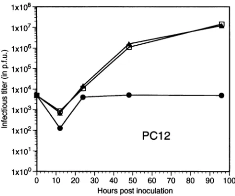
[image:9.612.66.304.75.274.2]FIG. 9. Replication cycles of theUL5rescuant, l7syn>' and R13-1
viruses in PC12 cells. Cultures were infected at a multiplicity of
infectionof 0.1 PFU with therescuant(A),17syn`(Li),orR13-1 (0),
andatthetimes indicated cellswereharvested and viruswastitrated.
adfragment in the left-hand region of the genome (12). This
region was subcloned, and four EcoRV subclones which spanned the fragmentwere transfected and assayed for their
abilitiesto restorevirulence. The results of these experiments
are summarized in Fig. 8 where it can be seen that the
left-handEcoRI-EcoRV fragment (open reading frames UL5 and UL6) restored the virulent phenotype. Thefragment was
furthersubclonedtoanEcoRI-Mlul fragment(containing only
UL5) and a MluI fragment (containing UL6). These were
transfected andassayed, and the results indicatedthattheUL5 ORFwassufficienttorestorevirulence.UL5 isacomponentof the helicase-primase complex of HSV, is essential for DNA replication, and interacts withtwoothervirally encoded
com-ponentsin ordertoform this complex (4). Inthecaseof R13-1,
this gene has been replaced with the homologous type 2
sequence, thus forming a chimeric complex with the other
virally encoded subunits of the origin-binding complex. It should be noted that in R13-1, the origin-binding complex would be predicted to be composed of UL5 and UL8 from HSV-2 and UL52 from HSV-1 (12).Thepredicted amino acid changes between type 1 and type 2 UL5 genes and their
relationshiptothese results are presented in the nextsection. Rescued viruses are virulent in mice. Two plaque-purified
isolates from separate transfection experiments were plaque
purified fromthe brains of mice that diedfrom encephalitis in themarkerrescueexperiments. Asdescribedin Materials and
Methods, these viruseswerescreened withlabeled
oligonucle-otidesspecific for thetype 1 UL5gene.Toassayforvirulence,
the viruses were then inoculated intracranially into mice. In thistest,thetworecombinants demonstrated
LD50s
of1 x 2.5 and 1 x 10 PFU, while R13-l killed at 5 x 10⁴ PFU. Inaddition, in order to ensure that in vivo passage of the
transfections did not select for viruses with additional muta-tions elsewhere in the genome that restored virulence, two additional recombinantswere selected directly fromseparate transfection experiments, again with the oligonucleotide probes specific for HSV-1 UL5. These isolateswere shown to
have LD5sSof 10 and 8 PFU, respectively.
Rescued virus shows normal replication in neurons and
PC12 cells. Since the marker rescue experiments demonstrated that the HSV-1 UL5 fragment was sufficient to restore the virulence to R13-1, replication in PC12 cells was studied. As shown in Fig. 9, the replication kinetics in PC12 cells of one rescuant isolated in cell culture are identical to those of wild-type virus. It should also be noted that this virus replicates in a manner similar to that of the wild type in Rat-2 cells, and immunofluorescence analyses of infected ganglia indicate that it and the wild type express similar amounts of viral antigen and that the cellular distribution of these products is identical. That is, unlike R13-1, significant numbers of neurons demon-strate antigens (data not shown).
DISCUSSION
This study has exploited the HSV intertypic recombinant R13-1 to determine the genetic basis of a mutation that confers on this virus a novel phenotype-the ability to replicate to high titers in the CNS without being virulent. Our analysis has shown that the replacement of UL5 from HSV-1 with its homolog in HSV-2 renders the recombinant virus defective for replication in neurons without affecting replication in other cells. Several unique biological properties of this mutant provide insight into certain aspects of virus-neuron interac-tions, and these are discussed below.
A number of replication-deficient mutants are restricted for replication in nervous tissues, and prototypes of one major class are represented by those with alterations in TK and UTPase genes. These genes have been implicated in the regulation of nucleotide pools, and the mutations presumably interfere with supplemental nucleotide pools needed for HSV replication in cells of the nervous system that are not actively dividing. R13-1 does not fall into this class because the physiologic state of the cell does not relate to restriction of R13-1. R13-1 replicates well in the CNS and in serum-starved cells, while TK- mutants are restricted under these conditions. Inaddition,R13-1 is restricted in both primary neurons and in PC12 cells; the latter represents an established cell line which activelydivides. From these considerations, we suggest that the mutation in R13-1 defines a replication-minus genotype spe-cific for neurons that is characterized by high-titer replication in other cells of the CNS and attenuation in neurovirulence. This suggests that there are at least two classes of neuroviru-lencemutations: one in genes which regulate nucleotide pools, andone at or near the level of DNA polymerase activity. In the latter regard, although involved cells have not been identified, HSV-1mutants with lesions in the polymerase gene have been shown to be avirulent when inoculated into the CNS (7). The relevant implication is that mutants deficient in nucleotide pool metabolism act at a general level by interfering with the replication of the virus in almost all cells of the CNS that are not actively dividing. The class represented by R13-1, and possibly the polymerase mutants, would represent a class that exhibits a more specific restriction within neurons that is linked to a difference in the interaction of the viral replication components withthose supplied by the neuron.
Neuron-specific influences on herpesvirus replication have also been suggested by Kosz-Vnenchak et al. (14). In their experiments, immediate-early and early gene expression was stimulated by viral DNA replication in neurons, a phenome-non not observed in other cell types. Although translational effects have not been ruled out, ourresultsare compatible with these findings, since when compared with
17syn+,
ganglionic neurons infected with R13-1 possessed decreased levels of immediate-early andearlyproteins.VOL.68, 1994
on November 9, 2019 by guest
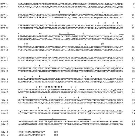
http://jvi.asm.org/
MAAAGGERQLDGQKPGPPHLQQPGDRPAVPGRAEAFLNFTSMHGVQPILKRIRELSQQQLDGAQVPHLQWFR MAAAGGERQLDGQKPGPPHLQQPGDRPAVPGRAEAFLNFTSMHGVQPILKRIRELSQQQLDGAQVPHLQWFR
I + +
DVAALESPAGLPLREFPFAVYLITGNAGSGKSTCVQTINEVLDCVVTGATRIAAQNMYAKLSGAFLSRPINT DVAALESPAGLPLREFPFAVYLITGNAGSGKSTCVQTLNEVLDCVVTGATRIAAQNMYVKLSGAFLSRPINT
+
+I+ + + +# F #
IFHEFGFRGNHVQAQLGQYPYTLTSNPASLEDLQRRDLTYYWEVILDLTKRALAASGGEELRNEFRALAALE
IFHEFGFRGNHVQAQLGQHPYTLASSPASLEDLQRRDLTYYWEVILDITKRALAAHGGEDARNEFHALTALE
+ ++ + II
RTLGLGQGALTRLAPATHGALPAFTRSNVIVIDEAGLLGRHLLTAVVYCWWMINALYHTPQYAARLRPVLVC QTLGLGQGALTRLASVTHGALPAFTRSNIIVIDEAGLLGRHLLTTVVYCWWMINALYHTPQYAGRLRPVLVC
III + + IV
VGSPTQTASLESTFEHQKLRCSVRQSENVLTYLICNRTLREYARLSYSWAIFINNKRCVEHEFGNLMKVLEY VGSPTQTASLESTFEHQKLRCSVRQSENVLTYLICNRTLREYTRLSHSWAIFINNKRCVEHEFGNLMKVLEY GLPITEEHMQFVDRFVPENYITNPANLPGWTRLFSSHKEVSAYMAKLHAYLKVTREGEFWFTLPVLTFVS GLPITEEHMQFVDRFVVPESYITNPANLPGWTRLFSSHKEVSAYMAKLHAYLKVTREGEFVWFTLPVLTFVS
+ + + + + +
VKEFDEYRRLTHQPGLTIEKWLTANASRITNYSQSQDQDAGHMRCEVHSKQQLVVARNDVTYVLNSQIAVTA VKEFDEYRRLTQQPTLTMEKWITANASRITNYSQSQDQDAGHVRCEVHSKQQLVVARNDITYVLNSQVAVTA
+ + #F ++ + +
RLRKLVFGFSGTFRAFEAVLRDDSFVKTQGETSVEFAYRFLSRLIFSGLISFYNFLQRPGLDATQRTLAYAR
RLRKMVFGFDGTFRTFEAVLRDDSFVKTQGETSVEFAYRFLSRLMFGGLIHFYNFLQRPGLDATQRTLAYGR
+ ++##f ++####f+######ftf + +
MGELTAEILSLRPKSSGVPTQASVMADAGAPGERAFDFKQLGPRDGGPDDFPDDDLDVIFAGLDEQQLDVFY LGELTAELLSLRRDAAGASATRAADTSDRSPGERAFNFKHLGPRDGGPDDFPDDDLDVIFAGLDEQQLDVFY
#T#+ #F #+ + + #f +
CHYTPGEPETTAAVHTQFALLKRAFLGRFRILQELFGEAFEVAPFSTYVDNVIFRGCEMLTGSPRGGLMSVA
CHYALEEPETTAAVHAQFGLLKRAFLGRYLILRELFGEVFESAPFSTYVDNVIFRGCELLTGSPRGGLMSVA
+ + + +
LQTDNYTLMGYTYARVFAFADELRRRHATANVAELLEESPLPYVVLRDQHGFMSVVNTNISEFVESIDSTEL LQTDNYTLMGYTYTRVFAFAEELRRRHATAGVAEFLEESPLPYIVLRDQHGFMSVVNTNISEFVESIDSTEL
V VI +
AMAINADYGISSKLAMTITRSQGLSLDKVAICFTPGNLRLNSAYVAMSRTTSSEFLRMNLNPLRERHERDDV AMAINADYGISSKLAMTITRSQGLSLDKVAICFTPGNLRLNSAYVAMSRTTSSEFLHMNLNPLRERHERDDV
ISEHILSALRDPNVVIVY ISEHILSALRDPNVVIVY
72 72 144 144 216 216 288 288 360 360 432 432 504 504 576 576 648 648 720 720 792 792 864 864 882
[image:10.612.77.522.66.513.2]882
FIG. 10. Comparison of thepredictedamino acidsequencesof the UL5genesof HSV-1and HSV-2. Thepredictedamino acid sequence of UL5wastranslated from the nucleic acidsequenceof HSV-1(18)andHSV-2(17)and isrepresentedinone-letter code. Conservative aminoacid
differences are indicatedby a +, and nonconservative differencesare shown by a #. Thesix major highlyconserved helicase motifs(30) are
indicatedbyaline above thesequence.
Ourstudyindicates that UL5playsaspecificrolefacilitating viralreplication inneurons.We postulate that replacementof
UL5 with its HSV-2counterpartresultsinthe formation ofan
alteredhelicase-primase complex. UL5 normallyinteracts with twoother viralproteins, UL8 and UL52, inordertoform this complex (4, 29),and inthecaseofR13-1, the HSV-2 compo-nentwould formachimericcomplex that is functional for viral
DNAreplication inmostcelltypes.However, it is possible that the helicase-primase complex is more elaborate and also containsone or morecellularproteins.Ifthiswerethecase,in
neuronstheprotein could either be absentorpossess
proper-ties unique to this cell type. The nature of the phenotype observed in R13-1 suggests the existence of a cellular factor that isacomponentof theorigin-binding complex requiredfor
replication. As examples, this could be a polymerase or an
accessory factor.
As to a potential domain in UL5 which may be involved, alignmentof thepredictedamino acidsequencesof UL5 from HSV-1
(17syn')
and HSV-2 (HG52) showsa high degreeof conservation (Fig. 10). Ofparticularinterest in thiscompari-son are the six conserved helicase domains that have been
reported for theHSV-1 UL5geneand thatwereshowntohave essential roles in the function of the UL5protein (30).There isonlyonepredicted amino acid changebetween HSV-1 and HSV-2 within thesemotifs, and this is aconservative replace-mentofavaline foran isoleucine in the first residue ofmotif
II.From thiscomparison, itseemsunlikelythatchangesinthe activityof the critical helicase domainsareresponsiblefor the HSV-1
HSV-2
HSV-1 HSV-2 HSV-1 HSV-2 HSV-1 HSV-2 HSV-1 HSV-2 HSV-1 HSV-2 HSV-1 HSV-2 HSV-1 HSV-2 HSV- 1 HSV-2 HSV-1 HSV-2 HSV-1 HSV-2 HSV- 1 HSV-2 HSV-1 HSV-2
on November 9, 2019 by guest
http://jvi.asm.org/
HSV NEUROVIRULENCE AND UL5 3771
restriction exhibited byR13-1. Instead, the comparison focuses attention on theamino acid substitutions which lie between the conservedmotifs. While themajority of these substitutionsare conservativechanges, there is a region of divergence between residues 589 and607 where 17 of the 18 residues are different between the two strains. Of potential significance is the observation that 12 of these 18 residues are nonconservative changes and result in an overall change in the predicted net charge of the region of -1.4 to -4.8. This shift is more suggestive of alpha-helix formation than of the beta-sheet structure predicted from the 17syn+ sequence. These changes could result in differences in the interactions of the HSV-2 UL5 component with the other components of the helicase-primase complex either by disrupting the contact sites of protein-protein interactions or bychanging the tertiary struc-ture of the protein. We would suggest that studies of the chimericcomplexassociated with R13-1 might prove useful for defining interactions of components of the primase-helicase complex.
With respect to more general features of herpetic disease, an important component of HSV pathogenesis is illustrated by studies of R13-1. R13-1 doesnotreplicate well inneuronsbut isunaltered in capacity to replicateinglial cells. Clearly, viral replication in neurons is a requirement for the development of clinically apparent neurologic disease, including encephalitis.
Finally, the ability of this recombinant to replicate in non-neuronal cellssuggests its potential application as a vector for the introduction of genes into the CNS. As mentioned earlier, even though this virus does not replicate well in neurons, we know thatit establishes latent infections efficiently. This result was predictable, since we and others have demonstrated that viral geneexpressionand DNAreplication are notrequired to establish latent infection (5, 15, 23, 27). The lack of virulence exhibitedby this mutant, coupled with the factthatitreplicates to high titers in the CNS, would allow the virus to establish latent infections relatively efficiently. This eliminates a prob-lem associated with many current nonreplicating HSV-based vectors. In those systems, latent infection is limited to the neuronsinitially infected by input virus.
ACKNOWLEDGMENTS
Thisworkwassupported byagrantfrom the W. M. Keck Founda-tion,PHS grantsAI-06246 and NS-30420 from the National Institutes of Health, and grant RG 1647-C-3 from the National Multiple SclerosisSociety.D.C.B.wassupported by postdoctoralfellowshipFG 909-A-I from the NationalMultiple Sclerosis Society.
Wethank D.Knipe for providing antiseratoICP8 and T. Wen for providing the clonecontaining the TK gene used forTKprobes. We thank L. Bloom, L. Feldman, B. Mitchell, G. Rall, T. Wen, and S. Yuhasz for criticalreading of the manuscript. The technical assistance of S. Comora is alsoacknowledged.
REFERENCES
1. Ausubel, F. M., R. Brent, R. E. Kingston, D. D. Moore, J. G. Seidman, J.A.Smith,and K.Struhl(ed.). 1993. Currentprotocols inmolecularbiology. CurrentProtocols, Boston.
2. Barker,D.E.,and B. Roizman. 1990.Identification of three genes nonessential forgrowth in cell culture nearthe right terminus of theunique sequences oflongcomponentofherpessimplexvirus1. Virology177:684-691.
3. Chou, J., E. R. Kern, R. J. Whitley, and B. Roizman. 1990. Mapping of herpes simplex virus-I neurovirulence to gamma 134.5,agenenonessential forgrowth in culture. Science 250:1262-1266.
4. Crute, J. J.,T.Tsurumi,L. A.Zhu,S. K.Weller,P. D.Olivo,M. D. Challberg, E. S. Mocarski, and I. R. Lehman. 1989. Herpes simplex virus 1 helicase-primase: a complex of three
herpes-encoded geneproducts.Proc.Natl. Acad.Sci. USA 86:2186-2189. 5. Dobson, A. T., T. P.Margolis, F.Sedarati, J. G. Stevens, and L. T. Feldman. 1990. A latent, nonpathogenic HSV-1-derived vector stably expresses beta-galactosidase in mouse neurons. Neuron 5:353-360.
6. Efstathiou, S.,S.Kemp, G.Darby,and A. C. Minson. 1989.The roleof herpes simplex type 1thymidinekinase inpathogenesis.J. Gen.Virol. 70:869-879.
7. Field,H.J., and D.M.Coen. 1986.Pathogenicityofherpessimplex virus mutants containing drug resistance mutations in the viral DNApolymerasegene.J. Virol. 60:286-289.
8. Field, H. J., andP.Wildy. 1978. Thepathogenicityof thymidine-kinase deficient mutantsof herpes simplexvirusin mice. J. Hyg. 81:267-277.
9. Fitzgerald, S. C. 1989.Dissociatedspinalcord-dorsalrootganglia cultureson plastictissue culture dishes and glass coverslipsand wells, p. 219-222. InA.Shaharetal.(ed.),Adissection and tissue culture manual of the nervoussystem., Alan R. Liss, Inc., New York.
10. Greene,L.A., J.M.Aletta,A.Rukenstein,andS.H.Green. 1987. PC12 pheochromocytomacells:culture,nervegrowthfactor treat-ment,andexperimentalexploitation.MethodsEnzymol. 147:207-216.
11. Izumi, K. M., and J. G. Stevens. 1990. Molecular and biological characterization ofaherpes simplexvirus type 1 (HSV-1) neuro-invasiveness gene. J.Exp. Med. 172:487-496.
12. Javier, R. T., K.M.Izumi, and J. G. Stevens. 1988. Localization of a herpes simplex virus neurovirulence gene dissociated from high-titervirus replication in the brain. J. Virol. 62:1381-1387. 13. Johnson, G. D., and G.M.C.N.Araujo. 1981.Asimplemethodof
reducingthefading of immunofluorescenceduring microscopy.J. Immunol.Methods 43:349-350.
14. Kosz-Vnenchak, M., J. Jacobson, D. M.Coen,and D. M.Knipe. 1993.Evidence foranovelregulatory pathwayforherpessimplex virus gene expression in trigeminal ganglion neurons. J. Virol. 67:5383-5393.
15. Leib,D.A.,D. M. Coen,C. L.Bogard,K. A.Hicks,D.R.Yager, D. M. Knipe,K. L.Tyler,and P. A. Schaffer. 1989. Immediate-early regulatory gene mutants define different stages in the establishment and reactivation ofherpessimplexviruslatency.J. Virol. 63:759-768.
16. Margolis, T. P., F. Sedarati,A. T. Dobson, L. T. Feldman, and J. G. Stevens. 1992.Pathwaysof viral geneexpression duringacute neuronalinfection with HSV-1.Virology 189:150-161.
17. McGeoch,D.J., C.Cunningham,G.McIntyre,and A. Dolan.1991. Comparativesequenceanalysisof the longrepeatand adjoining partsof thelong uniqueregionsin thegenomes ofherpes simplex viruses types 1 and 2. J. Gen.Virol. 72:3057-3075.
18. McGeoch,D.J.,A.J. Dalrymple,A.J.Davison,A.Dolan,M. C. Frame,D. McNab,L.J. Perry, J.E. Scott,and P.Taylor. 1988. The complete DNA sequenceof the long unique region in the genome ofherpes simplex virus type 1. J. Gen. Virol.
69:1531-1574.
19. Nishiyama, Y., Y. Yamada, R. Kurachi, and T. Daikoku. 1992. Construction ofa US3 lacZ insertion mutant ofherpes simplex virus type 2 and characterization of itsphenotype in vitro and in vivo.Virology 190:256-268.
20. Purves, F. C., D. Spector, and B. Roizman. 1991. The herpes simplexvirus 1proteinkinase encodedbytheUS3genemediates posttranslational modification of thephosphoproteinencodedby the UL34 gene.J. Virol. 65:5757-5764.
21. Pyles, R.B., N. M.Sawtelle,andR.L.Thompson. 1992. Herpes simplexvirus type 1 dUTPase mutantsare attenuated for neuro-virulence, neuroinvasiveness, and reactivation from latency. J. Virol.66:6706-6713.
22. Reed,L.J.,and H. Muench. 1938.Asimplemethod ofestimating fiftypercentendpoints.Am.J. Hyg.27:493-497.
23. Sedarati, F., T. P. Margolis, and J. G. Stevens. 1993. Latent infection can be established with drastically restricted transcrip-tionandreplicationof the HSV-1 genome.Virology192:687-691. 24. Sedarati, F.,andJ. G. Stevens.Unpublisheddata.
25. Thompson, R. L.,andJ. G. Stevens. 1983. Biological character-ization ofaherpessimplexvirus intertypicrecombinant which is VOL. 68, 1994
on November 9, 2019 by guest
http://jvi.asm.org/
completely andspecifically non-neurovirulent. Virology 131:171- 28. Yuhasz, S. A., and J. G. Stevens. 1993. Glycoprotein B is aspecific
179. determinant of herpes simplex type 1 neuroinvasiveness. J. Virol.
26. Thompson, R. L., E. K. Wagner, and J. G. Stevens. 1983. Physical 67:5948-5954.
location of a herpes simplexvirus type-i gene function(s) specif- 29. Zhu, L.,and S. K. Weller. 1992.The UL5 geneofherpessimplex
ically associated with a 10 million-foldincrease in HSV neuroviru-
virus
type1:isolationofalacZ insertionmutantandassociation oflence. Virology 131:180-192.
us
geneprolation
of
memertiof
theandiassociase
27. Valyi-Nagy, T., S. L. Deshmane, J. G. Spivack,I. Steiner, C. I.Ace, the UL5 geneproduct with other members of the helicase-primase C. M. Preston, and N. W. Fraser. 1991.Investigation ofherpes complex. J. Virol. 66:458-468.
simplex virus type 1 (HSV-1)geneexpression and DNAsynthesis 30. Zhu,L. A., and S. K. Weller. 1992. The six conserved helicase during the establishment of latent infection by an HSV-1 mutant, motifs of the UL5 gene product, a component of the herpes in 1814, that does not replicate in mouse trigeminal ganglia. J. simplexvirus type 1helicase-primase,areessential for its function.
Gen.Virol. 72:641-649. J.Virol. 66:469-479.