0022-538X/94/$04.00+0
Copyright © 1994,American Society for Microbiology
Simian Virus
40
Small-t
Antigen
Stimulates Viral DNA
Replication in Permissive
Monkey
Cells
CLAUDIA
CICALA,1
MARIA L.AVANTAGGIATI,'
ADOLPH GRAESSMANN,2 KATHLEEN RUNDELL,3 ARTHUR S.LEVINE,I
AND MICHELECARBONE`*
SectiononDNAReplication, Repair, andMutagenesis, National InstituteofChild Health and HumanDevelopment, Bethesda, Maryland
208921;
InstitutfOirMolekularbiologie und Biochemie der Freien UniversitatBerlin,Berlin 33, Germany2; and DepartmentofMicrobiologyand
Immunology,
NorthwestemUniversity, Chicago, Illinois
60611-30083
Received 22 September1993/Accepted31January 1994
The simianvirus 40 (SV40) large-Tantigen is essentialfor SV40DNA replicationand for late viral gene expression, but the role of the SV40 small-t antigen in these processes is still unclear. We have
previously
demonstratedthat small tinhibits SV40 DNAreplicationin vitro.In this study,weinvestigated theeffect ofsmall t onSV40 replication in culturedcells.CV1 monkey cell infectionexperiments indicated that mutant
virusesthat lack small t replicateless
efficiently
than thewild-type virus. Wenextmicroinjected
CV1cellswith SV40DNAwith and withoutpurified small-tproteinandanalyzed viral DNAreplicationefficiency
bySouthern blotting. Replication of eitherwild-type SV40orsmall-tdeletionmutantDNAwasincreased three-tofivefoldincellscoinjected with purified small t. Thus,incontrastto our invitroobservation, smalltstimulated viral DNA replication in vivo. This result suggests that small t has cellular effects that are not detectable in a
reconstituted in vitro replication system. We also found that small t stimulated progression of permissive monkey cells-but not ofnonpermissive rodent cells-from
Go-G1
to the Sphase ofthe cell cycle, possiblyleading toanoptimal intracellularenvironmentfor viralreplication.
Inmonkey cells infected by simian virus 40(SV40),large-T antigen directs an ordered sequence of events leading from the
early phaseofinfection,in which viralDNAreplicationstarts,
tothe latephase, when viral particles are produced(2,44,46).
The early phase is devoted to subversion of cellular control mechanisms topreparethecell for the late phase. T antigen is
an autoregulated phosphoprotein that accumulates in the nucleus during the early phase ofinfection; it alters cellular transcription patterns (6, 43) and stimulates cellular DNA
synthesis in quiescent cells (11, 19, 20, 21, 30, 45). Upon transition to the late phase, T antigen sustains viral DNA
replication and stimulates late gene expression and virion production.
The second early SV40 protein, the small-t antigen, is a
17-kDa polypeptide found predominantly in the cytoplasm of infected cells (13, 46). Small t shares 82 amino acids at its amino terminus with the large-T antigen; the remaining 92 amino acids areunique. Although small t can enhance trans-formationby the virus (3, 7, 8, 41), particularly when growth-arrested cells are used (28), little is known about its role in
permissiveinfections ofcycling cells. However, enhancement ofpermissiveinfectionsby small t is suggested by the reduced growth yields and small plaque sizes induced by viruses that carrymutations in the small-t antigen (40, 46, 47). Recently, it has been found that small-t antigen binds cellular protein
phosphatase2A(PP2A)(33, 50),inhibiting its activity toward
several substrates, including large-T antigen itself (39, 51). Withahighlypurified system for replication of SV40 DNA in vitro, PP2A was shown to dephosphorylate T antigen and
stimulate replication (26, 49). It was shown that addition of small-t antigen to an in vitro cell-free replication system
*Correspondingauthor. Mailing address: Section on DNA
Repli-cation, Repair,and Mutagenesis, NIH, Bldg. 6, Rm.lAl1,Bethesda, MD20892.Phone:(301)496-8912. Fax: (301) 402-0105.
decreasesSV40 DNAreplication (5), aresultconsistent with the inhibition of PP2A by small t. To explore this apparent paradox between in vivo and in vitro experiments,weexamined the effect of smalltonreplication of microinjected viral DNA and the effects of small-tantigenonthe cellcycle inpermissive
(monkey) cells. Given its role in transformation, we also studied the effect of small t on cell DNA synthesis in nonper-missive(rodent) cells.
MATERIALS AND METHODS
Cells and viruses.African green monkeykidney (CV1) cells
were grown in Dulbecco modified Eagle medium (DMEM)
containing 5% fetal bovineserum.Virus stockswereharvested fromlysates initiated with a low multiplicity of virus, and titers weredetermined on CV1 cells.Lysates preparedby subjecting even uninfected cells to freeze-thaw cycles contain factors which induce cellular DNA synthesis. Consequently, serum-free mock and virus stocks wereprepared. Cells were infected with 10 PFU per cell inmedium containing serum. Medium was then replaced with serum-free DMEM until cytopathic effects were apparent by extensivevacuolization of the cells, but most cells remained attachedto the dishes.Atthis time, mediumcontaining virus was collected and the small number of detached cells was removed by centrifugation. Titers of these serum-free viruses were two- to threefold lower than titers obtainedfollowing freezingandthawing of infected cells. Mocklysates prepared in this way were unable to induce DNA synthesis in growth-arrested cells, in contrast to mock lysates prepared byfreezing andthawing.
Rates of viral DNA synthesis. Confluent cultures of CV1 cells in 3.5-cm-diameter disheswereinfected with 10 PFU of virus per cell or equivalent volumes of lysates of uninfected cellsfor 2 h and thenkeptin serum-free medium.Atvarious times postinfection, 10,Ci of[3H]thymidinewas added for 1 h. Cellswerewashed withphosphate-bufferedsaline(PBS)and
3138
on November 9, 2019 by guest
http://jvi.asm.org/
then extracted by the method of Hirt (23) to separate viral and cellular DNAs. Radiolabeled viral DNA from supernatant fractions was precipitated with 10% trichloroacetic acid, sus-pended in alkali, neutralized, and counted (see Table 1).
Flow cytometry. CV1 cells were grown to confluence in 10-cm-diameter dishes and then kept in serum-free medium
for 96 h. Cells were then infected at 5 PFU per cell with serum-free virus prepared from cells infected in the absence of serum.Control and viral stocks were prepared without freezing and thawing of cells. The medium removed from the cells was added back at the end of the 2-h infection period. Theophylline wasadded in a finalconcentration of 1.8 mM to some cultures. At 36 h postinfection, cells were trypsinized to single cell suspensions, washed, and suspended in PBS containing 0.5% Nonidet P-40. Cells were over 90% viable as determined by trypan blueexclusion. Nuclei were collected by centrifugation, fixed, stained with propidium iodide, and analyzed by flow
cytometry.
Parallel control dishes were pulsed with 2,uCi of[ H]thymidine from 36 to 48 h after infection. Cells were washed with PBS and then extracted by the method of Hirt
(23) to separate viral and cellular DNAs. Radiolabeled viral DNA from supernatant fractions was precipitated with 10% trichloroacetic acid, suspended in alkali, neutralized, and counted (see Table 2).
Microinjection.
CV1 cells, primary mouse kidney cells, and3T3 cells, grown in DMEM supplemented with 10% fetal bovine serum and 1% gentamicin on glass slides (3 by 3 mm or 1by4cm) with an imprinted grid, were used for microinjection experiments. Plasmid DNA (0.2
,ug/,ul)
in 10 mM Tris-HCl(pH
7.5)-i
mMEDTA was loaded into glasscapillaries pulledfrom 1.2-mm-diameter glass with a capillary puller. Cells were microinjected as previously described (17). Small-t antigen used for microinjection was purified from bacterial sources as described previously (16), by using the dialyzable detergent N-octylglucopyranoside in the final stages of purification (48). Small-t preparations at 0.1 to 0.2 mg/ml were dialyzed
exten-sivelyagainst PBS before injection.
Immunocytochemistry.
After microinjection, cells werein-cubated for2-hintervals in DMEM containing [3H]thymidine
(0.5 ,OCi/ml of medium; Dupont, NEN). Thereafter, the cells werewashed in PBSsolution, fixed in methanol at -20°C for 10 min, and air dried. The cells were then stained to detect T antigen with a mouse anti-T antibody (Ab-1; Oncogene
Sci-ence) for 30 min at 37°C, washed with PBS, and further incubated with fluorescein-conjugated goat mouse
anti-body(Cappel Laboratories)for 30 min at 37°C. Slides with the fixed cells were then dipped in emulsion (Ilford K.2 emulsion in gel form; Polyscience Inc.) and further processed for autoradiography(21).
DNAextraction and Southern blot analysis. Viral DNA was extracted from the microinjected cells as previously described
(17).After agarose gel electrophoresis, DNA was transferred
to a nylon membrane (GeneScreen Plus; NEN) and UV cross-linked with a UV-Stratalinker (Stratagene). The DNAs were hybridized with nick-translated, 32P-labeled SV40 DNA by incubation with 2 x 10⁸ cpm/,ug as previously described
(27). Filterswerewashed and exposed to X-ray film.
Densitometric analysis. To quantify theSV40 DNA synthe-sized in the microinjected cells, the Southern blot autoradio-grams weresubjected to densitometric analysis with the Image program, version 1.3 (available from the National Technical Information Service), on an Apple Macintosh Ilci equipped withaSierraScientific MS-4030 high-resolution video camera anddataTranslation Quick Capture DT2255 Frame Grabber Board. Tryptic phosphopeptide maps of T antigen were
ana-lyzed inthesameway.
Metabolic labeling. Confluent cultures of
CV1
cells(106)
were infected with wild-type (WT) SV40 or the 884 small-t deletion mutant (dl-884) at a final concentration of 25 PFU per cell. At different times postinfection (12 to 40 h), the medium was removed and cells were preincubated withPi-free
DMEM for 30min.Cells were labeled with 3mCi
of
32p,
(Amersham) per ml for 4 h inPi-free
DMEM supplemented with 5% fetal bovine serum. After labeling, cells were washed three to five times with ice-cold PBS, pelleted, and suspended at106/ml
of lysis buffer containing 20 mM NaPO4 (pH 7.8), 250 mM NaCl, 5mMMgCl2, 1 mM dithiothreitol, and 0.01% sodium dodecyl sulfate supplemented with freshly prepared protease and phos-phatase inhibitors (10 mM sodium fluoride, 0.1 mM sodium vanadate, 1 mM phenylmethylsufonyl fluoride, and leupeptin, pepstatin, and aprotinin at a10-,ug/ml
final concentration). After 30min of incubation on ice, cell extracts were clarified by centrifugation (Eppendorf 5415C centrifuge) at 12,000 rpm and then immunoprecipitated with anti-T-antigen antibody (Ab-1; Oncogene Science) preadsorbed to protein A-Sepha-rose beads (Pierce) for 2 h at4°C.
Immunoprecipitated proteins were washed six times in lysis buffer, resolved by sodium dodecyl sulfate-8% polyacrylamide gel electrophore-sis, and blotted onto nitrocellulose filters (27).Phosphopeptide analysis of proteins. Phosphopeptide map-ping of T antigen was carried out as described elsewhere (4, 9, 25, 36-39). Briefly, T antigen was localized on the blotting membranes by autoradiography and the corresponding bands were cut out. T antigen was digested with trypsinand pronase E. Digested phosphopeptides were oxidized with formic acid (90% formic acid and 10% hydrogen peroxide), lyophilized, and applied to thin-layer chromatography plates. Electro-phoresis in the first dimension was carried out in 6% formic acid-1.25% acetic acid-0.25% (vol/vol) pyridine (pH 1.9) for 25min at 13kV.Ascending chromatography was performed in isobutyric acid-pyridine-aceticacid-butanol-H20 (65:5:3:2:29). In comparative analyses of the phosphoproteins, approxi-mately equal amounts of radioactive material were loaded onto the thin-layer plates. Alternatively, exposure times were varied.
RESULTS
Levels of viral DNA replication in mutant infections. The finding that small-t antigen could inhibit DNA replication in reconstituted in vitro replication systems was in apparent contradiction to initial reports (40, 46, 47) and observationsof many laboratories, including ours, that small-t mutants grew more slowly and to lower yields than
WVT
SV40. As shown in Fig. 1, levels of viral DNA which accumulate in mutant-infected cells were lower than levels found inWT infections. The data shown are for Hirt supernatant DNA obtained 72 h postinfection with mutant dl-888 and WT viruses. Similar patterns were obtained at 48 h postinfection. In addition, less mutant DNA was consistently obtained when extracted viral DNAs were purified on CsCl gradients (data not shown).Rates of viral DNA synthesis were also analyzed at several times postinfection with WT and mutant viruses. As shown in Table 1, following infection of CV1 cells with 10 PFU ofWTor mutant virus per cell, incorporation of
[3H]thymidine
into Hirt supernatants following 1-h pulses was consistently reduced indl-888-infected cells, suggesting that lower DNAlevels didnot
simply reflect a delayed time course of the mutant infection. Maximum rates of viral DNA synthesis occurred between 35 and 39 h postinfection. The earlier peak of viral DNAsynthesis by the mutant virus in data shown in Table 1 was not
reproducible, and the exact time of peaksynthesis varied from
on November 9, 2019 by guest
http://jvi.asm.org/
5pg 1 pg 0' 8h 1Oh 13h 15h 17h 22h 27h
A
DNAII
DNA I
A.U. FIG. 1. Accumulation of viral DNAin WTand dl-888 infections.
CV1cellsweremock infected(A)orinfectedat10 PFUpercellwith dl-888(B)orWTSV40 (C).At72 hpostinfection,cellswereextracted by the method ofHirt (23) and supernatant DNA was recovered following phenol extraction and ethanol precipitation. DNA was suspendedin 10 mM Tris-HCl(pH 7.5)-i mMEDTA,diluted50-fold in 6x SSC(1x SSC is 0.15MNaClplus0.015 M sodiumcitrate),and then further diluted three- and ninefold. A 100-,ul volume of each dilutionwasfilteredontonitrocellulosewithaslot blot manifold. After baking of the filterandprehybridization,viral DNAwashybridizedto
a 32P-labeled SV40 BamHI-BstXI fragment prepared by random oligonucleotide priming.
experiment to experiment. Rates of synthesis of WT DNA
exceeded those ofthe mutant even when the multiplicity of
infectionwasreducedfourfold. Thirtytothirty-fivepercentof
theacid-precipitable radioactivitywasfoundtobe form I DNA
byagarose gelelectrophoresis. Theamountof labeled form I DNAalways paralleled the total radioactivityin high-molecu-lar-weight materials.
Effect of microinjected purified small t on viral DNA
replication. To confirm that small twas responsible for the
stimulationof DNAreplication observed in infectedcells,we used purified small-t antigen to microinject CV1 cells along
with viral DNA. In the firstsetof experiments,WTSV40DNA was microinjected into the nuclei of growing CV1 cells and viral DNAreplicationwasassayedatvarioustimes afterDNA transfer by Southern blot analysis. For each test point, 100 CV1 cellsweremicroinjected. At the timepointsindicated in
Fig. 2, glass slides with the injected cellswere removed from the culture medium and washed with PBS and DNA was extracted bythe modifiedHirt extraction method (17).After
TABLE 1. Ratesof viral DNAsynthesis'
[3Hlthymidineincorporation/105cells(cpm, 104) Time (h)
postinfection dl-888 WT WT(1:4) Mock
infection
22 1.23 1.19
30 1.49 2.78
35 1.90 3.62 2.81 0.37
39 1.49 3.86 2.85 0.27
43 1.43 2.92
47 1.17 2.89
55 0.86 1.74
aCV1cells wereinfected withWTordl-888 at a multiplicity of 10 or withWT
SV40 diluted 1:4 at a multiplicity of 2.5. Total precipitable counts are shown, with
values for uninfectedcells included for comparison.
5pg 1pg
B
; i.:.1-11 <. :.s..e.t..4;=9.,
DNAII
DNA I
A.U.
0.05 0'
0.1 0.3 0.6 1.2 8h 1Oh 13h 15h 17h 22h 27h
0.03 0.03 1.5 1.6 3.8
FIG. 2. CV1 cells grown on smallglass slides (3 by3 mm) were microinjectedwith WTSV40DNA(A)orWTSV40DNAmixed with purifiedsmall-tantigen (B). Eachtestpoint is basedon 100injected cells. At the time indicated (0to27 h afterinjection),the slides with the cellswere removed from the culture medium and the DNAwas extracted andseparatedonagarosegelsasdescribed in Materials and Methods.The blotswerehybridizedwith 32P-labeledSV40DNA. The relativeDNAconcentrationsareshown inarbitraryunits(A.U.).The positions ofcovalently closed circular DNA(I) and relaxed circular DNA(II)areindicated.
agarosegelelectrophoresis,Southernblotting,and autoradiog-raphy,theamount ofviral DNAwasestimatedby densitome-try. The datawereconverted toarbitraryunits relativeto the
amount of microinjected DNA (DNA extracted from cells
immediately after microinjection). In the experiment whose results areshown inFig. 2A, SV40DNAreplicationwasfirst detectable 13 h afterinjection.Atthistime,theamountof viral DNA extracted from the cells was larger (0.1 arbitrary unit
[AU]) than the amountof DNA injected into the cells (0.05 AU). Levels of viral DNA then increased over thecourse of theexperiment (27 h) to 1.2 AUs.
The effect ofpurified small t on viral DNA synthesis was examined inparallel experiments. Smalltwasthencoinjected with 0.1,ugof viralDNAperml.As shown inFig. 2B,the time ofonsetof viral DNAsynthesiswassimilarbut the extentof
replicationwasfargreaterinthepresenceofsmall t,reaching
alevel of 3.8AUs. It isunlikelythat stimulationwasduetoa
contaminating bacterial product in the small-t preparation; comparable preparationsfrom bacteriacarryingtheexpression plasmid but lacking the small-t insert did not stimulate viral
replication following microinjection (data notshown).
Similar microinjection experiments were performed with small-tmutantdl-884DNA(Fig. 3).As for WTDNA,purified
A
B
C
on November 9, 2019 by guest
http://jvi.asm.org/
[image:3.612.319.558.75.379.2] [image:3.612.75.288.77.239.2]dl884 dl884+t dl884+t dl884+t 8h 15h 24h 8h15h 24h 8h15h24h 8h15h24h
DNAII
DNA
A.U. 0.6 0.6 3.0 3.5 2.8 2.6 3.1 3.5 FIG. 3. Southern blot showing replicationefficiency of dl-884 DNA anddl-884 DNA mixed with small-t antigen (resultsof three indepen-dentinjection experimentsareshown) after injection intoCV1 cells.
Fordetails,seethelegendtoFig. 2.
small-tantigenstimulated replication of dl-884DNAby about fourfold. Initially,wemighthavepredicted that the enhance-ment of replication would be greater when a mutant which could not synthesize its own small-t antigen was studied.
However, in these experiments purified small-t antigen was
coinjected with the template DNA, allowing highintracellular
concentrationsofsmalltlong before similarlevels could have
beenreached byde novotranscription and translation of the WTviral genome. Also, it is conceivable that the high intra-cellular levels ofmicroinjected small tsaturatedthe viral and
cellular responses tosmallt.
Phosphorylation pattern ofTantigen. Invitro experiments
have shown that the small-t-antigen binds to the regulatory
subunit of PP2A and inhibits the catalytic activity of the
enzyme.Inhibition ofdephosphorylationwasdemonstrated by
usingseveralsubstrates,includingtheSV40Tantigen (31, 39, 51). The PP2A-sensitive amino acids on the large-T antigen areSer-120-Ser-123andSer-677-Ser-679 (36, 38, 39, 49). The
phosphorylationstatusof thesetwoclustersofserinestogether
with Thr-124 seems to be critical for large-T-antigen-specific
DNAreplication activity (22, 36, 38, 39, 49).
To determine whether the small-t antigen mediates its
stimulatoryeffectonviral DNAreplication throughalteration of thephosphorylationstatusof thelarge-T antigen,wecould
not use microinjection experiments; the large number of
SV40-infectedcells necessaryto obtain enoughTantigen for detectionby trypticmapexperimentsprecludedthisapproach. Consequently,weturnedtostudies of cells infected with either WTvirusorsmall-tmutantvirusdl-884. Cellswerelabeled for 4hwith32p;at 16, 24,or36 h after infection. At these times,
cell proteins were extracted and T antigen was
immunopre-cipitatedwithamonoclonalantibody. Phosphorylated T
anti-genswere isolated and digestedwith trypsin and pronase E,
and peptides were separated as described in Materials and
Methods. Thepeptide mapsobtained from Tantigenlabeled
between 16 and 20 h following infection with dl-884 showed
somewhat reducedphosphorylation ofone region of large T
believedtoplayarolein DNAreplication.Forexample,inthe
mapshown inFig.4(obtained 16h afterinfection),Tantigen
from dl-884-infected cells (Fig. 4B) appeared to have less
phosphorylation ofpeptide7andmoreofpeptides 11 and 12
thanthemapof WTTantigen (Fig.4A).Peptides 7, 11,and12
arerelatedtoeach otherand differmainly by charge (36, 38). Peptide7 is labeledonSer-120, Ser-123,andThr-124;peptide
11 is labeledonSer-120orSer-123andonThr-124;peptide12
containsphosphate only onThr-124 (36, 38).The differences
[image:4.612.313.553.78.178.2]inthe WT andmutanttrypticmapswerenotstrikingandwere
FIG. 4. Phosphopeptide analyses of large-Tantigenobtained from CV1 cellsinfected withWTSV40 (A) and dl-884 (B). CV1 cellswere
labeledat 16 hpostinfection for 4 h in thepresenceof 5 mCi of32p.
Peptide analysiswascarriedoutasdescribed in Materials and Meth-ods.
limited mainly to peptide 7, which densitometric analyses indicated was twofold more phosphorylated in theWTtryptic map(Fig.4A) than in the mutant tryptic map (Fig. 4B). These differences were observed only at this early time (16 h) after infection. No differences were found in maps of T antigens obtained at 24 and 36 hpostinfection (data not shown).
Effect ofsmall t on cellcycle progression. Since the
differ-encesinT-antigen phosphorylationin the presence or absence of smallt werenotimpressive, we considered that small t might
exert someothereffects, not detectable in a cell-free replica-tion system, responsible for the stimulation of viral DNA
synthesis in vivo. In transformation assays, it has been
sug-gestedthat smalltis neededmainly whengrowth-arrestedcells are assayed, suggesting that one function of smallt might be stimulation of cellcycleprogression. Inpermissive cells, such growthstimulationmight account for the increased viral DNA
synthesis observed in the presence of small t. To test this possibility, we analyzed cells infected with WT and mutant
viruses by flow cytometry in parallel with thymidine incorpo-ration.
Ithasbeenextremelydifficulttoarrestmonkeykidneycells, and high levels of thymidine incorporation continue when these cells reachconfluence. Toreduce thymidine
incorpora-tion, it was necessary to maintain cells at confluence in serum-free medium forprolonged periods of time. Such cells
undergo some morphological alteration and are easily
dam-aged by microinjection; however, they maintain viability as
measuredbytrypanblue exclusion.
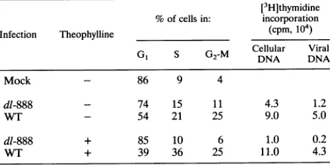
As shown in Table 2, cellskeptin serum-free medium and then infected with serum-free mocklysate showed largely G1 DNA content (see Materials and Methods for technical
de-tails). It is not clear whether the cells with G2 or S DNA
TABLE 2. Cellcycleanalysis of uninfected and infectedCV1 cells
[3H]thymidine %of cells in: incorporation
Infection Theophylline (cpm,104)
Cellular Viral
G1 S G2-M DNA DNA
Mock - 86 9 4
dl-888 - 74 15 11 4.3 1.2
WT - 54 21 25 9.0 5.0
dl-888 + 85 10 6 1.0 0.2
WT + 39 36 25 11.0 4.3
on November 9, 2019 by guest
http://jvi.asm.org/
[image:4.612.53.296.78.197.2] [image:4.612.316.556.607.727.2]content were actually cycling or whether they represented polyploidcells in thepopulation.When cellswereinfectedwith lowmultiplicitiesof dl-888orWTvirus(5PFUpercell),itwas
possible to detect an influence ofsmall t on host cell DNA induction.Forty-sixpercent of thecells infected withWTSV40
showed SorG2DNA content,comparedwith26% of thecells infected with dl-888. These differences wereeven more
pro-nounced when infectionswere carried out in the presence of
1.8 mM theophylline, a methylxanthine which causes growth
arrest inCV1 cells(35).We haveshownpreviouslythatsmall tallows CV1 cellstoovercome atheophylline-induced growth arrest which may result from theophylline inhibition of the
Na+/H+ antiporter (32).When infected cellsweremaintained inthepresenceoftheophyllinebefore flowcytometricanalyses
at 36 h, cells infected with small-tmutantviruses showed no
evidence ofprogression through the cell cyclewhile most of
the cells infected withWTSV40had S orG2DNA content.
Some of the increase in DNAcontent may have resulted
from accumulation of viral genomes. However, significant
cellular DNAsynthesisoccurred ininfectedCV1 cellsaswell.
Parallel dishes of infected cells were analyzed for [3H]thymi-dineincorporationtodetermine thestatusofongoingviral and
cellular DNAsynthesis. As shown in Table2, about twice as much cellular DNA synthesis was detected in cells infected withthe WTvirus than in cells infected with the small-tmutant
virus. Theophylline resulted in a 75% decrease in thymidine incorporation inmutant-infectedcells,whiletheophyllinehad noeffectoncells infected with WTSV40.Viral DNAsynthesis
showed the same general patterns, with little effect of
theo-phyllineonviral orcellularDNAsynthesis in WTinfections.
Althoughnotshownhere,theophyllinedidnotreduce levels of
large-T antigensignificantlyin mutant-infected cells(34a).
Effect ofmicroinjected smallton host DNAsynthesis.We
nextattemptedtorepeat the results described in theprevious paragraph (i.e., stimulation of cellular DNA replication by
small t) by microinjectionofpurified small t into CV1 cells.
However,wewereunabletomeasurehostcell DNAsynthesis
inthepermissive CV1 cellssuccessfullyusedtomeasureviral DNAsynthesis. Growth ofCV1 cells is extremelydifficult to arrest. When CV1 cells were maintained in the absence of serumfor several days, conditions required to reduce
thymi-dine incorporation, theywere easily damaged by microinjec-tion. Consequently, we examined the effect of smallt on the induction of hostcell DNAsynthesis bylargeTin
nonpermis-sive cells. Because no viral DNAsynthesis occurs in
nonper-missive rodent cells, autoradiography following thymidine
in-corporationwas usedas a measureofongoing cellularDNA
synthesis.
Primarymousekidneycellswerechosenbecausethese cells are terminally differentiated and do not have a proliferative
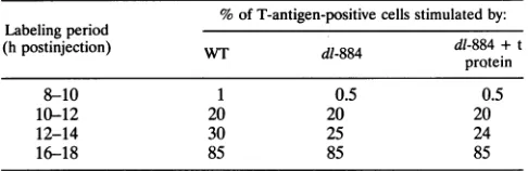
capacity.ViralDNA(atadilution of0.01 ,ug/,ul, corresponding to approximately 20 to 40 molecules) was injected into the nucleiofgrowth-arrestedcells,whichwerethenpulsed for2-h intervalsasindicated inTable3. After these labeling periods, cellswerefixed,stainedfor Tantigen, and thenprocessedfor
autoradiography (18). Dataarepresentedasthepercentageof
T-antigen-positive cells showing ongoing incorporationof thy-midine into DNA. In these cells, DNA from small-t mutant
virusdl-884inducedcellularDNA synthesisasefficientlyasdid WT SV40DNA and 85% of injected cells underwent DNA
synthesisby16to18 hfollowingmicroinjection. Coinjectionof
small-t antigenhad noeffectonthe time ofonsetorextentof
cellular DNA synthesis. Similar resultswere obtained witha second nonpermissive cell type, mouse 3T3 cells (data not shown). The inability ofpurified small t to stimulate cellular
[image:5.612.323.566.98.177.2]DNAsynthesis maybeaconsequenceofthecell typewehad
TABLE 3. Stimulation of DNAsynthesisinprimarymousekidney
cellsmicroinjectedwithviralDNA"
% ofT-antigen-positivecells stimulatedby:
Labelingperiod
(hpostinjection) WT dl-884 dl-884 + t
protein
8-10 1 0.5 0.5
10-12 20 20 20
12-14 30 25 24
16-18 85 85 85
aEachvalue shown is based on threeindependentexperiments.Lessthan1%
of themock-injectedcellsincorporatedthymidine.
to use in these microinjection experiments (nonpermissive
rodentcells). It is possible that smalltis able to stimulate cell DNAsynthesisonly in permissive cells.
DISCUSSION
SV40 T antigen is a multifunctional protein with several intrinsic biochemical activities required for virus production. The T antigen regulates the timing of the infection cycle in permissive cells, it represses its own transcription, and it initiates viral DNA replication. Initiation of viral DNA
repli-cation requiresbinding of the T antigen to the viral origin of replication (for a review, see reference 44). T antigen also induces cellular DNAsynthesis in quiescent cells; it transforms tissue culture cells and induces tumor formation in animals (46). T antigen forms stable complexes with a number of cellular proteins that have been implicated ingrowth control (e.g., Rbl and p53) and with the DNA polymerase-DNA primase complex(14).
Recently reported evidence indicates that the phosphoryla-tion state of the T antigen is of importance for various T-antigen functions, such as viral DNA replication and cell transformation(9, 14, 31). The T antigen contains two clusters of phosphorylated serine and threonine residues. One is adjacent to its DNA-binding domain (106, 111, Ser-112,Ser-120,Ser-123, andThr-124), and the second isnearthe carboxy-terminal region of theprotein(639, 676, Ser-677, Ser-679, and Thr-701) (36-39). Some of these serine residues are selectively dephosphorylated by PP2A, and in vitro dephosphorylation of these amino acids increases the bindingaffinity of the T antigen for the viralDNA-binding site (II) and, hence, viral DNA replication (31, 34). Underphos-phorylation of some of these amino acids is also associated with reduced transformation capacity of the large-T antigen (9). The SV40 small-tantigen binds cellular PP2A, reducing its activity against avariety of substrateswhich include the viral large-T antigen. Because PP2A has been shown to stimulate viralreplication when added to highly purified cell-free DNA replication systems (26, 49), it was not surprising that small t
reduced viral DNA synthesis in vitro by cellular extracts (5). Theparadoxaddressed by these experiments is that small-t
antigen is known to increase virus yields and plaque size in infected monkey kidney cells, suggesting that it enhances rather than depresses viral replication. The data presented here suggest that reduced viral yields can be related directly to reduced viral DNA synthesis in the absence of functional small-tantigen. One possible explanation derives from obser-vations of others that small t could induce continued cell
cyclingin nonpermissivecells(24)andthat itplayed arole in transformation ofnonpermissive cellsprimarily when growth-arrested cells were studied (28). These reports suggest that
on November 9, 2019 by guest
http://jvi.asm.org/
smalltfunctionstopromotecellcycle progression in infected cells.
Support
for this hypothesiscomesfromourflowcytometricanalyses
of infected CV1 cells, which showed that at lowmultiplicities,
more cells infected with WT SV40 than thoseinfected with the small-t mutant virus had DNA contents
equivalent
to the S or G2 level. Because of the prolongedserum starvation needed with CV1 cells and the low virus
multiplicities
used, host cell inductionwas quiteinefficient inthese
experiments
and not all cellsexitedG1.
However, these conditions did allowaneffect of small-tantigentobeobserved.Additional evidence that small t can promote cell cycle
pro-gression
was provided using theophylline to arrest cells. Intheophylline-treated
cells,smalltisessential for release of cellsfrom
Go.
Wehave shownpreviouslythattheophyllinedoes not appearto arrestcellsthrough changesincyclicAMPlevels but does affect activation of the Na+/H+ antiporter (32), a key enzymeingrowth
induction.Aneffect of smalltinstimulatingprogression
of cells into the cell cycle mightalso explain ourobservation that
coinjection
of small t with either WT ormutant
template
DNAresultedinstimulationofreplicationtosimilarextents.
Following microinjection,
smalltmight triggereventswhich
normally
occur inGO-G,
progression leadingtoexpression
of enzymes which promotereplication
atG1-S
andthe S
phase.
In this case, cells would be in anoptimal
environment for viral DNAreplication
oncesufficient levels oflarge-T antigen
wereexpressed
frommicroinjected
genomes.InadditiontoenzymessuchasDNApolymerase,invitro DNA
replication
studies(12)
have alsoshown that thecyclinA-cdc2complex
presentinS-phase
extracts canstimulate SV40repli-cation, possibly by promoting
therequired
Thr-124phosphor-ylation
oflargeT.Data shown in this report confirm those of several others
(10, 15, 19, 21, 45)
thatlarge
Tis sufficienttoinducecellularDNA
synthesis
innonpermissive
cells.Although
we wereunable to
study
the effect ofmicroinjected
smalltoncellularDNA
synthesis
inpermissive monkey
cells because of thefragility
of the cells under thelong
serumstarvationconditionsrequired
forgrowth
arrest,wedid carryoutsuchstudies withnonpermissive
cells. Wewereunabletodemonstrateaneffectof
purified
small-tprotein
oneither thetiming
ortheextentofinduction of cellularDNA
synthesis
inmicroinjected
nonper-missive cells. As in transformation(1),
a role for small t instimulating
cellcycle progression might
be apparentonly
whenlower concentrations ofT
antigen
arepresent.Alternatively,
it has been shown inmouseembryo
cells(24)
that smalltwas notrequired
to induce aninitial roundof cellularDNAsynthesis
butwas
required
forsubsequent
rounds of division to occur.However,
experiments
withtemperature-sensitive
mutantsin-dicated that in human cells
permissive
forSV40,
induction of cellular DNAsynthesis
and mitosisdepends
only
on thelarge-T
antigen (46).
Our studies have demonstrated that under certain
condi-tions,
small t influences cellcycle
progression.
It should benoted, however,
thatevenunderconditions in which CV1 cellsare not
severely growth arrested,
levels of viralDNAreplica-tion were
consistently
lowerin the absence of small t.Thus,
small t may promote viral DNA
replication through
some mechanism in addition topromotion
ofprogression
through
the cellcycle.
Alternatively, large
TmayinduceG1progression
by
activating
functions whichnormally
occur inmid-G,,
by-passing
some of the earlier steps ofGo-to-G1
transitions orearly
Gl.
Thismight
create anintracellular environment whichsuffices for host DNA
synthesis
but is limited inkey
factorsrequired
for theadditional demandsplaced
onthe cellby
viralDNA
synthesis.
The major finding of this study is that purified small t can stimulate DNA replication of microinjected WT or dl-884 genome. It does notseem likely that small t stimulates viral DNAsynthesis in microinjected cells only by altering levels of large-T antigen. Similar levels of total large T appeared early afterinfection witheither the WT or mutant virus, although T
antigenfrom mutantinfections appeared less phosphorylated
atveryearlytimes postinfection, even in one-dimensional gels ofimmunoprecipitates. Furthermore, peptide maps suggested that early after infection, the large T isolated from mutant infections was less
phosphorylated
than that of the WT in peptide 7. Results from several laboratories suggest thatdephosphorylation should correlate with increased, not de-creased, viral DNA synthesis (22, 29, 34, 49). However, it should bepointedoutthat these results were obtained from in vitro experiments, which do not necessarily reflect in vivo conditions. Our flowcytometric studies of infected CV1 cells suggest that small t can promote cell cycle progression in
permissive cells. This effect is probably mediated by the stimulation of the MAP kinase cascade, as reported very
recently by Sontagetal.(42).The advantages of having cells in the late G1 or S phase, if promoted by the small-t antigen,
mightthenoutweighwhateverinfluence small t might have on the phosphorylation state of proteins, such as the large-T antigen.
ACKNOWLEDGMENTS
WethankLucio Miele and WilliamSanslonefor criticalreading of the manuscript; Wolfang Deppert, Karl-Heinz Scheidtmann, and Gernot Walter foradviceon thepreparationof thephosphopeptide
maps of large-T antigen; Karl-Heinz Scheidtmann for help in the
interpretation of the phosphopeptidemaps;and E.Appellaforhelp in
conductingthephosphopeptide experiments.
This workwas supported in part by Public Health Service grant CA-21327(to K.R.)andaDeutscheForschungsgemeinschaftgrant(to
A.G.).
REFERENCES
1. Bikel, L.,X.Montano, M.Agha, M. Brown, M. McCormack, J.
Boltax, and D.Livingston. 1987. SV40small-t antigen enhances the transformation activity of limiting concentrations of SV40
large-T antigen.Cell48:321-330.
2. Borowiec, J. A.,F. B.Dean,P. A.Bullock, and J. Hurwitz. 1990.
Bindingandunwinding-howTantigenengagestheSV40origin
of DNA replication.Cell 60:181-184.
3. Bouck, N.,N.Beales,T.Shenk,P.Berg, and G. di Mayorca. 1978. Newregionof simian virus 40 genomerequiredforefficientviral transformation. Proc.Natl.Acad. Sci. USA75:2473-2477. 4. Boyle,W.S.,P.vanderGeer,and T. Hunter.1991.
Phosphopep-tidemappingandphosphoaminoacidanalysis bytwodimensional
separationonthin-layercelluloseplates.MethodsEnzymol.202: 110-149.
5. Carbone, M., J. Hauser,M. C.Carty,K.Rundell,K.Dixon,and A.S. Levine.1992. Simian virus 40(SV40)small-tantigeninhibits
SV40DNAreplicationinvitro. J. Virol. 66:1804-1808.
6. Casaz, P.,R.Sundseth,and U. Hansen.1991. Transactivation of the simian virus 40 late promoter by large T antigen requires
bindingsitesfor thecellulartranscriptionfactorTEF-1. J. Virol. 65:6535-6543.
7. Chang,L.S.,M.Pater,N.Hutchinson,andG.diMajorca. 1985. Transformationbypurifiedearlygenesof simian virus 40. Virol-ogy 146:246-261.
8. Cicala, C.,F.Pompetti,P.Nguyen,K.Dixon,A.S.Levine,and M. Carbone.1992.SV40small-tdeletionmutantspreferentially
trans-form mononuclearphagocytesand Blymphocytesin vivo. Virol-ogy 190:475-479.
9. Deppert,W.,M.Kurth,M.Graessmann,A.Graessmann,and U.
Knippschild. 1991. Altered phosphorylationatspecificsites
con-fersa mutantphenotypetoSV40wildtypelarge-T antigenin flat
on November 9, 2019 by guest
http://jvi.asm.org/
revertantsofSV40-transformed cells.Oncogene6:1931-1938.
10. Dobblestein, M., A. D. Arthur, S. Dehde, K. van Zee, A. Dick-manns, and E. Fanning. 1992. Intracistronic complementation
reveals a new function ofSV40Tantigenthat co-operates with Rb and p53 binding to stimulate DNA synthesis in quiescent cells. Oncogene 7:837-847.
11. Dora, S., C. Schwarz, M. Baack, A. Graessmann, and R. Knippers. 1989. Analysis of a large-T-antigen variant expressed in simian virus 40-transformed mouse cell line mKS-A. J. Virol. 63:2820-2828.
12. D'Urso, G., R. Marracino, D. Marshak, and J. Roberts. 1990. Cell cycle control of DNA replication by a homologue from human celis of the p34 cdc2 protein kinase. Science 250:786-791. 13. Ellmann, M., I. Bikel, J. Figge, T. Roberts, R. Schlossman, and D.
Livingston. 1984. Localization of the simian virus small t antigen in the nucleus and cytoplasm of monkey and mouse cells. J. Virol.
50:623-628.
14. Fanning,E. 1992. Simian virus 40 large-T antigen: the puzzle, the
pieces, andthe emerging picture. J. Virol. 66:1289-1293.
15. Floros, J., G. Jonak, N. Galanti, and R. Baserga. 1981. Induction ofcellularDNA replication in Gl-specific Ts mutants by micro-injectionofSV40 DNA. Exp. Cell Res. 132:215-223.
16. Goswami,R., B. Turk, K.Enderle,A. Howe, and K. Rundell. 1992. Effect ofzinc ions on the biochemical behavior of simian virus 40
small-t antigenexpressed in bacteria. J. Virol. 66:1746-1751.
17. Graessmann, A., and M. Graessmann. 1983. Microinjection of tissueculture cells. Methods Enzymol. 101:482-492.
18. Graessmann,A., M. Graessmann, E.Guhl, and C. Mueller. 1978.
Quantitative correlation between simian virus 40 T-antigen syn-thesis and late viralgene expression in permissive and
nonpermis-sive cells. J. CellBiol. 77:1-8.
19. Graessmann,A., M. Graessmann, and C. Mueller. 1981.
Regula-tionofSV40 gene expression. Adv. Cancer Res. 35:111-149.
20. Graessmann,M., E. C. Bumke-Vogt, and A. Graessmann. 1984. The second large simian virus 40 T-antigen exon contains the information for maximal cell transformation. J. Mol. Biol. 180: 111-129.
21. Graessmann, M., and A. Graessmann. 1976. Early simian virus 40-specific RNA contains information for tumor antigen
forma-tion and chromatin replication. Proc. Natl. Acad. Sci. USA 73:366-370.
22. Grasser, T., K. Mann, and G. Walter. 1987. Removal of serine phosphates from simian virus 40 large-T antigen increases its
ability tostimulate DNA replication in vitro but has no effect on
ATPase and DNAbinding. J. Virol. 61:3373-3380.
23. Hirt, B. 1967.Selective extraction of polyoma DNA from infected
mouse cellcultures.J. Mol. Biol. 26:365-369.
24. Hiscott, J. B., and V. Defendi. 1981. Simian virus 40 gene A regulation ofcellularDNAsynthesis. II. In nonpermissive cells. J.
Virol.37:802-812.
25. Knippschild, U., J. Kiefer, T.Patschinsky, and W. Deppert. 1991.
Phenotype-specificphosphorylation of simian virus 40 tsA mutant large TantigensintsAN-type and A-type transformants. J. Virol. 65:4414-4423.
26. Lawson, R., P. Cohen, and D. P. Lane. 1990. Simian virus 40
large-T-antigen-dependent DNA replication is activated by
pro-tein phosphatase2Ainvitro. J. Virol. 64:2380-2383.
27. Maniatis, T., E. F. Fritsch, and J. Sambrook. 1982. Molecular cloning: a laboratory manual. Cold Spring Harbor Laboratory,
ColdSpringHarbor, N.Y.
28. Martin, R., V. Setlow, C. Edwards, and D. Vembu. 1979. The roles of thesimian virus 40 tumorantigens in transformation of Chinese hamsterlung cells. Cell 17:635-643.
29. Mohr, I., B.Stillman,andY.Gluzman. 1987. Regulation ofSV40 replicationbyphosphorylation of T antigen. EMBO J. 6:153-160.
30. Mueller, C., A. Graessmann, and M. Graessmann. 1978. Mapping of earlySV40specificfunctions by microinjection of different early
viral DNA fragments. Cell 15:579-585.
31. Mumby, M. C., and G. Walter. 1991. Protein phosphatase and DNA tumor viruses: transformationthrough the back door? Cell Regul. 2:589-598.
32. Mungre, S., C. Renz, and K. Rundell. 1991. Inhibition of the Na+/H+-antiporter by theophylline in growth-arrested monkey
kidneycells.Exp. Cell Res.193:236-239.
33. Pallas,D. C., L. K. Shahrik, B. L.Martin,S. Jaspers, T. B.Miller,
D. L. Brautigan, and T. M. Roberts. 1990. Polyoma small and
middleTantigensand
SV40
small-tantigen form stablecomplexes with protein phosphatase 2A. Cell 60:167-176.34. Prives, C. 1990. The regulation functions of
SV40
T antigen areregulated by phosphorylation. Cell 61:735-738. 34a.Rundell, K. Unpublished data.
35. Rundell, K., and J. Cox. 1979. Simian virust antigen affects the sensitivity of cellular DNA synthesis to theophylline. J. Virol. 30:394-396.
36. Scheidtmann, K. H., M. Buck, J. Schneider, D. Kalderon, E.
Fanning, and A. E. Smith. 1991. Biochemical characterization of phosphorylation site mutants of simian virus 40 large T antigen: evidence for interaction between amino- and carboxy-terminal domains. J. Virol. 65:1479-1490.
37. Scheidtmann, K.-H., B. Echle, and G. Walter. 1982. Simian virus 40 large T antigen is phosphorylated at multiple sites clustered in two separate regions. J. Virol. 44:116-133.
38. Scheidtmann,K. H., M. Hardung, B. Echle, and G. Walter. 1984.
DNA-binding activity of simian virus 40 large T antigen correlates with a distinct phosphorylation state. J. Virol. 50:1-12.
39. Scheidtmann, K. H., M. C. Mumby, K. Rundell, and G. Walter. 1991. Dephosphorylation of simian virus 40 large-T antigen and p53 protein by protein phosphatase 2A: inhibition by small-t antigen. Mol. Cell. Biol. 11:1996-2003.
40. Shenk, T., J. Carton, and P. Berg. 1976. Construction and analysis of viable deletion mutants of simian virus 40. J. Virol.18:664-671.
41. Sleigh, M., W. Topp, R. Hanich, and J.
Sambrook.
1978. MutantsofSV40with an altered small-t protein are reduced in their ability to transform cells. Cell14:79-88.
42. Sontag, E., S. Fedorov, C. Kamibayashi, D. Robbins, and M. Mumby. 1993. The interaction of
SV40
small t tumor antigen with protein phosphatase 2A stimulates the Map kinase pathway and induces cell proliferation. Cell 75:887-897.43. Srikumar, C., V. B. Kraus, B. Kroger, K. Munger, P. M. Howley, W. C. Phelps, and J. R. Nevins. 1992. Adenovirus ElA, simian
virus 40 tumor antigen, and human papillomavirus E7 protein share the capacity to disrupt the interaction between transcription factor
E2F
and the retinoblastoma gene product. Proc. Natl. Acad. Sci. USA 89:4549-4553.44. Stillman, B. 1989. Initiation of eukaryotic DNA replication in vitro. Annu. Rev. Cell Biol. 5:197-245.
45. Tjian, R., G. Fey, and A. Graessmann. 1978. Biological activity of
purified simian virus 40 T-antigen proteins. Proc. Natl. Acad. Sci. USA 75:1279-1283.
46. Tooze, J. 1981. Molecular biology of the DNA tumor viruses, 2nd ed. Cold Spring Harbor Laboratory, Cold Spring Harbor, N.Y. 47. Topp, W. C. 1980. Variable defectiveness for lytic growth of the dl
54/59 mutants of simian virus 40. J. Virol.33:1208-1210.
48. Turk, B., A. Porras, M. C. Mumby, and K. Rundell. 1993. Simian virus 40 small-t antigen binds two zinc ions. J. Virol. 67:3671-3673.
49. Virshup, D. M., M. G. Gauffman, and T. J. Kelly. 1989. Activation
ofSV40DNA replication in vitro by cellular protein phosphatase 2A. EMBO J. 8:3891-3898.
50. Walter, G., R. Ruediger, C. Slaughter, and M. Mumby. 1990.
Association of protein phosphatase 2A with polyoma virus me-dium tumor antigen. Proc. Natl. Acad. Sci. USA 87:2521-2525.
51. Yang, S. I., R. L. Lickteig, R. C. Estes,K. Rundell,G. Walter, and M. C. Mumby. 1991. Control of protein phosphatase 2A by simian
virus 40 small-t antigen. Mol. Cell. Biol. 64:1988-1995.