0022-538X/94/$04.00+0
Copyright © 1994,AmericanSocietyforMicrobiology
Induction
In
Vitro of Primary
Cytotoxic T-Lymphocyte Responses
with
DNA
Encoding
Herpes
Simplex Virus
Proteins
RICHARDJ. D. ROUSE, SMITA K. NAIR,t SHARI L. LYDY, JOANNE C. BOWEN,
ANDBARRY T. ROUSE*
DepartmentofMicrobiology, College of Veterinary Medicine, University of Tennessee,
Knoxville,
Tennessee37996-0845Received 3 February 1994/Accepted 27 May 1994
Vaccines which successfully protect against virus infections usually need to induce a broadly reactive immuneresponsewhichincludes the inductionof cytotoxicTlymphocytes (CTL). In this study, we have used aconvenient in vitroapproachtoinvestigate ifplasmidDNAs encoding proteins of herpessimplexvirus(HSV) are capable of inducing primary CD8+ CTL. Dendritic cells or macrophages were transfected with either plasmidDNAencoding glycoprotein Bor DNA encoding the immediate-earlyprotein ICP27. These antigen-presentingcells (APC)were thenused tostimulate enrichedpopulations of naive T cells in microcultures for 5days in vitro. Antigen-specific CD8+ CTL which reacted both with specific protein-expressing targetsand with syngeneic targets infected with HSV could be demonstrated. Dendritic cells, as APC, generated the maximal responses, but such cells needed to be transfected with DNA in the presence of a cationic lipid. However,macrophages could act as APC when they were exposed to purified DNA. HSV-primedsplenocytes were also shown to generatespecific CTL responses when they were stimulatedwith purified DNAencoding ICP27. The novel approach described in this paper promises to be extremely useful, since defining
immunogenicityprofilesandidentifyingepitopes on viral proteins should be easier and moreconvenientwhen workingwith DNA and investigatingvariables in vitro.Thisisparticularlythe casewithcomplexviruses such asHSV, mostof whose encodedproteins haveyet to beisolatedinsufficient quantityorpurity to perform in vivoimmunological studies.
The recent observation that protective immunity can be inducedfollowing genetic immunization with DNA (7, 21-23, 26) opens up a new approach to vaccination. This novel immunization strategy promises some advantages, such as resistance to neutralization by any existing antibody, gene expression in host cells so that broadly reactive immunity should be induced, and the potential ease of generating combination vaccines. However, many parameters require detailed analysis before genetic immunization becomes an acceptable procedure. These includedefining the proteins and peptides toencode, particularly when aiming toachieve opti-malimmunity against complex viruses, suchasherpes simplex virus (HSV), that expresses multiple proteins (17). Other issues include the evaluation ofmeansofadministration that will optimally induce componentsofimmunity that best cor-relatewithprotection.
With many viruses, the attainment of protective immunity usually means that an effective response of cytotoxic T-lym-phocytes (CTL)mustbeinduced(6, 19). Suchresponses occur only if antigens are appropriately processed by antigen-pre-senting cells (APC), and this may fail to occur with certain typesofvaccines, inparticular with those whichareinactivated (4, 9, 20). As regards the role of antigens and presentation systemsthat result inCTLinduction, it ismoreconvenientto
perform studies in vitro, particularly since approaches found effective in the moredemanding in vitrosystem are invariably also successful in vivo (5, 10, 14). Accordingly,in thepresent
communication, we have useda primaryin vitro CTL induc-tionsystem (12-14)to establishconditions under which
prep-*Corresponding author. Phone: (615) 974-4026. Fax: (615)
974-4007.
tPresent address: Department of Experimental Surgery, Duke University Medical Center, Durham, NC 27710.
arations ofDNA thatencode HSVproteinscan induce virus-specificCTL. Wedemonstrate that twoproteins, especiallyif expressed in dendritic cells (DC) as antigen-presenting cells (APC), induce potentprotein andvirus-specific CTL.
MATERIALSAND METHODS
Mice.Female7- to 8-week-old C57BL/6
(H-2b)
andBALB/c(H-2")
mice were obtained from Harlan Sprague-Dawley, Indianapolis, Ind.,orfrom Sasco,Omaha, Nebr.Inconducting theresearch described in thisarticle, theinvestigators adhered to the guidelines proposed by the Committee on the Care of Laboratory AnimalResources, Commission onLife Sciences, National Research Council. The facilities arefully accredited by the American Association for Accreditation of Laboratory Animal Care.DNApreparation andprotein expression. ThecDNAclone of the HSV type 1 (KOS) (HSV-1) gB gene in pSR175 (designated gBpSR175)was kindlyprovided by Martin Mug-geridge.The3.7-kbgBcDNAwascutfromgBpSR175 by using HindIII (5')andBamHI (3') andwassubsequentlysubcloned into the plasmid vectors pUC19 and pcDNAI (Invitrogen). The 2.4-kb ICP27gene wascloned intoM13mpl8asdescribed previously (3) and wassubsequently subcloned into the
pcD-NAI vector
(HindIII-XbaI
sites) oriented 5'->3'totheplasmid cytomegalovirus promoter. The vector pcDNAI was chosen because it containsacytomegaloviruspromoterand enhancer for gene expression in eucaryotic cells and a ColEl-like high-copy-number plasmid origin ofreplication, allowing thepreparation oflarge amounts ofrecombinant
plasmid
DNA. Recombinant plasmids containing the gB or ICP27 gene of HSV-1 were used to transform Escherichia coli(strain
MC1061) underampicillin andtetracyclineselection. Recom-binant plasmid DNAwasisolated from transformed bacterial 5685
on November 9, 2019 by guest
http://jvi.asm.org/
colonies and was prepared by standard techniques using alka-line lysis, two purifications on CsCl gradients, and ethanol precipitation (18). The purity and concentration of DNA were analyzed by A260 and A280 and by agarose gel electrophoresis and ethidium bromide staining.
Expression of the cloned gB and ICP27 genes in vitro was confirmed by transfecting splenic macrophages
(M+)
and DC (5 x 10⁶ cells) from naive BALB/c female retired breeders with 5 pLg of recombinant plasmid DNA and 15Fig
of the cationic lipid DOTAP (Boehringer Mannheim Biochemicals, Indianapolis, Ind.). Transfected cells were lysed with 0.5 ml of RIPA buffer (150 mM NaCl, 1% Triton X-100, 0.5% deoxy-cholate, 0.1% sodium dodecyl sulfate [SDS], 50 mM Tris [pH 8.0]). Cell lysates were immunoprecipitated with either mouse anti-ICP27 monoclonal antibody (MAb) (1:500) or mouse anti-gB polyclonal antibody (1:500) and protein A Sepharose (Pierce). The precipitates were analyzed by gel electrophoresis under reducing conditions on 10% gels with a 29.2:0.8 acryl-amide-to-bisacrylamide ratio. Proteins were visualized by Western blotting (immunoblotting) using goat anti-mouse immunoglobulin conjugated to alkaline phosphatase (Bio-Rad).The relative efficiencies of transfection ofICP27 DNA in Mf and DCwere determined by comparing relative amounts ofICP27 protein expressed to input DNA. Cells (2x
106)
were transfectedwith either5,ugof vector control DNA or 1, 5, 25, or 50pg
ofICP27 DNA using DOTAP as described previously. Afterovernight transfection, cells were lysed and proteins were processed by immunoprecipitation, SDS-polyacrylamide gel electrophoresis (PAGE), and immunoblotting. Alternatively, M4 and DC were incubated in the presence of exogenous ICP27 DNA in culture medium overnight in the absence of cationic lipid, andICP27 protein expression was analyzed as described above. Protein band intensities on nitrocellulose blots wereanalyzed by scanning densitometry using Molecular Analyst software (Bio-Rad).Virus and peptides. HSV-1 strain KOS was propagated on Vero cell monolayersand stored as infectious cell preparations at -70°C. Viral titersfor HSV-1 KOS were expressed as 50% tissueculture infectiousdoses. TheH-2b-specificpeptide (ami-noacids 498 to505 [SSIEFARL])was synthesized on resin to have unblocked (free) amino and carboxyl ends (Research Genetics, Birmingham, Ala.).
APC and responderT cells. Splenocytes obtained from naive C57BL/6 or BALB/c female mice were treated with ammo-nium chloride-Tris buffer for 3 min to deplete erythrocytes. Splenocytes(3 ml) at 2x 10⁷ cells per ml were layered over a 2-ml metrizamide gradient column (Nycomed Pharma AS, Oslo, Norway; analytical grade, 14.5 g added to 100 ml of
phosphate-bufferedsaline [PBS], pH 7.0) and were centrifuged at600x gfor 10min. Cells from the interface were collected, and analysis with a fluorescence-activated cell sorter (FACS) showed on average 60 to 70% DC (MAb 33D1; kindly pro-vided by Ralph Steinman, The Rockefeller University) (15), 1% M4 (MAb F4/80)(2), 15 to 20% T cells, and 10 to 12% B cells. Thepellet was resuspended and allowed to adhere for 1 h. More than 75 to 80% of the adherent population was identified as M4P by FACS analysis, with approximately 5% lymphocytes and <5% DC. B cells were separated from the
nonadherentpopulation by panning on anti-immunoglobulin-coatedplates. The separated cell population, which consisted of >80% T lymphocytes by FACS analysis, was used as responder naive T cells.
Pulsing of APC with DNA preparations. DC or
M(
were exposed topurified DNApreparations either without or in the presence of the cationic lipid DOTAP. For transfection withthis cationic lipid, DNA preparations (50 to 75 jig) were reacted with 15
jig
of DOTAP in 200,ul
of PBS (pH 7.2) for 10min
at room temperature in a 4-ml polystyrene tube (Falcon, Lincoln Park, N.J.). The APC (5x10⁶
cells) were transfected with a DNA-DOTAP mixture in 1 ml ofRPMI-5% fetal calf serum (FCS) medium at37°C
for 3 h with occasionalshaking. At the end of the incubation, the cells were washed and used as stimulators. The procedure was not visibly toxic to APC, as determined by trypan bluestaining.Induction of primary CTL in vitro. Purified T cells (1.0x
107
cells per ml) from naive BALB/c orC57BL/6
mice were cultured with DC or M4 (1.0 xl05
cells per ml) to give responder-to-stimulator ratios ranging from 100:1 to 6.25:1 in 200RI
of LDA medium (NCTC 109 and RPMI 1640 [1:1]), supplemented with 10% heat-inactivated FCS, 10mM
L-glutamine, 1 mM oxalacetic acid, 0.2 U of bovine insulin per ml, and 50RI
of 2-mercaptoethanol in 96-well U-bottom plates. The plates were incubated at37°C
under 5%CO2
for5 days. CTL assays were performed on the 5th day by taking 100RI
of medium from each200-plI microculture well and adding 100 pA of target cells(104).
Alternatively, T cells (5x10⁶
cells per ml) and APC (2x10⁵
cells per ml) were cultured in LDA in 96-well U-bottom plates to give a responder-to-stimulator ratio of 25:1. After 5 days, the cells were used as effectors in a standard 4-h 51Crrelease assay.Restimulation of HSV-primed effectors with DNA. Spleens from mice immunized with infectious HSV three times at 2-week intervals were collected 10 to 14 days after the last immunization, and bulk cultures were set up in six-well plates (Corning Glass Works, Corning, N.Y.) as described elsewhere (3). Briefly, 1.25x
107
splenocytes were cultured per well in 5 ml of LDA medium. The splenocytes were restimulated with DNA at a concentration of 5,ug/ml
for 5 days at37°C.
Target cells for cytotoxicity assays. EMT6 cells (H-2" [mam-mary adenocarcinoma cells provided by Ed Cantin, City of the Hope National Medical Center, Duarte,
Calif.]),
EL4
cells(H-2*
lymphoma[American
Type Culture Collection]), and YAC-1 cells (H-2,lymphoma) were all cultured in Dulbecco modified Eagle medium (Gibco, Grand Island, N.Y.) supple-mented with 10% heat-inactivated FCS. EMT6 cells were transfected with the recombinant plasmid pJF24, containing the HSV-1 ICP27 gene (EMT6-27), by Frank Jenkins, Uni-formed Services University, Bethesda, Md. This plasmid con-tains theICP27gene and its own promoter as well asthe G418 resistance gene under the control of the HSV thymidine kinase promoter. These cells were grown in Dulbecco medium with 10%FCS containing 300,ug
of G418 per ml. EMT6 cells were also transfected with pcDNAgB by using DOTAP as described previously. These cells were transfected overnight prior to51Cr
labeling and were used as targets.
Cytotoxicity assays. All target cells were labeled with5tCrby reacting 2 x
106
cells in 500 plA of RPMI 1640 plus 10% FCS with 100,uCi
ofNa25'CrO4
for 90min at37°C.
The cells were washed three times prior to addition to effector cells. To detect HSV-specific lysis, target cells were infected with HSV (at a multiplicity of infection of 5) at the same timeas
5tCrlabeling. To detectICP27-specific lysis, EMT6-27 cells were used. To detect gB-specific lysis, two systems were used. ForH-2d-specific responses, EMT6 cells were transfected with pcDNAgB in the presence of DOTAP, whereas in theH-2b
system, gB killing was detected by adding the CTLepitope
peptide (amino acids 498 to 505 [SSIEFARL]) (12) to 'Cr-labeled EL4 cells(105/ml) at a concentration of 10
,ug/ml
to 2 x 10⁶ cells. All5'Cr
release assays were run for 4 h at various effector-to-target cell ratios in 96-well V-bottom plates. Prior to harvesting, the plates were centrifuged and 100pAl
of theon November 9, 2019 by guest
http://jvi.asm.org/
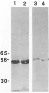
12 3 4
65-
56-
30-FIG. 1. Expression of gB andICP27 in splenic MX and DC. Lysates from cells transfected with recombinant plasmidswere
immunopre-cipitated with anti-gB oranti-ICP27 antibody andwere analyzed by SDS-PAGE andWestern blotting. Truncated gB in Mf (lane 1) and DC (lane 2) and full-lengthICP27expressed in MX (lane 3) and DC
(lane 4)at24 hposttransfectionareshown.
culture supernatant was collected to measure radioactivity.
Percentspecific releasewascomputedas(experimental release
- spontaneous release)/total release x 100.
Each assay was performed in triplicate, and only
experi-ments with spontaneous release values of 20% or less were
reported.
In vitro antibody depletions. Effector cells generated by stimulating naive T cells with DC pulsed with pcDNAgB or
pcDNAICP27-DNA-DOTAPat aresponder-to-stimulator
ra-tio of 25:1 were depleted of either T cells, CD8+ cells, or
CD4+ cells by using anti-Thy-1.2, anti-CD8, and anti-CD4 MAbsalongwithcomplement, respectively. Ratanti-Thy and anti-CD8 and rabbit Low-tox-m complement were obtained
from CedarlaneLaboratories (Accurate Chemical and Scien-tific, Westbury, N.Y.).The GK1.5 (anti-CD4) hybridoma cell linewasobtained from the AmericanType Culture Collection
(Rockville, Md.) andwasusedasascites fluid (40to50,ug/5 x 106 cells). Effectorswere incubated with the required volume
of MAbat4°C for 1h. The cellswerewashed twice andwere
resuspended in complement ataconcentration which didnot
cause lysis of the cells when they were treated with
comple-mentalone. After 60minofincubationat37°C,thecellswere
washed and used aseffectors in theCTLassay.
RESULTS
Expression ofHSVproteins.Following transfection of both M4 andDC with DNAencoding gBandICP27,viralproteins
weredetectable,asshownby SDS-PAGE andWesternblotting
(Fig. 1). The ICP27 proteinappearedasfull length in both cell
types, whereas gB was truncated to a 55-kDa protein rather
than the normal 1 10-kDaprotein.Itisnotuncommontodetect truncations of gB, since this protein is highly sensitive to
proteolytic cleavage (25). Densitometer analysis of ICP27 protein expression after transfection of
M4
and DC did notreveal major differences (within 20%) in the total amount of protein made by the two cell types (data not shown). In addition, Mf exposedto25,ugofpurified pcDNAICP27inthe absence oftransfectingagentexpressed asmall but detectable
amountofICP27 protein; however, DCsimilarly exposed to
[image:3.612.133.214.73.225.2]pcDNAICP27DNAfailed toexpress detectableprotein.
TABLE 1. Primary induction ofCTLwith
plasmid DNA-transfectedAPC'
Percentspecificlysis APC Stimulant
EMT6-27 EMT6 HSV EMT6 EL4 HSV
DC None <1 <1 <1 <1
DC pcDNAICP27 46 4 11 + 1.5 <1 1 + 1
DC pcDNA <1 <1 <1 <1
M4 ~pcDNAICP27 27±2 <1 <1 <1
M( pcDNA <1 <1 <1 <1
aNaive enriched T cells fromBALB/cmice were cultured for 5 days in vitro with syngeneic APC(M4 orDC) that had been transfected with plasmid DNA
viathe DOTAP procedure (see Materials and Methods). Various responder-to-stimulator ratios(100:1to6.25:1)wereused,but the data shown in the table were for the ratio 50:1. Background release from targets during the 4-h51Crrelease assay was15%orless.
Primary CTL induction with APC transfected with DNA. Splenic
M4
and DCwere purified fromyoung immunologi-cally naive BALB/c mice and were transfected with either plasmid DNA or plasmid DNA encoding ICP27. Following transfection, washed cellswereculturedatvarious stimulator-to-responder ratios with naive enriched Tcells for 5 days.At the end ofculturing, the cytolytic activities of the cells were testedagainstarangeof target cells. As is evident in Table 1, both APC types exposed to pcDNAICP27 induced protein-specific primary CTL responses. However, responses were higher when DCwereusedasthe APC.Inaddition, when DC were used as APC for naive T cells, cytolytic activity was detectable by using HSV-infected syngeneic target cells.Furtherexperimentswereperformedtotestthespecificity of CTL induced invitro. Forthis purpose, DCweretransfected with either pcDNAICP27 or pcDNAgB in the presence of DOTAP, and these cells were used as APC. Cultures were performed at optimal responder-to-stimulator ratios (25:1). The results, depicted in Table 2, clearly show that both gB-specific and ICP27-specific CTL were generated. Thus, gB-induced CTL thatwerecytotoxic for gB but thatwere not ICP27-expressing targets. Similarly, the ICP27-specific cells failed to kill gB-expressing targets. In this same experiment, control uninfected EMT6 cells and theNKcell target YAC-1 cellswere notkilled.Inotherexperiments, primarygB-specific CTL generated by induction with pcDNAgB-transfected DC wereshown to kill syngeneic but not allogeneic HSV-infected targets cells(datanotshown). InTable3, the data show that the cytotoxicity generated in the primary in vitro induction systemwasmediatedby CD8+ Tcells.
TABLE 2. Primary induction of CTL in vitro with DCtransfected with plasmid DNAa
Percentspecific lysis
Stimulant :
EMT6-27 EMT6-gB YAC-1
None 50:1 <1 <1 1± 1
pcDNAICP27 80:1 28+2 <1 2± 1
pcDNAICP27 20:1 15 1 <1 1± 1
pcDNAgB 80:1 <1 45±2 0±1
pcDNAgB 20:1 <1 10±1 1±1
pcDNA 80:1 <1 <1 2 1
pcDNA 20:1 <1 <1 1 1
aDC asAPCfromBALB/cmiceweretransfected withplasmidDNAin the presence of DOTAP andwerecultured withsyngeneicnaive enriched T cells for 5daysat aresponder-to-stimulatorratio of 25:1. Onday 5,effectorswerepooled
andcytotoxicityassayswere setupatvariouseffector-to-targetcell ratios.
bE:T,effector-to-targetcell ratio.
on November 9, 2019 by guest
http://jvi.asm.org/
[image:3.612.313.557.94.176.2] [image:3.612.313.556.582.680.2]TABLE 3. Evidence that CTLgenerated in primary induction byDNA areCD8+Tcellsa
Percentspecific lysisEMT6-27 Treatment
75:1 37:1 16:1
C' 16+ 1.8 12±3 3±2
Anti-Thy-1+C' <1 <1 <1
Anti-CD8+C' <1 <1 <1
Anti-CD4+ C' 15.5 ±1 ND ND
aDC from BALB/c mice were transfected with pcDNAICP27 in the presence
ofDOTAP and were used to induceprimary CTL in naive syngeneicT-cell cultures. In anexperiment of similardesign,inwhich effector cells from T-cell cultures were stimulated with DCtransfectedwithpcDNAgBin the presence of
DOTAP,the percentspecificlysisofsyngeneicHSV-infected target effector cells of 18% (effector-to-target cell ratio, 50:1)was abolished by treatment with
anti-CD8+C'.C',complement;ND, no data.
[image:4.612.64.304.91.164.2]The experiments recorded in Tables 1 to3 all demonstrate primary CTL responses with APC transfected with DNA in the presenceof DOTAP. However, as shownin Table 4, cytotoxic responses could be obtained in a naive T-cell population by using APC exposed to purified DNA (in the absence of DOTAP). Thus, splenic Mf were exposed to pcDNAgB or pcDNAICP27 inmicrocultures for 24h, after which they were addedtonaive T-cell and DC cultures (responder-to-stimula-tor ratio, 25:1). After 5 days of incubation, the cytotoxicities against different target cells were measured. As is evident in Table 4, this procedure resultedin thegeneration ofspecific CTL in both the gB and the ICP27 systems. Similar results wereobtained in three otherexperiments. Inthecaseof
M4
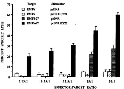
exposed topurified pcDNAgB, theC57BL/6 systemwasused and the target peptide forgB-specific CTL(amino acids 498 to 505) (12) was used to detect specific lysis. Not shown is the observation that when DCwere similarly exposed topurified DNA without the cationic lipid, CTL responses were not induced. As mentioned previously, although M( exposed to purified DNA in the absence of DOTAP do produce some detectableprotein,suchprotein couldnotbe detected in DC. Restimulation ofHSV-primed splenocytes. Unfractionated spleen cells from mice previously infected with HSV-1 were cultured in bulk cultures for 5days with purified pcDNAICP27 orwith control pcDNA, and the cellswere tested for cytotox-icity against antigen-expressing target cells. As is evident in Fig. 2, potent cytotoxicactivity against ICP27-expressingtarget cells was demonstrated with the pcDNAIPC27-exposed splenocytes. Normal unfractionated splenocytes exposed toTABLE 4. Generation ofprimaryCTL from naive cellsbyM4 exposedtopurifiedDNAW
Stimulant Percent specificlysis
Expt t (
Exptto
Mf
EL4-498-505 EL4 EMT6-498-505 EMT6-271 pcDNAgB 28±1 <1 1.2 ND
1 pcDNA <1 <1 <1 ND
1 None <1 <1 <1 ND
2 pcDNAICP27 ND ND <1 15± 1
2 pcDNA ND ND <1 <1
2 None ND ND <1 <1
a M (5 x10⁵cells perml)weretreated withpurifiedDNA(5
p.g/ml)
for 24h,and then DCmicrocultures of T cells(responder-to-stimulatorratio of 25:1) wereadded. CTL assays were done after 5daysofculture. In experiment 1,M4
were exposed to pcDNAgB in the H-2b system (C57B1J6 mice), while in
experiment 2, H-2d Mw were exposed to pcDNAICP27 (BALB/c mice). In
experiment 3,M4 exposedtopcDNAgB generated gB-specificCTLin T cells
(DCcultures in theH-2d[BALB/c]system).ND, no data.
v.)
I-P3 u) U. Eu
3
3.13:1 6.25:1 12.5:1 25:1 50:1
EFFECTOR:TARGET RATIO
FIG. 2. Cytotoxicity resulting from stimulation in vitro of HSV-primedsplenocyteswithpurifiedDNA. Notshownarevaluesoflysis forunstimulated cells primed against EMT6 and EMT6-27 targets. Levels were approximately the same as those shown for pcDNA-stimulated cells.
pcDNAICP27under the same bulk culture conditions failed to generate detectable CTLactivity (datanotshown).
DISCUSSION
Our resultsdemonstrate that CTL responsescanbe gener-ated invitro fromnaive ormemoryTcellsupon exposureto APC transfected with plasmid DNA encoding HSV genes drivenbyacytomegalovirus promoter.PrimaryCTLresponses were investigatedwith twoproteins, theglycoprotein gB and the immediate-early protein ICP27. The responses generated were not only protein specific butwere also reactive against HSV-infected target cells. From the genotype of the target cells killed and that of the effector cells,the responses being induced were mediated by major histocompatibility complex class I-restricted CD8+ CTL. However, the possibility that other functional aspectsof T-cellimmunitywere alsoinduced was not fully evaluated, and this aspect is under further investigation.
The approach that we havedescribed appears to be novel.It promises to be extremelyuseful, since defining immunogenic-ity profiles and identifying component epitopes onviral pro-teins should be easier andmoreconvenient whenworkingwith DNA. This is particularly the case when studying complex viruses suchasHSV,mostofwhose encodedproteins have yet tobeisolated insufficientquantityorpurityforimmunological studiestobeperformed.Moreover, sincethere isnow alively interest ingenetic vaccines(7, 21-23, 26), screeningcandidate DNA constructs for various aspects ofimmunogenicity by in vitro analyses is likely to be a more efficient approach than extensive in vivo studies. Interestingly, both of the HSV protein-encoding plasmid DNA constructs that we have de-scribed were, in preliminary studies, immunogenic in vivo when administered inan appropriatefashion (17a).
In most of the experiments described in the present com-munication, plasmid DNA wastransfected into APCby using the cationic lipid DOTAP. Moreover, as observed with ap-proachesinwhichproteins areintroduced into APCwith, for example,pH-sensitive liposomes (13, 14),DC were invariably superiortoMpasAPC inprimaryin vitro CTL responses from naiveTresponder cells(10,11, 13, 14).Toobtain induction of CTLwith DC, it was necessary to transfect with DNA that formed a complex witha cationic lipid, and APC activityfor
on November 9, 2019 by guest
http://jvi.asm.org/
[image:4.612.62.304.573.662.2]primaryCTLresponses has yet to be obtained when DC were exposed topurified DNA. Interestingly, however, this was not the case with M( as APC. Accordingly,
M4)
exposed to purified DNA without a cationic lipid were shown to express APCactivity for both primary and secondary CTL responses. How M( take up purified DNA and subsequently express proteininsuch a way that it becomes available for MHC class I processing and CTLrecognition remains to be established. It is curious that naked DNA injected in vivo seems to be best expressed when it is delivered to muscles (1, 24). Muscle cells are not generally considered as APC for the immune system; however, geneticvaccines injected intramuscularly may induce effectiveprotective immunity that appears to correlate with the induction of the CTL response (22). Conceivably, muscle cells maytake up DNA and express protein; however this material orsomemetabolic fragment may be subsequently taken up by APC such as DC which then, in turn, express it in a form appropriate for CTL induction. We have preliminary evidence from in vitro studies that M4 may take up naked DNA and release peptides thatcan be presented by DC to achieve CTL induction (13a). There is also evidence from several in vivo studies that M4 and DC may interact during antigen presen-tation (8, 16). Thus, for example, we have observed that the APC activity of DC after in vivo administration of liposomal antigen failed to materialize when M4 in the recipient werepreviouslyinactivated by exposure to the selective
M+-inacti-vating agentdichloromethylenedisphosphonate (13). It will be important to establish the role of different types of APC and how best to deliver antigen to them in order to achieve optimal T-cell response with genetic vaccines.
In conclusion, our data show that primary in vitro CTL induction can be achieved with plasmid DNA encoding viral proteins. This approach should be valuable to monitor the immunogenicity of proteins and peptides encoded by plasmid DNA constructs and should help guide the development of genetic vaccines that optimally induce long-term protective
immunity
in vivo.ACKNOWLEDGMENTS
This study wassupportedbyPublicHealthServicegrantsAl 14981 and Al 24762.
We sincerely thank Audrey Williams and Paula Keaton for their patiencewith themanuscript.
REFERENCES
1. Acsadi, G., G. Dickson, D. R. Love, A. Jani, F. W. Walsh, A. Gurusinghe, J. A.Wolff,and K. E. Davies. 1991. Human dystro-phin expressionin ndxmice after intramuscular injection of DNA constructs. Nature(London) 352:815-817.
2. Austyn, J. M., and S.Gordon. 1981. F4/80: a monoclonal antibody directed specifically against the mouse macrophage. Eur. J. Im-munol.10:805-815.
3. Banks, T. A., S.Nair, and B. T. Rouse. 1993. Recognition by and in vitro induction ofcytotoxic T lymphocytes against produced epitopesoftheimmediate-early proteinICP27 of herpes simplex virus. J. Virol.67:613-616.
4. Brodsky, F. M.,and L. E. Guagliardi. 1991. The cell biology of antigenprocessingandpresentation. Annu.Rev. Immunol. 9:707-744.
5. DeBruijn,M. L. H., J. D.Nieland,T. N. M. Schumacher, W. M. Kast, and C. J. M. Melief. 1992. Mechanisms of induction of primary virus-specific cytotoxic T lymphocyte responses. Eur. J. Immunol.22:3013-3020.
6. Doherty, P.C., W.Allen, M.Eichelberger,and S. R. Carding. 1992. Roles ofcot and-yf Tcellsubsets in viral immunity. Annu. Rev. Immunol. 10:123-151.
7. Fynan, E. F., R. G. Webster, D. H. Fuller, J. R. Haynes, J. C. Santoro, and H. L. Robinson. 1993. DNA vaccines: protective immunizationby parenteral, mucosal, and gene-gun inoculations. Proc. Natl. Acad. Sci. USA 90:11478-11482.
8. Holt, P. G., J. Oliver, N. Bilyk, C. McMenamin, P. G. Mc-Menamin, G. Kraal, and T. Thepen.1993. Down-regulation of the antigen presenting cellfunction(s) of pulmonary dendritic cellsin vivo byresident alveolar macrophages. J. Exp.Med. 177:397-407. 9. Knight,K. C., and A. J. Stagg. 1993.Antigen-presenting cell types.
Curr. Opin. Immunol. 5:374-382.
10. Macatonia, S. E., S. Patterson, and S. C. Knight. 1991. Primary proliferative and cytotoxic T-cell responses to HIV induced in vitro by human dendritic cells. Immunology 74:399-406.
11. Macatonia, S. E., P. M. Taylor, S. C. Knight, and B. A. Askonas. 1989. Primary stimulation by dendritic cells induces antiviral proliferative and cytotoxic T cell responses in vitro. J. Exp. Med. 169:1255-1264.
12. Nair, S., J. S. Babu, R. G. Donham, P. K.Kanda,R. L. Burke, and B. T. Rouse. 1993. Induction of primary, antiviral cytotoxic, and proliferative responses with antigens administered via dendritic cells. J. Virol. 67:4062-4069.
13. Nair, S., A. M. J. Buiting, N. V. Rooijen, L. Huang, and B. T. Rouse. Role of macrophages anddendritic cells in primary invitro cytotoxic T lymphocyte responses. Submitted forpublication. 13a.Nair, S., and R. J. D. Rouse. Unpublished data.
14. Nair, S., F. Zhou, R. Reddy, L. Huang, and B. T. Rouse. 1992. Soluble proteins delivered to dendritic cells via pH-sensitive liposomes induce primary cytotoxic T lymphocyte responses in vitro. J. Exp. Med. 175:609-612.
15. Nussenzweig, M. C., R. M. Steinman, M. D. Witmer, and B. Gutchinov. 1982. A monoclonal antibody specific for mouse dendritic cells. Proc. Natl. Acad. Sci. USA 79:161-165.
16. Pfeifer, J. D., M. J. Wick, R. L. Roberts, K. Findley, S. J. Normark, and C. V. Harding. 1993. Phagocytic processing of bacterial antigens for class I MHC presentation to T cells. Nature(London) 361:359-362.
17. Roizman, B., and A. E. Sears. 1990. Herpes simplex viruses and their replication, p. 1795-1841. In B. N. Fields and D. M. Knipe (ed.), Virology. Raven Press, Ltd., New York.
17a.Rouse, B. T., and S. Nair. Unpublished observations.
18. Sambrook, J., E. F. Fritsch, and T. Maniatis. 1989. Molecular cloning: a laboratory manual, 2nd ed. Cold Spring Harbor Labo-ratory Press, Cold Spring Harbor, N.Y.
19. Schmid, D. S., and B. T. Rouse. 1992. The role of T cell immunity in control of herpes simplex virus. Curr. Top. Microbiol. Immunol. 179:57-74.
20. Steinman, R. M. 1991. The dendritic cell system and its role in immunogenicity. Annu. Rev. Immunol. 9:271-296.
21. Tang, D., M. DeVit, and J. A. Johnston. 1992. Genetic immuniza-tion is a simple method for eliciting an immune response. Nature (London) 356:152-155.
22. Ulmer, J. B., J. J. Donnelly, S. E. Parker, G. H. Rhodes, P. L. Felgner, V. J. Dwarki, S. H. Gromkowski, R. R. Deck, C. M. Dewitt, A. Freidman, L. A. Hawe,K.R. Learder, D. M. Martinez, H. C.Perry,J. W. Shiver, D. L.Montgomery,and M. A. Liu. 1993. Heterologous protection against influenza by injection of DNA encoding a viral protein. Science 259:1745-1749.
23. Wang, B., K. E. Ugen, V. Srikanton, M. G. Agcoliganyan, K. Dang, Y. Refaeli, A.I.Sato, S. Boyer, W. V. Williams, and D. B. Weiner. 1993. Gene inoculation generates immune responses against hu-man immunodeficiency type 1. Proc. Natl. Acad. Sci. USA 90: 4156-4160.
24. Wolff, J.A., R.W. Malone, P. Williams, W. Clong, G. A. Acsadi, A. Jani, and P. L. Felgner. 1990. Direct gene transfer into mouse muscle in vivo. Science 274:1465-1468.
25. Zezulak, K. M., and P. G. Spear. 1984. Limited proteolysis of herpes simplex virus glycoproteins that occurs during their extrac-tion from Vero cells. J. Virol. 50:258-262.
26. Zou, N., D. Liggitt, Y. Liu, and R. Debs. 1993. Systemic gene expression after intravenous DNA delivery into adult mice. Sci-ence 261:209-211.