0022-538X/97/$04.00
1
0
Copyright © 1997, American Society for Microbiology
Identification of Two Surface Proteins from C6/36 Cells That
Bind Dengue Type 4 Virus
JUAN S. SALAS-BENITO
ANDROSA M.
DELANGEL
1*
Departamento de Patologı´a Experimental, Centro de Investigacio
´n y de Estudios Avanzados del IPN,
Mexico City 07360, Mexico
Received 10 March 1997/Accepted 18 June 1997
Dengue viruses infect cells by attaching to a surface receptor, probably through the envelope (E)
glycopro-tein, located on the surface of the viral membrane. However, the identity of the dengue virus receptor in the
mosquito and in mammalian host cells remains unknown. To identify and characterize the molecules
respon-sible for binding dengue virus, overlay protein blot and binding assays were performed with labeled virus. Two
glycoproteins of 40 and 45 kDa located on the surface of C6/36 cells bound dengue type 4 virus. Virus binding
by total and membrane proteins obtained from trypsin-treated cells was inhibited, while neuraminidase
treatment did not inhibit binding. Periodate treatment of cell proteins did not reduce virus binding, but it
modified the molecular weight of the polypeptide detected by overlay assays. Preincubation of C6/36 cells with
electroeluted 40- and 45-kDa proteins or with specific antibodies raised against these proteins inhibited virus
binding. These results strongly suggest that the 40- and 45-kDa surface proteins are putative receptors or part
of a receptor complex for dengue virus.
Dengue viruses, mosquito-borne members of the
Flaviviri-dae
family, are the causative agents of dengue fever and its
associated complications dengue hemorrhagic fever and
den-gue shock syndrome (6, 24). These potentially lethal conditions
may be caused by any of the four virus serotypes (types 1 to 4)
identified so far (4). Virus attachment to a cell surface receptor
is the first step in viral infection, initiating events that lead to
release of the viral nucleic acid into the cytoplasm.
Under-standing of how viral proteins and host cell receptors mediate
this initial interaction is required for an understanding of late
events, such as viral replication and pathogenesis.
The viral 11-kb RNA genome encodes three structural
pro-teins (capsid, precursor of membrane protein, and envelope)
and seven nonstructural proteins (NS1, NS2a, NS2b, NS3,
NS4a, NS4b, and NS5) (10, 11, 27, 41).
Dengue virus attachment to the host cell surface is mediated
by the viral attachment protein (VAP), which seems to be the
glycoprotein E present on the viral membrane. This is
sup-ported by the correlation found between the degree of E
pro-tein binding and the susceptibility of various cell lines to viral
infection (1, 30). The presence of a glycine-rich internal
ele-ment (amino acids 98 to 111) that is conserved among
flavivi-rus E proteins and involved in low-pH-catalyzed membrane
fusion and the evidence of pH-dependent conformational
changes (9, 14, 15, 18–20, 33) further suggest that E protein
corresponds to the VAP. In addition, a recombinant envelope
protein inhibits infection of Vero cells by dengue virus, and the
binding motif of this recombinant protein has been identified
as being between amino acids 281 and 423 (5).
Dengue virus can infect its host cells via two mechanisms:
through the binding of virus complexes to the Fc receptor or
through the direct interaction of viral proteins with a specific
host cell receptor (8, 23). The first mechanism has been studied
extensively because the increase in viral load observed in
den-gue hemorrhagic fever and denden-gue shock syndrome has been
associated with an increase in virus-antibody complexes that
bind to Fc-
g
receptor-positive cells via the Fc portion of
im-munoglobulin G (IgG) (16, 23, 25, 28). However, the second
mechanism, which produces the primary infection in mosquito
or human cells, has been poorly studied, and the cellular
re-ceptors have not been yet identified.
Peripheral blood human monocytes and C6/36 cells (an
Aedes albopictus
cell line derived from the larval stage) have
proven to be useful models to study early events in dengue type
2 virus infection, such as virus attachment and penetration.
Electron microscopy analysis with the cells has shown that
attachment is a temperature-independent process which
oc-curs at both 4 and 37°C, while viral penetration proceeds only
at 37°C and can occur by membrane fusion in C6/36 cells or by
receptor-mediated endocytosis in monocytes (2, 17). Trypsin
treatment of adherent human monocytes greatly reduces the
ability to support dengue virus replication (8).
The capability of dengue virus to infect human and mosquito
cells suggests the presence of a dengue virus receptor. Thus,
the identification of the nature, number, and distribution of
host cell receptors for dengue virus is important to understand
tissue tropism, pathogenesis, and viral replication in the host.
Vertical transmission of dengue virus in the genus
Aedes
has
been demonstrated, indicating that the virus, and probably its
receptor, are present in several mosquito tissues during
differ-ent stages of the life cycle (12, 22, 34, 35, 37). The C6/36 cell
line, which can be infected by dengue virus, was used to
par-tially characterize a putative dengue virus receptor.
In this paper we report that dengue type 4 virus specifically
bound to two glycoproteins of 40 and 45 kDa located on the
surface of C6/36 cells. Binding was inhibited in total and
mem-brane proteins obtained from trypsin-treated cells, while
neur-aminidase treatment did not alter virus binding, and neither
did sodium periodate, although it modified the molecular
weight of the polypeptide. Polyclonal antibodies raised against
the 40- and 45-kDa proteins identified the proteins on the
surface of C6/36 cells in immunofluorescence assays and
blocked the binding of radiolabeled virus to intact C6/36 cells.
These data indicate that the 40- and 45-kDa proteins bind
* Corresponding author. Mailing address: Departamento de
Pato-logı´a Experimental, Centro de Investigacio
´n y de Estudios Avanzados
del IPN, Av. I.P.N. 2508, Col. San Pedro Zacatenco, Me
´xico, D.F. C.P.
07360, Mexico. Phone: (525) 747-7000, ext. 5647. Fax: (525) 747-7107.
E-mail: [email protected].
7246
on November 9, 2019 by guest
http://jvi.asm.org/
gfor 10 min. Viruses were pelleted at 100,0003gfor 2 h at 4°C and stored at
220°C in GTNE buffer (50 mM Tris-HCl, 200 mM glycine, 100 mM NaCl, 1 mM EDTA) supplemented with 50% fetal calf serum. The virus titer was determined by plaque assay in CV-1 cells, as described previously (13). As a control, labeled proteins from uninfected cells were prepared by the same protocol described above.
Binding assay.Direct binding assays were carried out to characterize the attachment of dengue virus to C6/36 cells. For these studies, 12-well plates with 53106C6/36 cells per well were placed at 4°C for 2 h before incubation with different amounts of radiolabeled dengue virus. Virus-cell interaction was carried out at 4°C to avoid viral penetration. At different times, the medium was re-moved and the cells were washed twice with fresh medium and lysed with IP buffer (10 mM phosphate-buffered saline [PBS] [pH 7.2], 0.15 M NaCl, 1% Triton X-100, and 0.1% sodium dodecyl sulfate [SDS]) (1). The radioactivity present in both the medium and the cells was measured in a scintillation counter. Assays were conducted in triplicate. Bound counts per minute due to specific binding were obtained after subtracting the bound counts per minute due to nonspecific binding (obtained after cell incubation with labeled proteins from uninfected cells) from the bound counts per minute obtained after incubation with labeled dengue virus. For calculations of nonspecific binding for the satu-ration experiment, cells were preincubated with 280mg of unlabeled dengue type 4 virus prior to the addition of different concentrations of labeled dengue virus. Binding assays were performed twice. Each point was determined in duplicate, and values from independent experiments varied by 2 to 5% from the average. For trypsin treatment, 53106C6/36 cells were incubated with a 0.075% solution of pancreatic bovine trypsin for 30 min at 34°C (8), followed by incu-bation for 45 min with fresh medium supplemented with 10% fetal calf serum to inactivate trypsin. Finally, cell monolayers were washed twice with fresh medium, and binding assays were performed as described above.
For sodium periodate treatment, 53106C6/36 cells were treated with dif-ferent concentrations of sodium periodate (Sigma) in PBS (pH 5.6) at 4°C for 15 min. The cells were washed twice with PBS and incubated for 15 min with fresh medium supplemented with 10% fetal calf serum. Binding assays were per-formed as described above.
Preparation of total cell proteins.C6/36 cells were pelleted at 843gfor 10 min and washed three times with PBS (pH 7.5). The pellet was resuspended in RSB-NP40 (1.5 mM MgCl2, 10 mM Tris-HCl, 10 mM NaCl, and 1% Nonidet P-40) in the presence of protease inhibitors (2 mM EDTA, 0.5 mM phenylmeth-ylsulfonyl fluoride, 2 mM benzonidine, 5mg of aprotinin per ml, 5mg of pepstatin per ml, 5mg of leupeptin per ml, and 5mg of chymostatin per ml). Nuclei and debris were removed by centrifugation at 9,000 3gfor 10 min at 4°C. The amount of protein was quantified by the Bradford method (3).
Trypsin treatment was performed as described above, but incubation was increased to 60 min. After incubation, cells were washed three times with cold PBS, and cell extract was prepared as described above.
For sodium periodate treatment, cells were incubated with 10 mM sodium periodate in PBS (pH 5.6) for 15 min at 4°C. After treatment, cells were washed and cell extract was prepared as described above.
Cell membrane protein preparation.To obtain cell membrane proteins, a phase partitioning with Triton X-114 was performed. C6/36 cells were washed three times with Tris-buffered saline (TBS) (10 mM Tris-HCl [pH 7.5], 150 mM NaCl). Cells were lysed in 4 cell pellet volumes of 1% Triton X-114 in TBS in the presence of the protease inhibitor cocktail at 4°C and centrifuged at 4,0003gfor 30 min. The supernatant was incubated overnight at220°C and for 10 min at 37°C and centrifuged at 603gfor 15 min. The pellet was resuspended in the same volume of TBS in the presence of protease inhibitors and incubated at 4°C for 30 min. To ensure good recovery of membrane proteins, the procedure was repeated starting with the incubation at 37°C. Acetone precipitation was carried out to eliminate the detergent, and the amount of protein was quantified by using Bradford’s reaction (3).
VOPBA.To identify cell polypeptides involved in virus binding, a virus overlay protein-binding assay (VOPBA) was carried out. VOPBA was performed as described by Jin et al. (21), Ludwig et al. (26), and Crane et al. (7) with some modifications. Briefly, 200mg of total or membrane proteins from C6/36 cells was
subjected to SDS-polyacrylamide gel electrophoresis (SDS-PAGE) and trans-ferred to nitrocellulose membranes by using a semidry blotting apparatus in 48 mM Tris–39 mM glycine–20% (vol/vol) methanol (39). After overnight renatu-ralization of transferred proteins with 4% bovine serum albumin (BSA) in PBS at 4°C, the membranes were blocked for 1 h at room temperature with 5% low-fat milk in PBS and washed three times with PBS. Membranes were incu-bated overnight with 53105cpm of radiolabeled dengue type 4 virus in MEM supplemented with 10% fetal calf serum at room temperature with gentle rock-ing. Afterwards, membranes were washed four times for 15 min with 2% BSA in PBS and once with 0.1% Nonidet P-40 in PBS at room temperature. Finally, membranes were dried and autoradiographed. To determine the specificity of the virus-cell protein interaction, VOPBAs with a high salt concentration were performed. Briefly, before incubation with the virus, the membranes were washed once for 5 min with PBS–1% skim milk and once in high-salt washing solution (PBS, 1% skim milk, 220 mM NaCl). The incubation with virus was performed under the same conditions described above but in the presence of high-salt washing solution. Finally, membranes were washed three times with high-salt solution prior to exposure to X-ray film (26).
Protein purification.Cell membrane proteins were subjected to SDS–10% PAGE. The 40- and 45-kDa bands were cut from the gel and electroeluted overnight at 25 V in an electroelution apparatus (Blue Tank [ISCO]). The integrity of both proteins was monitored by SDS-PAGE.
DSP cross-linking assay.Cross-linking assays were performed with DSP [di-thiobis(succinimidylpropionate)] (Pierce), 100mg of dengue type 4 virus and 100
[image:2.612.345.526.75.431.2]mg of labeled membrane proteins or 100mg of labeled 40- and 45-kDa proteins obtained after electroelution, according to the manufacturer’s protocol. After the cross-linking reaction, samples were treated with 5%b-mercaptoethanol to cleave the DSP reagent.
FIG. 1. Binding of35S-labeled dengue type 4 virus (specific activity, 43104 cpm/mg) to the surface of C6/36 cells. (A) Saturation experiment with different amounts of labeled dengue type 4 virus in the presence of a constant number of cells (53105). Bound virus (B) and free virus (F) were quantitated. (B) Com-petition of unlabeled dengue virus preincubated with C6/36 cells for 2 h before the addition of labeled dengue virus.
on November 9, 2019 by guest
Incubation of cell proteins with lectins.Cell membrane proteins and electro-eluted 40- and 45-kDa proteins were subjected to SDS-PAGE and transferred as described above. Membranes were blocked overnight at 4°C in TBS containing 3% (wt/vol) BSA and washed three times in 0.5% (wt/vol) Tween 20 in TBS. Biotinylated aglutinin I (Ricinus communis) (Vector) was diluted at 20mg/ml in TBS and incubated for 2 h at room temperature. Streptavidin coupled to alkaline phosphatase was diluted 1:5,000 and incubated for 1 h at room temperature in TBS. Color was developed with BCIP (5-bromo-4-chloro-3-indolylphosphate toluidinium) and NBT (nitroblue tetrazolium chloride), and the reaction was stopped after 15 min with water.
Polyclonal antibody production.BALB/c mice were immunized six times sub-cutaneously with 80mg of the 40- and 45-kDa proteins, obtained by electroelu-tion, emulsified in Freund’s complete adjuvant for the primary immunization and in Freund’s incomplete adjuvant for the other five immunizations, at 15-day intervals. Mouse sera were obtained 6 days after the last immunization, and immunoglobulins were purified in protein G columns (Gibco BRL), dialyzed against PBS, and lyophilized. Sera were tested by Western blot assays.
Indirect immunofluorescence assay.C6/36 cells (5 3104) were plated in 16-well plates (Lab-Tek), and indirect immunofluorescence assays were per-formed as described by Meerovitch et al. (31). The assays were done in the presence or absence of Tween 20.
Western blot assay.C6/36 cell proteins were subjected to SDS-PAGE and transferred as described above. Membranes were blocked at room temperature for 1 h in PBS containing 5% (wt/vol) skim milk and washed three times in 0.5% (wt/vol) Tween 20 in PBS. The anti-40- and 45-kDa protein serum and a mono-clonal antibody against dengue type 4 virus E protein (1H10) were diluted 1:1,000 and 1:250 in PBS, respectively, and incubated overnight at 4°C. The second antibody, anti-mouse IgG conjugated to alkaline phosphatase, was di-luted 1:4,000 in PBS and incubated at room temperature for 1 h. Color was developed with BCIP and NBT, and the reaction was stopped after 1 h with water.
RESULTS
Binding of dengue type 4 virus to C6/36 cells.
To determine
the importance of receptor-ligand interactions for the
attach-ment of dengue virus to C6/36 cells, a series of virus-binding
experiments was performed. Initially, a constant number of
cells was incubated with different amounts of
35S-labeled
den-gue virus (Fig. 1A). Bound virus and free virus were quantified
in triplicate, and counts were averaged. The data show that
under isotonic conditions, C6/36 cells bound 12 to 13% of the
total input counts per minute. The counts of cell-bound virus
increased proportionally with viral input to a plateau,
indicat-ing saturation of cellular bindindicat-ing sites. Moreover, the data
show that the binding of dengue virus to C6/36 cells was dose
dependent and saturable.
To determine the specificity of cell-virus binding,
competi-tion experiments using unlabeled dengue virus were
per-formed. Different amounts of unlabeled dengue virus were
preincubated with C6/36 cells, followed by the addition of
labeled virus. After incubation with 280
m
g of unlabeled
den-gue virus, a 90% reduction of specific binding was observed
(Fig. 1B), suggesting that the interaction between dengue virus
and C6/36 cells is specific.
[image:3.612.140.475.70.382.2]Identification of dengue virus-binding proteins on cells.
To
determine the molecules on C6/36 cells which bind to dengue
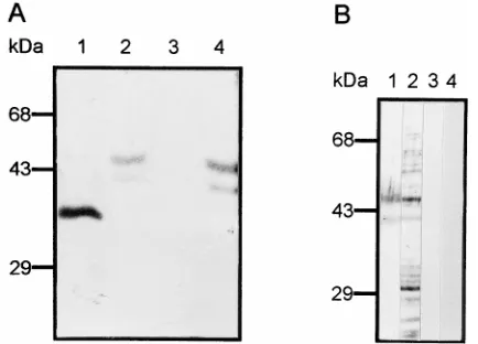
FIG. 2. VOPBA with [35S]methionine-labeled dengue virus. (A) Total C6/36 cell proteins were subjected to SDS–10% PAGE, transferred to a nitrocellulose membrane, and incubated with 53105cpm of labeled dengue virus (lane 1) or 53105cpm of labeled proteins from uninfected cells (lane 2). (B) C6/36 cell membrane proteins were run in the presence (lanes 1 and 3) or in the absence (lanes 2 and 4) ofb-mercaptoethanol and incubated with 53105cpm of labeled dengue type 4 virus under isotonic (lanes 1 and 2) or hypertonic (lanes 3 and 4) conditions. (C) C6/36 cell membrane proteins (lanes 1, 19, 3, and 39) and electroeluted 40- and 45-kDa proteins (lanes 2, 29, 4, and 49) were subjected to SDS-PAGE, transferred to a nitrocellulose membrane, and incubated with labeled proteins from uninfected cells (lanes 1, 19, 2, and 29) or with labeled dengue virus (lanes 3, 39, 4, and 49). The membrane was exposed to X-ray film (lanes 1, 2, 3, and 4) and then incubated with the monoclonal antibody 1H10 against dengue type 4 virus E protein (lanes 19, 29, 39, and 49). (D) DSP cross-linking assay. Labeled membrane proteins from C6/36 cells (lanes 1 and 4) and labeled 40- and 45-kDa proteins (lanes 2 and 5) were cross-linked in the presence (lanes 1 to 3) or absence (lanes 4 and 5) of unlabeled dengue virus. Cross-linked proteins obtained after centrifugation were treated with 5%b-mercaptoethanol, subjected to SDS–10% PAGE, and exposed to X-ray film.
on November 9, 2019 by guest
http://jvi.asm.org/
virus, [
35S]methionine-labeled viruses were incubated with
proteins subjected to SDS-PAGE and transferred to
nitrocel-lulose membranes. Under isotonic conditions, labeled dengue
virus bound to two molecules of approximately 40 and 45 kDa
present in cell lysates from C6/36 cells (Fig. 2A, lane 1) and to
other proteins with lower molecular masses. When labeled
proteins from uninfected cells were incubated with transferred
proteins from C6/36 cells, no reaction was observed (Fig. 2A,
lane 2). Similar bands of 40 and 45 kDa were detected when
the binding assay was performed with membrane proteins (Fig.
2B). The presence (Fig. 2B, lanes 1 and 3) or absence (Fig. 2B,
lanes 2 and 4) of
b
-mercaptoethanol did not alter the binding
pattern. More stringent conditions with 220 mM NaCl (Fig.
2B, lanes 3 and 4) reduced the nonspecific binding observed
with lower-molecular-weight proteins. These results suggest
that the 40- and 45-kDa membrane proteins, which do not
appear to be disulfide-linked subunits, bind specifically to
den-gue virus.
To corroborate that the E protein from dengue virus binds
to the 40- and 45-kDa molecules, a VOPBA-Western assay was
performed. Membrane proteins (Fig. 2C, lanes 1, 1
9
, 3, and 3
9
)
and electroeluted 40- and 45-kDa proteins (Fig. 2C, lanes 2, 2
9
,
4, and 4
9
) were incubated with labeled proteins from
unin-fected cells (Fig. 2C, lanes 1, 1
9
, 2, and 2
9
) or with labeled
dengue virus (Fig. 2C, lanes 3, 3
9
, 4, and 4
9
). The bands of 40
and 45 kDa were detected only after incubation with labeled
dengue virus (Fig. 2C, lanes 3 and 4), as is observed in Fig. 2A
and B. When a monoclonal antibody against dengue type 4
virus E protein (1H10) was used, the 40- and 45-kDa proteins
were revealed (Fig. 2C, lanes 3
9
and 4
9
), suggesting that E
protein binds the two molecules. No bands were detected in
the presence of labeled proteins from uninfected cells (Fig. 2C,
lanes 1, 1
9
, 2, and 2
9
).
To demonstrate the specificity of the binding, a cross-linking
assay was performed.
35S-labeled membrane proteins and
la-beled 40- and 45-kDa proteins purified by electroelution were
incubated and cross-linked to unlabeled dengue virus. Under
these conditions, a 45-kDa protein present in the membrane
fraction cross-linked to unlabeled dengue virus (Fig. 2D, lane
1), and 45- and 50-kDa proteins from the electroeluted
frac-tion were observed cross-linked to unlabeled virus (Fig. 2D,
lane 2). No bands were detected in the absence of unlabeled
dengue virus (Fig. 2D, lanes 4 and 5) or in the absence of
labeled cell proteins (Fig. 2D, lane 3).
Characterization of dengue virus-binding proteins.
To
ini-tially characterize these molecules, binding assays were
per-formed with preincubation of C6/36 cells with trypsin and
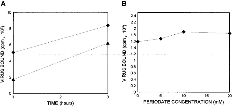
sodium periodate. Trypsin digestion for 30 min reduced the
binding by 50% after 1 h of incubation with labeled viruses
(Fig. 3A), indicating a peptidic nature of the binding
mole-cules. However, after 3 h of incubation, binding was reduced by
only 20%, suggesting an active replacement of the binding
proteins on the cell membrane.
To analyze the possible role of carbohydrates in dengue
virus binding, C6/36 cells were treated with sodium periodate.
However, no inhibition of binding was observed (Fig. 3B),
suggesting that carbohydrates are not essential for virus
bind-ing.
The analysis of the binding molecules was continued with
overlay assays, using total proteins obtained after cell
treat-ment with trypsin, neuraminidase, and periodate. No bands
were detected when cells were treated with trypsin (Fig. 4A,
lane 3), but a doublet of 40- and 45-kDa proteins was observed
after neuraminidase treatment (Fig. 4A, lane 2). When
radio-labeled virus was incubated with cell proteins obtained after
sodium periodate treatment, a single band of 38 kDa was
revealed (Fig. 4A, lane 1), suggesting that the 40- and 45-kDa
proteins are glycoproteins, although the carbohydrates are not
essential for virus binding. To support the hypothesis that the
40- and 45-kDa proteins are glycoproteins, membrane cell
pro-teins and electroeluted 40- and 45-kDa molecules were
incu-bated with aglutinin I (Fig. 4B). Aglutinin I reacted with the
40- and 45-kDa proteins present in the electroeluted fraction
and in membranes (Fig. 4B, lanes 1 and 2, respectively),
sug-gesting the presence of
b
-galactose in these proteins.
[image:4.612.116.497.73.247.2]Specificity of virus binding.
The specificity of the virus
in-teraction with the 40- and 45-kDa proteins was demonstrated
by competition assays with unlabeled dengue virus and
polio-virus. The overlay mixture was incubated with 280 or 560
m
g of
unlabeled dengue virus or with 280
m
g of poliovirus (Fig. 5,
lanes 2, 3, and 4, respectively) before incubation with labeled
dengue virus. Labeled virus binding was competed out when
dengue virus but not when poliovirus was used as a competitor,
FIG. 3. Binding of35S-labeled dengue type 4 virus to the surface of C6/36 cells. (A) C6/36 cells nontreated (}) and treated with trypsin for 30 min (Œ) were incubated for up to 3 h with labeled dengue virus. (B) C6/36 cells treated with different concentrations of sodium periodate were incubated with 53103cpm of labeled dengue virus.
on November 9, 2019 by guest
indicating that the interaction between dengue virus and the
40- and 45-kDa proteins is specific.
To further analyze the specificity of the interaction between
the 40- and 45-kDa proteins and dengue virus, a binding assay
with preincubation of labeled dengue virus with the
electro-eluted 40- and 45-kDa proteins was performed. Preincubation
of dengue virus with both proteins prevented by 90% the
bind-ing of labeled dengue virus to the C6/36 cells (Fig. 6).
Antibodies against the 40- and 45-kDa proteins block
den-gue virus binding.
Polyclonal antibodies against the 40- and
45-kDa proteins obtained after immunization of mice with the
electroeluted proteins were able to detect mainly a 45-kDa
protein and to a lesser extent 40-, 32-, 28-, and 20-kDa proteins
in Western blot assays (Fig. 7A, lane 1), while no bands were
observed after incubation with preimmune serum (Fig. 7A,
lane 2). To determine if the antibodies recognized the same
proteins that bound dengue type 4 virus, a binding assay was
performed. IgG against the 40- and 45-kDa proteins blocked
by 85% the binding of labeled virus to C6/36 cells, while
pre-immune IgG did not alter virus binding (Fig. 7B). A virus
binding inhibition similar to that observed with the polyclonal
antibodies was also detected after preincubation with an excess
of 110
m
g of unlabeled dengue virus (Fig. 7B).
[image:5.612.317.555.69.265.2]Localization of the 40- and 45-kDa proteins on C6/36 cells.
The localization of the 40- and 45-kDa proteins on C6/36 cells
was analyzed by immunofluorescence. Polyclonal antibodies
against both proteins were able to react with the surface of
nonpermeabilized C6/36 cells (Fig. 8A) and, to a lesser extent,
with the cytoplasm of permeabilized C6/36 cells (Fig. 8B),
FIG. 4. Characterization of the binding molecule by VOPBA with [35 S]me-thionine-labeled dengue virus. (A) Total proteins obtained from C6/36 cells untreated (lane 4) or treated with sodium periodate (lane 1), neuraminidase (lane 2), or trypsin (lane 3) were subjected to SDS–10% PAGE, transferred to a nitrocellulose membrane, and incubated with 23106cpm of labeled dengue virus. (B) Electroeluted 40- and 45-kDa proteins (lanes 1 and 3) and cell mem-brane proteins (lanes 2 and 4) were subjected to SDS-PAGE, transferred to a nitrocellulose membrane, and incubated in the presence (lanes 1 and 2) or absence (lanes 3 and 4) of biotinylated aglutinin I and streptavidin coupled to alkaline phosphatase.
[image:5.612.71.288.71.227.2]FIG. 5. Total proteins from C6/36 cells subjected to SDS–10% PAGE were preincubated in the absence of virus (lane 1) or in the presence of 280 or 560mg of unlabeled dengue virus (lanes 2 and 3, respectively) or 280mg of poliovirus (lane 3) prior to incubation with 3.53105cpm of labeled dengue type 4 virus.
[image:5.612.319.558.475.650.2]FIG. 6. Competition experiment with unlabeled 40- and 45-kDa proteins. Labeled dengue type 4 virus was preincubated with different amounts of elec-troeluted 40- and 45-kDa proteins prior to the incubation with C6/36 cells. After virus-cell interactions, the counts per minute for bound and free virus were determined.
FIG. 7. Binding inhibition induced by polyclonal antibodies against the 40-and 45-kDa proteins. (A) Western blot assay of total proteins from C6/36 cells with the 40- and 45-kDa protein immune (lane 1) and preimmune (lane 2) serum. (B) Binding inhibition induced by the presence of antibodies to 40- and 45-kDa proteins. C6/36 cells were preincubated in the presence of different dilutions of immune (}) or preimmune (h) IgG against the 40- and 45-kDa proteins at 4°C for 2 h prior to incubation with labeled dengue virus. Bound and free counts per minute were determined. The bar shows the binding inhibition observed after preincubation with 100mg of unlabeled dengue type 4 virus.
on November 9, 2019 by guest
http://jvi.asm.org/
while preimmune serum was negative with Tween 20-treated
or nontreated cells (Fig. 8D and C, respectively). This result
indicates that the 40- and 45-kDa proteins are located on the
surface of C6/36 cells and thus could function as a receptor for
dengue virus.
DISCUSSION
The basis of the cell and tissue tropisms of viruses is often
related to the ability of the VAP to bind a specific viral
recep-tor. Viral binding is followed by membrane fusion and leads to
productive infection in suitable hosts. The envelope protein
(E) is the major structural protein exposed on the surface of
dengue viruses and has been identified as the VAP (1, 5). An
internal domain of the E protein, located between amino acids
281 and 423, contains the binding motif, although its
counter-part, the cellular receptor, remains unknown (5). The
identi-fication of the receptor is required to understand tissue
tro-pism, pathogenesis, and viral replication in the hosts. To
partially characterize the putative dengue virus receptor, we
used a mosquito cell line, C6/36, derived from the larval stage
of
A. albopictus
, which is susceptible to dengue virus infection.
Our results show that dengue type 4 virus specifically binds
to C6/36 cells in a dose-dependent and saturable manner,
which is characteristic of specific receptor-ligand interactions,
suggesting the presence of a specific receptor for dengue virus
on the surface of C6/36 cells. This has also been described for
dengue type 1 virus on Vero and HepG2 cell lines (30). Trypsin
treatment of the cells inhibited virus binding, while sodium
periodate and neuraminidase treatment did not. Protease
sus-ceptibility and neuraminidase resistance have also been
re-ported when dengue virus was used in binding assays with
Vero, HepG2, and K562 cells and monocytes (8, 29, 30).
These results were supported by overlay assays, where we
could demonstrate the binding of labeled dengue type 4 virus
the 38-kDa protein and its relation to the 40- and 45-kDa
proteins is currently being performed in our laboratory.
Virus-cell interaction was resistant to neuraminidase
treat-ment in binding (data not shown) and overlay assays,
suggest-ing either that sialic acid is not essential for virus bindsuggest-ing or
that the 40- and 45-kDa proteins do not contain sialic acid. In
support of the absence of sialic acid in the 40- and 45-kDa
proteins, the lack of a sialyl transferase activity in a cell line
from
A. albopictus
has been reported (36, 38, 40). Although
this analysis was performed with a different cell line, both lines
were obtained from the same insect, and it has been suggested
that all insect cell proteins lack sialic acid (40). In addition,
resistance of other viral receptors to neuraminidase treatment
has also been reported for the minor group receptor of human
rhinovirus, where neuraminidase treatment increases
rhinovi-rus binding, suggesting that sialic acid is not required for virhinovi-rus
binding (32).
Dengue virus cross-linked with a 45-kDa membrane protein,
while 45- and 50-kDa proteins were cross-linked when labeled
electroeluted 40- and 45-kDa proteins were used. The 40-kDa
protein detected when an overlay assay was performed with
membrane proteins was not detected by the cross-linking assay.
This could be explained in two different ways. First, even
though in the overlay assays proteins of 40 and 45 kDa were
able to bind dengue virus, the native foldings of the two
teins are different, with the conformation of the 45-kDa
pro-tein being more appropriate for viral attachment. Second,
al-though both proteins are present in a membrane fraction, it is
possible that only the 45-kDa protein is exposed on the surface
of C6/36 cells. The 50-kDa protein, which was observed when
electroeluted 40- and 45-kDa proteins were used in the
cross-linking assay, could correspond to a contaminant of the
elec-troeluted 40- and 45-kDa proteins, and its presence in the
cross-linked proteins has two explanations. First, this may be a
cytoplasmic protein that binds to dengue virus, since it was not
detected in our membrane fraction. Second, since DSP
cross-links interacting molecules, it is possible that the 50-kDa
pro-tein binds to the 45-kDa propro-tein and not to the virus. In
support of the second idea, the 50-kDa protein present in total
cytoplasmic extracts was not detected in overlay assays (Fig.
2A), suggesting that this protein does not bind directly to
dengue virus.
The 40- and 45-kDa proteins do not appear to be
disulfide-linked subunits, since in the presence or absence of
b
-mercap-toethanol both protein bands showed the same molecular
weight in VOPBA. Moreover, dengue virus binding does not
require a folding dependent on disulfide bridges.
[image:6.612.61.301.70.319.2]The immunofluorescence assays indicate that 40- and/or
45-kDa proteins are localized on the surface of C6/36 cells. This
result correlates with the presence of both proteins in a
mem-brane fraction obtained by Triton X-114 extraction and with
FIG. 8. Immunofluorescence assay with C6/36 cells. Nonpermeabilized (A and C) and permeabilized (B and D) C6/36 cells were incubated with immune (A and B) or preimmune (C and D) serum against the 40- and 45-kDa proteins. As a second antibody, goat anti-mouse IgG coupled to fluorescein was used.
on November 9, 2019 by guest
their susceptibility to trypsin and sodium periodate treatments,
which were performed with intact cells and not with cell
ex-tracts. Monoclonal antibodies raised against each of the
pro-teins are being currently prepared in our laboratory to
deter-mine their differential localizations in the cell.
The presence of the 40- and 45-kDa proteins on the surface
of the C6/36 cells and the fact that both electroeluted proteins
and antibodies against them inhibit dengue virus binding
strongly support the idea that the 40- and 45-kDa proteins are
putative receptors or part of a receptor complex for dengue
virus.
We are aiming our efforts at the complete characterization
of the DNA and amino acid sequences of both proteins, to
confirm that one or both molecules are indeed the dengue
virus receptor. If the 40- and 45-kDa proteins are receptors for
dengue virus, it would be of interest to determine their
distri-butions in some susceptible and resistant mosquito tissues (37).
ACKNOWLEDGMENTS
We thank Fernando Medina for the cell cultures and Salvador
Cha-varrı´a for technical assistance. We also thank Goro Kuno for the C6/36
cell line, David Vaughn for the 1H10 monoclonal antibody against
dengue type 4 virus E protein, and Lorena Gutie
´rrez and Martha
Espinosa-Cantellano for critical comments on the manuscript.
This work was supported by a grant from Consejo Nacional de
Ciencia y Tecnologı´a. Juan Salas has a scholarship from Consejo
Na-cional de Ciencia y Tecnologı´a.
REFERENCES
1.Anderson, R., A. D. King, and B. L. Innis.1992. Correlation of E protein binding with cell susceptibility to dengue 4 virus infection. J. Gen. Virol.
73:2155–2159.
2.Barth, O. M.1992. Replication of dengue viruses in mosquito cell cul-tures—a model from ultrastructural observations. Mem. Inst. Oswaldo Cruz
87:565–574.
3.Bradford, M. M.1976. A rapid and sensitive method for the quantitation of microgram quantities of protein utilizing the principle of protein-dye bind-ing. Anal. Biochem.72:248–254.
4.Chambers, T. J., C. S. Hahn, R. Galler, and C. M. Rice.1990. Flavivirus genome organization, expression, and replication. Annu. Rev. Microbiol.
44:649–688.
5.Chen, Y., T. Maguire, and R. M. Marks.1996. Demonstration of binding of dengue virus envelope protein to target cells. J. Virol.70:8765–8772. 6.Chungue, E., V. Deubel, O. Cassar, M. Laille, and P. M. V. Martin.1993.
Molecular epidemiology of dengue 3 viruses and genetic relatedness among dengue 3 strains isolated from patients with mild or severe form of dengue fever in French Polynesia. J. Gen. Virol.74:2765–2770.
7.Crane, S. E., J. Buzy, and J. E. Clements.1991. Identification of cell mem-brane proteins that bind visna virus. J. Virol.65:6137–6143.
8.Daughaday, C. C., W. E. Brandt, J. M. McCown, and P. K. Russell.1981. Evidence for two mechanisms of dengue virus infection of adherent human monocytes: trypsin-sensitive virus receptors and trypsin-resistant immune complex receptors. Infect. Immun.32:469–473.
9.Despre`s, P., M.-P. Frenkiel, and V. Deubel.1993. Differences between cell membrane fusion activities of two dengue type-1 isolates reflect modifica-tions of viral structure. Virology196:209–219.
10. Deubel, V., R. M. Kinney, and D. W. Trent.1986. Nucleotide sequence and deduced amino acid sequence of the structural proteins of dengue type 2 virus, Jamaica genotype. Virology155:365–377.
11. Deubel, V., R. M. Kinney, and D. W. Trent.1988. Nucleotide sequence and deduced amino acid sequence of the structural proteins of dengue type 2 virus, Jamaica genotype: comparative analysis of the full-length genome. Virology165:234–244.
12. Freier, J. E., and P. R. Grimstad.1983. Transmission of dengue virus by orally infectedAedes triseriatus. Am. J. Trop. Med. Hyg.32:1429–1434. 13. Gould, E. A., and J. C. S. Clegg.1991. Growth, titration and purification of
alphaviruses and flaviviruses, p. 43–78.InB. W. J. Mahy (ed.), Virology: a practical approach. IRL Press, Oxford, United Kingdom.
14. Guirakhoo, F., F. X. Heinz, and C. Kunz.1989. Epitope model of tick-borne encephalitis virus glycoprotein E: analysis of structural properties, role of carbohydrate side chain, and conformational changes occurring at acidic pH. Virology169:90–99.
15. Guirakhoo, F., A. R. Hunt, J. G. Lewis, and J. T. Roehrig.1993. Selection and partial characterization of dengue virus mutants that induce fusion at elevated pH. Virology194:219–223.
16. Halstead, S. B., K. Larsen, S. Kliks, J. S. M. Peiris, J. Cardosa, and J. S. Porterfield.1983. Comparison of P388D1 mouse macrophage cell line and human monocytes for assay of dengue-2 infection-enhancing antibodies. Am. J. Trop. Med. Hyg.32:157–163.
17. Hase, T., P. L. Summers, and K. H. Eckels.1989. Flavivirus entry into cultured mosquito cells and human peripheral blood monocytes. Arch. Virol.
104:129–143.
18. Heinz, F. X., G. Auer, K. Stiasny, H. Holzmann, C. Mandl, F. Guirakhoo, and C. Kunz.1994. The interactions of the flavivirus envelope proteins: implications for virus entry and release. Arch. Virol.9:339–348.
19. Heinz, F. X., K. Stiasny, G. Pu¨schner-Auer, H. Holzmann, S. L. Allison, C. Mandl, and C. Kunz.1994. Structural changes and functional control of the tick-borne encephalitis virus glycoprotein E by the heterodimeric association with protein prM. Virology198:109–117.
20. Helenius, A.1995. Alphavirus and flavivirus glycoproteins: structures and functions. Cell81:651–653.
21. Jin, Y.-M., I. U. Pardoe, A. T. H. Burness, and T. I. Michalak.1994. Iden-tification and characterization of the cell surface 70-kilodalton sialoglycop-rotein(s) as a candidate receptor for encephalomyocarditis virus on human nucleated cells. J. Virol.68:7308–7319.
22. Khin, M. M., and K. A. Than.1983. Transovarial transmission of dengue 2 virus byAedes aegyptiin nature. Am. J. Trop. Med. Hyg.32:590–594. 23. Kliks, S.1990. Antibody-enhanced infection of monocytes as the pathogenic
mechanism for severe dengue illness. AIDS Res. Hum. Retroviruses6:993– 998.
24. Kurane, I., A. L. Rothman, P. G. Livingston, S. Green, S. J. Gagnon, J. Janus, B. L. Innis, S. Nimmannitya, A. Nisalak, and F. A. Ennis.1994. Immunopathologic mechanisms of dengue hemorrhagic fever and dengue shock syndrome. Arch. Virol.9:54–64.
25. Littaua, R., I. Kurane, and F. A. Ennis.1990. Human IgG Fc receptor II mediates antibody-dependent enhancement of dengue virus infection. J. Im-munol.144:3183–3186.
26. Ludwig, G. V., J. P. Kondig, and J. F. Smith.1996. A putative receptor for Venezuelan equine encephalitis virus from mosquito cells. J. Virol.70:5592– 5599.
27. Mackow, E., Y. Makino, B. Zhao, Y. M. Zhang, L. Markoff, A. Buckler-White, M. Guiller, R. Chanock, and C.-J. Lai.1987. The nucleotide sequence of dengue type 4 virus: analysis of gene coding for non-structural proteins. Virology159:217–228.
28. Mady, B. J., D. V. Erbe, I. Kurane, M. W. Fanger, and F. A. Ennis.1991. Antibody-dependent enhancement of Dengue virus infection mediated by bispecific antibodies against cell surface molecules other than Fcg receptors. J. Immunol.147:3139–3144.
29. Mady, B. J., I. Kurane, D. V. Erbe, M. W. Fanger, and F. A. Ennis.1993. Neuraminidase augments Fcg receptor II-mediated antibody-dependent en-hancement of Dengue virus infection. J. Gen. Virol.74:839–844. 30. Marianneau, P., F. Me´gret, R. Olivier, D. M. Morens, and V. Deubel.1996.
Dengue 1 virus binding to human hepatoma HepG2 and simian Vero cell surfaces differs. J. Gen. Virol.77:2547–2554.
31. Meerovitch, K., Y. V. Svitkin, H. S. Lee, F. Lejbkowics, D. J. Kenan, E. K. I. Chan, V. I. Agol, J. D. Keene, and N. Sonenberg.1993. La autoantigen enhances and corrects aberrant translation of poliovirus RNA in reticulocyte lysate. J. Virol.67:3798–3807.
32. Mischak, H., C. Neubauer, E. Kuechler, and D. Blaas.1988. Characteristics of the minor group receptor of human rhinoviruses. Virology163:19–25. 33. Rey, F. A., F. X. Heinz, C. Mandl, C. Kunz, and S. C. Harrison.1995. The
envelope glycoprotein from tick-borne encephalitis virus at 2 A resolution. Nature (London)375:291–298.
34. Rosen, L., D. A. Shroyer, R. B. Tesh, J. E. Freier, and J. C. Lien.1983. Transovarial transmission of dengue viruses by mosquitoes:Aedes albopictus
andAedes aegypti. Am. J. Trop. Med. Hyg.32:1108–1119.
35. Rosen, L.1987. Sexual transmission of dengue viruses byAedes albopictus. Am. J. Trop. Med. Hyg.37:398–402.
36. Smith, G. W., and P. J. Wright.1985. Synthesis of proteins and glycoproteins in Dengue type 2 virus-infected Vero andAedes albopictuscells. J. Gen. Virol.66:559–571.
37. Sriurairatna, S., and N. Bhamarapravati.1977. Replication of dengue-2 virus inAedes albopictusmosquitoes. Am. J. Trop. Med. Hyg.26:1199–1205. 38. Stollar, V., B. Stollar, R. Koo, K. A. Harrap, and R. W. Schlesinger.1976. Sialic acid contents of Sindbis virus from vertebrate and mosquito cells. Virology69:104–115.
39. Towbin, H., T. Staehelin, and J. Gordon.1979. Electrophoretic transfer of proteins from polyacrylamide gels to nitrocellulose sheets: procedure and some applications. Proc. Natl. Acad. Sci. USA76:4350–4354.
40. Warren, L.1963. The distribution of sialic acid in nature. Comp. Biochem. Physiol.10:153–171.
41. Zhao, B., E. Mackow, A. Buckler-White, L. Markoff, R. M. Chanock, C.-J. Lai, and Y. Makino.1986. Cloning full-length dengue type 4 viral DNA sequences: analysis of genes coding for structural proteins. Virology155:77– 88.
![FIG. 2. VOPBA with [35membrane, and incubated with 5S]methionine-labeled dengue virus](https://thumb-us.123doks.com/thumbv2/123dok_us/1255677.77880/3.612.140.475.70.382/fig-vopba-membrane-incubated-methionine-labeled-dengue-virus.webp)