PLACENTA AND ITS CLINICAL
CORRELATIONS
Dissertation submitted for
M.S.DEGREE EXAMINATION
BRANCH - V
(ANATOMY)
THANJAVUR MEDICAL COLLEGE
THANJAVUR
THE TAMILNADU Dr. M.G.R. MEDICAL UNIVERSITY
CHENNAI
CERTIFICATE
This is to certify that dissertation entitled “Placenta and its
clinical correlations” is the bonafide record of work done by
Dr.M.Elangovan in the Department of Anatomy, Thanjavur medical
college, Thanjavur, during his post graduate course from 2004-2007.
This is submitted as partial fulfillment for the requirement of
M.S. Degree Examination – Branch V (Anatomy) to be held in
March 2007.
The Dean, Professor and Head,
CONTENTS
INTRODUCTION 01
AIM OF STUDY 03
HISTORICAL PERSPECTIVE 04
NORMAL ANATOMY 05
REVIEW OF LITERATURE 20
MATERIALS AND METHODS 37
OBSERVATION 39
DISCUSSION 55
SUMMARY 63
CONCLUSIONS 65
ACKNOWLEDGEMENT
I am very grateful with deep sense of gratitude to Professor
Dr. T . SIVAKAMI, M.S., Head of the Department of Anatomy, Thanjavur
Medical College, Thanjavur for her invaluable guidance, without whose help
this study would not have been a reality. I am thankful to her for her advice
and guidance in designing and enabling me to do this study with creative
suggestions and constructive criticisms.
I am extremely grateful to Professor. Dr. SORUPARANI
SELVARAJ M.D., D.G.O., Head of the Department of Obstetrics and
Gynaecology and Assistant Professor. Dr. S. PRADEEPA M.D. (OG), for
graciously giving me permission to collect placenta from their department.
I am very much grateful to Assistant Professors Dr. S. SUMATHI
M.S., Dr. R. MANORANJITHAM M.S., Dr. K. MOHAN M.S., and
Dr. P.S. CHITRA M.S., for their valuable suggestions and help.
I deem it a pleasure to acknowledge with gratitude my colleague post
graduates Dr. B . SANTHI and Dr. N. ISAI VANI for their valuable help.
My sincere thanks are due to the teaching and non-teaching staff of the
Department of Anatomy, Thanjavur Medical College, Thanjavur.
I profoundly thank THE DEAN, Thanjavur Medical College,
INTRODUCTION
The word ‘placenta’ connotes a functional union between fetal and
maternal structures, and in the human this union has developed to a
remarkable degree, as a consequence of the evolutionary development which
has followed internal fertilization of a single ovum and the retention of the
embryo within the mother for a relatively long time.
Placenta is a diplomatic intermediary between mother and child; ten
months nourisher of the still helpless fetus; the tender supplier of fetal
requisites; eminent emissary of fetal waste; the wise interpreter of harmful
intruders ( to the child); the physiological parasite; siphoning off blood not
for itself, but for the growing fetus – which in turn as a token of gratitude
carries in it’s body during extrauterine life, the only scar a human being is
never ashamed of – “the umbilicus”.
The placenta is the most accurate record of the infant’s prenatal
experiences. The fetus, cord and the placenta constitute genetically identical
parts of a unit that share the uterine environment.
Physicians, generally are uncomfortable with the task of examining the
organ to reasonably knowledgeable look and touch can provide much insight
into prenatal life. It helps in caring for the neonate; a record for neonatologist
to plan for MCH care, otherwise this valuable information would be discarded
along with the organ.
Remarkable advances have occurred in our understanding of normal
human embryology, placentation, and fetal growth. In an increasing number
of pregnancies, an abnormality may be detected early in gestation, and its
etiology determined by investigations using sonographic and genetic analysis.
Knowledge of the human placenta is now reasonably well in hand; not
only the gross and light microscopic features. With the noninvasive
investigations like sonography, we have entered an era of diagnosing
anomalies of placenta even before delivery and there is a scope for
interventional procedures to correct them. A placental examination with
gross and microscopic studies will yield additional information about the
course of the pregnancy.
AIM OF STUDY
Placental examination offers a lot of information of prognostic
significance for the newborn. This enables the neonatologist to handle the
babies much more efficiently.
This prompted me to study the placental shapes, cord insertion, lobes
and gestational outcome and to correlatre the morphological / morphometric
HISTORICAL PERSPECTIVE
Placenta derives its name from Greek & Latin. The Latin root
“Placentos” means a cake; Greek root “Plakios” means flat.
The placenta – a dynamic organ, which is unique in its development
has been known to early man. The reason being it was obvious after any
childbirth & hence it was called “afterbirth”. It was renamed ‘Placenta” by
Realdus Columbus in 1559.
There are two school of thought about who coined the term. One set of
people claim that it was Gabriele de Fallopsis (1523 – 1562) whereas the
popular view is that it was Realdus Columbus (1516 – 1559) who named it.
NORMAL ANATOMY
At full term the placenta is disc like, and presents foetal and maternal
surfaces, and peripheral margin.
Foetal surface is smooth, covered by amnion and presents the
attachment of the umbilical cord close to its centre. Beneath the amnion,
umbilical vessels radiate from the cord. Sometimes the extra-embryonic part
of the yolk sac, known as the umbilical vesicle is found beneath the foetal
surface close to the umbilical cord and is connected by a fibrous remnant of
the vitello-intestinal duct.
Maternal surface is rough and irregular, and is mapped out into 15 to
30 polygonal areas known as the cotyledons which are limited by fissures.
Each fissure is occupied by a placental septum.
Peripheral margin is continuous with the foetal membrane which
consists from outside inwards of fused decidua parietalis and capsularis,
Measurements – At full term the placenta presents the following
measurements :
Diameter – 15 to 20 cm
Thickness – 3 cm ( at the centre )
Weight – 500 gms
Proportional weight between placenta and foetus at various stages of
pregnancy :
1st month – placenta : foetus = 6:1
4th month – placenta : foetus = 1:1
At birth – placenta : foetus = 1:7
At birth placenta occupies about 30%of the uterine wall.
Structure:
The placenta consists of chorionic plate on the foetal side, basal or
decidual plate on the maternal side, stem villi extending between the plates,
and the intervillous space between the stem villi filled with the maternal
blood.
Chorionic plate is composed of the following structures from within
outwards (foetus to mother):
(i) Primary mesoderm containing branches of umbilical vessels
(ii) Cytotrophoblast;
(iii) Syncytiotrophoblast;
Basal plate consists from outside inwards of :
(i) stratum spongiosum of decidua basalis;
(ii) outer layer of syncytiotrophoblast (Nitabuch’s layer) which
undergoes fibrinoid degeneration;
(iii) outer shell of cytotrophoblast;
(iv) inner layer of syncytiotrophoblast; outer zone of this layer also
undergoes fibrinoid degeneration and is known as Rohr’s
Fibrinoid stria.
The basal plate is perforated by the spiral branches of uterine arteries
and veins; eventually the intervioous space is filled with maternal blood.
Numerous placental septa project from the basal plate into the intervillous
space but they fail to reach the chorionic plate. Each placental septum
consists of a central core of the decidua basalis containing decidual cells,
remnants of endometrial glands and some maternal vessels, and is covered by
the cyto- and syncytiotrophoblasts. The areas between the adjacent placental
septa are known as the cotyledons which are observed from the maternal
surface. Each cotyledon presents in the central axis one or more stem villi.
basal plate. The origin of these cells is disrupted, but giant cells are believed
to secrete placental hormones.
STEM VILLI connect the chorionic and basal plates, and consist of
primary, secondary and tertiary villi with the progress of development.
A primary villus consists of a central core of cytotrophoblast and is
covered by the cells of syncytiotrophoblast.
A secondary villus contains a central core of primary mesoderm and is
covered successively by cyto- and syncytiotrophoblasts.
A tertiary villus contains in the centre the foetal blood vessels which
are surrounded successively from within outwards by primary mesoderm,
cyto- and syncytiotrophoblasts. From each tertiary stem villus numerous
branching villi project into the intervillous space. The branching villi may
fuse with the adjacent villi and the outer and inner walls .of the intervillous
space. Therefore, the intervillous space is converted into a sponge-like
network of villous type of labyrinthine structure and is filled with maternal
blood. The blood vessels within the branching villi do not anastomose with
the neighbouring ones. The branching or terminal villi are the functional
trophoblastic epithelium – inner cytotrophoblast and outer
syncytiotrophoblast, resting on a basement membrane.
The central core of the villus contains one to six foetal capillaries, and
stromal cells. The stroma consists of primitive mesenchymal cells,
fibroblasts, collagen fibres, some phagocytic and reniform Hofbauer cells.
Syncytial buds invaginate from the trophoblasts and project into the villous
stroma.
In early part of pregnancy, about 800 to 1000 villi radiate from the
entire chorionic wall. Later, with the regression of chorion leave only 60
stem villi persist in human placenta. Since the number of maternal cotyledons
is 15 to 30, it is suggested that each cotyledon contains 2 to 4 major stem villi.
A major stem villus and the vessels derived from it form the foetal cotyledon
or placentome.
The compartments of the intervillous space are not water-tight, and
communicate with one another around the stem villi. The inner wall of the
space is formed by chorionic plate and its outer wall by basal plate. The
space is lined internally by syncytiotrophoblast, and is filled with maternal
blood which never communicates directly with the foetal blood. About 500ml
the volume of the intervillous space is about 140 ml, the blood within the
space is exchanged about four times per minute.
The placental barrier consists of tissues which intervene between foetal
blood in the chorionic villi and the maternal blood in the intervillous spacee.
Through this barrier exchange of gaseous and metabolic products takes place
between the foetus and the mother. Upto the third month of pregnancy the
barrier consists of the following four layers from foetus to mother –
endothelium of foetal capillaries resting on a basement membrane, a core of
primary mesoderm, a basement membrane upon which rest cytotrophoblast
and syncytiotrophoblast. From the fourth month onwards, the barrier is
practically reduced into two layers – endothelium of the foetal capillaries
resting on a basement membrane, and syncytiotrophoblalst which presents
numerous micro villi in the intervillous space increasing the total surface area
to about 14 sq. metres at full term.
In some areas of terminal villi the syncytium fuses with the stromal
capillary wall, forming ‘vasculo-syncytial membrane’. The membranous
areas are known as ‘alpha zones’ which are the sites of all materno-foetal
exchanges. Rest of the areas of terminal villi are non-membranous and are
called ‘beta zones’ where stroma cells and cytotrophoblasts persist. Beta zone
Therefore, human placenta is discoid, deciduate, haemo-chorial, and
villous type of labyrinthine organ.
DEVELOPMENT OF PLACENTA :
Placenta is developed from two sources – foetal part from chorion
frondosum, and maternal part from decidua basalis.
Stages of development:
When the blastocyst is embedded in the endometrial wall of the
uteruss, the endometrium is changed into decidua ( due to decidual reaction of
stromal cells). The decidua consists of basalis, capsularis, and parietalis.
Decidua basalis is situated at the embryonic pole of the blastocyst, the
capsularis envelops the rest of the blastocyst and the parietalis lines the
uterine cavity. Syncytiotrophoblast proliferates into multilayered and
multinucleated protoplasmic mass and advances towards the decidua basalis
and capsularis by proteolytic reaction. Meanwhile the cytotrophoblast
differentiates internally into a layer of primary mesoderm. Trophoblast and
primary mesoderm together form the chorion.
A number of lacunar spaces appear within the syncytiotrophoblast
Each lacuna communicates with the adjacent one around cords of syncytial
cells which are known as the trabeculae.
The lacunae enlarge, and erode the branches of uterine arteries and
veins. Therefore, lacunae are now filled with maternal blood establishing the
utero-placental circulation.
Trabeculae are converted into primary chorionic villi by the invasion
of cytotrophoblast in the central axis of each trabecullae. Lacunar spaces are
now called the intervillous spaces. At the tips of the primary villi the cells of
cytotrophoblast spread outwards and form the outer cytotrophoblastic shell.
The primary mesodermal cells of the chorion invade the central axes of
the primary villi and convert them into secondary chorionic villi. The cells of
the primary mesoderm extend upto the distal ends of the villi,. And do not
form the outer wall of the intervillous space.
Secondary villi are converted into tertiary villi when foetal bnlood
vessels derived from umbilical vessels appear within the primary mesoderm.
Therefore, all tertiary villi are vascular. From each tertiary villus numerous
branching villi project into the intervillous space converting the latter into
The chorionic villi attached to the embryonic pole of the blastocyst
proliferate more rapidly, they are called the chorion frondosum. Rest of the
chorionic villi attached to the abembryonic pole are known as the chorion
leave.
During the 3rd month of pregnancy, the chorion leave degenerate and
disappear due tom fusion of decidua capsularis and parietalis.
As a result, the persistent chorion frondosum and decidua basalis form
together a human placenta.
CLINICAL CORRELATIONS :
GROSS ABNORMALITIES IN PLACENTATION:
1. PLACENTA EXTRACHORIALIS
In this placenta, the area of the chorionic plate is less than that of the
decidual plate and some of the placenta is outside the chorionic plate. Since
the superficial decidua is largely spared it appears as a ring or band on the
surface of the placenta (placenta marginata) or the membranes may be folded
to enclose decidua (placenta circumvallata). Both types may be found on the
surface of placenta and may partially or completely encircle the placenta. The
2. PLACENTA MEMBRANACEA
The entire fetal membranes are covered with a thin but functioning
placenta and the thickness of the placenta is in inverse proportion to its area.
3. PLACENTA SUCCENTURIATA
In this variety, the main placenta is connected to an accessory and small
placenta by an artery and a vein which pass through membranes. The clinical
significance of placenta succenturiata is that the accessory lobe may be
overlooked at delivery and left in utero, and give rise to haemorrhage,
infection and the clinical finding of a placental polyp.
4. PLACENTA ACCRETA, INCRETA AND PERCRETA
In a few instances, the trophoblastic villi penetrate deeply into the decidua,
or even through the layer and into the muscle without disturbing the gestation.
It can only be diagnosed by examining the removed uterus. In such studies
portions of the decidua basalis are seen to be absent and the chorionic villi
attach directly onto myometrial cells (placenta accreta), penetrate the cells to
some degree (placenta increta), or even penetrate through to the serosal
surface of the uterus (placenta percreta).
5. HEMANGIOMATA OF THE PLACENTA
These tumours are relatively common, being found in approximately 1
significance but a few are large and are associated with hydramnios,
antepartum haemorrhage and premature labour.
6. HYDATIDIFORM MOLE :
A hydatidiform mole is a noninvasive condition in which many of the
chorionic villi are characterized by nodular swellings, giving them an
appearance almost like bunches of grapes. Commonly, much of the villous
surface of the placenta takes on this appearance, in addition the embryo is
either absent or not viable. The villi show no evidence of vascularization.
Genetic analysis has determined that hydatiform moles represent the
results of paternal imprinting where the female pronucleus of the egg does not
participate in development. The chromosomes of hydatidiform moles are
paternally derived 46 XX, since the number of lethal genes in 46 YY embryos
is not compatible with tissue survival.
7. CHORIOCARCINOMA ;
They are malignant tumors derived from embryonic cytotrophoblast
and syncytitrophoblast. These tumors are highly invasive into the maternal
decidual tissues and blood vessels. Like hydatidiform moles, most
choriocarcinomas, contain only paternally derived chromosomes and are thus
CLINICAL APPLICATIONS OF PLACENTA
1. CHORIONIC VILLOUS SAMPLING :
A sample of chorion frondosum can be obtained and used for prenatal
diagnosis. The procedure is undertaken in trimester 1, after 6 post ovulatory
weeks. Usually biopsy forceps of a catheter is inserted through a
transcervical route under ultrasonic guidance. Recombinant DNA
oprocedures are increasingly being used to identify the molecular defects
associated with many heritable diseases. DNA can be obtained either by
chorionic villous sampling (trophoblast) or by amniocenteses (and culture of
amniocytes). Moreover, DNA can be amplified by the polymerase chain
reaction (PCR)
Limb defects have been reported after CVS, the most frequent type
being transverse deficiency of either fingers or toes. Vascular disruption
following bleeding from the chorion is a likely cause. Another possibility in
some instances in inadvertent puncture of the amniotic sac, producing
2. AMNIOTIC MEMBRANE
Amniotic Membrane is very thin and transparent and can be used to
apply over large areas of burn wound. The role of amniotic membrane in
covering partial thickness burn wounds has been well established ever since J.
Pigeon (1960) made use of it. Its effective reduction of bacterial count in
infected burn wounds was shown by Robson and Krizek (1973) and in
contaminated granulation tissue by Bade (1958). It is quite effective due to its
covering effect (Morris et al 1966) and due to the presence of Lysozyme,
Oestrogen and Progesterone (Glask and Snyder 1970). Amniotic membrane
alleviates pain, promotes epithelialisation and is also economical (Thompson
and Parks 1982; Heberal et al., 1987). Amniotic membrane as an effective
burn wound cover for partial thickness burns as has been convincingly noted
by Pigeon J (1960), Dino et al (1966), Golocco et al (1974), Bose (1979),
Piserchia and Akenzua (1981), Ramakrishnan and Rao (1983), Ramakrishnan
et al (1995).
Amniotic membrane is derived from the ectoderm. Amniotic
membrane has a basement membrane similar to the one in the wound bed. It
consists of type IV collagen, which is essential in healing wounds. The
membrane is transparent and has many pores; through which exudation of
USE OF AMNIOTIC MEMBRANE DRESSING
Earlier, amniotic membrane was used over contaminated wound
surfaces and was found that the colony count decreased considerably. Also
the amnion and the chorion layers were not separated initially. Later, amnion
when separated was found to be very thin and transparent and was
advantageous as a temporary wound cover. This thin amnion was often used
on superficial partial thickness burn wounds and it is advantageous to use on
deep partial thickness burn wounds also in that it prevents infection and
bacterial colonization. In larger burn area, when the areas after excision is
not suitable for skingrafting, the excised area is covered with amniotic
membrane again and this prepares the wound ideally for skingrafting.
Advantage of Amniotic Membrane as a Wound Cover
The following advantages of amniotic membrane as a wound cover are noted:
1. Amnion, when separated is very thin, transparent compared to other
wound covers.
2. It is easy to handle the amnion and it is flexible.
3. Amnion is easy to apply, spreads easily and can be used without
hurting the patient.
4. Adherence of the membrane to the wound surface is very good and due
5. Under the amnion dressing, one could visualize the progress of sound
healing, due to its transparency.
6. Pain is much less after the application of the membrane.
7. The large quantity of estrogen, progesterone in the membrane also
hastens epithelialisation.
8. When heparin was used as a spray over the burn wound covered with
membrane, the healed scar was very satisfactory due to early collagen
remodeling. It is easy to combine pharmacological agents like heparin
and silver sulphadiazine with the membrane.
9. The cost of burn management and duration of hospital stay was
considerably reduced, which is a great advantage in developing
countries.
In a developing country with large incidence of burns like in India, amniotic
REVIEW OF LITERATURE
Evolution of knowledge of placenta starts from the Biblical times.
Egyptian believed that the placenta was external soul.
The placenta – a dynamic organ, which is unique in its development
has been known to early man. The reason being it was obvious after any
childbirth & hence it was called “afterbirth”. It was renamed ‘Placenta” by
Realdus Columbus in 1559.
There are two school of thought about who coined the term. One set of
people claim that it was Gabriele de Fallopsis (1523 – 1562) whereas the
popular view is that it was Realdus Columbus (1516 – 1559) who named it.
Columbus borrowed the Latin term which means “flat cake”.
According to Henry Gray the normal parameters of placenta are –
weight 500 grams (Range 200 – 800 gm), thickness – 23 mm (Range 10 – 40
mm). Thickness is greater at center than periphery.
Hamilton classifies placenta according to the distribution of chorionic
zonary placenta [villi grouped in bands around the equator of the chorio all
antonic placenta], Bidiscoidal [villi disappear from all except a circumscribed
area over the chorion] & placenta succenturiata [accessory lobule].
He also classifies placenta according to the “pattern of distribution of
the umbilical vessels after piercing the chorionic plate”
a) The Disperse type [umbilical vessels successively undergo
dichotomous division & diminish in caliber]
b) The Magistral type [side branches are given off and reduction in
caliber starts in margin. According to Stordania latter type favours
fetal growth due to better haemodynamics.
c) Mixed type. – Combination of both the above patterns.
Grosser (1909) provides an acceptable classification of placental
morphology, based on maternal & fetal tissue layers interspersed between the
fetal and maternal blood streams.
(i) Haemochorial - contains three layers which are trophoblast,
connective tissue & endothelium. This is further subdivided into labyrinthine
& villous.
(ii) Endothelio chorial – contains four layers, the maternal endothelium
(iii) Syndesmo chorial - contains five layers, the endometrial
connective tissue being the fifth layer.
(iv) Epithelio chorial – contains six layers, lining endometrium the
sixth layer.
Mossman (1937) added one more category to this haemoendothelial –
endometrial tissue & trophoblast is destroyed & only the fetal capillary
divides the circulation.
Enders suggested the most useful classification of haemochorial
placenta depending on the complete layers of trophoblast namely
haemomonochorial, haemodichorial & haemotrichorial.
Mossman attributes the name chorio-allantoic to the vascularity
homologous to the allantoic vessels of lower animals; haemochorial to the
nature of placental membrane ; villous to its villi; decidua to its shedding at
birth; discoidal to its circular shape.
The mature placenta consists of two plates, the chorion, and the
decidua basalis, between which is the much thicker labyrinth of chorionic villi
and intervillous spaces. The cut surface is dark red and spongy. It shows
The fetal placenta is made up of a number of subunits that are now
generally known as lobules. The injection studies of Wilkin (1965) have
shown that the primary stem villi break up just below the chorionic plate into
a number of secondary stem villi which, after running parallel to the chorionic
plate for a short distance, divide into a series of tertiary stem villi. The
lobules are derived from these tertiary stem villi, which sweep down through
the intervillous space to anchor into the basal plate.
The placental septa appear during the third month of gestation : they
protrude into the intervillous space from the basal plate and divide the
maternal surface of the placenta into 15 – 20 lobes. These septa are simply
folds of the basal plate, formed partly as a result of regional variability in
placental growth and partly by the pulling up of the basal plate by the
anchoring columns, which have a poor growth rate (Boyd & Hamilton 1970).
The human placenta at term weighs around 500 g. It may range
between 250-700gm. (das Gupta, Mathur and Gupta, 1974) in Indian
subjects. Armitage et al (1967) reported that the average weight of the term
placenta was 508gms with extremes of 310gms and 880 gms. There is a
positive correlation between the weight of the baby and the placental weight.
Strahl classifies placenta into placenta Vera or voll placenta in which
the deciduas is in part sloughed off at full term & semi placenta or half
placenta – in which the maternal blood space remains intact.
According to Robinson, placentas are of two types – (i) apposed
placenta where chorion and the uterine mucosa are apposed and (ii) conjoined
placenta where chorion uterine mucosa is fused together.
Whereas, Assheton describes another way of classifying placenta
a) Placenta plicatae – with a simple and unspecialized chorionic
epithelium
b) Placenta cumulatae – with highly specialized trophoblast
containing lacunae for maternal blood.
The attachments for umbilical cord and vessels to the fetal aspect are
variable. According to Boyd, the attachments can be eccentric, marginal and
midcentral. Usually the umbilical vessels remain close to each other.
Occasionally, they are separated by some distance from the placenta
Arey says that the maternal surface shows irregular rough reddish gray
lobular areas which correspond to cotyledons.
Langhmann states that cotyledons receive their blood through 80 –
100 spiral arteries that pierce the decidual plate and enter the intervillous
spaces at more or less regular intervals. The lumen of the spiral artery is
narrow resulting in an increased blood pressure as blood enters intervillous
space.
In villi from term placentas the syncytial nuclei are irregularly
dispersed and often appear aggregated to form multinucleated protrusions
from the villous surface, these being known as syncytial knots. Syncytial
knots should be differentiated from syncytial sprouts which are present from
the early stages of pregnancy and mainly represent the initial stages in the
development of lateral villi.
Placental barrier is not a true barrier according to Arey as many
substances pass through freely. At times pathogens gain entry.
As per Langman, the main functions of the placenta are exchange of
metabolic and gaseous products between maternal and fetal blood streams and
The clinical significance of placental anomaly has been much debated
but it is now clear that the circummarginate form is devoid of clinical
importance, and that although circumvallate placentation is associated unduly
frequently with a rather small baby, and possibly with a slight excess of
congenital malformation (Lademacher et al 1981), it does not appear to be
associated with an increased perinatal mortality (Fox 1978). Other aberrant
forms of placentation are either of no functional significance, e.g. bilobate
placenta, accessory lobe, or so rare that they can, in practical terms, be
ignored e.g. placenta membranacea, girdle placenta.
There have been claims that marginal or velamentous insertion of the
cord is related to a high perinatal mortality rate but recent studies agreed that
the site of cord insertion is unrelated to the incidence of premature labour, low
birth weight or fetal hypoxia. (Uyanwah – Akpom & Fox 1977, Woods &
Malan 1978). Velamentous insertion does of course present a small risk
because of the danger of traumatic damage to, and bleeding from the
unprotected vessels running through the membranes.
There appears to be a clear correlation between the presence of a
to attribute the deficient fetal growth to damage inflicted on the placenta by
the inflammatory processes.
Remarkably little is known about the possible injurious effects on the
placenta of toxins, drugs or environmental pollutants. The only example of
toxic damage which has been adequately studied is that of the effects of
maternal cigarette smoking and low birth weight. The placentae of cigarette
smokers show evidence of ischaemic damage which is probably due to
vasoconstrictive effects of nicotine on the uterine vasculature (Van der Veen
& Fox 1982).
The most important role of the placenta is to transfer oxygen and
nutrients from the maternal circulation to the fetal blood. Many have thought
that the placenta frequently fails to meet the demands placed upon it and that
the resulting condition of placental insufficiency is responsible for many
instances of foetal hypoxia, growth retardation or death. In reality, the
placenta rarely becomes insufficient for it has a very considerable functional
reserve capacity (Fox 1997 a).
Williams et al describe the umbilical cord or the funis as the cord
extending from fetal umbilicus to the fetal surface of the placenta. Its exterior
vessels are seen. Its diameter is 0.8 to 2 cms. With an average length of 55
cms. (Range 30 – 100 Cms). Cord length less than 32 Cms is considered
abnormally short.
Henry Gray observes that the vessels of the umbilical cord are rarely
straight but usually show a twisted configuration. It exists as a right or left
handed cylindrical helix. Spiraling occurs in a clockwise (dextral) or anti –
clockwise (sinistral) manner.
Arey describes false knots as the blood vessels that are curled up which
look like external bulgings.
According to A.L. Mudaliar, placental anomalies can be in the size,
form, numbers, attachment of cord and position in uterus.
He classifies diseases of placenta into adherent placenta, placental
infarcts, placentitis & tumors of placenta. He also classifies the anomalies of
umbilical cord according to its length & insertion apart from knots and
torsions of cord.
Teasdale F et al say that placental dysfunction can be evaluated
placental structures that are intimately related to the transfer function of the
placenta. He concludes that morphometry is presented as an indirect and
non-invasive approach to study the physiology and physiopathology of gestation
in the human.
Fox.H describes formation of placenta as under : Trophoblast
differentiates into two layer : Outer layer, syncytiotrophoblast and inner layer
of cytotrophoblast. A lacunar network develops around the
syncytiotrophoblast and fills with maternal blood and endometrial secretions
forming primitive uteroplacental circulation. The villi on endometrial side
regress leaving smooth surface called chorion leave.
Milovanov A.P.et al suggest a functional system ‘mother – her target
organs – uteroplacental area – placenta, fetus and newborn’ to be borne in
mind in case of autopsies of maternal deaths.
Emmrich.P. Horn.L.C. and Seifert.U say that definite and possible
causes of fetal death and abortion can be due to placental changes such as
infection of fetal membranes, disturbances of uteroplacental circulation and
Biagiotti.R.et al report that significant reduction in the proportion of
villous tissue occupied by the peripheral villi are consistent with the theory
that failure of normal development of terminal villous is responsible for
increased vascular resistance in IUGR pregnancies.
Stoz.F., Schumann.R.A. and Schulltz.R. observe that significant
differences in placental retardation are between diabetics and control.
Las Heras et al have found that the lumen to whole diameter of fetal
arteries is reduced in toxemia of pregnancy.
Jauniaux E et al describe the pathological features of placenta obtained
from HIV positive mothers. They include chorioamnionitis and a relative
villous hypercellularity.
Because of association of HIV and Non Hodgkin lymphomas, Raphael
N. Pollack suggest a careful examination of placenta in a pregnancy
complicated by maternal HIV infection to detect any evidence of malignancy.
In this connection, the morphological studies of the chorion & placenta
The rest continue to branch and proliferate known as chorion
frondosum and forms the bulk of the placenta. Placental septa divide the
maternal surface into 15 – 20 lobules that have no physiological significance.
Patten.Z says that the greatest relative size is reached during fifth
month, when the placental area is roughly half that of the interior of the
uterus.
Pirino.A. et al in their study confirm the existence of single syncytial
units joined to constitute the syncytial layer that completely fuse and have
functional relationship with the underlying Langhans cells.
Matsubara . S et al have studied the morphology of the mitochondria
and endoplasmic reticulum of chorion leave cytotrophoblast and they report
that chorion leave cytotrophoblasts afre metabolically active cells as villous
and syncytiotrophoblasts have same function relating to fetal membrane
physiology.
Potter & Adair have studied the morphology of placenta and estimated
Hamilton, Mossmann & Boyd claim that placental barrier is thinned
nearing them to enable more transmission to meet increased nutritional
requirement.
According to Henry Gray the normal parameters of placenta are weight
500 gms, thickness 23mm. Thickness is greater at centre than periphery.
There are multiple shape and forms of human placenta & a variety of
types of umbilical cord insertion.
William observes that maternal surface has raised elevated convex
areas called lobes – cotyledons separated by grooves of variable depth.
Crawford suggests that the number of cotyledons remains the same
throughout gestation. But individual cotyledons grow less actively in the final
weeks.
According to Gray & Hamilton, fetal surface is covered by a smooth,
shiny transparent chorion. The amnion is closely applied to the chorion on
Crawford observes that the number of trunci as high as 200 although
its normal is only 60.
The size, weight and shape of the placenta are all subjected to wide
variations.
According to Ramsey, the maternal blood enters the intervillous space
in spurts produced by the maternal blood pressure. The blood flows in
discrete streams towards the chorionic plate until the pressure is reduced.
Lateral spread then occurs.
Placental barrier is a structure of considerable physicochemical
complexity, made of four layers, separating maternal and fetal blood. It does
not behave like a simple semi permeable membrane.
Getzowa and Sadowsky call it vasculo-syncytial membrane especially
around fourth month when it is very thin and there is close association of fetal
vessels and the syncytium.
According to Mossmann, the placenta is not a lung, a digestive
apparatus, a liver, a kidney nor an endocrine gland. It is an organ in its own
functions of these organs. In other words, it is analogous to the organs that
have been named, but is not in any way homologous with them.
The average length of the normal umbilical cord is between 54 and 61
cm (Fox, 1997) whilst it is thought that a cord length of 32 cm or less should
be regarded as abnormally short (Haines and Taylor, 1999). Naeye (1985)
has shown that there is a correlation between an unduly short cord and an
increased frequency of subsequent childhood mental and motor impairment.
Berg and Rayburn (1995) defined an unusually long cord as being more than
80cm whilst Heifetz (1999) regarded a length of 60 cm or more as excessive.
True knots can be formed in the umbilical cord and these are to be
distinguished form ‘false knots’, which are either local dilatations of
umbilical vessels or focal accumulations of Wharton’s jelly. True knots are
found in about 0.5% of all deliveries (McLennan et al., 1988; Heifetz, 1999).
According to Ronald W. Dudek and James D. Fix (2002), the
definitive umbilical cord at term is pearl-white, 1 to 2 cm in diameter, 50 to
60 cm long, and eccentrically positioned. It contains the right and left
umbilical arteries, left umbilical vein, and mucous connective tissue
Chacko and Reynolds state that when fixed in the normally distended
state, umbilical arteries exhibit transverse intimal folds called valves of
Hoboken.
Teasdale F et al say that placental dysfunction can be evaluated
through quantitative analysis of the morphological changes in the placental
structures. Morphometry is an indirect and noninvasive approach to the study
of physiology and physiopathology of gestation in the human.
The placental parameters probably do not reflect the biochemical
activity of the trophoblast say Distler.W et al. They further say that a
sufficient capacity of the placenta for estriol synthesis must be taken into
account independently of its morphological substrate.
In different conditions complicating pregnancy, placental morphology
is deranged.
Aleschchenko I.E. et al state that morphofunctional state of the
placenta is altered in hyperthyroidosis in pregnancy.
YinL.Liu states that placental weight and function is reduced as
Keith L. Moore and TVN Persaud (2004) consider placenta as an
allograft with respect to the mother. According to them the foetal part of the
placenta is a derivative of the conceptus, which inherits both paternal and
maternal genes. They postulated theories regarding protection of the placenta
from rejection from the mother’s immune system. The syncytiotrophoblast of
the chorionic villi, although exposed to maternal immune cells within the
blood sinusoids, lacks major histocompatibility antigens, and thus do not
evoke rejection responses. However, extravillous trophoblast (EVT) cells,
which invade the uterine deciduas and its vasculature (spiral arteries), express
class I MHC antigens. These antigens include HLA-G, which being
nonpolymorphic (class Ib) is poorly recognizable by T lymphocytes as an
alloantigen, as well as HLA-C, which being polymorphic (class Ia), is
recognizable by T cells. In addition to averting T cells, trophoblast cells must
also shield themselves from potential attack by natural killer (NK)
lymphocytes and injury inflicted by activation of complement.
Hu L.Lytras et al after their study conclude that the expression of
terminal placental differentiation markers such as hGH/CS genes, is altered in
term placentas from these diabetics reflecting either impaired placental
differentiation or post differentiation impairment of normal placental
MATERIALS AND METHODS
A total number of 100 placentae were collected from Raja Mirasudhar
Hospital ‘s Maternity ward and operation theatre.
The placentae collected were from normal deliveries & caesarean
sections. The collected placentae were washed in tap water and membranes
examined. The specimens were transported to the Anatomy Department in
10% Formalin filled bucket.
Following parameters were taken into consideration for the study of
placenta & umbilical cord.
a) Weight was measured using a weighing scale.
b) The shape was observed & noted by naked eye examination.
c) The Diameter was measured by a measuring tape,
d) The thickness was measured using a Webers compass.
e) The number of cotyledons were counted visually and
f) The colour of the membranes was noted and presence of any cyst was
As far as the umbilical cord,
a) The length was measured from the umbilicus of the baby the cut end.
The length of cord upto its insertion on the foetal surface of placenta
was also measured using a measuring ape. Cord length was calculated
by adding both. This was done prior to transporting placenta.
b) The thickness was measured using a measuring tape.
Apart from these morphometric analysis, following were also noted.
(i) Presence or Absence of placental calcification.
(ii) Presence or absence of retroplacental clots.
(iii) Abnormalities of wharton’s jelly.
(iv) Abnormalities of umbilical vessels such as single umbilical
artery, vessel constriction and segmental thinning were
looked for
(v) Type of insertion of umbilical cord was noted.
(vi) Presence of true knots and false knots were looked for as
clinical correlation was made.
Details from case sheets were noted which included age, parity &
complication of any such as pregnancy induced hypertension, gestational
Diabetes, Anemia and Rh In-compatibility.
The following particulars of the babies were obtained, viz
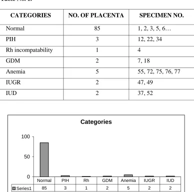
OBSERVATION
Of the 100 placentae collected, 53 were from primi and 47 were from
multi gravida. 85 were from uncomplicated pregnancies and 15 were from
complicated pregnancies. The 15 cases included pregnancy induced
hypertension 3, anemia 5, Gestational Diabetes mellitus 2, Rh incompatability
[image:43.612.120.509.312.696.2]1, IUGR 2 and IUD 2.
Table No. 1.
CATEGORIES NO. OF PLACENTA SPECIMEN NO.
Normal 85 1, 2, 3, 5, 6…
PIH 3 12, 22, 34
Rh incompatability 1 4
GDM 2 7, 18
Anemia 5 55, 72, 75, 76, 77
IUGR 2 47, 49
IUD 2 37, 52
Categories
0 50 100
Series1 85 3 1 2 5 2 2
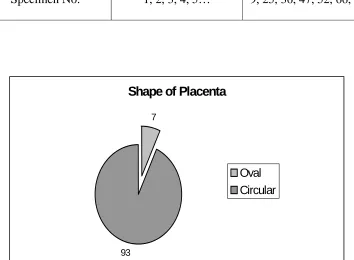
MORPHOLOGICAL PARAMETERS OF PLACENTA
a) SHAPE
[image:44.612.136.490.316.576.2]Out of 100 specimens 93 were circular and 7 were oval in shape.
Table No. 2.
SHAPE CIRCULAR OVAL
No. of Placenta 93 7
Specimen No. 1, 2, 3, 4, 5… 9, 25, 36, 47, 52, 66, 87
Shape of Placenta
7
93
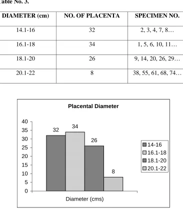
b) DIAMETER & THICKNESS :
In the present study, the range of the placental diameter was from 14
cms to 22 cms, average being 17.5.
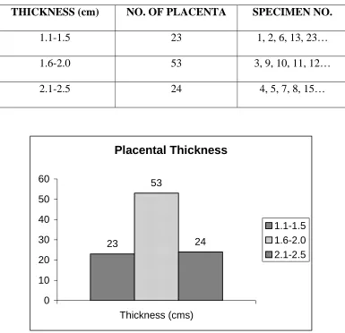
The range of the placental thickness was from 1.1 cms to 2.5 cms,
[image:45.612.126.492.283.701.2]average being 1.8 cms.
Table No. 3.
DIAMETER (cm) NO. OF PLACENTA SPECIMEN NO.
14.1-16 32 2, 3, 4, 7, 8…
16.1-18 34 1, 5, 6, 10, 11…
18.1-20 26 9, 14, 20, 26, 29…
20.1-22 8 38, 55, 61, 68, 74…
Table No. 4.
THICKNESS (cm) NO. OF PLACENTA SPECIMEN NO.
1.1-1.5 23 1, 2, 6, 13, 23…
1.6-2.0 53 3, 9, 10, 11, 12…
2.1-2.5 24 4, 5, 7, 8, 15…
Placental Thickness
23
53
24
0 10 20 30 40 50 60
Thickness (cms)
1.1-1.5
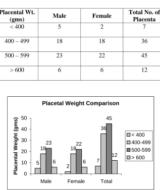
c) WEIGHT :
The minimum weight of placenta in case of male baby was 250 gms
and maximum was 760 gms with an average of 504.8 gms.
The minimum weight of placenta in case of female baby was 230 gms
[image:47.612.153.470.290.667.2]and the maximum was 680 gms with an average of 512.7 gms.
Table No. 5.
Placental Wt.
(gms) Male Female
Total No. of Placenta
< 400 5 2 7
400 – 499 18 18 36
500 – 599 23 22 45
> 600 6 6 12
Placetal Weight Comparison
5 2 7 18 18 36 23 22 45 6 6 12 0 10 20 30 40 50
Male Female Total
Placental Weight (gms)
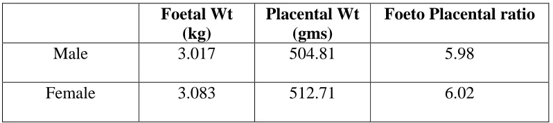
d) FOETO-PLACENTAL RATIO :
It is the ratio of foetal weight to placental weight. It is normally 6:1
according to Hamilton & Boyd. In the present study it was 5.98:1 in case of
[image:48.612.114.513.276.365.2]male babies and 6.02:1 in case of female babies.
Table No. 6.
Foetal Wt
(kg)
Placental Wt (gms)
Foeto Placental ratio
Male 3.017 504.81 5.98
Female 3.083 512.71 6.02
Fetal Weight/Placental Weight
0 100 200 300 400 500 600 700 800
0 1 2 3 4 5
Fetal Weight (Kg)
Fetal Weight/Placental Weight (Male)
0 100 200 300 400 500 600 700 800
0 1 2 3 4 5
Fetal Weight (Kg)
Placental Weight (gm)
Fetal Weight/Placental Weight (Female)
0 200 400 600 800
0 1 2 3 4 5
Fetal Weight (Kg)
e) PLACENTAL COEFFICIENT :
It is ratio of placental weight to foetal weight. It is 0.15 normally,
according to Little.
In the present study, the placental coefficient in male babies was 0.17
[image:50.612.131.496.281.651.2]and in case of female babies was also 0.17.
Table No. 7.
Foetal Wt
(kg)
Placental Wt (gms)
Placental Coefficient
Male 3.017 504.81 0.17
Female 3.083 512.71 0.17
Placental Coefficient
0 1 2 3 4 5
0 200 400 600 800
Placental Weight (gm)
Placental Coefficient (Male)
0 1 2 3 4 5
0 200 400 600 800
Placental Weight (gm)
Fetal Weight (Kg)
Placental Coefficient (Female)
0 0.5 1 1.5 2 2.5 3 3.5 4 4.5
0 200 400 600 800
Placental Weight (gm)
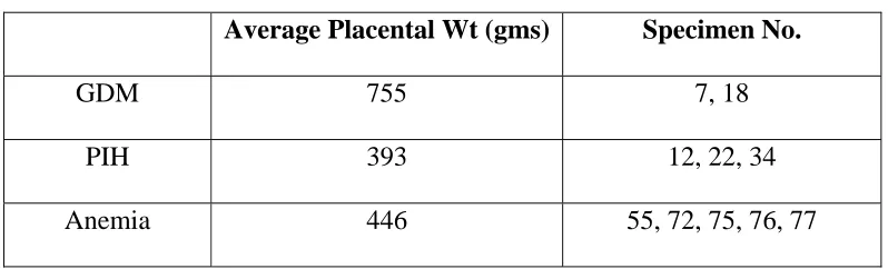
f) PLACENTAL WEIGHT IN VARIOUS FACTORS COMPLICATING
PREGNANCY :
The average placental weight in case of gestational diabetes was 755
gms. In case of anemia complicating pregnancy, it was 446 gms. It
Pregnancy Induced Hypertension, it was 393 gms.
In case of gestational diabetes, the fetoplacental ratio was 5.2 whereas
placental coefficient was 0.19.
In case of anemia complicating pregnancy, fetoplacental ratio was 5.8
and placental coefficient was 0.17.
In case of PIH, fetoplacental ratio was 5.8 and Placental coefficient
[image:52.612.114.513.526.647.2]was 0.17.
Table No. 8.
Average Placental Wt (gms) Specimen No.
GDM 755 7, 18
PIH 393 12, 22, 34
g) NUMBER OF COTYLEDONS :
The number of cotyledons in normal pregnancy ranged from 10 - 29,
average being 20. In cases of pregnancy induced hypertension, it was 18. In
gestational Diabetes mellitus, it was 21 and in anemia it was 16. In Rh
[image:53.612.114.515.276.423.2]incompatability, it was 12.
Table No. 9.
NO. OF
COTYLEDONS NO. OF PLACENTA SPECIMEN NO.
10 – 14 13 4, 12, 16, 21, 23…
15 –19 28 2, 3, 5, 8, 13…
20 – 24 43 6, 7, 9, 10, 14…
25 – 29 16 1, 11, 22, 32, 39…
Cotyledon Number
13
28
43
16
0 10 20 30 40 50
10-14
h) CORD LENGTH :
The umbilical cord length ranged from 47-70 cms, average being 54.4
[image:54.612.154.476.417.613.2]cms.
Table No. 10.
CORD LENGTH (cm) NO. OF PLACENTA SPECIMEN NO.
41 – 50 22 2, 12, 13, 22, 33…
51 – 60 65 1, 3, 4, 5, 6, 7…
61 – 70 13 10, 19, 24, 74, 76…
Cord Length
22
65
13
0 10 20 30 40 50 60 70
Length (cm)
41-50
i) DIAMETER OF UMBILICAL CORD :
[image:55.612.112.514.193.310.2]It was from 0.7 cms to 1.8 cms and the average was 1.24 cms.
Table No. 11.
CORD DIAMETER (cm)
NO. OF PLACENTA SPECIMEN NO.
0.7 – 1.0 29 1, 2, 5, 6, 8…
1.1 – 1.4 48 4, 13, 14, 15, 16…
1.5 – 1.8 23 3, 7, 24, 25, 26…
Cord Diameter
29
48
23
0 10 20 30 40 50 60
Diameter (cms)
0.7-1.0
j) CORD ATTACHMENT :
The following types of cord attachment were observed. Most of the
cords were attached eccentrically and it was found to be 66. The next
common attachment was central, totaling 27. Marginal attachment was seen
[image:56.612.147.478.285.646.2]only in 7 cases.
Table No. 12.
ATTACHMENT NO. OF PLACENTA SPECIMEN NO.
Eccentric 66 1, 3, 4, 5, 6…
Central 27 7, 11, 22, 23, 24…
Marginal 7 2, 17, 18, 21, 26, 28, 42
Cord Attachment
27
66 7
k) UMBILICAL CORD VESSELS :
Normally umbilical cord contains two umbilical arteries and one
umbilical vein. In our study also all the umbilical cord contained two
[image:57.612.111.512.251.367.2]umbilical arteries and one umbilical vein.
Table NO. 13.
C/S OF UMBILICAL CORD NO. OF PLACENTA
2 Umbilical Arteries 100
Single Umbilical Artery 0
Double Umbilical Vein 0
l) KNOTS :
In the present study, false knots were observed in 6 cords. No true
knots were observed.
Table No. 14.
KNOTS NO. OF PLACENTA SPECIMEN NO.
No Knots 94 1, 3, 4, 5, 6…
False Knots 6 2, 7, 16, 28, 33, 68
[image:57.612.115.513.558.676.2]m) PLACENTAL CALCIFICATION :
[image:58.612.115.509.196.282.2]No placental calcification were observed in any of the 100 placenta.
Table No. 15.
PLACENTAL CALCIFICATION NO. OF PLACENTA
Absent 100
DISCUSSION
This study on morphometric analysis of human placenta was carried
out as the placenta gives an accurate record of the infant’s intrauterine life.
From this study, it can be said that a ten minutes careful examination of the
placenta reflects the ten months environment from where the child has come.
This is an era of non-invasive techniques and placenta can very much
be examined thoroughly during routine ultrasonogram of expectant mothers.
During Ultrasonogram test, due care has to be taken to study the
various aspects of placenta and umbilical cord.
In morphometric study of placenta, measurement of its shape, size
weight, thickness and the number of cotyledons were measured.
The umbilical cord length, circumference, diameter were measured and
its attachment were also noted down.
Details of the mother and child were also obtained for clinical
This study was done using 100 placentae which were obtained from
the labour ward and operation theatres of Rajah Mirasudhar Govt. Hospital.
Of these 100, 85 were from uncomplicated pregnancies and 15 were
from factors complicating pregnancy. This 15 cases included 2 cases of
gestational Diabetes, 3 cases of Pregnancy Induced Hypertension, 1 case of
Rh incompatability, 5 cases of anemia complicating pregnancy, 2 cases of
Intra uterine growth retardation and 2 cases of Intra uterine death.
About 93 placentae were circular in shape, while 7 were found to be
oval. This finding fits with the normal range seen in other studies.
Three cases of Pregnancy Induced Hypertension were seen. The
weight of placentae in these cases respectively 390 gms, 310 gms and 480
gms. The average was 393 gms. There is no difference between this and the
average from normal pregnancies. However, these were moderate cases only
and severe cases were not encountered.
The average weight of placenta in gestational Diabetes was higher than
The reason for big babies in diabetes remained a matter of controversy.
One possible reason cited in literature is dysfunction of Anterior Pituitary
gland due to excessive production of diabetogenic and growth producing
hormones of Anterior pituitary.
According to Y in Liu Y, the placental weight gradually decreased in
prolonged pregnancy as according to them, the placental function is lowered
in them. But, in this study no such observation was made.
The placental weight is low in case of mothers who had anemia.
Maternal diseases including anemia have their effects on the fetus by altering
placental metabolism and transfer mechanisms. The observations made in
this regard was consistent with the study of Woohing et al 1976. The average
weight of placenta in anemic mothers in this study was 446 gms and 360 gms
in previous study of Woodling et al.
The weight of placenta is normally 500 gms. In this study, the
minimum weight of placenta was 230 gms & the maximum weight of the
PLACENTAL DIAMETER & THICKNESS :
In this study the placental diameter in case of male babies was found to
be 17.9 cms and female babies was 17.5 cms. The term human placenta is 15
to 20 cms according to A.L. Mudaliar. The result of this study falls within the
normal range. So also the thickness of placenta at term was 1.88 cms in case
of male and 1.71 cms in case of female babies. This parameter also fell
within the normal suggested by AL Mudaliar.
The maternal surface of placenta bears cotyledons ranging from 12 to
24 with an average of 18. The average number of cotyledons in present study
was 20.
The paucity of lobes in cases of pregnancy induced hypertension and
low birth weight as seen by Nordenvall et al was not seen in present study.
These cotyleldons were ill defined in cases of anemia complicating
pregnancy.
There is no correlation between the number of cotyledons and the sex
OTHER OBSERVATIONS :
The present study shows translucent membrane in 92 cases, opaque in
2 cases and meconium stained in 6 cases. In one of the 2 opaque membranes,
the baby had cord around the neck three times and mildly asphyxiated. It was
resuscitated immediately. All the 6 cases of meconium stained membranes
were associated with foetal distress.
The fetal surface of placenta was steel blue in colour in most of the
cases. Sub-chorionic fibrosis and Tasselations were present in all term
placentae. Tasselations are sclerosed vessel below the chorion forming a
criss-cross pattern on the foetal surface.
No calcification was noted in any of the 100 placenta. In the present
study, retroplacental clots were noticed in 4 cases and all were Abruptio
placentae. No congenital anomaly was present in the new born. The average
weight of the retroplacental clot was 100 gms.
PLACENTAL WEIGHT IN RELATION TO WEIGHT OF THE BABY :
The ratio of foetal weight to placental weight if 6 : 1, according to
Hamilton & Boyd. In the present study, it was 5.98:1 in case of male babies
and 6.02:1 in case of female babies. Both were nearer to normal ratio. The
induced hypertension was 5.8 and in anemia complicating pregnancy 5.8. All
these values are also almost nearer to the normal ratio suggested by Hamilton
& Boyd.
The otherway to correlate the weight of the baby and placenta is
placental coefficient. The placental weight in grams divided by foetal weight
in grams is the placental coefficient as defined by Little.
The normal placental coefficient is 0.12 to 0.2. The placental
coefficient in the present study for male babies was 0.17 and for female
babies also. This placental coefficient in Gestational diabetes was 0.19,
Pregnancy Induced Hypertension was 0.17 and in anemia complicating
pregnancy was 0.17.
MORPHOLOGICAL PARAMETERS OF THE UMBILICAL CORD :
The umbilical cord is also called the Funiculus Umbilicalis. The
minimum cord length in the present study was 47 cms and the maximum was
70 cms. The average cord length was 54.4 cms.
The average length of umbilical cord in African population was 58.4
others as well as this established the fact that the cord length are not
determined by the racial groups.
This study also observed that there is no significant relation between
cord length and parity of mother.
The diameter of the cord varied from 0.7 cms to 1.8 cms in the present
study. The average was 1.24 cms.
Out of 100 cases, Eccentric type of cord insertion was seen in 66 cases,
central type of cord insertion in 27, marginal insertion in 7. Velamentous
type and Battledore type of insertion were not encountered.
The mode of umbilical cord insertion has no significant effect on
compartmental volume of placental parenchyma.
The cord insertion is visualized in more than half of the cases in
Ultrasonography in clinical practice and the knowledge is useful for planning
obstetrical management.
The calculated length of umbilical cord is almost equivalent to actual
The thickness (circumference) of the umbilical cord includes the
umbilical vessels and the connective tissue called as Whartons Jelly. All
these are surrounded by a sheath formed by the amnion. For a short distance
from its junction with anterior abdominal wall, the cord is covered by an
epithelial tissue.
Marginal cord insertion is usually related to bilobate placenta. Major
fetal anomalies with normal Karyotype are found in Velamentous type.
The cord insertion is probably the effect of biological variations during
normal placental and fetal development. Marginal cord insertion may evolve
into velamentous insertion as pregnancy progresses.
In the present study, no true knot (nodi veri) was observed. However,
false knots (nodi spurii) was observed in 6% of cases. They are formed by
umbilical Vein twisting around the umbilical artery, causing a localized
thickening of Wharton’s Jelly and they lack any clinical significance.
In the present study, Whartons jelly was present in all the cases. If
absent, they are associated with other congenital anomalies.
SUMMARY
The summary of the study of morphology & morphometric analysis of
placenta is as follows:
A total of 100 specimens of placenta and umbilical cord were studied
in the present study.
The placenta were derived from 53 primigravida and 47 multigravida.
85 were from uncomplicated cases and 15 were from factors complicating
pregnancy. Out of 100 specimens, 52 were from male, 48 from female.
Out of 100 specimens studied, 93 were circular, 7 were oval.
The weight of the placenta ranges from 230 gms to 680 gms in case of
female with an average of 512.71 gms & 250 gms to 760 gms with an average
of 504.81 gms in case of male children.
The diameter of the placenta in the present study ranges from 14 cms
The placental thickness ranges from 1.1 cms to 2.5 cms, with an
average of being about 1.8 cms.
The ratio between the foetal weight and placental weight in case of
male was 5.98 and in case of female was 6.02.
The placental coefficient in the present study is 0.17 both in male and
female.
The average number of cotyledons was 20.
Subchorionic fibrosis and Tasselations seen in all placentae.
No placental calcification seen.
Retroplacental clots seen in 4 cases and all of them were associated
with Abruptio placentae.
The average length of the umbilical cord was 54.4 cms.
Present study showed eccentric type of cord insertion in 66, central
type in 27, Marginal type in 7.
False knots noticed in 6 cases.
Diameter of the umbilical cord on an average was 1.24 cms.
CONCLUSIONS
Morphological studies of placenta in high risk group and subsequent
cytogenetic analysis will prove presence or absence of chromosomal foetal
malformations in terms of 8 – 24 weeks when termination is possible.
Morphometry is an indirect and non-invasive approach to the study of the
physiology and physiopathology of gestation in the human. Morphometry is a
good adjuvant to histopathology.
Gross examination was more informative, though microscopic
examination would have been necessary.
Triage is the sorting and allocation of treatment according to a system of
priorities in order to maximize treatment. Placental triage promptly after
delivery of the placenta with documentation of the findings in the medical
record only takes a few minutes, and allows for the identification of abnormal
placentae to be submitted for detailed gross and microscopic examination. It
requires familiarity with normal gross placental anatomy.
Our study concludes with a recommendation that a systematic procedure
has to be adopted and documentation for comprehensive examination of the
placental disk as a whole, the umbilical cord, the extra placental membranes,
the fetal surface, the maternal surface and the parenchyma. Otherwise, the
BIBLIOGRAPHY
1. Alesllchenko IE, Milovanov AP,V,al’tseva IUIU. Morphological
characteristics of placenta & hormonal balance in
hyperthyroidism of pregnancy. Arkh Patol 2001 May – June 63
(3): 26-30.
2. Archakova TM, Banyliak IR, Nikitchyana TV, Zaderozhna TD
Tsitol. Morphological and cytogenetic aspects in a study of
chorionic and placental biopsies in pregnant women who are over
the afe of 35. Genetics 1998 Jan-Feb 32 (1) 96-107.
3. Arey Placentation & Decidual – Text book of embryology.
4. Asim Kumar Datta; The Placenta; Essentials of Human
Embryology; 1978.
5. Biagiothi R, Sgambaki E, Brizzi E. Placental morphometry in
pregnancies, complicated by intrauterine growth complicated by
intrauterine growth retardation with absent or reversed end
diastolic flow in the umbilical artery. Anatomy Embryology,
1999 Oct-Dec;104 (4);201-7.
6. Bruce M. Carlson, M.D, Ph.D; Placenta and Extraembryonic
Membranes; Human Embryology and Developmental Biology;
7. Derek Llewellyn-Jones; The Placenta; Fundamentals of Obstetrics
and Gynaecology; 1982.
8. Distler W, Bender HG, W Geburtr Shilfe, Placental morphometric
parameters correlated with serum Oestriol. Perinatology, 1983
Jan-Feb 187(1); 23-7.
9. Durand A, Descamps P, Vieyres P, Menigault E, Gregoire JM,
Pour celot D, Fiechrer G, Lansac J Body G, Pourcelot C. In utero
measurement of the umbilical cord in full term pregnancy.
Journal of Gynaecology & Obsteritics Reproductive Biology
1996;25 (1) 78-86.
10. Emrich P, Horn LC, Seifert U Zentrabe, Morphological findings
in fetuses & placentas of late abortion in the 2nd trimester.
Gynaecology 1998;120 (8); 399-405.
11. Grebesa D Durst Zivkovic B P flugers. The neonatal & placental
factors in relation to the mode of umbilical cord insertion Arch
1996;431 R 205-6.
12. Haines and Taylor; Development and anatomy of the placenta;
General pathology of the placenta, umbilical cord and fetal
membranes; Obsterical and Gynaecological Pathology; 1999.
13. Hamilton, Mossman and Boy; Implantation, fetal membrane &
placentae. The Human placenta, Cambridge England : W Heffer