1
A
A
s
s
s
s
e
e
s
s
s
s
m
m
e
e
n
n
t
t
o
o
f
f
d
d
i
i
a
a
g
g
n
n
o
o
s
s
t
t
i
i
c
c
v
v
a
a
l
l
i
i
d
d
i
i
t
t
y
y
o
o
f
f
P
P
r
r
o
o
c
c
a
a
l
l
c
c
i
i
t
t
o
o
n
n
i
i
n
n
i
i
n
n
b
b
u
u
r
r
n
n
s
s
s
s
e
e
p
p
s
s
i
i
s
s
p
p
a
a
t
t
i
i
e
e
n
n
t
t
s
s
–
–
A
A
p
p
r
r
o
o
s
s
p
p
e
e
c
c
t
t
i
i
v
v
e
e
s
s
t
t
u
u
d
d
y
y
A dissertation submitted to the Tamil Nadu Dr M.G.R Medical
University in partial fulfillment of the requirement of the award Of
2
CERTIFICATE
I hereby declare that this dissertation entitled “Assessment of diagnostic validity of Procalcitonin in burns sepsis patients – A prospective study” is a bonafide research work carried out by Dr. Naveen Kumar H R in partial fulfillment of the requirement for the degree of M.Ch. in Plastic Surgery.
Guide:
Dr Kingsly Paul M
Professor
Department of Plastic Surgery Christian Medical College Vellore
Co Guide:
Dr Elvino Barreto
Associate Professor
3
ENDORSEMENT BY THE H.O.D. OF PLASTIC SURGERY
This is to certify that this dissertation entitled “Assessment of diagnostic validity of Procalcitonin in burns sepsis patients – A prospective study” is a bonafide and genuine research work carried out by Dr. Naveen Kumar H R under the guidance of
Dr. Kingsly Paul M M.S, MCh,Professor and unit head, Department of Plastic Surgery, Christian Medical College, Vellore.
Head of Department:
Dr. Ashish Kumar Gupta
Professor and Head
4
ACKNOWLEDGEMENT
I wish to express my deepest sense of gratitude to my teacher Prof. Kingsly Paul M who has played an active role in this work and also was a constant source of inspiration and encouragement to me throughout.
I sincerely thank The Director and the members of the IRB committee , Christian Medical College, Vellore for approving my thesis topic and gave permission to conduct the study.
I owe my sincere thanks to my teachers, Prof. Ashish Kumar Gupta, Dr Elvino Barreto, Dr Shashank Lamba and Dr Rahul Shetty who gave valuable advice, support and encouragement throughout the work.
I sincerely thank Prof. and Head Dr R . Selvakumar , Department of Clinical Biochemistry for extending the support in conducting the tests.
I sincerely thank Prof. Sampath Karl Department of Pediatric surgery for allowing me to do the study in their department.
I sincerely acknowledge the help of Miss. Visali, Department of Biostatistics in performing the statistical analysis of data and Mr Sathiya Murthi, Dept CEU in formatting the thesis.
I am also grateful to Mrs Kalaivani ward incharge and all the staff nurses of Q1 WEST ward for all their support.
I am also grateful to other faculty members and all my post-graduate colleagues for their practical suggestions and help rendered in the preparation of this work.
I also acknowledge the constant support of my family throughout the course of my education.
5
INDEX
S.NO. CONTENTS PAGE NO.
1
Introduction
6
2
Aims and Objectives
9
3
Materials and Methods
11
4
Review of Literature
17
5
Results and Analysis
33
6
Discussion
45
7
Summary
55
8
Conclusion
58
9
Bibliography
60
10
Annexure-1 Proforma & consent form
66
11
Annexure-2 Master chart
75
ABSTRACT
TITLE OF THE ABSTRACT : To asses the Diagnostic validity of Procacitonin in Burns Sepsis patients.
DEPARTMENT :Plastic and Reconstructive surgery NAME OF THE CANDIDATE :Dr. Naveen Kumar H R
DEGREE AND SUBJECT :Mch , Plastic surgery NAME OF THE GUIDE :Dr. Kingsly Paul M
AIM / OBJECTIVE:
To asses the Diagnostic validity of Procacitonin in Burns Sepsis patients.
MATERIAL AND METHODS:
This was a prospective diagnostic study done for a period of 9 months involving 36 cases who sustained > 20% TBSA of second degree burns. The criteria explained by the American Burn Association (ABA – 2007) were used as the reference standard to diagnose sepsis. Patients were continuously monitored for the development of signs and symptoms of sepsis. Procalcitonin (PCT) estimation was done at the time of sepsis and without sepsis. The values of PCT correlate with the sepsis parameters of American Burn Association. Data was analysed using Receiver operating curve analysis, Fisher exact test, t test, Mann whitney U test.
RESULTS:
The predictive ability of the PCT was determined by using the ROC (Receiver operating curve) and the area under the curve was calculated with 95% confidence intervals. Analysis of the coordinate points reveled the best threshold values of PCT for the prediction of sepsis based on the combination of sensitivity and specificity of each proposed threshold. The cut off value was 5ng/ml based on the ROC analysis at the sensitivity of 88.9% and specificity of 83.3% with area under the curve was 91%.
CONCLUSION:
6
7
Introduction:
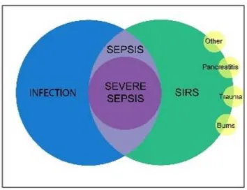
Sepsis is one of the major cause of mortality in burns patients.1 Sepsis is defined as systemic inflammatory response with documented infection. If not treated, then the infection will lead to organ dysfunction, hypoperfusion and hypotension called as severe sepsis. Later it will end up with septic shock. Systemic inflammatory response syndrome (SIRS) can be due to either infectious or non infectious cause. In major trauma and burns patients it is always difficult to differentiate whether SIRS is a result of injury itself or due to superimposed infection. Most of the clinical signs of infection such as fever, tachycardia , leucocytosis were also present in SIRS.
Thus to overcome this a consensus panel was formed by American burns association and guidelines were framed to define sepsis in burns patient. In this the definition of SIRS and severe sepsis was excluded and the range of vital parameters were raised, platelet count and patient glucose level included. Inability to feed the patient enterally for more than 24 hrs was also one of the criteria. To diagnose it as sepsis, infection should be documented or the infection should respond to the antibiotic started empirically. These guidelines were based on the consensus and not on any prospective clinical studies.
8
explained in the literature eg., CRP – C reactive protein, TNF , IL-6 etc. But Cytokine and non cytokine markers are not used routinely in burned patients. The marker that have been consistently elevated in patients with infection is procalcitonin(PCT). Studies have shown that increased plasma levels of PCT are sensitive and specific marker of infection.3
The marker is more specific for the bacterial infection and increase several folds within 4 hrs of infection, reaches peak by 6 hrs and plateau by 8-24hrs, then return to baseline by 2-3 days. Its half life is 24-30hrs. These qualities of the marker made it clinically suitable and to repeat at regular intervals.
9
10 Aims and objective:
1) To find the diagnostic validity (sensitivity and specificity) of procalcitonin in burns sepsis patients.
11
12
Materials and methods :
This is a prospective study done for a period of 9 months from March 1st , 2012 to November 31st , 2012 in the Department of Plastic Surgery, Christian Medical College and Hospital, Vellore. This study was approved by the Institutional Review Board. There were 36 patients involved in this study.
Inclusion criteria : All patients with more than or equal to 20% burns.
Exclusion criteria : All patients with less than 20% burns.
All patients presented with 20% or more of burns admitted in the burns ward of CMC Hospital were included in the study. If the patient got admitted within 24 hr of injury the resuscitation protocol of our burns unit was started which involves
- Admission to the burns unit.
- Insert a central line preferably in non burnt areas. - To place a Foleys catheter to measure urine output. - To start fluid (Ringer lactate) as per parkland formula. - Burn wound care and silver sulfadiazine dressing. - Insert naso gastric tube in major burns
- I.V. morphine as analgesic round the clock. - To send all base line blood investigations.
13
On the first day of resuscitation period we maintained a urine output of 30-50ml/kg/hr. we titrated the fluid based on urine output. On second day of resuscitation colloid was started. We infused 0.5ml/kg/%burns of plasma with 5% dextrose to maintain urine output of 1ml/kg/hr. The Patients were monitored continuously. Every 4th hourly Temperature, pulse rate and respiratory rate were recorded daily from the day of admission. Patient daily calorie intake was documented. Twice a week baseline blood investigations were done. Pus culture of the wound, blood culture, urine culture and sputum culture were done from third day of burns depending on the patient condition. All the parameters necessary to diagnose burns sepsis according to American Burn Association (ABA) consensus were documented in a Chart to diagnose sepsis (Chart attached in Annexure 1). This was the reference standard of our study.
American burn association consensus definition of sepsis and infection : 3 or more of the following :
1. Temperature >39°C or <36.5°C 2. Progressive tachycardia (> 110/min) 3. Progressive tachypnoea (>25/min)
4. Thrombocytopenia < 1 lac (only applies 3 days after initial resuscitation) 5. Hyperglycemia (in the absence of pre-existing diabetes mellitus)
6. Inability to continue enteral feeding >24 h.
Plus documented infection with one or more of the following: 1. Culture positive infection.
14
15
Procalcitonin estimation:
Procalcitonin measurement in our study was done using the test kits obtained from B.R.A.H.M.S (Kryptor method) Germany. The results were obtained within 19 min of the test. The analytical sensitivity of the test was 0.019ng/ml and the functional assay sensitivity of 0.06ng/ml with a probability of 95%. The Kryptor method consists of sheep polyclonal anti – calcitonin antibody and a monoclonal anti – katacalcin antibody which binds to the calcitonin precursor molecules. The antibodies used in this assay do not show cross reaction with human calcitonin (up to 2.5ng/ml) and katacalcin (up to 10ng/ml) , human a-CGRP and b-CGRP (up to 4 mcg/ml). The interfering substances like icteric , haemolytic, hyperlipemic samples and also samples with turbid or contain fibrin if present then it is signalled by Kryptor.
Measuring principle :
16
17
18
Thesis : Review of literature
American College of Chest Physicians/Society of Critical Care Medicine (ACCP/SCCM) in 1992 described the Systemic inflammatory response syndrome (SIRS) based on clinical and experimental results which is independent of cause.4
According to this two or more following criteria should be fulfilled to diagnose as SIRS
1. Temperature > 38 or < 36 degree 2. Heart rate > 90/min.
3. Respiratory rate > 20/min or Paco2 <32mmHg
4. Leucocyte count >12000/<4000/>10% immature (band) forms.
The criteria explained were more sensitive and less specific. In order to improve the specificity there was a meeting regarding this issue in second conference in 2001. Additional criteria were added that defined metabolic, biochemical and functional alterations associated with SIRS. Those criteria were hyperglycemia, edema, elevated plasma C reactive protein, coagulation abnormalities, thrombocytopenia, hyperbilirubinemia and ileus.5 SIRS can occur in infective and non infective cases. For eg., in burns , multiple trauma, pancreatitis etc. sepsis was defined as SIRS with documented infection.
19
But the extensive wounds in burns patients had chronic exposure to inflammatory mediators which causes fever, tachycardia, tachypnea and resetting of baseline metabolic rate considered as the normal physiologic response. So in order to define sepsis in burns the SIRS category was dropped as this was considered as the protective response of the body towards exaggerated inflammatory condition. Thus in 2007 American Burn Association consensus conference defined sepsis and infection in burns with the following guidelines.6
SIRS:
Not applicable in burn patients Sepsis:
3 or more of the following triggers search for infection 1. Temperature >39°C or <36.5°C
2. Progressive tachycardia (>110 /min) 3. Progressive tachypnoea (> 25/min)
4. Thrombocytopenia (only applies 3 days after initial resuscitation) 5. Hyperglycemia (in the absence of preexisting diabetes mellitus) 6. Inability to continue enteral feeding >24 hrs
Plus documented infection with one or more of the following 1. Culture positive infection
20
Severe sepsis:
Not applicable in burns patients Septic shock:
Sepsis with persistent hypotension despite adequate fluid resuscitation.
The aim of this guidelines is to have a consistent diagnosis in all burn centres and to help in future research. Still the term SIRS is used to explain the pathogenesis of post burn injury and sepsis. SIRS is also called as hypermetabolic response in burn injury which is more meaningful.
Two hit hypothesis of sepsis :
The pathophysiology of SIRS and sepsis was explained by Two hit hypothesis. The injured host manifest an exaggerated inflammatory response if exposed to secondary inflammatory stimulus during the post injury period. The initial insult resulted in production of lymphokine interferon alpha (IFN ) acts as first signal and prime macrophages for increased inflammatory response. A second stimulus causes the primed macrophages to secrete increase amount of TNF . This was not produced in large amount during the initial inflammatory insult. If the primed macrophages were exposed to even small amount of endotoxin at the time of second stimulus then TNF was produced enormously. T- lymphocytes also become hyper responsive at the post injury period.
21
studies by different authors. When a week old thermally injured sheep exposed to systemic bacterial challenge found increase pulmonary hypertension and hyperdynamic response.8 Also when combined administration of low dose endotoxin and TNF in rat model resulted in hypotension and metabolic effects that was seen after giving highly lethal dose of each compound alone.9
Inflammatory Mediators of SIRS :
The initial stimulus for the activation and release of inflammatory mediators of SIRS is either major injury or infection. The various mediators were mentioned in brief.
Cytokines: have unique role in causing SIRS after injury. The most important were TNF , IL 6, IL 1 and interferon (IFN ).
22
IL 1 actions are similar to TNF except it doesn’t induce tissue injury or apoptosis by itself instead it potentiates the injurious effects of TNF .13
The function of IL 6 is to induce production of acute phase proteins and acts as growth and differentiation factor for B lymphocytes. It is produced by macrophages, endothelial cells and fibroblasts.
IFN is the primary cytokine produced at the initial insult by T lymphocytes and NK cells in response to antigen presentation and induction from IL 12 and IL18. The primary functions of IFN is to amplify inflammatory response of macrophages, induce secretion of inflammatory mediators (TNF , IL 1) and potentiates the antigen presentation by HLA II complex. The blockade of IFN markedly decrease the inflammatory effects induced by the bacterial endotoxin.14
Chemokines :
The other group of proteins responsible for SIRS is chemokines. Primarily they function as chemotactic factor for leucocytes. Among various chemokines which were described IL 8 is the potent chemoattractant for neutrophils and major factor in recruiting neutrophils to inflammatory foci. IL 8 also mediate tissue injury in lung in response to trauma and burns.15
Non cytokine factors : also have role in the pathogenesis of SIRS they were platelet activating factor ( PAF) , Leucotrienes (LT) ,Thromboxane A2.
23
permeability and acts as chemotaxis of leukocytes. PAF can trigger inflammation, thrombosis and also mediate molecular and cellular interactions.
Leukotriens (LT) : Synthesized from leucocytes and acts on G- protein coupled receptors. It causes contraction of endothelial cells and encourage capillary leakage thus sustaining the inflammatory response.
(Thromboxane A2)TXA2 : It belongs to eicasanoids family, synthesized from platelets. It promotes platelet aggregation, vasoconstriction and tissue thrombosis.
Systemic manifestations of the burns patients
This is divided in to early hypovolemic shock phase and delayed phase of systemic inflammation (SIRS) in which monocytes, macrophages and T cells were activated to release various mediators of inflammation mainly cytokines. The activated T cells divided in to Th1 cells secrets proinflammatory cytokines and Th2 cells secrets anti inflammatory cytokines. The cytokines were differentiated from the hormones in a way that these were not secreted by a specialized cells arranged in glandular tissue, it has broad spectrum of activity when compare to hormones and these were either positive or negative regulators of cell cycle, its differentiation, cell survival, apoptosis and transformation.
There are three important factors which determine the effects of SIRS in burns patients:
1. Severity of initial SIRS is proportional to severity of injury. 2. Prolonged SIRS leads to higher complication rate.
24
So lesser the initial insult lesser the SIRS. Prolongation of SIRS can be reduced by adequate fluid resuscitation, excision of necrotic tissue and enteral feeding. The inflammation is mediated by the cytokines which are released in response to mechanical, thermal and ischemia reperfusion injury at the cellular level. The proinflammatory mediators were TNF and IL- 6. Worsening or prolongation of SIRS were due to inadequate fluid resuscitation, repeated insults by infection, tissue necrosis and endotoxin migration from the intestine. At the same time body responds by producing more anti inflammatory mediators. These were TGF and IL 10. Producing excess of these anti inflammatory mediators will cause immunosupression and patient will exposed to uncontrollable infections.
Till the early 90s the cytokines and acute phase proteins were the markers used to assess the inflammatory response. The rise in the level of these markers is non specific and increase in both infective and non infective foci. The half life of these markers is very low thus making it not suitable for clinical conditions. Also it is not possible to assess the severity of the inflammation and after any therapeutic intervention these markers take longer period to reach the baseline.
25
Induction and synthesis of PCT :
Procalcitonin belongs to CAPA (Calcitonin gene related peptide amylin procalcitonin adrenomedullin) protein family. Calcitonin gene related peptides (CGRP ) I, II and procalcitonin belong to this group. CGRP I and the mRNA of calcitonin precursor were encoded on CALC- I gene on chromosome 11. All these proteins were usually produced in pro – pro form consisting of approximately 100 amino acids to gain access to golgi system. These prtotein has two cysteine residues to form di sulphide bridge and two protein cleavage sites to form final core protein of 35 amino acid which can be amidated.
PCT mRNA is synthesized by CALC- I gene present on chromosome 11 exclusively during inflammation and sepsis. Calcitonin is also product of CALC- I gene secreted from C cells of thyroid gland in normal individuals. PCT producing cells secrete two different types of PCT mRNA responsible for forming PCT – I and PCT – II which has differences at eight C- terminal amino acids. CALC - I gene produce three sequence of proteins and after processing there are nine different proteins. These proteins differ not only with mRNA splicing and procession , but also with respect to their regulation.
26
concentration in the plasma of healthy individuals is 10-50 pg/ml. In humans PCT I and PCT II mRNA is found mainly in liver, lung, kidney and testis.17 Ex vivo it is possible to produce PCT mRNA in immunocompetent cells like mononuclear cells when it is stimulated by endotoxin.18
PCT has secondary and tertiary structure that is modified by post translational processing. Protein modification occurs by glycosylation. Di peptidyl peptidase enzyme located on renal, epithelial and endothelial cells induced by proinflammatory mediators and endotoxin and is responsible for deamidation of PCT.19 Till now the function of biologically active PCT molecule is not known.
27
Procalcitonin (PCT) :
Procalcitonin is a peptide precursor of calcitonin hormone which is secreted mainly by the parafollicular C cells of the thyroid gand . Calcitonin is responsible for the calcium metabolism and to maintain the calcium homeostasis in the body. PCT is made of 116 amino acids which is also secreted by the neuro endocrine cells of the liver, lung and intestine only at the time of inflammatory process. The increased PCT at the time of inflammation does not have any role in calcium metabolism.
PROCALCITONIN - ng/ml
Normal 0.05
Local infection < 0.5 Systemic infection 0.5 - 2
Sepsis 2 – 10
Severe Sepsis >10
28 Studies of PCT in sepsis other than Burns
PCT has been utilised to differentiate bacterial from viral meningitis in children. This will helps in avoiding antibiotics and its adverse effects in meningitis due to viral cause.22
PCT is a diagnostic biomarker which can be utilised for the assessment of disease severity and will be the guidance for treating bacterial infections. This diagnostic biomarker will be a complementary for the clinical diagnoses of the infection. So the cut off values will be based on many diagnostic studies. Biomarker should never be utilised alone and always should be considered along with the clinical signs and symptoms of infection.23
29 PCT evaluation related to burns:
There are studies done in the past related to procalcitonin in burns patient. Most of the studies were done for evaluating the parameters like CRP, ESR and WBC. The changes in these parameters at the time of burns has been compared to the procalcitonin levels and concluded that procalcitonin has higher specificity when the sepsis starts due to bacterial origin. Also it is known that the changes in other markers are not specific as they keep rising in both hypermetabolic response and sepsis. So it is not possible to consider a cut off margin of these markers above which we can consider it as established sepsis.
Normal value of PCT in adult male : 0.05ng/ml. The level of procalcitonin for the diagnosis of sepsis vary in different studies from >0.5 ng/ml to 3ng/ml. Previously one of the criteria for diagnosing burns sepsis was leucocyte count, which may be either leucocytosis or leucopenia. According to the American burn association these counts are very unreliable and thus it has been excluded from the newer definition of burns sepsis.26
30
A Study done by Athina Lavrentieva et al , evaluated serum PCT, CRP, leukocyte count and temperature as markers of sepsis and divided the cases in to two groups as sepsis with SIRS and sepsis without SIRS and concluded that PCT is a better marker of sepsis than other inflammatory markers and the area under ROC curve has an acceptable accuracy.28
Another study done by D. von Heimburg et al Procalcitonin - a sepsis parameter in severe burn injuries, they assessed the PCT values and compared with the Baltimore sepsis score (BSS). There was a significant correlation between the BSS and rise in PCT level. A PCT value of 10ng/ml and increasing suggestive of severe systemic infection even when the blood culture was negative. A cut off value of 3ng/ml and above suggestive of bacterial infection and less than this patients will have better prognosis.29
31
32
The following statistical analysis were used:
1) Fisher exact test for categorical data.
2) ROC curve to assess the diagnostic performance. 3) t test
4) Mann whitney U test to know the significance.
33
34
RESULTS AND ANALYSIS:
Data of 36 patients were analysed with mean age of 27.97 yr (2-59 yr). Among them 44% were male, 42% female and 14% children sustained burns of > 20% TBSA. The average total body surface area of burns was 47.16 (20 – 97 %). Majority of the patients, 86% (31) sustained thermal burns and remaining 14% (05) had electrical burns. Out of 18 patients five patients (27.77%) had lung infection, 08 (44.44%) patients had positive growth of organism in blood culture and 05 (27.77%)patients had local wound sepsis with growth of multiple organisms. The organisms grown in blood culture was Pseudomonas in 3 patients, Klebsiella in 2, Acinetobacter in 01, MRSA in 2.
35
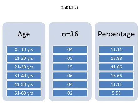
DEMOGRAPHIC PROFILE
[image:36.595.84.524.193.524.2]Age distribution : Fifteen patients (41.66% ) were in the age group of 21-30yrs with mean age of 27.97
TABLE : 1
Spearmans rank correlation coefficient did not reveal any relationship between age and PCT values.
Age
0 - 10 yrs
36
DISTRIBUTION OF SEX :
Total of 36 cases 16(44%) were male, 15(42%) were female and 05(14%) were children.
TABLE : 2
Patients Percentage
MALE 16 44
FEMALE 15 42
CHILDREN 05 14
[image:37.595.93.537.226.677.2]TOTAL 36 100
Fig: 1
Analysis with Mann whitney U test did not reveal any significant relationship between distribution of sex and rise in PCT level.
44%
42% 14%
Distribution of sex
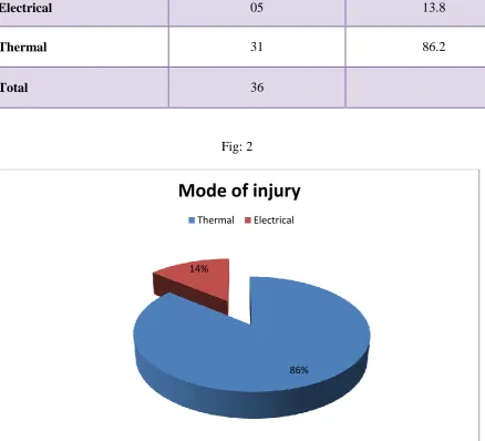
37 Mode of injury
Majority of the patients sustained thermal burns (86%) followed by electrical burns (13.9%).
[image:38.595.92.531.301.699.2]TABLE : 3
Fig: 2
86% 14%
Mode of injury
Thermal Electrical
Number of patients Percentage
Electrical 05 13.8
Thermal 31 86.2
38
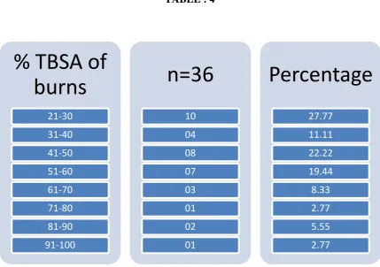
PERCENTAGE OF BURNS
[image:39.595.92.522.301.603.2]Twenty seven percent had burns within 30% of Total body surface area. There was no correlation between the percentage of burns and PCT values though higher values were noted in early phase of electrical injuries.
TABLE : 4
39
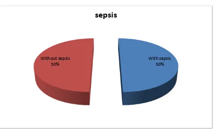
[image:40.595.86.530.195.348.2]Development of sepsis in study group: TABLE : 5
Number of patients Percentage
With sepsis 18 50
Without sepsis 18 50
Total 36
Half of the cases 18 (50%) fulfil the criteria for sepsis. Fig: 3
With sepsis 50% Without sepsis
50%
[image:40.595.92.531.447.712.2]40
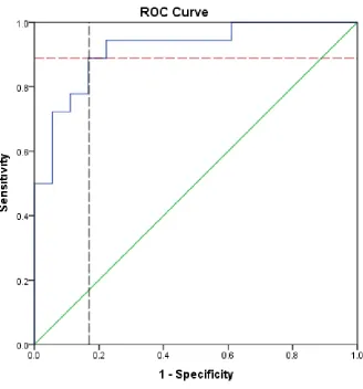
[image:41.595.159.488.206.559.2]Finding cut off value of Procalcitonin using ROC Curve
Fig 4, Receiver operating characteristic curve (ROC) showing the diagnostic performance of PCT in sepsis. (Y axis – Sensitivity, X axis – 1- Specificity).
For each PCT value plotted on ROC curve sensitivity and specificity is calculated. The value which is plotted most nearer to the 1 is the cut off value (5ng/ml). Two
perpendicular lines were drawn to join X and Y axis which shows sensitivity and 1-specificity to that value.
41
Co-ordinates of the curve :
TABLE : 6
PCT cut off value Sensitivity 1 - specificity
3.812 0.889 0.222
4.903 (=5) 0.889 0.167
5.729 0.833 0.167
42
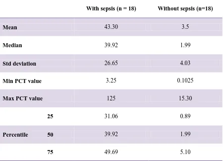
[image:43.595.90.540.174.498.2]Procalcitonin and sepsis – Testing the significance of association TABLE : 7
With sepsis (n = 18) Without sepsis (n=18)
Mean 43.30 3.5
Median 39.92 1.99
Std deviation 26.65 4.03
Min PCT value 3.25 0.1025
Max PCT value 125 15.30
25 31.06 0.89
Percentile 50 39.92 1.99
43
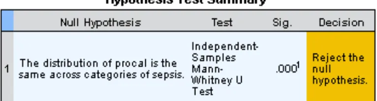
[image:44.595.133.516.196.299.2]Mann Whitney U test
TABLE 8
44
[image:45.595.110.503.157.424.2]Diagnostic performance of PCT in sepsis :
TABLE : 9
Variables sepsis
Cut off value (ng/ml) 5
Sensitivity (%) 88.9
Specificity (%) 83.3
PPV (%) 84.2
NPV (%) 88.2
AUC 0.917
ROC significance 0.0001
95% CI Lower 0.825
Upper 1.000
45
46
Discussion :
How sepsis in burns differ from the sepsis due to other causes ?
[image:47.595.131.486.185.457.2]
47
One of the most common cause of sepsis in general population is infection. The cause of infection may be bacterial , viral or fungal. The development of sepsis follows sequence of events. Any infection localized or systemic if not treated leads to development of SIRS. The criteria to define SIRS were mentioned earlier and is due to activation of various inflammatory mediators. SIRS is basically a normal physiologic response of the human body towards a noxious stimulus. If the causative agent is not removed this will cause more exaggerated SIRS and ultimately leads to development of sepsis , severe sepsis and end up with MODS (Multi Organ Dysfunction Syndrome) followed by death as shown in Fig 2.
48
Fig : 7 The events following untreated infection
49
Markers which are used as sepsis indicators:
1) C- reactive protein 2) Cytokines
3) Adrenomedullin
4) Atrial natriuretic peptide 5) Protein C
6) Endocan 7) Neopterin
C- reactive protein :
50
coronary syndromes, malignancy and after surgery.33 But one of the promising role of CRP is in assessing the antibiotic response when used to treat localized infections.
Cytokines :
Any inflammatory insult cytokines were the primary mediators. These are glycoproteins which are released by macrophages, monocytes, lymphocytes, and endothelial cells. Efforts were made to assess the level of cytokines which can reflect the severity of the inflammation. Tumor necrosis factor-a, IL-1, IL-6, IL-8, and IL-10 are the important cytokines which are considered to assess the sepsis. Among these IL-6 and IL-8 increase by 1000 fold and suppose to be a marker of sepsis.34 But the major drawback in clinical use is its very short half life of few minutes and the circulating levels falls when it binds with the receptor antagonist.
Adrenomedullin:
51 Atrial natriuretic peptides:
This secreted by the distension of atria caused by myocardial depression. This plays an important role in regulation of fluid volume. This may become a potential marker but need further evaluation.36
Protein C:
This is one of the important molecule in coagulation cascade. The concentration of protein c decrease many folds about 12 hrs before the onset of septic shock.37 It has also been documented that infusion of protein c helps in decreasing 28 day mortality in patients with severe sepsis. In association with prothrombin time, antithrombin activity, and D-dimer which causes worsening of the coagulation cascade on the first day of severe sepsis helps in predicting the organ failure.
Endocan :
52 Neopterin:
it is released from monocytes after stimulated by interferon. It function is associated with the cytotoxic reactivity of activated macrophages. It is not clinically valuable as marker as the specificity is less between infectious and non infectious causes of sepsis39 and also long time of induction and accumulation in patients with renal failure.
Thus to define an ideal sepsis marker:
1) It should add to the clinical findings to confirm the diagnosis.
2) It should be able to increase several folds at the earlier stage of the sepsis. 3) The change in its concentration should able to assess the severity of the sepsis 4) It should reflect the response to the treatment
5) It shoud be specific for the infection/sepsis due to bacterial origin. 6) It should differentiate the infectious from non infectious causes.
7) The half life of the marker should be suitable to repeat at regular intervals which are suitable for clinical condition.
So the ideal sepsis marker doesn’t exist but which can be more reliable than other markers is the Procalcitonin.
53
differentiate infection from bacterial to non bacterial origin. This was proved by many studies in the past like to differentiate between bacterial from viral meningitis, bacterial pneumonia, fever of unknown origin to rule out etiology of bacterial infection and to rule out sterile v/s infected necrosis secondary to acute pancreatitis. There were various cut off values for each study and sensitivity and specificity were calculated. PCT will also rise in non infective cases as cardiogenic shock, burns, early post operative period, polytrauma, heat shock, severe systemic inflammation e.g., secondary to multiple organ dysfunction syndrome (MODS).
54
[image:55.595.102.544.159.589.2]ALGORITHM OF THE STUDY PLAN
Fig : 8
Step 1
• Cases included in the study
Step 2
• Monitor daily for the diagnoses of sepsis according
to American Burn Association sepsis guidelines
Step 3
• Collection of sample for PCT level at the time of
admission, sepsis and after recovers from sepsis.
Step 4
• Data analysis
55
56
Summary :
This was a prospective study done for a period of 9 months to assess the diagnostic validity of procalcitonin (PCT) in sepsis patients due to burns. The criteria given by the American burn association (ABA) for sepsis in burns were used as reference standard. Most of the time in the presence of chronic inflammatory stage the diagnoses of sepsis based on ABA criteria was underestimated and delayed in starting the antibiotic which may lead to mortality of the patient or overlooked the sepsis condition and started antibiotics when it was actually not necessary. This may result in development of resistant organisms. To over come this we need a marker which can reflect the infection status and also add to the diagnostic criteria of sepsis. Various markers had been explained in the past but the PCT was more specific to the infection due to bacterial cause.
A total of 36 patients were included in the study who had burns of > 20%. The vital parameters and necessary investigations which fulfill the criteria of sepsis was done regularly. To know the diagnostic validity we estimated the baseline PCT level at the time of admission or when the patient doesn’t have sepsis. When the patient was diagnosed as sepsis one more estimation was done. Data of PCT was collected at the time of sepsis and without sepsis were analysed.
57
The etiology of sepsis in burns patients were local wound sepsis, blood stream infection, urinary infection and respiratory infection. The PCT value in our study was below 5ng/ml when the infection was localized. If the patient was immunologically stable and responded to the antibiotics the PCT level was below 5ng/ml. The onset of sepsis should be suspected when the PCT level started increasing and the level more than 10ng/ml was always had a higher mortality rate which suggests the worsening of sepsis which may led to MODS. The initial PCT level in patients sustained high voltage electrical burns was higher due to trauma and not because of infection.
58
59
Conclusion :
1) PCT has high diagnostic validity to diagnose sepsis in burns patients
Sensitivity – 88.9%, Specificity – 83.3%.
60
61
Bibliography :
1. Sharma BR, Harish D, Singh VP, Bangar S. Septicemia as a cause of death in burns: an autopsy study. Burns. 2006 Aug;32(5):545–9.
2. Lodise TP Jr, Patel N, Kwa A, Graves J, Furuno JP, Graffunder E, et al. Predictors of 30-day mortality among patients with Pseudomonas aeruginosa bloodstream infections: impact of delayed appropriate antibiotic selection. Antimicrob. Agents Chemother. 2007 Oct;51(10):3510–5.
3. Braithwaite S. Procalcitonin: new insights on regulation and origin. Crit. Care Med. 2000 Feb;28(2):586–8.
4. Bone RC, Balk RA, Cerra FB, Dellinger RP, Fein AM, Knaus WA, et al. Definitions for sepsis and organ failure and guidelines for the use of innovative therapies in sepsis. The ACCP/SCCM Consensus Conference Committee. American College of Chest Physicians/Society of Critical Care Medicine. Chest. 1992 Jun;101(6):1644– 55.
5. Levy MM, Fink MP, Marshall JC, Abraham E, Angus D, Cook D, et al. 2001 SCCM/ESICM/ACCP/ATS/SIS International Sepsis Definitions Conference. Crit. Care Med. 2003 Apr;31(4):1250–6.
6. Greenhalgh DG, Saffle JR, Holmes JH 4th, Gamelli RL, Palmieri TL, Horton JW, et al. American Burn Association consensus conference to define sepsis and infection in burns. J Burn Care Res. 2007 Dec;28(6):776–90.
7. Paterson HM, Murphy TJ, Purcell EJ, Shelley O, Kriynovich SJ, Lien E, et al. Injury primes the innate immune system for enhanced Toll-like receptor reactivity. J. Immunol. 2003 Aug 1;171(3):1473–83.
8. Dehring DJ, Fader RC, Traber LD, Traber DL. Cardiopulmonary changes occurring with pulmonary intravascular clearance of live bacteria in sheep. Circ. Shock. 1989 Nov;29(3):245–56.
62
Feb;33(2):108–20.
10. Spooner CE, Markowitz NP, Saravolatz LD. The role of tumor necrosis factor in sepsis. Clin. Immunol. Immunopathol. 1992 Jan;62(1 Pt 2):S11–17.
11. Torre-Amione G, Bozkurt B, Deswal A, Mann DL. An overview of tumor necrosis factor alpha and the failing human heart. Curr. Opin. Cardiol. 1999 May;14(3):206– 10.
12. Voss M, Cotton MF. Mechanisms and clinical implications of apoptosis. Hosp Med. 1998 Dec;59(12):924–30.
13. Van der Poll T, Van Deventer SJ. Cytokines and anticytokines in the pathogenesis of sepsis. Infect. Dis. Clin. North Am. 1999 Jun;13(2):413–426, ix.
14. Doherty GM, Lange JR, Langstein HN, Alexander HR, Buresh CM, Norton JA. Evidence for IFN-gamma as a mediator of the lethality of endotoxin and tumor necrosis factor-alpha. J. Immunol. 1992 Sep 1;149(5):1666–70.
15. Laffon M, Pittet JF, Modelska K, Matthay MA, Young DM. Interleukin-8 mediates injury from smoke inhalation to both the lung endothelial and the alveolar epithelial barriers in rabbits. Am. J. Respir. Crit. Care Med. 1999 Nov;160(5 Pt 1):1443–9. 16. Assicot M, Gendrel D, Carsin H, Raymond J, Guilbaud J, Bohuon C. High serum
procalcitonin concentrations in patients with sepsis and infection. Lancet. 1993 Feb 27;341(8844):515–8.
17. Russwurm S, Stonans I, Stonane E, Wiederhold M, Luber A, Zipfel PF, et al. Procalcitonin and CGRP-1 mrna expression in various human tissues. Shock. 2001 Aug;16(2):109–12.
18. Oberhoffer M, Stonans I, Russwurm S, Stonane E, Vogelsang H, Junker U, et al. Procalcitonin expression in human peripheral blood mononuclear cells and its modulation by lipopolysaccharides and sepsis-related cytokines in vitro. J. Lab. Clin. Med. 1999 Jul;134(1):49–55.
63
dipeptidyl peptidase IV (DP IV). FEBS Lett. 2000 Jan 21;466(1):155–9.
20. Bracq S, Machairas M, Clement B, Pidoux E, Andreoletti M, Moukhtar MS, et al. Calcitonin gene expression in normal human liver. FEBS Lett. 1993 Sep 27;331(1-2):15–8.
21. Gabay C, Kushner I. Acute-phase proteins and other systemic responses to inflammation. N. Engl. J. Med. 1999 Feb 11;340(6):448–54.
22. Marc E, Ménager C, Moulin F, Stos B, Chalumeau M, Guérin S, et al. [Procalcitonin and viral meningitis: reduction of unnecessary antibiotics by measurement during an outbreak]. Arch Pediatr. 2002 Apr;9(4):358–64.
23. Schuetz P, Albrich W, Christ-Crain M, Chastre J, Mueller B. Procalcitonin for guidance of antibiotic therapy. Expert review of anti-infective therapy. 2010;8(5):575–87.
24. Guven H, Altintop L, Baydin A, Esen S, Aygun D, Hokelek M, et al. Diagnostic value of procalcitonin levels as an early indicator of sepsis. Am J Emerg Med. 2002 May;20(3):202–6.
25. Fernández Lopez A, Luaces Cubells C, García García JJ, Fernández Pou J. Procalcitonin in pediatric emergency departments for the early diagnosis of invasive bacterial infections in febrile infants: results of a multicenter study and utility of a rapid qualitative test for this marker. Pediatr. Infect. Dis. J. 2003 Oct;22(10):895– 903.
26. Chipp E, Milner CS, Blackburn AV. Sepsis in burns: a review of current practice and future therapies. Ann Plast Surg. 2010 Aug;65(2):228–36.
27. Barati M, Alinejad F, Bahar MA, Tabrisi MS, Shamshiri AR, Bodouhi N, et al. Comparison of WBC, ESR, CRP and PCT serum levels in septic and non-septic burn cases. Burns. 2008 Sep;34(6):770–4.
64
29. Von Heimburg D, Stieghorst W, Khorram-Sefat R, Pallua N. Procalcitonin--a sepsis parameter in severe burn injuries. Burns. 1998 Dec;24(8):745–50.
30. Bargues L, Chancerelle Y, Catineau J, Jault P, Carsin H. Evaluation of serum procalcitonin concentration in the ICU following severe burn. Burns. 2007 Nov;33(7):860–4.
31. Morley JJ, Kushner I. Serum C-reactive protein levels in disease. Ann. N. Y. Acad. Sci. 1982;389:406–18.
32. Brunkhorst FM, Heinz U, Forycki ZF. Kinetics of procalcitonin in iatrogenic sepsis. Intensive Care Med. 1998 Aug;24(8):888–9.
33. Meisner M, Tschaikowsky K, Hutzler A, Schick C, Schüttler J. Postoperative plasma concentrations of procalcitonin after different types of surgery. Intensive Care Med. 1998 Jul;24(7):680–4.
34. Wakefield CH, Barclay GR, Fearon KC, Goldie AS, Ross JA, Grant IS, et al. Proinflammatory mediator activity, endogenous antagonists and the systemic inflammatory response in intra-abdominal sepsis. Scottish Sepsis Intervention Group. Br J Surg. 1998 Jun;85(6):818–25.
35. Ueda S, Nishio K, Minamino N, Kubo A, Akai Y, Kangawa K, et al. Increased plasma levels of adrenomedullin in patients with systemic inflammatory response syndrome. Am. J. Respir. Crit. Care Med. 1999 Jul;160(1):132–6.
36. Morgenthaler NG, Struck J, Christ-Crain M, Bergmann A, Müller B. Pro-atrial natriuretic peptide is a prognostic marker in sepsis, similar to the APACHE II score: an observational study. Crit Care. 2005 Feb;9(1):R37–45.
37. Mesters RM, Helterbrand J, Utterback BG, Yan B, Chao YB, Fernandez JA, et al. Prognostic value of protein C concentrations in neutropenic patients at high risk of severe septic complications. Crit. Care Med. 2000 Jul;28(7):2209–16.
65
21;276(51):48341–9.
39. Shaw AC. Serum C-reactive protein and neopterin concentrations in patients with viral or bacterial infection. J. Clin. Pathol. 1991 Jul;44(7):596–9.
40. Müller B, Becker KL, Schächinger H, Rickenbacher PR, Huber PR, Zimmerli W, et al. Calcitonin precursors are reliable markers of sepsis in a medical intensive care unit. Crit. Care Med. 2000 Apr;28(4):977–83.
41. Vincent JL, Moreno R, Takala J, Willatts S, De Mendonça A, Bruining H, et al. The SOFA (Sepsis-related Organ Failure Assessment) score to describe organ dysfunction/failure. On behalf of the Working Group on Sepsis-Related Problems of the European Society of Intensive Care Medicine. Intensive Care Med. 1996 Jul;22(7):707–10.
66
67
Annexure 1 : proforma and consent form:
CONSENT FORM Patient information sheet
68
Please read the details carefully and clarify your queries if any before deciding on consenting for the study.
What is this study about?
Study about assessing the diagnostic validity in detecting the sepsis in burns patient. Does participating in the study alter the treatment of the patient?
No. The patient shall be given the same treatment as planned irrespective of your decision to agree or disagree to participate in the study.
Does doing this investigation have any side effects? No
What investigation will be done if I consent for the study?
After taking consent a routine blood sample will be taken from the patient to assess the level of Procalcitonin at the time admission, sepsis and post sepsis period.
Will I get compensation if I suffer damage due to the study? You are not likely to have any damage because of the study. What do I have to do?
You are asked to read this consent form in detail, clarify your doubts if any and sign at the end of the form if you decide to participate in the study.
What will I have to do if I participate in the study?
You will have to allow to collect a blood sample of about 3 ml for each test during the time of admission for an average of about 2-3 samples and have to respond to the follow-up calls as and when required.
Can I say NO to the study?
69
Will my treatment details be kept confidential?
Your personal and medical records will be kept confidential and shall be used only for academic and research purposes and may be presented or published in academic circles. However, you will not be identified personally.
Can I withdraw from the study once I consent?
Yes. You can opt out of the study if you want. However, your medical records shall be available for the academic review even if you discontinue the participation.
Can my participation in the study be cancelled by the investigators?
Yes. Your participation in the study can be rejected or cancelled without your permission or information at any stage during the study period if the investigators wish so for any reason.
70
Informed Consent form to participate in clinical trial
Study Title: Prospective analysis of Procalcitonin (PCT) in burns sepsis patients
Subject’s Name: Age:
(Subject)
(i) I confirm that I have read and understood the information sheet dated _________ for the
above study and have had the opportunity to ask questions. [ ]
(ii) I understand that my participation in the study is voluntary and that I am
Free to withdraw at any time, without giving any reason, without my medical care or legal
rights being affected. [ ]
(iii) I understand that the Sponsor of the clinical trial, others working on the Sponsor’s behalf,
the Ethics Committee and the regulatory authorities will not need my permission to look
at my health records both in respect of the current study and any further research that may
be conducted in relation to it, even if I withdraw from the trial. I agree to this access.
However, I understand that my identity will not be revealed in any information released
to third parties or published. [ ]
(iv) I agree not to restrict the use of any data or results that arise from this study provided such
a use is only for scientific purpose(s) [ ]
(v) I agree to take part in the above study. [ ]
Signature (or Thumb impression) of the Subject/Legally Acceptable
Representative:_____________
Date: _____/_____/______
Signatory’s Name: _________________________________
71
Date: _____/_____/______
Study Investigator’s Name: _________________________
Signature of the Witness: ___________________________
Date:_____/_____/_______
72
STUDY :Prospective analysis of Procalcitonin (PCT) in burns sepsis patients .
NAME : AGE :
HOSPITAL NO : ADDRESS :
DIAGNOSIS :
TBSA OF BURNS :
DOA :
73
DATA COLLECTING SHEET :
74
CULTURE REPORT SHEET :
DAY BLOOD CULTURE/URINE CULTURE/SPUTUM CULTURE/PATHOLOGICAL TISSUE
75
76
MASTER CHART
Hosp no Age Sex Date of burns Date of admission Percentage of burns Diagnosis Pct 1st sample Pct 2nd sample Pct 3rd sample
028192f 25 F 9/11/2012 11-sep-12 27 Thermal 0.2982 013401f 35 M 9/3/2012 4-sep-12 22 Electrical 0.1025
064524f 59 F 10/5/2012 7-oct-12 61 Thermal 0.3951 7.969 064523f 55 F 11/5/2012 7-nov-12 58 Thermal 0.5045
615516d 49 M 12-may-12 12-may-12 50 Thermal 3.564 8.274 739071d 31 F 7/17/2012 7/17/2012 54 Thermal 0.244 4.374 162377c 47 M 8/31/2012 8/31/2012 44 Thermal 1.698 5.432 089472f 48 M 10/28/2012 4-nov-12 30 Thermal 2.1541
098769f 30 F 11/23/2012 24-nov-12 33 Thermal 1.6441 244196d 23 M 3/1/2012 2-mar-12 21 Electrical 1.0261
133413f 24 F 3/21/2012 22-mar-12 50 Thermal 3.0531 10.5 956996a 20 F 3/16/2012 16-mar-12 66 Thermal 6.8111 41.34 174371f 22 F 5-apr-12 5-apr-12 70 Thermal 3.2511
188435f 9 M 4/28/2012 29-apr-12 60 Thermal 7.289
207329f 29 F 5/19/2012 20-may-12 90 Thermal 10.110 125.000 218428f 29 M 6/4/2012 5-jun-12 20 Thermal 3.147 0.523
936807 35 M 6/11/2012 11-jun-12 27 Thermal 15.770 6.026 1.937 888851d 49 M 3/11/2012 11-mar-12 55 Electrical 65.600 46.620
558485c 13 F 7/21/2012 22-jul-12 25 Thermal 1.172 1.120 777334d 2 M 8/6/2012 7-aug-12 25 Thermal 2.400
77
261273f 9 M 7/8/2012 1-aug-12 50 Thermal 1.840 0.307 239446f 21 M 7/4/2012 5-jul-12 50 Electrical 43.130 0.694 064053f 40 M 10/27/2012 10/27/2012 58 Thermal 2.398 10.276 631005d 32 M 8/24/2012 8/24/2012 78 Thermal 1.964 26.512 299400f 30 F 9/17/2012 9/17/2012 50 Thermal 7.791 15.300
309392f 22 F 9/23/2012 28-sep-12 40 Thermal 5.893 1.351 31.930 299612f 19 M 8/30/2012 14-sep-12 25 Electrical 0.270
347792f 25 M 11/22/2012 22-nov-12 25 Thermal 18.330 42.430 17.970 338769f 17 F 11/5/2012 11/5/2012 35 Thermal 7.674 9.432 1.318 341968f 5 M 11/11/2012 20-nov-12 40 Thermal 2.939
338364f 26 M 11/4/2012 11/4/2012 45 Thermal 1.155 0.216 857543d 22 F 3/16/2012 16-mar-12 60 Thermal 3.267 69.014
Month
k
p
.
Year GRAPHIC (T.P.R.) CHARTService Ward
Sex Marital Statu
.c.>:
Bed MRD No
SUGANTHI S.
C PU:31 Q1W Ward
I
Hospital Number2994~0F ~:r;0 . St t
7'">9 - ,.(lon aus
-Name Age
-
..(. (-126ICUL T~
~f~~
J
~2
-\~ \\\'}. . US
l~
\~\~,
<} II-v f21i~(~DATE IV\/ I\,)
NO.: OF DAYS \ ()~
v,
~ ~F-'"'
DAYS POST -OP. " ~
TIME I",111~ kl~m.::h let: I,..c:;
II'>- i l'\ IL ~ I'D
,~
2- I ~'\. (20112-[ii::I~ [l;~ to '~I
-1\.1"rVl Iv .•.Ik l'll:.III. ~
-PULSE TEMP
.
-h
-•
CO•
FO210 41.1 106
200 40.6 105
,
190 40.0 104
180 39.4 103
170 38.9 102
160 38.3 101
.
'
-
-150 37.8 100
..
•
.''.
",
=r--140 37.2 99 I -v . , III
~
.
36.7
~
.
•
.
-.; ~.,130
V
120 36)1 97
110 35.6 96 I ~
I~ ~
rft!; -.l. ..• V
.
~35.0 95/ .
100
90
/
0
80 .[;7 50
70 40
f...
60 30
.2
-50 20 ./
.
IIIr" "7-
-.
-
i_17/q/,,
,
,
I
Ir/1/r"
"1 ,~~
~o,';,~
J.
..
c/1fi'
"
pc.J' ~
I
1
~
I
:
: ~
.
1 .
: ~
:
-
1.
~
~
~_
L
:
Ll
_
:
_.:
.
-
~~
.~
.
-:J
.:
"
_~_
~11J
__
L
.
:
t.
,
L
.
T<;:/.oc.. 1j,COO TG-, •..Lt1-oO
r
1SeX> 1_ . __ •.••..:l)optLi .•.•·S.-SL.AL. 3·f:?t} LAL- 1 . 2..0\ IAL
g
.
.
BS..
.1?-3. : .2-.1.0 _l~r
__
~'c)_L_ ~t
-
~At
:
38-0 0. ~.roQ__--: 3~
.
Q
__
,
_
:
J..
_
~
~
9=
=
=
-
==:~
=~::
:
..
~_gc:>_ __ .('
.
/s
-
-- ..
---,._.
e
~t
_
:_
~E4'::!rJ,... _. _OAY
eN"!,
~..ti'1 9r~W\~.e. &-'GlA)r:>C.1- ptl) <A l.Ct.,.<> "II)J pI..f' ...p~ '1'ft('T
-r
<
V
o« - T vT<4 f.. '1.ors: f2Jt. ( - ~M £0.>"'U
t,A'"c o-»IVT Ic-.c..l. - PA n.y r:.rrA ~E
, M.-~HPOA/J€ "'-0 ~ ""
Month
==&Er
~
~
ar
g
o
12.- .Name Age Sex Marital Status
I
Hospital NumberSt t
Ser . W d B d MRD N
~/t/f"'
"
~/'1lt'&
-rC. T ~
1 L ~ 1
I 1 ~ , I 1 :T
~ . -
,- I
:
~~(
)
~~
_
_
l~
_~ __
ll
L~
_
~
.~:
~
,
1 1
L
LITUb,", &'9.CO 3~oo
~
~o
____
_
_
!>L; 1..·~bw... _. _ ... _
((.$~ 1(,0
!£.CU- 3Jool~ ~·AI...
(,/~
DAY
pc.., - p,.v eA1..C·f .,.., " '"
7o/0( • -rvr4ltt. It ",~
COuNT
3~oo
vice ar e o. I
72't8':>61 F 30 FIon a us
l.
.
DATE ~8l
_
£1
\'2..-- ~ 1(\ \1\cy-
-
~
~414
WhOR
I 2~'\ q
NO.: OF DAYS II,.J h- ~
-
qDAYS POST -OP. ••... V
TIME ~ ~ \..,.~IW ,I- ~ [t( II- ~\)(It..I~
,,.
,
~'"
.
~ .~ I•.• Ih-~ ~ •••.1\,
J;...
IV K"~,.
!
"
_h \.. 11reto'Il\.Ilet
'PULSl- TEMP ,
•
Co•
FO210 41.1 106
2()O 40.6 105
1q() 40:0 104
180 39.4 103
170 38.9 102
,f 38.3 101
.
1~{J 37.8 100 I "
'(:
140 37.2 99 II '.... l- II 'I ~
'1'1.
"
13f 367
9y
i,.
.
/
t20 36.1 97 I.,
110 35.~ ~ ,
100 35.0 95
RE SP
9t)
•
60
80 50
Iv 40
30
-
--t·\.; 20
/
£: 1
/
0
I--~ ••
~
---
.
.J
.jl.ElPOlo.(l€ "'0 ~ ""
J .e:•••.r-tv•.~./
r~.,
s,'-M~ ) .\.•.•.11\@.,
PL.,- _ pt..Af~l",
~~ _JZ.ANo"M IJ..O<)(} Cvt,AtL
Ju:,.L - f)A 7c..y ~ J!t.£
---~---GRAPHIC (T.P.R.) CHART
Year
-rvice
Age
Ward
Sex SATHYA . B
C PLSB r~lW Ward
B9883:iD 25
57893171 I.ABOUHER
F t· Hospital Number Occupation Status IV'"
,,-Bed13 l ""
...
.
L
,
DATE •
NO.: OF DAYS DAYS POST - c}P.
TIME
\PULSE TEMP
• Co. FO
210 41.1 106
200 40.6 105
190 40.0 104
180 39.4 103
170 38.9 102
,60 38.3 101
150 37.8 100
140 37.21/
130
\
;
6
98I
/
o
36.1 97// 110
~/
V
;
6
~ 35.0 95
RE SP
•
60
80 50
70 40
60 3~
Y
/
20
40 10 90
r
r.
I~ I~ ,..
I'\.
,.
II" ...,
J
9JO v-~ y 0lC9 \:{
.nfh ear
Sex Marital Status
I
GRAPHIC (T.P.R.) CHART
!'...me.
/,0 : 11 :
!
~,~
~
,
«/
Oe
..
Jf 0oo
fL'T
R-B~.
ILeAl•.
t
·
Ab
.
Lis
DAY
DATE ~
NO.: OF DAYS DAYS POST -<8P. TIME
PULSE TEMP
•
Co • Fa210 41.1 106
200 40.6 105
190 40.0 104
180 39.4 103
170 38.9 102
160 38.3 101
150 37.8 -100
140 37.2 99
130
~
W
e
1~ 36.1 97
110 35.6 96
100 35.0 95
RE SP
90
•
60
80
~
I
~
V 4060 30
50 20
40~
l,
,
'/3//t.
.-
·
3D/S!2tOII"L~
~
6t.
:
~
'
!
: :
I } , :~.1
L~~
_~
.
:
11
i
_
~
~.qg.Q_.f~...:~- ., ~&too,_
.1.'Ol-W' _ 9foo,?
~}~---..,..---.
-__..-1~!?O~O~__ .,..--___ {
r-r
-
0,HIL.,_ _ __._~-j ~ - ~
_________ I--_\\..1.>-'1.9_.:JcL~.u.!1v=:.!.'I L.:::.,-__ ..Ll ----'!....--
---N~I/e.
pl-r- pu\na.rr
~ r_/l..AtNOttM ~fi'\>" Cv'tA'"
;u:.
f,.- f>A''
'
Y ~
.
JC.Ef:>c.1- P ~tJeA1.1C f"to "", '"
""-c;o( ~-rt.Y1".,:f (.. l, ",~
co
..
,,,,'"
A
1-" IA
I'"
]