STUDY OF THE DUCTAL PATTERN OF
PANCREAS IN 50 SPECIMENS
Dissertation submitted in partial fulfillment of the requirement
for the award of
M.S. DEGREE EXAMINATION (ANATOMY)
BRANCH V
MARCH – 2010
Institute of Anatomy Madurai Medical College
Madurai - 625 020
THE TAMILNADU Dr. M.G.R. MEDICAL UNIVERSITY
CHENNAI- 600 032
CERTIFICATE
This is to certify that the dissertation entitled “STUDY OF THE
DUCTAL PATTERN OF PANCREAS” submitted by Dr.DH.Gopalan,
postgraduate in Anatomy to the faculty of Anatomy, The Tamilnadu
Dr. M.G.R Medical University, Chennai in partial fulfillment of the
requirement for the award of M.S. Degree in Anatomy, is a bonafide work
carried out by him under my direct supervision and guidance.
Place: Madurai Dr. V. Rajaram D.L.O., M.S.,
DECLARATION
I, Dr.D.H.GOPALAN solemnly declare that the dissertation entitled
“STUDY OF THE DUCTAL PATTERN OF PANCREAS IN 50
SPECIMENS” has been prepared by me under the guidance and
supervision of Dr.V.RAJARAM D.L.O., M.S., Director & Professor I/C,
Institute of Anatomy, Madurai Medical college, Madurai in partial
fulfillment of the requirement for the award of M.S. (Anatomy) Degree
Examination of The Tamilnadu Dr. M.G.R Medical University, Chennai
to be held in March 2010. This work has not formed the basis for the award
of any other degree to me from any other university.
Place: Madurai Dr. D.H.Gopalan
ACKNOWLEDGEMENT
I sincerely thank the Dean, Madurai Medical College, Madurai, for
permitting me to use the college and department facilities to my study.
I profoundly thank Dr.V.Rajaram, D.L.O., M.S., Director
& Professor I/C, Institute of Anatomy, Madurai Medical College, Madurai
for his constant guidance, encouragement and help rendered throughout the
period of the study
I express my sincere thanks to Dr.K.Meiyazhagan, M.D., Professor
& Head of the department, Department of Forensic Medicine, for providing
the autopsy specimens to my study.
I also thank Dr.T.Jeeva, Dr.S.Sundari, Dr.K.Parthiban,
Dr.P.G.Ananthi, and Dr.M.Shobana the assistant professors and the non
faculty members in the Institute of Anatomy for their valuable help in the
CONTENTS
CHAPTER TITLE PAGE
NO.
1 INTRODUCTION 1
2 AIM OF THE STUDY 5
3 REVIEW OF LITERATURE 6
4 MATERIAL AND METHODS 25
5 OBSERVATIONS 30
6 DISCUSSION 42
7 CONCLUSION 55
BIBLIOGRAPHY
ANNEXURE
INTRODUCTION
Pancreas is a Compound racemose gland ,analogous in its structure
to the salivary glands, though softer and less compactly arranged than the
above organs. It is composed of two quite separate types of glandular
tissue which are however in intimate topographic association with each
other. The main mass of the tissue is exocrine part of pancreas embedded
in which clusters of endocrine cells constituting the Pancreatic Islets.
Ductal pattern of Pancreas is important because of its implications
in various fields of Medicine. It is an interesting topic for the Anatomists
because most of the clinically important variations will be clearly
analyzed by them under Embryological basis.
The knowledge about the Normal Ductal pattern as well as the
congenital variations are very much important for the Surgeon according
to which he can modify the surgical procedure in a more satisfactory way.
This will help him to prevent most of the common post operative
retrograde Pancreaticography itself can leave chronic recurrent
pancreatitis.
More than 75% of the adenocarcinomas, which is the most
common pancreatic malignancy arise from the ducts. Operative loosening
of proximal part of duodenum may sometimes injure the accessory
pancreatic duct. In cases where the accessory duct is the main excretory
route this may cause post operative pancreatitis which is an acute
emergency. A patient with an obstructive lesion of main pancreatic duct
either with a tumor or stone may not develop any symptom if the
accessory duct is patent.
As science advanced procedures like Percutaneous
Pancreatography and Endoscopic Pancreatography (EPR) became widely
accepted invaise techniques. Ultrasound guided Percutaneous
Pancreatography with fine needle may be under taken when EPR fails to
demonstrate pancreatic ducts. At present Magnetic Resonance
Pancreatography, a non invasive technique is very commonly being used.
This is used to delineate the segments of ducts which are not
The condition called Pancreatic Divisum is associated with chronic
pancreatits. There is no doubt that the patient will yield abnormal
pancreatograms which may be mistaken for a pancreatic disease. This
may lead to misinterpretation of ultrasonic and CT findings. The
knowledge of the existence of these anomalies and the ductal pattern in
them is helpful for an endoscopist for the correct interpretation and is
helpful to the surgeon contemplating pancreatic surgery and may be a
factor in deciding the surgical procedure to be carried out. In Pediatric
Patients some of the serious conditions like Mongolism, Cardiac defects,
Intestinal Malrotation, Ductal Atresia and Tracheo Oesophagial fistula are
being noted in association with pancreatic divisum, iatrogenic pancreatitis
also is associated with this condition.
Retro pancreatic position of common bile duct is clinically
important because it is being often subjected to operative exploration.
This topic is of primary interest to every biliary surgeon for as stated by
the Late Lahey of Bostin- “We urgently need more investigation on the
retro pancreatic part of bile duct”.
Pancreatico biliary ductal union is a complex anatomical and
common duct with more that 12mm length is reported to be associated
with cystic dilation of gallbladder and carcinoma of gallbladder.
Common channel with more than 3mm in length is always
associated with reflux pancreatitis in a case of block.
Because of the above clinical significance the topic for chosen for
AIM
To study
a) Ductual pattern of pancreas
b) Retro Pancreatic Positioning of bile duct
c) Pancreatico biliary ductal union
REVIEW OF LITERATURE
DUCTAL PATTERN OF PANCREAS:
Johann George Wirsung (1589-1643), the prosector of Padua-Italy,
discovered the human pancreatic duct in 1642 during the dissection of an
executed murderer. Instead of publishing his work he engraved a drawing
of the duct in a copper plate, from which seven or more imprints were
taken. The copies were sent to the leading Anatomists in Europe with the
question-‘Should I call it an artery or vein? I never found blood in it’
Wirsung was assassinated subsequently. Three and half a Centuries later
Haward J.M.HessW & Transverso.W traced Six copies of “Ductus
Wirsungianus”. Copper plate remains well preserved. His findings had
opened a new field of Medicine.
Santorini (1775), the Italian Anatomist was the first to describe the
Anatomy of accessory pancreatic duct and minor duodenal papilla. He
gave the first accurate concept regarding the relations of bile duct and
pancreatic duct to each other and two papillae through which they
Since from the time of reporting, the Anatomy of pancreatic ducts
were studied by so many devoted workers.
Claud Bernard (18560 resuscitated Santorini’s work and by
injections of metallic mercury into bile and pancreatic ducts, he
determined their mode of termination and function.
Opie (1903), one of the great American pathologists stated that in
10% of 100 cases the duct of Santorini was functionally as well as
structurally the chief outlet of exocrine pancreatic secretion.
Baldwin of university of Cornell (1911), presented a major pioneer
investigate work on the pancreatic ducts. According to him in 82% of 100
cases the accessory duct is patent ventral to that of main pancreatic duct
and is restricted to its cephalic and ventral segments of head, its orifice
(minor papilla) being cephaloventral to that of the major duodenal papilla
on a transverse mucous membrane fold.
Shwartz of University of Heiddilberg (1926) after an examination
of 64 cases, reported that santorini’s system was missing in 25 cases and
was the sole outlet in 3 cases and was rudimentary in another 3. It
in 8 cases. He said that in all cases the duct of Santorini was demonstrated
in the head of pancreas ventral to the duct of Wirsung.
Simkin’s (1931) studied in detail the duct of santorini in 25
specimens and quoted that in 10% cases the duct of santorini was the
chief outlet of pancreatic exocrine secretion. In a study conducted in the
Philadelphia general hospital he described 3 groups of Santorini.
1) Santorini’s duct present only in Cephalic part of head.
2) Santorini’s duct confined to the Caudal part of head.
3) Distributed both in Caudal and Cephalic part of head.
Naetamen of the University of Helsinki (1941) investigated the
ductal pattern in 100 specimens in which he injected a coloured fluid into
the ductal system. In 20% only he demonstrated a patent accessory
pancreatic duct. So according to him if the duct of Wirsung is getting
obstructed by a stone or tumour 1 in every 5th case only the gland was
able to excrete the secretions into the duodenum through the above patent
duct.
The ductal pattern was studied in detail again by Rienhaff and
Pickrell (1945). They observed that only in 4 cases out of 100 the
Santorini’s duct lies on a plane ventral to that of main duct and
communicates with it in the head of pancreas near the neck region.
As years advanced the dedicated workers started using different
colouring fluids and dyes for pancreatographic techniques and they started
using the radiological methods apart from the routine postmortem
dissection or the cadaver dissection.
Hjorth (1947) claimed a sex difference in the patency of santorini.
He found an open communication of Santorini with duodenum in 44%
cases in men and 14% in women. He tried to explain the association with
the occurrence of biliary diseases more in women. Santorini’s duct in
open communication with duodenum would act like a safety valve for
increased pressure in the pancreatic duct and this reduces the possibility
of reflux of pancreatic juice into the common bile duct.
In 1950s the study was conducted by so many Anatomists and most
presented a common percentage of 90 where the Duct of Wirsung is the
main excretory route. One important among this was of Berker (1950) and
in his study he pointed out that the junction of ducts of Wirsung and
Santorini represents a weak point in the pancreatic drainage system and is
duodenum, it will empty its contents into the main duct against the direct
of flow.
Erik Millbourn of University of Laud, Sweden (1950) has presented
the most extensive investigation on the Anatomy of pancreatic ducts. To
get the full display of pancreatic ducts he made a closure of pancreatic
orifice in duodenum by sewing it up. There upon injected a 20% Barium
sulphate solution into the bile duct and gave supplementary injections into
the main duct from the tail of pancreas. The contrast injected specimens
were studied in X-ray films and in gross dissection.
From his studies Erik Millbourn concluded that the sole or main
excretory channel of pancreatic parenchyma is the duct of Wirsung in
about 90% cases and the duct of Santorini in about 10% cases. Santorini’s
duct invariably courses ventral to the duct of Wirsung and terminate at the
lesser papilla situated Cephalo ventral to the greater papilla. He found no
sex difference as claimed by Hjorth (1947).
Berman (1960) studied multiple variations of pancreatic ductal
pattern in relation to the common bile duct. His findings were based on
the study of ductal system in Man by the use of vinyl acetate casts of
specimens. According to his report in 90% cases the major pancreatic duct
was the main excretory route and in 10% of cases the accessory pancreatic
duct was the chief excretory route.
Dawson W. and L.Langman (1961) did a study about the ductal
pattern in 100 specimens and he divided them into 3 groups.
1) Where both ducts are patent
2) Where ansa pancreaticus is present
3) Where the accessory duct is obliterated either proximally or
distally.
Recently the development of techniques like Endoscopic
Retrograde Cholangio Pancreatography (ERCP) and Magnetic Resonance
Cholangio Pancreatography (MRCP) had enabled the demonstration of
pancreatic duct system in a large number of patients.
Reports by Cotton and Kiju (1976) had shown that the pancreatitis
was present in a high proportion of patients with an unfused pancreatic
duct system and suggested that the anomaly might be the cause of
Varly P.F and Rohman C.A (1976) using Endoscopic
pancreatograms studied the precise details of the ductal course in Thai
people.
Willarumec. C & Pongichirecks. P (1999) studied the ductal pattern
in Thai people by the injection of methylene blue and showed that in
majority (90%) the main route of excretion is the main duct. The
accessory pancreatic duct functions as the main channel in 10% people.
Morgan DE & Logan K (1999) in a study based on ERCP stated
that in a group of people referred for ERCP, the prevalence of pancreatitis
was very high in patients with pancreatic divisum and was limited to a
doral distribution.
Yokohate.K & Shirakana K (2000) stated that the dilatation of
ductal branches depicted by MRCP might be a hint for early detection of
pancreatic malignancy.
Ductal patterns in clinically important congenital anomalies were
studied by so many workers. In annular pancreas from the head of
pancreas an extension forms a complete or incomplete ring around the
described by Tiedemann, Professor of Anatomy at Hiedelberg University,
Germany in 1818, Ecker (1862) gave it the name Annular Pancreas.
In their case report Mc Naught and Cox of the Standard University
School of Medicine (1935) presented pancreatographic method for
visualizing the duct system and noted that in 88 percent of 40 reviewed
cases, the duct of annular pancreatic ring is a part of duct of Wirsung, this
being a strong indication that annular pancreas is a developmental
anomaly of ventral pancreas.
Lehman of University of Virginia School of Medicine (1942)
analysed 48 cases of annular pancreas. Analysis showed that there was a
constant anterior point of origin of the duct of the ring which was
subsequently coursing to the right over duodenum, then posteriorly and to
the left behind the duodenum, finally entering the head of pancreas in
close relation to the common bile duct and joint the main pancreatic duct.
Millbourn (1950) from his observations suggested that most of the
affected individuals are males in relation to Annular Pancreas.
Shapinker (1954) studied the duct pattern in annular pancreas and
stated that modification of surgical procedure is needed to avoid injury to
satisfactory bypass operation. Other reported anomalies related to ductal
system of pancreas are,
1) Inversion of pancreatic ducts: Here the embryonic pattern of
duct persists and greater part of pancreas is drained through minor
duodenal papilla.
2) Pancreatic divisum (Divided pancreas): The parts of pancreas
derived from the dorsal and ventral buds fail to fuse with each other.
Distance between major & minor duodenal papillae:
It was studied by so many workers as about the ductal pattern.
Lerulle and Nattan Larrier (1898) found variations of distance from
10 to 35 mm, the average being 20mm.
According to Baldwin (1911) the distance from the lesser to the
greater papillae varies from 9 to 35 mm, the average being 20mm.
Sice of France (1911) reported a distance of 29mm in one case.
Claimant of Switzerland (1923) reported a distance of 3mm
between the two papillae in one case.
Maeda (1950) found the distance to vary most frequently from 20
Millbourn (1950) found the lesser papilla to lie oroventrally to the
greater papilla. In his 200 specimens he found an average distance of
20mm between the two papillae.
Recently, Hughes and Kernutt of Australia (1954) found the
distance between two papillae to be an average 21mm.
Retropancreatic positioning of common bile duct:
Accessibility of the retro pancreatic bile duct for surgical
exploration was studied by so many great workers.
Beginning with Lettille and Nattan Larrier of France (1898), who
were among the first to talk about the laminar extension of pancreatic
head over the retropancreatic part of bile duct, so many investigators
started studying the Anatomy of Pancreatic part of Choledochus.
Nuboer, of the University of Utrecht (1931), reported that in 67%
cases the common bile duct passed through a tunnel in pancreatic tissue,
meaning that it was being covered by only a connecting tissue and in 33%
it was not freely accessible.
In an examination of 100 specimens, Nattemen of University of
1) It coursed in a furrow on the dorsal surface of the pancreas covered
by connective tissue (53%).
2) It is covered by a thin sheath of pancreatic tissue (34%).
3) It coursed through a tunnel of pancreatic tissue (12%) which was
closer to the dorsal surface of the pancreas.
Up until about 1945, it was believed generally that, as the common
bile duct approached the second part of the duodenum in an oblique
direction, in most of the individuals it passed through the head of pancreas
in a complete or incomplete tunnel and thereby became an intraglandular
structure not readily accessible or explorable without cutting the
pancreatic tissue. But Dejhi & Fritas (1945) studied this topic and
reported that in 60% of 100 specimens the retro pancreatic bile duct was
easily accessible.
As shown by Smanio, of the Department of Anatomy of the School
of Medicine of University of Sao Paulo, Brazil (1954) in 60% of cases the
retropancreatic bile duct was easily accessible. In his summary Smanio
stated that the choledochus in the retro pancreatic portion was easily
accessible, either because it was not covered by pancreatic tissue or was
by two ‘lingulae’ (laminar extension of pancreatic tissue) the lips of
which are juxtaposed to the level of posterior face of the duct.
In 40% cases the lingual was thick. Under these circumstances the
isolation of bile duct was very difficult.
According to Smanio observations in both sexes showed no
significant statistical variations related to position and accessibility of
retropancreatic Bile duct.
Pancreatico bile union:
The concept where by the common bile duct and main pancreatic
duct are converted into a common channel is well established by most of
the anatomists who investigated this region.
Whether the distal end of pancreatic duct and common bile duct are
really forming a common channel or these are separated by a thin
membrane till their openings into the duodenum was studied by so many
workers.
The first accurate description and illustration of Ampulla of Vater
in the rat was made by Professor H. gage of Coinell University (1878) and
he published the same in the American Quaterly Microscopic Journal in
Little and Nattamlarrier (1898) of Paris among the first to classify
and illustrate the various types of openings of the common bile duct and
pancreatic duct into the duodenum. They published their article in the
Bulletin of the Anatomical Society of Paris. They investigated 21
specimens previously treated for 24 hrs with Mullers fluid. The ductal
openings according to them form so many groups.
1) The Two ducts joining in an acute angle but separated internally by
a thin membrane and distally opening through a true common
channel.
2) The two ducts are opening separately on a depression i.e., no ‘true’
common channel.
3) Two ducts are opening separately on a same plane.
4) No ampulla present.
Opie (1903) studied the mode of union in 100 specimens. He stated
the presence of common channel in 89% and separate opening in 11%.
Archibald (1919) noted the average length of the common channel
as 2mm. variations were from 2 to 10mm.
Baldurin (1911) carried out routine dissection in 90 specimens and
Pedro Belou, the professor of Anatomy at the University of Buenos
Aires, Argentina in his classical monograph published in 1915 classified
the terminal endings of bile and pancreatic duct as observed in 50
specimens in two groups.
1) Cases in which there was a common opening for the ducts in 62%.
2) The common channel was not demonstrated in 38%. The length of
the common channel when it was present was from 3 to 7mm.
Cameron and Noble (1924) studied the pattern in 75 specimens by
routine dissection method. They demonstrated common channel in 76%
and separate openings in 19%. In the remaining specimens the main
pancreatic duct became a fibrous strand and accessory pancreatic duct was
the draining channel.
Couveliare (1934) after studying 100 specimens by routine
dissection method found out that only in 49% cases had a common
channel. All other specimens showed separate openings for the common
bile duct and the main pancreatic duct.
According to Pfuhl (1936), the common channel is formed in the
following manner. As the bile duct passes obliquely through the
tapered, then after receiving the pancreatic duct it ends in a dilated
ampulla.
Boyden (1937) on the embryological basis stated that the
confluence of bile duct and pancreatic duct starts outside the duodenum,
but in the course of development the zone of junction becomes drawn into
the duodenal wall. He did a comparative study in opossums, guinea pig,
dog and in man.
Naatanan (1941) worked out 100 specimens and described a
common channel in 67% and demonstrated a separate channel in 33%. He
noted the average length of common channel as 6mm with variations from
2 to 15mm.
Neboer of Berlin city Hospital (1931) reported a true common
channel in 76% and separate openings in 19% out of 75 cases he studied.
Rienholff and pickrell (1945) of John Hopkins University after a
study of 250 autopsy specimens noted the following types.
1) No union of pancreatic and bile duct, both entering the duodenum
2) The ducts were contiguous with the dividing thin septum
terminating within 3mm from the apex of the duodenal papilla
(37%).
3) True common channel varying from 3mm to 14mm in length from
the apex of duodenal papilla and having an average diameter of
3mm (32%).
4) The main pancreatic duct is reduced into a fibrous strand (2%).
Hjorth (1947) from California studied 100 specimens with the help
of cholangiographic methods. He injected a contrast medium under low
pressure into the pancreatic duct and noting its reflux, course and its
relations were visualised. He demonstrated a common channel in 86% and
separate openings in 14%. Out of the 14% cases with separate openings
5% showed openings in the common major papilla. 9% showed separate
points of opening into the duodenum.
Millbourn (1950) studied 200 specimens by injecting contrast
material into the bile duct and using cholangrographic method. He
reported a common duct in 85% and separate openings in 9%. In the rest
Hotzapel of Dootmund of Germany (1950) reported the following
types of duct orifices for bile and pancreatic duct.
1) Separate openings of two ducts in one papilla.
2) Separate openings of two ducts in two papilla.
3) Common opening in one papilla.
Hughes and Kernutt (1954) studied 30 specimens and demonstrated
a common channel in 57% and separate openings in 37%. In the rest the
pancreatic duct was a fibrous cord.
Brue, Walmikey & Ross have described in the Manual of Surgical
Anatomy (1979) three variations in the termination of the ducts.
1) Two ducts unite to form a true common channel of length less than
3mm i.e. the Ampulla is absent.
2) The common channel length is more than 3mm i.e., ampulla can be
defined.
3) The two ducts open separately either into a summit of a papilla or
in a slight depression.
Variations were also described by Skandalkis (1979) after studying
the pancreatico biliary ductal union. He described 3 groups.
- A short common channel.
- Open separately into duodenum.
A Japanese study group on Pancreatico Biliary Maljunction
(1994) defined Anomalours Pancreatico Biliary Ductal Union (APBDU)
as a congential anomaly where there is a connection of pancreatic duct
and bile duct with an obviously long common channel (>12mm) or their
union in an apparently anomalous form. In their studies they noted
Anomalous Pancreatico Biliary Ductal Union (APBDU) has a prevalence
of 1.5%-8.7% in Thai people. They divided the APBDU into 2 groups.
a) Biliary-pancreatic type (B-D) where common bile duct is joining
the pancreatic duct.
b) Pancreatic-biliary type (P-B) where pancreatic duct is joining the
bile duct.
K.B.Chauch, Y.K.Yap and H.S.Nag (2000) in a study based on
Singapore population noted the average length of common channel as
Ronald A Bergan PhD (2001) studied the Pancreatico Biliary
Ductal Union in 100 specimens. By routine dissection method in 63
specimens a common channel was demonstrated. 30 specimens in this
study showed separate openings. And in the rest the main pancreatic duct
was reduced into a fibrous cord.
MATERIAL AND METHODS
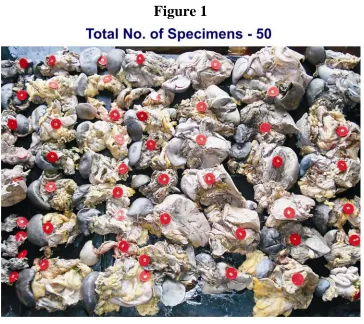
Materials: 50 adult pancreas specimens were taken for the study.
20 specimens were collected from the cadavers in the Institute of
Anatomy, Madurai Medical College. 30 specimens were collected from
the Department of Forensic Medicine, Madurai Medical College,
Madurai.
Specimens were collected from the cadavers in the dissection table.
Pancreas was removed along with the duodenum and retropancreatic part
of the bile duct.
Specimens collected from Forensic Medicine Department were
cleaned in tap water and were put in 10% Formalin solution and were
taken for dissection.
Materials Used:
•
Stainless steel student’s scalpel.
•
Stainless steel forceps- toothed and non- toothed.
•
Stainless steel long and short straight scissors.
•
Black cream sheet, Rubber sheet, Graduated scale, HB pencil,
0.4mm thread and Cotton.
•
Gloves and Apron
•
Covered container for preserving specimens in formalin.
•
10% formalin.
•
20 ml syringe.
Methods of Study
1. Routine dissection of the Specimens to see the pattern of pancreatic
ducts and mode of their opening into the 2nd part of duodenum.
2. Injection of air into the pancreatic duct in underwater dissection
method to study the patency of the ducts.
3. Measurement of the distance between the major and minor
duodenal papillae.
4. Measurement of the length of the common pancreatico-biliary
Method I (Routine Dissection)
Specimens were cleaned by removing the remains of peritoneum
and vessels which are closely attached to it and then washed thoroughly in
tap water. On the posterior aspect of the pancreas the bile duct was traced
down to the pancreaticobilliary junction at the 2nd part of Duodenum.
While tracing down, retropancreatic positioning of the bile duct was also
noted down. The major pancreatic duct was traced towards the tail end.
Some of the main branches were also traced. The ducts were also traced
up to the duodenal wall. The accessory pancreatic duct was traced up to
the duodenal wall.
After this, a longitudinal incision was made on the posterior aspect
of the 2nd part of the duodenum towards the right margin at the level
where pancreaticobilliary ductal union approaches the duodenal wall. This
incision was extended upwards and downwards. Flap was reflected
medially by putting two horizontal incisions at the ends. Duodenal lumen
was cleaned thoroughly in running water and the two papillae were
Method II (patency testing by injection of air in underwater
dissection method )
After the dissection, air was injected from the distal end of the
ducts and patency of the major and minor ducts were noted with the
appearance of air bubble at the papilla in underwater dissection method. If
the air injected in the main duct was not entering the accessory duct
separate injections were made in the distal end of that duct.
Method III: Measuring the distance between 2 papillae
In the lumen of duodenum the distance between the two papilla was
measured using a thread and scale. The thread was placed on the major
duodenal papilla and the other was stretched towards the level of minor
papilla and markings were made on the thread. The marked distance was
measured using a scale.
Method IV: Measuring the length of common channel
A clean slit was made on the bile duct little away from the union of
pancreatic duct the incision was extended down and the duct was traced
up to the opening in the duodenal mucosa. The following things were
a. Whether there is any septum separating the two ducts till a very
small distance from the papilla.
b. Whether the two ducts are opening separately into the duodenal
lumen.
c. The length of the true common channel was measured using a
thread and scale. The thread was marked after keeping it straight
from the duodenal papilla to the point of starting of the true
OBSERVATIONS
The total 50 specimens were studied by dissection method.
The following observations were made during the study.
A) Mode of Formation of Pancreatic duct
In all the specimens the main duct was formed in the tail end of
pancreas by the union of smaller ducts alternatively from either side
giving a ‘herring bone’ pattern (Figure 3). As it goes towards the head end
of the pancreas the caliber of the duct was increasing. Branches of the
main duct showed similar pattern. The main pancreatic duct was receiving
branches from tail, body, neck, postero inferior aspect of head and
uncinate process of pancreas. The accessory duct was receiving branches
from superior part and antero inferior part of head of the pancreas.
B) Position of pancreatic duct
Inside the pancreatic tissue the main pancreatic duct was placed
posteriorly towards the superior border. Accessory duct when present was
C) The Mode of Drainage of Exocrine pancreas
1. Main mode of drainage of exocrine pancreas (Table No.I)
The observations made in this aspect during the present study were
tabulated in the Table No.1. From this the following data were obtained.
Out of total 50 specimens studied, duct of wirsung is the main mode of
drainage in 47 of the total specimens. Duct of santorni is the main mode
of drainage in 3 specimens out of Total 50 specimens.
Table No: I
Mode of drainage of exocrine pancreas
Percentage
Number of specimens studied 50 100%
Duct of wirsung as the main mode 47 94%
2) In specimens where the duct of wirsung is the main mode of
drainage, the duct of Santorini was also observed during the dissection.
For this study only dissected specimens were used.
Among the total 50 specimens where the main mode drainage was
Table No: II
Duct of santorini
Percentage
Specimens studied 50 100%
Duct of santorini observed 23 46%
54%
46%
DUCT
OF
SANTORINI
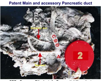
D) Patency of the Pancreatic ducts
1) Main pancreatic duct was observed as patent in all cases.
2) Among the specimens where the accessory was not the main mode
of drainage its patent communication with duodenum was observed
as follows (Table No. III). For this study only dissected specimens
were used . In total, out of 50 specimens studied in this group 12
showed a patent communication of Accessory pancreatic duct with
duodenum (Figure 4).
Table No. III
Patency of Accessory duct with duodenam
Percentage
Specimens studied 50 100%
Accessory having patent
communication with duodenum
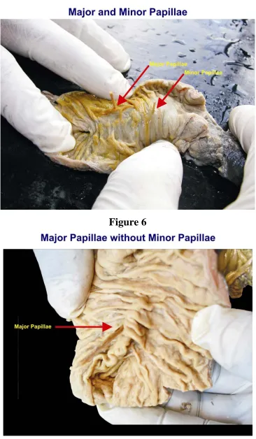
E) Distance between major and minor duodenal papillae
For measuring the distance between the two duodenal papillae only
the dissected specimens were taken. Among 50 specimens studied the
average distance between the two papillae was calculated as 15mm. The
distance varies from 10mm to 20mm (Figure 5).
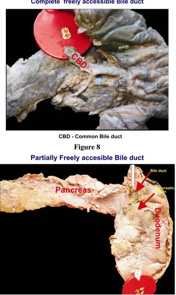
F) Retropancreatic positioning of bile duct
1) Accessibility of bile duct (Table No. IV)
Out of the total 50 specimens studied in this category 30 specimens
showed a freely accessible bile duct in the retropancreatic position and 20
specimen showed not freely accessible bile duct (Figure 9).
Table No. IV
Retro pancreatic positioning of bile duct
Percentage
Specimens studied 50 100%
Freely accessible 30 60%
II) According to the observations the freely accessible bile duct which
was noted in 30 specimens can be divided in to 2 groups (Table No.V).
1) Completely free readily accessible retropancreatic bile duct.
2) Retropancreatic bile duct which was freely accessible but
covered by a thin pancreatic tissue.
Among the thirty specimens 1) Retropancreatic bile duct was
completely free in twenty specimen i.e. group 1 (Figure 7). Ten specimens
showed a thin pancreatic tissue covering the retro pancreatic bile duct i.e.
group II (Figure 8).
Table V
Freely accessible retro pancreatic bile duct
Percentage
Specimens studied
Freely accessible retro pancreatic bile duct 30 100%
Completely free (Group-I) 20 66%
Covered by thin pancreatic tissue
(Group-II)
40%
20%
40%
RETRO
PANCREATIC
POSITION
OF
BILE
DUCT
NOT FREELY
ACCESSIBLE
COMPLETELY FREE
G) Mode of termination of common bile duct and pancreatic
duct (Table No.VI)
Table No. VI
Mode of termination of common bile duct and pancreatic
ducts
Percentage
Specimens studied 50 100%
Common channel 42 84%
For this study only dissected specimens were used. The main
pancreatic duct and the common bile duct opened through a common
channel in 42 out of 50 specimens.
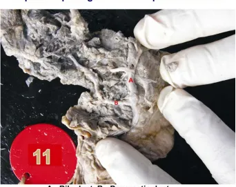
Separate openings were noted in 8 specimens out of 50 specimens
constituting of total specimens (Figure 10 & 11).
H) Length of the common channel (Table No.VII )
Only dissected specimens were used for measuring the length of
common channel. In this study out of total 50 specimens 42 specimens
showed a common channel for the bile duct and pancreatic duct. So for
measuring the length of common channel only 42 specimens were taken.
The observations were tabulated in 3 groups.
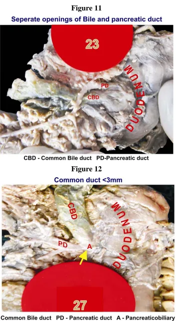
The Group I is where the length of common channel was measured
to a maximum of 3 mm (Figure 12 & 13). This was observed in 24
specimens.
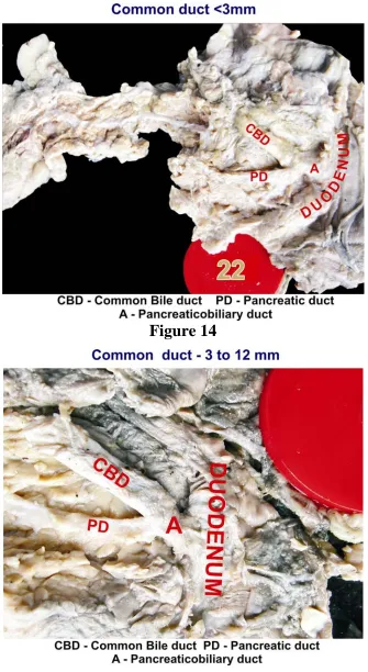
In the Group II the length measured was more than 3mm and to a
maximum of 12 mm (Figure 14). The length of common channel
measured in 17 specimens came under this group.
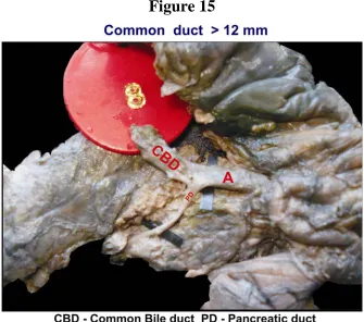
The Group III is where the common channel length has to be more
From the Table No.VII it is evident that totally 18 specimens
showed a common channel length of more than 3 mm length.
In one specimen the length of the common channel was measured
as 15mm. In this type the junction was noted as of a P-B type
(Pancreatic-Biliary type).
Table No.VII
Measured length of the common channel
Percentage
Specimens studied 42 100%
Length of the common channel
Upto 3 mm (Group –I) 24 56%
More than 3mm and less than or
equal to 12 mm (Group – II)
17 41%
DISCUSSION
Ductal pattern of Pancreas
A) Mode of formation and position of Pancreatic Ducts
In the present study, the main pancreatic duct is noticed, as it is
commencing at the tail end of the pancreas and is nearer to the posterior
surface towards the superior border. The secondary ducts are draining at
the right angle to the main duct to form the characteristic ‘herring bone’
pattern. Main duct is receiving branches from tail, body, neck and postero
inferior part of head and uncinate process of pancreas. Accessory duct
when it is noticed is placed anterior as well as cranial to the Main
pancreatic duct. It receives branches from antero inferior part and upper
part of the head.
The same pattern was described by Cameron (1924), Nebour
(1931) Reinholf and Pickrell (1945) and Erik Millbourn (1950). Similar
observation was described by Willasrumec and Pongichireek (1999) in
Thai people. The patterns observed in the present study is correlating with
the pattern described in the Gray’s Anatomy (39th Edition) and is the
B) Mode of drainage of Exocrine Pancreas
Table No. VIII
Mode of drainage of exocrine pancreas – Comparison study of with previous study results
Study Wirsung duct as the
main duct
Accessory duct as the main duct
OPIE (1903) 90% 10%
SIMKINS (1911) 90% 10%
RIENHOFF & PICKRELL (1945) 96% 4%
ERIK MILBOURN (1950) 90% 10%
WILLASRUMEC (1999) 90% 10%
85%
90%
95%
100%
90%
90%
96%
90%
90%
94%
10%
10%
4%
10%
10%
6%
MODE OF DRAINAGE OF EXOCRINE PANCREAS – COMPARISON STUDY
RESULTS
In 1903 Opie had pointed out that in 10% cases the duct of
Santorini is acting as the chief outlet and in 90% of cases the Duct of
wirsung is the main mode of excretion. Same observation was made by
Simkin’s in 1931 and Erik Millbourn in 1950. In a study conducted in
Thai people (1999) by Willasurmec and Pongichirecks.P, the report
wirsung and in the remaining 10% cases the Duct of Santorini is the main
mode of excretion.
Present study shows that in 94% cases the Duct of Wirsung is the
main mode of drainage of exocrine pancreas and in 06% cases duct of
santorini is the main mode of drainage.
Thus the present study results are correlating with the above
mentioned work results.
Rienhoff and Pickrell (1945) in their study showed that in 96% of
cases the main mode of drainage is the Duct of Wirsung and in only 4%
of Duct of Santorini is the main excretory route. The present study result
shows a higher value (06%) where the duct of santorini is the main
excretory route compared to the Rienhoff study. And compared to his
study the percentage of cases where the Duct of wirsung is the main mode
of drainage shows a lower value in the present study (94%).
Surgical Importance
In cases where the Accessory Duct is the chief outlet care should be
taken to avoid injuries to it when mobilizing the proximal part of
where the Accessory Duct is the chief outlet, may not be symptomatic in
obstructive lesions of Main Pancreatic duct.
Present study shows in cases where the Duct of Wirsung is the
main excretory route, the presence of Duct of Santorini is noted by
dissection in 46% cases. This result is in close approximation to the result
shown by Erik Millbourn (1950) where he noted the same in 50% cases.
C) Patency of Pancreatic Ducts
Main Pancreatic duct was found to be patent in all specimens in this
present study.
Apart from the specimens where it is acting like the chief outlet, the
accessory pancreatic duct is having a patent communication with
duodenum in 24% of specimens. The present study doesn’t show a strong
correlation of patency of accessory pancreatic duct as shown by Erik
Millbourn’s results who noted a percentage of 50 for the same. If the air
injected in the distal end of the main pancreatic duct and air bubble
appearing first at the minor duodenal papilla, accessory pancreatic duct is
Distance between the major and minor duodenal papillae
The average distance between major and minor duodenal papillae
was measured as 20mm in the present study. The distance varies from
15mm to 30mm.
The present study report coincides with results noted by Baldwin
(1911) and Millbourn (1950) where they noted the average distance
between major and minor duodenal papillae as 20mm in their studies.
The present study result is also is very close to the study result of
21mm noted by Higress and Kernelt of Australia (1954).
Retropancreatic position of common bile duct
Table No. IX
Retro pancreatic positioning of bile duct comparison of present results with previous study results
Study Easily
accessible
Not freely accessible
NEBUOR (1931) 67% 33%
NOGUERIE (1944) 66% 34%
SMANIO (1954) 60% 40%
0% 10% 20% 30% 40% 50% 60% 70%
NEBOUR NOGUERIE SMANIO PRESENT STUDY
67% 66%
60% 60%
33% 34%
40% 40% RETRO PANCREATIC POSITION OF COMMON BILE DUCT‐COMPARISON
STUDY RESULTS
EASILY ACCESSIBLE NOT EASILY ACCESSIBLE
Nebuor (1931) reported that in 67% cases the retropancreatic duct
was easily accessible and in 33% it was not freely accessible. In study
reports given by Nogueria (1944) and Fritas (1945) they showed in 66%
cases retro pancreatic bile duct was easily accessible and in 34% it was
cases the retro pancreatic bile duct was being covered by a thin lamina of
pancreatic tissue or only by connective tissue and so it was easily
accessible. In the remaining 40% he noticed ‘not freely accessible’
retropancreatic bile duct.
In the present study 60% showed an easily accessible
retropancreatic bile duct and in 40% it was not freely accessible.
Present study results are in close relation with study results showed
by the above mentioned workers.
Surgical Importance
From the present study results, it is evident that in 60% cases the
exploration of retropancreatic bile duct is very easy in Biliary surgeries
for an impacted gall stone or strictures in the bile duct.
Termination of Common Bile Duct and pancreatic duct
According to the present study results 84% of the specimens had a
common channel for biliary and pancreatic output and 16% showed
separate openings.
Opie (1903) noted the presence of a common channel in 89% and
the presence of common channel in 76% and separate openings in 19%
and in the remaining specimens main duct reduced to a fibrous strand.
Hjroth (1947) in his study result quoted a percentage of 86 for the
common channel and 14% for the separate openings.
Table No. X
Mode of termination of common bile duct and pancreatic duct –
comparison of present study with previous studies
Study Common
channel
Separate openings
Main duct reduced to a fibrous strand
OPIE (1903) 89% 11%
-
CAMERON (1924) 76% 19% 5%
NEBUOR (1931) 76% 19% 5%
NAATANEN (1941) 67% 33% -
HJROTH (1947) 86% 14% -
RONALD (2001) 63% 30% 7%
0%
10%
20%
30%
40%
50%
60%
70%
80%
90%
MODE
OF
TERMINATION
OF
BILE
&
PANCREATIC
DUCT
‐
COMPARISON
STUDY
RESULTS
The present study results are in close relation to the above
mentioned work results.
Naatanen (1941), in his study noted the presence of common
channel in 67% and in separate openings in 33%. In a study conducted by
Ronald A.Berger (2001) he showed that 63% of specimens had a common
channel for bile and pancreatic ducts and 30% showed separate openings.
These results are not coinciding with present study results.
Length of the Common Channel
The Common Channel formed by the union of main pancreatic duct
and common bile duct was measured and depending upon the length of
the channel, specimens were grouped into 3 categories.
In the present study 24 specimens (56%) showed a common
channel with the length less than or equal to 3 mm. All the other 18
specimens (4%) with a common channel showed a length more than 3
mm.
According to Reinhoff and Pickrell (1945) Bruce, Valmikey and
length is less than 3mm. So in the present study group only in 18
specimens ampulla can be defined.
Clinical Correlation
This people with a long common channel is prone for reflux
pancreatitis when there is an obstructive lesion in the common channel
either due to tumor or stone.
One more group is defined in this study where the common channel
is having a length more than 12mm. In this group only one specimen
where a common channel with the length of 15mm was noticed. A
Japanese study group (1994) described this condition, where common
channel length is more than 12mm, as Anomalous Pancreatico Biliary
Ductal Union (APBDU) which is a congenital anomaly. They described 2
types of APBDU.
1. Pancreatic – biliary type (P-B type)
2. Biliary – pancreatic type (B-P type)
In the present study APBDU noted was of P-B type.
The known associations of APBDU include bile duct cancer, gall
for B-P type with choledochal cyst and P-B (type noticed in this study)
with gall bladder cancer and biliary pancreatitis.
Finally, the average length of the common channel noted in this
study group is from 15mm.
Archibald (1919) noted the average length of common channel as 2
mm. The present study result is not coinciding with the above worker.
Naatnan (1941) noted the average length of common channel as
6mm in his study. K.B. Chauch C.K.Yap and H.S. Nag (2000) in their
study based on Singapore population quoted an average length of 4.5mm
for the common channel.
The present study result is closely related to the above mentioned
works.
CONCLUSION
The present study included postmortem specimens too. From the
study the following conclusions arrived.
1. The main pancreatic duct was commencing in the tail end of the
organ and lying in the posterior surface close to the superior border
in all the specimens.
2. Forty seven specimens showed the duct of wirsung as the main
route of drainage and the duct of santorini in three specimens.
This shows that surgical procedures in the proximal part of
duodenum should be carried out carefully in order to avoid injury to
the accessory duct, as sometimes it may be the chief outlet of
exocrine pancreas.
3. The accessory duct was found to have a patent communication with
duodenum in twelve specimens.
4. The average distance between major and minor duodenal papillae is
15mm. In fifteen cases it is 15-20mm, in eight cases it is 10-15mm
and in remaining twenty seven cases there is absence of minor
5. The bile duct was freely accessible in retro pancreatic position in
thirty cases and was deeply buried in the pancreatic tissue in twenty
cases. The above finding is useful to the surgeons during the
extraction of gall stones and in stricture surgeries.
6. Forty two specimens showed a common channel and eight
specimens showed separate opening in the duodenum for bile duct
and pancreatic duct. When the common channel was measured
seventeen specimens showed a channel length more than 3mm and
twenty four specimens showed a channel length upto 3mm. This
study results shows that almost half of the people with common
BIBLIOGRAPHY
Androulakis. J. Colborn G L.
Embroyologic and Anatomic basis of duodenal surgery Surgical clinics of North
America. 80 (1) : 171 – 99.
Avisse C, Filament T.B
Ampulla of vatar. Anatomic, embryologic and surgical aspects, Surgical clinics of
North America 80 (1) – 201 – 212.
Brno W.V & K, Raymond
Embryology, Anatomy, Histology and Anomalies of Pancreas (3rd Edition) (3835–
3842).
Bruce, Walmekey & Ross.
Manual of Surgical Anatomy (2nd Edition) 351 – 352.
C.P. Choudari, Stuart Sherman.
Success of ERCP at a referral center after a previously unsuccessful attempt.
Gastro Intestinal Endoscopy 52 (4) 478 – 484.
Classen, M., Hellwig, H and Rosch, W. (1973)
Anatomy of the pancreatic duct. A duodenoscopic. Radiological Study Endoscopy
5. (14-17).
J.Cochen
K.B. Chuach, C.K. Yap. H.SNg
Singapore Medical J. 2001 Vol 42 (40:165-169).
Dalvi. A.N. Pramesh. C.G.
Incomplete pancreatic Divisum with anomalous Choledochopancreatic duct
Achives of surgery 134 (10) : 1150-2.
David Sutten
Text Book of Radiology Imaging (Sixth Edition)
Dawar on. W.J.Langman.
An Anatomical Radiological study on pancreatic duct pattern in man.
Anat. Rec. 139:59-68.
Dongil
Ultrasonography of the pancreatic duct in normal children and those with
pancreatitis.
Korea Radiology:217,558-563.
Escallon. A.Sack
Choledochoduodenostomy 231(6)-635-642.
GRAY’S Anatomy (38th Edition)
Hardt. P.D
Pathological pancreatic exocrine function and duct morphology in patients with
cholelithiass.
Howard, J. and Jones, R. (1947).
The Anatomy of the pancreatic ducts. The etiology of acute pancreatitis.
Am.J.Med Sci 214: 617-622.
Langman’s
Medical Embryology (6th Edition)
Michel Trade, Sir David
Surgery of the Pancreas (2nd Edition).
Morera. C. Jaen D
Sphincter of Oddi Dysfunction in children with Recurrent pancreatitis.
Gastrointestinal Endoscopy. Aug 50 (194-9).
Morris
Human Anatomy (11th Edition) 57-71
Nicholas. A. Michal
Blood supply and anatomy of Upper Abdominal organs 123-124 (5th Edition).
Raval. B. Kramer LA
Advances in the imaging of common duct stones using magnetic resonance
cholangiography.
Rainhoff, W, F and Pickrell, K.L
Pancreatitis
An anatomy study of the pancreatic and extrahepatic biliary systems.
Arch.Surg.51:205-219.
Rehert, L. abd Mac Carty.
Retrograde pancreatography in Autopsy specimens. Am. J. of Roentgenology 23:
No.2. 359-366.
Ronald A. Bergman: Ph D Adel K.Afifi
Illustrated Encyclopedia of Human Anatomic variation
Virtual Hospital Opus IV organ Systems
Rourke. R.W
Pancreatic divisum and stenosis of major and minor papillae.
Journal of Paediatric Surgery 33(s) 789-91.
Sandy Nelles. Peter. B.Cotton
MVSC Digestive Disease Center-Case Studies
Skandalakis L.J. Rowe J.S.J Gray S.W Skandalakis J.E
Surgical Embryology Anatomy of the pancreas (67 ref)
American Journal of Roentenolgy 173(1):193-8 1999 Jul
Solo.J.A Alvarez.O
Traumatic disruption of the pancreatic duct
Sugiyamma. M
Endoscopic resection of Adenoma of minor papilla. Hepatogastroenterology
46(28):182-92.
The Japanese Study group on Pancreatico biliary Maljunction
Jhep Bil Panc Surgery 1994:1:219-21.
Figure 1
[image:67.612.129.493.70.394.2]Figure 3
[image:68.612.132.487.418.703.2]Figure 5
Figure 7
Figure 9
Figure 11
Figure 13
Figure 15
C NC <3mm 3‐12mm >12mm
1 Herring Bore PSB CV + - + - 15mm + - - + - - +
-2 Herring Bore PSB CV + - + + 20mm - - + + - - - +
3 Herring Bore PSB - + - - - - + - - + - + -
-4 Herring Bore PSB CV - + + + 17mm - + - + - + -
-5 Herring Bore PSB CV + - + - 18mm + - - - + - -
-6 Herring Bore PSB - + - - - - - - + + - + -
-7 Herring Bore PSB - + - - - - + - - + - + -
-8 Herring Bore PSB CV - + + + 15mm - - + + - + -
-9 Herring Bore PSB CV + - + + 18mm - - + - + - -
-10 Herring Bore PSB - + - - - - + - - + - + -
-11 Herring Bore PSB CV + - + - 20mm + - - - + - -
-12 Herring Bore PSB - + - - - - - - + + - + -
-13 Herring Bore PSB - + - - - - - + - + - + -
-14 Herring Bore PSB CV + - + + 13mm + - - + - + -
-15 Herring Bore PSB - + - - - - - - + + - - +
-16 Herring Bore PSB - + - - - - + - - + - + -
-17 Herring Bore PSB CV + - + - 12mm - - + + - - +
-18 Herring Bore PSB - + - - - - - - + + - - +
-19 Herring Bore PSB CV + - + + 14mm - + - - + - -
-20 Herring Bore PSB - + - - - - - - + + - + -
-21 Herring Bore PSB - + - - - - - - + + - + -
-22 Herring Bore PSB - + - - - - + - - + - - +
-23 Herring Bore PSB CV + - + - 19mm - - + - + - -
-24 Herring Bore PSB CV + - + + 20mm - + - + - - +
-25 Herring Bore PSB CV + - + - 16mm - + - + - - +
-26 Herring Bore PSB - + - - - - - + - + - - +
-27 Herring Bore PSB - + - - - - + - - + - + -
-28 Herring Bore PSB CV + - + - 18mm - + - + - + -
-29 Herring Bore PSB - + - - - - - - + + - + -
-30 Herring Bore PSB CV + - + - 16mm + - - + - - +
-31 Herring Bore PSB CV - + + - 10mm + - - + - - +
-32 Herring Bore PSB - + - - - - - - + + - + -
-33 Herring Bore PSB - + - - - - + - - + - - +
-34 Herring Bore PSB CV + - + + 15mm - + - - + - -
-35 Herring Bore PSB - + - - - - + - - + - - +
-36 Herring Bore PSB - + - - - - - - + + - - +
-37 Herring Bore PSB CV + - + + 13mm + - - + - + -
-38 Herring Bore PSB - + - - - - - - + + - + -
-39 Herring Bore PSB - + - - - - + - - + - - +
-40 Herring Bore PSB - + - - - - + - - + - + -
-Master Chart
Freely
Length of common channel Retropancreatic bile
duct Accessibility
Mode of termination of bile duct Main Mode of
Drainage
Distance b/w major & minor
41 Herring Bore PSB CV + - + + 18mm - - + + - + -
-42 Herring Bore PSB - + - - - - - + - - + - -
-43 Herring Bore PSB CV + - + - 16mm + - - + - - +
-44 Herring Bore PSB - + - - - - - - + + - + -
-45 Herring Bore PSB - + - - - - - - + + - + -
-46 Herring Bore PSB CV + - + + 13mm + - - + - - +
-47 Herring Bore PSB - + - - - - - - + + - + -
-48 Herring Bore PSB CV + - + + 11mm - + - + - + -
-49 Herring Bore PSB - + - - - - + - - - + - -
-50 Herring Bore PSB CV + - + - 19mm - - + + - - +
MASTER CHART ABBREVIATIONS
MPD - Main Pancreatic Duct
PSB - Posterior Surface and Superior Border
APD - Accessory Pancreatic Duct
C - Completely
NC - Not Completely
b/w - between