Copyright © 2000, American Society for Microbiology. All Rights Reserved.
Conserved Surface-Exposed K/R-X-K/R Motifs and Net Positive
Charge on Poxvirus Complement Control Proteins Serve as Putative
Heparin Binding Sites and Contribute to Inhibition of Molecular
Interactions with Human Endothelial Cells: a Novel
Mechanism for Evasion of Host Defense
SCOTT A. SMITH,
1NICK P. MULLIN,
2JOHN PARKINSON,
2SERGEI N. SHCHELKUNOV,
3ALEXEI V. TOTMENIN,
3V. N. LOPAREV,
4RATCHAPIN SRISATJALUK,
1DAVID N. REYNOLDS,
1KRISTEN L. KEELING,
1DAVID E. JUSTUS,
1PAUL N. BARLOW,
2ANDGIRISH J. KOTWAL
1*
Department of Microbiology and Immunology, University of Louisville School of Medicine, Louisville, Kentucky
1; The Edinburgh
Center for Protein Technology, The University of Edinburgh, Edinburgh EH9 3JJ, United Kingdom
2; Department of Molecular
Biology of Genomes, State Research Center of Virology and Biotechnology, Koltsovo, Novosibirsk Region, 633159 Russia
3;
and Poxvirus Section, Viral Exanthems and Herpesvirus Branch, Division of Viral and Rickettsial Diseases,
National Center for Diseases, Centers for Disease Control and Prevention, Atlanta, Georgia
4Received 6 December 1999/Accepted 6 March 2000
Vaccinia virus complement control protein (VCP) has been shown to possess the ability to inhibit both
clas-sical and alternative complement pathway activation. The newly found ability of this protein to bind to heparin
has been shown in previous studies to result in uptake by mast cells, possibly promoting tissue persistence. It
has also been shown to reduce chemotactic migration of leukocytes by blocking chemokine binding. In addition,
this study shows that VCP—through its ability to bind to glycosaminoglycans (heparin-like molecules) on the
surface of human endothelial cells—is able to block antibody binding to surface major histocompatibility
com-plex class I molecules. Since heparin binding is critical for many functions of this protein, we have attempted
to characterize the molecular basis for this interaction. Segments of this protein, generated by genetic
engi-neering of the DNA encoding VCP into the
Pichia pastoris
expression system, were used to localize the regions
with heparin binding activity. These regions were then analyzed to more specifically define their properties for
binding. It was found that the number of putative binding sites (K/R-X-K/R), the overall positive charge, and
the percentage of positively charged amino acids within the protein were responsible for this interaction.
Poxviruses are complex double-stranded DNA viruses
capa-ble of causing disease in a wide variety of animals, including
humans (22). Much of the success of this family of viruses is
due to the ability to encode several proteins that can subvert
the host’s immune response. Poxviruses produce two major
families of proteins involved in evasion of host defense:
cell-associated cytokine response-modifying proteins
(serpin-re-lated proteins) (14) and secreted proteins termed virokines
(13). The secreted protein family is made up of several
cyto-kine-chemokine-binding proteins (termed viroreceptors) (29),
a neurovirulence factor (10), and a complement control
pro-tein (13). We have been most interested in studying the many
properties of the vaccinia virus complement control protein
(VCP). VCP, the first identified soluble microbial protein to
have complement binding activity, is composed of four short
consensus repeats (SCR), making it and other homologous
proteins members of the complement control protein (RCA)
superfamily (13). VCP exhibits functional similarity to factor H
(fH), membrane cofactor protein, decay-accelerating factor,
and complement receptor one and is structurally similar to
human complement 4b binding protein. Biologically active
VCP has been shown to inhibit the classical pathway of
com-plement activation through its ability to bind C3 and C4 and
act as a cofactor for factor I cleavage of C3b and C4b (11, 19).
It has also been shown to inhibit the alternative pathway (to a
lesser extent) by using the same mechanism of action, resulting
in the cleavage of C3b into iC3b, thereby preventing the
for-mation of the alternative pathway C3 convertase (24). It has
been shown that VCP can prevent antibody-mediated virus
neutralization (8). Also, by blocking complement activation at
multiple sites, there is a large reduction of C3a, C4a, and C5a
proinflammatory chemotactic factors, resulting in reduced
cel-lular influx and inflammation. Support for this comes from in
vivo experiments using BALB/c mice injected in the footpad
with cowpox virus (CPV) expressing or lacking the VCP
ho-molog in CPV, termed the inflammation modulatory protein
(IMP) (21). Results showed a significantly greater influx of
inflammatory cells into the tissue when infected with CPV
lacking IMP than that found in IMP-expressing CPV-infected
footpads. Another experiment, using BALB/c congenically
matched C5-sufficient and C5-deficient mice injected in the
footpad with CPV, showed that C5-deficient mice exhibited a
significantly greater swelling response that persisted longer and
also showed signs of hemorrhage, edema, and ulceration (20).
In addition, the presence of IMP drastically diminished the
inflammatory response elicited by CPV (7, 12).
The most recently identified property of the multifunctional
VCP is its ability to bind heparin. It has been shown in previous
studies, as well as this one, to exhibit lysozyme-like heparin
binding activity. Because of this activity, VCP can be taken up
by mast cells and possibly persist in the tissue for extended
periods of time, helping to preserve the viral habitat (9). It has
* Corresponding author. Mailing address: Department of
Microbi-ology and ImmunMicrobi-ology, University of Louisville School of Medicine,
Louisville, KY 40202. Phone: (502) 852-5359. Fax: (502) 852-7531.
E-mail: [email protected].
5659
on November 9, 2019 by guest
http://jvi.asm.org/
also been shown to reduce chemotactic migration of leukocytes
in the presence and absence of the chemokine MIP-1
␣
(23).
This suggests that VCP can bind to heparin-like molecules
lining the surface of endothelial cells, blocking chemokine
binding and thereby blocking the chemotactic signal.
In this paper, we have attempted to further characterize the
biological significance of VCP’s ability to bind heparin.
Us-ing flow cytometry, the amount of specific antibody bindUs-ing
to human endothelial cells—in the presence and absence of
VCP—was measured. It was found that VCP was able to
in-hibit antibody binding to major histocompatibility complex
class I molecules on human endothelial cells. This suggests that
VCP can interfere with molecular interactions with infected
cells and could prevent antibody-dependent cell-mediated
cy-totoxicity as well as other cytotoxic cell interactions with target
cells. The ability of VCP to bind heparin-like molecules
sug-gests that it plays many roles and therefore may have a variety
of applications. It is for these reasons that we have been
in-terested in obtaining a better understanding of the molecular
basis for the VCP-heparin interaction. Through examination of
several recombinant VCP (rVCP) fragments, it has now been
determined that the percentage of positively charged amino
acids, overall charge, and the number of putative heparin
ing sites are all important factors governing the heparin
bind-ing ability of VCP.
MATERIALS AND METHODS
Flow microfluorimetric analysis.Human umbilical cord vascular endothelial cells (HUVECs) were obtained from the American Type Culture Collection (Manassas, Va.) at passage 13. Monolayer cultures were maintained by using F12K Ham’s media supplemented with 10% fetal bovine serum (Sigma), 30g of endothelial cell growth supplement (Sigma) per ml, and 100g of heparin (Sigma) per ml at 37°C in humidified air containing 5% CO2. Cells were cultured to approximately 80% confluency in 75-cm2vented flasks (Falcon, Lincoln Park, N.J.) coated with 1.5% gelatin (Sigma) in phosphate-buffered saline. Cells were trypsinized (0.25% trypsin, 1 mM EDTA; Sigma), and volumes at 4⫻105cells/ ml were placed in six-well flat-bottom culture plates (3.5-cm-diameter wells; Falcon) coated with 1.5% gelatin and incubated for 24 h in F12K media without growth factor (15). In order to determine the ability of rVCP, rVCP SCR (3, 4), and heparin binding protein (HBP) to bind HUVECs, duplicate wells (2 ml each) were trypsinized and washed with fluorescent treponemal antibody (FTA) hem-agglutination buffer (Becton Dickinson, San Jose, Calif.) and stained for 30 min on ice with either 20 g of fluorescein isothiocyanate (FITC)-labeled rVCP, 30g of FITC-labeled rVCP SCR (2, 3), or 30g of FITC-labeled HBP. For analysis of antibody interaction with cell surface class I HLA-ABC molecules, triplicate wells (2 ml each) were trypsinized and washed with FTA hemaggluti-nation buffer and stained for 30 min on ice with 0.25g of phycoerythrin-conjugated mouse anti-human HLA-ABC monoclonal antibody (MAb) (Caltag, Burlingame, Calif.) or a mouse immunoglobulin G2a (IgG2a) MAb (an isotype-match negative control) in the presence or absence of 2 or 5g of VCP that was 90 to 98% pure from natural infection, prepared as described before (11). After incubation, cells were washed three times in FTA buffer and then fixed in Hank’s balanced salt solution containing 2% paraformaldehyde. Before being stained, cell cultures were assessed for viability by trypan blue dye exclusion, and in all cases, the cells were found to be ⬎95% viable. The percentage of positively stained cells was determined by using a flow cytometer (Becton Dickinson FAC-Scan) equipped with a single 15-mW argon laser tuned to 488 nm. Forward and 90° angle light scatter and integrated log phycoerythrin and FITC fluorescence signals were collected and analyzed. Variability between duplicate samples was less than 10%. To compensate for any background fluorescence, the control threshold was set at less than 1% binding of control MAbs. Data were acquired from the analysis of⬎3,000 events. A single homogenous cell population was indicated as detected by forward and 90° light scatter.
Cloning of VCP.Fragments of VCP were expressed inPichia pastoriscells by using the secretory expression vector pPIC9. The fragments corresponded to amino acids 18 to 146 [rVCP SCR (1,2)], 82 to 204 [rVCP SCR (2,3)], 145 to 263 [rVCP SCR (3,4)], and 18 to 262 [rVCP SCR (1-4)]. Genomic DNA from vac-cinia virus was used as template for the amplification of the DNA fragments encoding the above protein fragments by PCR. In all cases except for rVCP SCR (3,4), the oligonucleotides used introduced a 5⬘EcoRI site and a 3⬘NotI site, which were used for cloning the fragments into the expression vector. For rVCP SCR (3,4), cloning was carried out as described earlier (30).
The selection of clones and expression of rVCP SCR (3,4) have been described previously (30). For rVCP SCR (1,2), rVCP SCR (2,3), and rVCP SCR (1-4),
clones transformed with the expression vectors described above were selected on the basis of their ability to grow on histidine-deficient medium. Small-scale expression screening was performed by inoculating 5 ml of buffered minimal glycerol (BMG) and growing it at 30°C until the optical density at 600 nm (OD600) reached between 6 and 10. The cells were harvested and resuspended in 2 ml (for KM71 cells) or 15 ml (for GS115 cells) of buffered minimal methanol (BMM). Cells were grown with vigorous shaking for 4 to 5 days with daily addition of methanol to reach a concentration of 0.5%. Media from these inductions were analyzed by sodium dodecyl sulfate-polyacrylamide gel electro-phoresis (SDS-PAGE), and the highest level expressers were selected. For rVCP SCR (1,2), (2,3), and (1-4), the KM71 cell line proved to produce the most protein. For large-scale growth of rVCP SCR (1,2) and rVCP SCR (1-4), 100 ml of BMG was inoculated with 5 ml of an overnight culture and grown at 30°C with vigorous shaking overnight. This culture was then used to inoculate several liters of BMG. The cultures were grown for 48 h until the OD reached approximately 20. The cells were spun down and resuspended in the same volume of BMM and grown for 4 to 5 days with vigorous shaking. Cells were fed methanol to a concentration of 1% every 24 h. For rVCP SCR (2,3), BMG media were inoc-ulated with an overnight culture and grown at 30°C with vigorous shaking until an OD of approximately 6 had been attained. The pellet was then harvested by centrifugation, resuspended in BMM media (40% of the total BMG volume), and grown for 5 days with daily addition of methanol to a concentration of 0.5%. Purification of rVCP SCR (3,4) has been described previously (30). For puri-fication of rVCP SCR (1,2), the medium was concentrated down to a small volume by using a combination of a Millipore Prep/Scale-TFF cartridge (3-kDa molecular mass cutoff) and an Amicon stirred cell (with 3-kDa molecular mass cutoff). The sample was exchanged into 50 mM Tris-HCl (pH 9.0) by using a Pharmacia PD-10 column and loaded onto a Pharmacia Mono-Q column equil-ibrated in the same buffer. The protein was then eluted with an NaCl gradient of 0 to 100% over 20 min. Fractions containing rVCP SCR (1,2) were collected and concentrated by using Amicon stirred-cell ultrafiltration and then loaded onto a Brownlee Aquapore C4 reversed-phase column, and the protein was eluted with an acetonitrile gradient. Fractions containing pure rVCP SCR (1,2) were col-lected and lyophilized. For purification of rVCP SCR (2,3), the medium was concentrated to approximately 50 ml by using an Amicon concentrator (3-kDa cutoff) before being centrifuged at 20,000⫻gfor 1 h, and the pellet was dis-carded. The sample was exchanged into 5 mM sodium acetate (pH 4.0) by using a PD-10 column and applied to a Mono-S cation-exchange column (Amersham Pharmacia, Uppsala, Sweden) equilibrated in the same buffer. Protein was eluted with an NaCl gradient, and fractions corresponding to pure rVCP SCR (2,3) were pooled and lyophilized. For purification of rVCP SCR (1-4), the medium was concentrated to a small volume by the same means as rVCP SCR (1,2). The protein was exchanged into 20 mM phosphate (pH 6) by using a PD-10 column and dried.
Heparin binding ability.In order to establish a basis for comparison, 10g each of bovine serum albumin (BSA), HBP (described earlier [5]), lysozyme, and MIP-1␣were pooled, dissolved in 1 ml of ultrapure water, and passed through a 1-ml HiTRAP heparin column, and the unbound material was collected. The bound proteins were washed with 1 ml of ultrapure water and then eluted with increasing sodium chloride concentrations ranging from 250 mM to 4.0 M. Next, 20g of purified rVCP SCR (1,2), rVCP SCR (2,3), rVCP SCR (3,4), and rVCP or 10g of wild-type VCP were each dissolved in 1 ml of ultrapure water and passed through separate 1-ml HiTRAP heparin columns, and the unbound ma-terials were collected. After the column was washed with 1 ml of ultrapure water, the proteins were eluted with sodium chloride concentrations ranging from 250 mM to 2.5 M. The fractions were then separated by using SDS-PAGE and silver stained. Densitometric readings were taken by using the AlphaImager 2000 soft-ware system. Finally, 0.5 ml of the fraction containing the protein was concen-trated by ultrafiltration and its activity was tested by using the hemolysis assay. Sequencing of the VCP homolog in MPV.Sequencing of the VCP homolog in monkeypox virus (MPV) was done as described previously (29).
Hemolysis assay.The biological activity of the proteins was determined by using the hemolysis assay, which was described earlier (12). In order to more specifically quantitate their activity, each protein was tested as a series of dilu-tions ranging from 0.5 to 5.0g.
Model of VCP.The structures of the two unknown VCP modules (VCP SCR 1 and VCP SCR 2) were modeled by homology with the four known VCP module structures, VCP SCR 3, VCP SCR 4, fH SCR 15, and fH SCR 16, by using the program MODELLER (25). Five models of each module were created. The model which was closest to the average structure (as measured by root-mean-square deviation) was used in the construction of the VCP SCR (1-4) model. Construction of VCP SCR (1-4) was undertaken by using the molecular visual-ization package InsightII (Msi Inc., San Diego, Calif.). This was achieved by simply bonding the two modeled VCP structures to the nuclear magnetic reso-nance-derived structure of the VCP SCR (3,4) module pair (30). No attempt was made to accurately model the junctions between modules 1 and 2 and modules 2 and 3. Rather, torsion angles in the two residues linking the module pairs were set to yield a relatively extended conformation.
5660
SMITH ET AL.
J. V
IROL.
on November 9, 2019 by guest
http://jvi.asm.org/
RESULTS
In order to determine the biological consequences of
hepa-rin binding by VCP, we tested whether VCP was able to block
functional molecular interactions with human vascular
endo-thelial cells. First, the ability of rVCP, rVCP SCR (3,4), and
HBP to bind the surface of HUVECs was tested by using flow
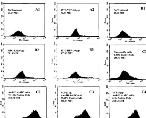
cytometric analysis. As shown in Fig. 1A and B, all three
proteins possess the ability to bind strongly to HUVECs. A
3.0-, 2.9-, or 5.4-fold increase in fluorescent intensity over that
of the control was observed when 20
g of FITC-labeled
rVCP, 30
g of FITC-labeled rVCP SCR (3,4), or 30
g of
FITC-labeled HBP, respectively, was added to the suspension
of HUVECs. Next, using flow cytometric analysis of HUVECs
treated with mouse MAbs to human major histocompatibility
complex class I molecules in the presence and absence of VCP,
VCP was shown to be capable of modulating antibody binding
in a dose-dependent manner. As shown in Fig. 1C, addition of
5
l (2
g) of VCP to antibody-treated HUVECs reduced
antibody binding from 91.5 to 75.4%. An additional 15
l (5
g
total) of VCP reduced antibody binding from 91.5 to 61.0%,
suggesting that it blocks in a dose-dependent manner.
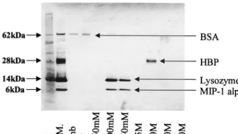
To calibrate the HiTRAP heparin column, several proteins
having different binding affinities for heparin were passed
through the column and eluted with increasing sodium
chlo-ride concentrations. SDS-PAGE analysis of the eluted
frac-tions containing these proteins of various heparin-binding
abil-ities can be seen in Fig. 2. Analysis suggests that BSA does not
bind heparin, since it was present only in the unbound and
wash fractions. Lysozyme and MIP-1
␣
, which bound heparin
with moderate and equal affinity, were contained within the
500 and 750 mM sodium chloride fractions. HBP, showing the
highest affinity for heparin, was eluted by a sodium chloride
concentration of 2.0 M.
[image:3.612.66.544.71.458.2]In order to better characterize the heparin binding ability of
the VCP molecule, VCP, rVCP, and various recombinant
seg-ments of VCP were passed through HiTRAP heparin columns
and analyzed by using SDS-PAGE, shown in Fig. 3. The results
suggest that the full-length natural and recombinant proteins
FIG. 1. Representative histograms showing that VCP is able to reduce mouse anti-human HLA class I antibody binding to HUVECs. The results of flow microflourimetric analysis are as follows. (A1) No treatment control. (A2) Binding of FITC-labeled rVCP to HUVECs. (B1) No treatment control. (B2) Binding of FITC-labeled rVCP SCR (3,4) to HUVECs. (B3) Binding of FITC-labeled HBP to HUVECs. (C1) Nonspecific mouse MAb (isotype-matched mouse IgG2a MAb) binding to HUVECs (negative control). (C2) Mouse monoclonal anti-human HLA-ABC antibody binding to HUVECs (positive control). (C3) Mouse monoclonal anti-human HLA-ABC antibody binding to HUVECs in the presence of 5l (2g) of VCP. (C4) Mouse anti-human HLA-ABC antibody binding to HUVECs in the presence of 20l (5g) of VCP.on November 9, 2019 by guest
http://jvi.asm.org/
bind heparin with an affinity close to or equal to that of
lyso-zyme and MIP-1
␣
; both VCP and rVCP were concentrated
primarily in the 500 and 750 mM sodium chloride fractions.
PAGE analysis of the various rVCP segments revealed even
more interesting data. rVCP SCR (1,2) and rVCP SCR (3,4)
bound heparin with the same strength as the full-length
pro-tein, eluting once again at 500 and 750 mM. rVCP SCR (2,3),
on the other hand, did not bind heparin at all and was found
primarily in the unbound and wash fractions. The activity of
the purified proteins was then tested by using the hemolysis
assay. The results indicate that only the full-length protein
inhibits lysis of sensitized sheep red blood cells—anywhere
from 60 to 90% inhibition. The rVCP segments showed no
inhibition of lysis, suggesting that the whole protein is needed
to block complement activation. However, the naturally
trun-cated VCP homolog produced by MPV (Fig. 4 and 5), which
lacks almost the entire fourth SCR, has been shown to inhibit
hemolysis of sensitized sheep red blood cells, thus indicating
that it blocks the classical pathway of complement activation.
To more precisely identify the molecular basis behind the
VCP-heparin interaction, the amino acid sequences were
an-alyzed by using the MacVector software system. Putative
[image:4.612.55.294.73.207.2]hep-FIG. 2. PAGE analysis of the heparin binding activity of BSA, HBP, lyso-zyme, and MIP-1␣. In order to calibrate the HiTRAP heparin column, various proteins (BSA, HBP, lysozyme, and MIP-1␣) with differing affinities for heparin were passed through the column and eluted with sodium chloride concentrations ranging from 250 mM to 4.0 M. Lanes: M, molecular mass marker; S.M., starting material; Unb, unbound fraction; W, wash.
FIG. 3. PAGE analysis of the heparin binding activity of VCP and rVCPs. VCP and various rVCPs were passed through separate HiTRAP heparin columns and eluted with sodium chloride concentrations ranging from 250 mM to 2.5 M. The fractions were collected, run on SDS-PAGE gels, and silver stained, and the band densities were measured. The results are shown as follows. (A) rVCP SCR (1,2). (B) rVCP SCR (2,3). (C) rVCP SCR (3,4). (D) rVCP. (E) VCP from the natural infection process. Lanes (A to E): M, molecular mass marker; S.M., starting material; Unb, unbound fraction; W, wash; VCP, VCP from natural infection. (F) Densitometric scanning of the gels (A to E).
5662
SMITH ET AL.
J. V
IROL.
on November 9, 2019 by guest
http://jvi.asm.org/
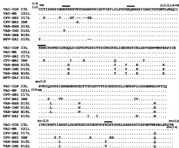
arin binding sites (K/R-X-K/R) were first identified—shown in
Fig. 4 along with the sequence alignment of different pox virus
VCP homologs—and predicted to be located on the surface of
the protein. The amino acid sequences were then scanned to
determine the total number of positive amino acids, the
per-centages of these positively charged amino acids, and the
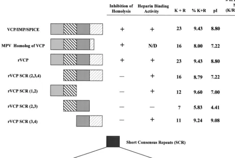
over-all pI of the protein, and the results are summarized in Fig. 4.
All of the proteins shown to bind heparin had an overall pI of
greater than 7.0 and, more importantly, were made up of more
than 9% positively charged amino acids. The results also
sug-gest that SCR 1 and SCR 4 contribute the most to this
inter-action. Although the results show there is no simple answer to
how VCP binds heparin, it is clear that the overall positive
charge, the number of putative binding sites, and the
percent-age of positive amino acids making up the protein have a
significant effect on its ability to interact with and bind heparin.
DISCUSSION
The ability of VCP to bind heparan sulfate proteoglycan
molecules adds a new dimension to its role in modulating the
host immune response. In previous studies, the ability of VCP
to bind heparin was found to permit uptake by mast cells,
possibly allowing for tissue persistence over extended periods
of time. In addition, earlier studies have shown that binding to
heparin-like molecules on the surface of endothelial cells can
block chemokine adherence, resulting in reduced chemotactic
signaling. The next step was to determine if VCP bound to the
endothelial cell surface could interfere with antibody binding.
In this study, flow cytometry was used to demonstrate that
VCP is able to bind the surface of endothelial cells, interfering
with antibody binding. More importantly, this study
demon-strates that addition of VCP significantly decreases the amount
of antibody able to attach to HUVECs in a dose-dependent
manner. It is postulated that VCP, binding to heparin-like
molecules on the surface of endothelial cells, prevents
anti-body adherence. This binding may therefore block antianti-body
attachment through steric interference and block both
molec-ular and cellmolec-ular interactions, which are dependent on target
cell adhesion molecules binding to cytotoxic cells (F.
Al-Mo-hanna, R. Parhar, and G. J. Kotwal, submitted for publication).
Furthermore, this novel mechanism can protect virus-infected
cells or the antigen-presenting cell from destruction by a host
defense mechanism. Interestingly, it was estimated that the
amount of VCP required for inhibition of roughly 5
⫻
10
5cells
is approximately the same as that produced by 2
⫻
10
5infected
cells, and therefore each infected cell can protect itself and one
or two surrounding cells. Thus, VCP enables orthopoxviruses,
all of which encode homologs of VCP, to evade the immune
system by multiple mechanisms, e.g., by blocking complement
pathways, by blocking antibody-dependent cytotoxic cell
activ-ity, or by interfering with attachment of cytotoxic T cells to
virus-infected cells. In addition, as explained below, VCP can
inhibit alternative pathway activation in a manner similar to
that of fH, another heparin binding protein.
[image:5.612.120.484.73.373.2]Due to the apparent importance heparin binding has in
many of the biological activities exhibited by VCP, we became
FIG. 4. Sequence alignment, including termini of rVCP constructs and putative heparin binding sites. Multiple alignment of the four SCR from orthopoxviruses VAC-COP (vaccinia virus, Copenhagen strain) (6), VAC-WR (vaccinia virus, Western Reserve strain) (10), CPV-GRI (cowpox virus, Russian isolate from human patient) (28), CPV-BRI (cowpox virus, Brighton strain) (20), VAR-BSH (variola virus, Bangladesh strain) (17), VAR-IND (variola major virus, India strain) (27), VAR-GAR (alastrim variola minor virus, Garcia strain) (18), and MPV-ZAI (MPV isolated from a human patient from Zaire in 1996). The putative heparin binding sites (K/R-X-K/R) are marked with solid bars, arrows indicate the termini of the rVCP constructs, and the cysteines are highlighted.on November 9, 2019 by guest
http://jvi.asm.org/
interested in better characterizing the molecular reasons for
this interaction. Recombinant segments of VCP, each
repre-senting two of the four SCRs, were generated by genetic
en-gineering vaccinia virus DNA encoding VCP into the
P.
pas-toris
expression system. The ability of these recombinant
segments to bind heparin was then tested. It was determined
that recombinant proteins rVCP SCR (1,2) and rVCP SCR
(3,4) retained the same binding affinity as the full-length native
protein while recombinant protein rVCP SCR (2,3) retained
no heparin binding ability at all. In order to explain this, the
amino acid sequences of the recombinant protein segments
were more closely examined. It was determined that the
num-ber and, more importantly, the percentage of positively
charged amino acids making up the protein contributed greatly
to its ability to interact with heparin. Also, the overall charge
and the number of putative binding sites (K/R-X-K/R) making
up the protein appear to play a role in heparin binding. For the
most part, the ability of VCP to bind heparin lies in its first and
fourth SCR. These two SCR possess within them the highest
percentage of positive amino acids and the greatest number of
putative binding sites and have a very strong overall positive
charge (data not shown).
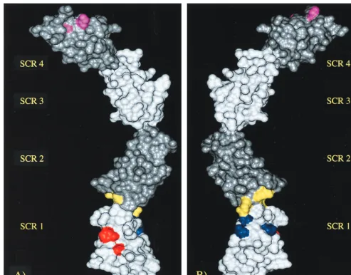
In order to better visualize the positioning of heparin
bind-ing sites in the VCP molecule, a model was developed by usbind-ing
four known VCP module structures: VCP SCR 3, VCP SCR 4,
fH SCR 15 and fH SCR 16 (Fig. 6). As can be seen in this
model, heparin binding exists primarily at the ends (SCR 1 and
4). The middle of the molecule therefore does not contribute
to the binding [hence, VCP SCR (2,3) showed no ability to
bind].
It is clear that the ability to bind polyanions, such as heparin,
is crucial for the function of many immune-regulating proteins.
fH, for example, binds to the polyanion sialic acid on the
surface of mammalian cells, preventing activation of the
alter-native pathway. Like VCP, fH is made up of SCR, two of which
have been shown to have heparin binding abilities (1). It is
postulated that these heparin-binding domains are responsible
for binding sialic acid, thereby preventing alternative pathway
activation. The heparin binding regions of many other proteins
exhibiting heparin binding activity have been mapped (2, 3, 16,
26, 31). In most cases (including ours), binding is the result of
dense regions of positively charged amino acids. These
argin-ine-rich and/or lysargin-ine-rich regions vary in sequence and length
among heparin binding proteins. Interestingly, the heparin
binding sequence used by VCP and its homologs is short and
relatively conserved.
[image:6.612.66.527.81.391.2]In summary, by examining the heparin-binding ability of
VCP, we have gained a better understanding of the molecular
basis by which this interaction occurs. In addition, the data
presented here further enhance our understanding of this truly
unique, yet compact, multifunctional viral protein. It resembles
in structure and function the giant complex human
comple-ment regulatory proteins human C4b binding protein and
com-plement receptor 1, respectively. Not only is VCP a miniature
version of these large molecules, it has retained all the
com-plement modulating functions and, as we have described here,
FIG. 5. Structure-function summary table of VCP, VCP homologs, and rVCPs. VCP/IMP/SPICE, the MPV homolog of VCP, rVCP, and four recombinant segments of VCP are shown along with whether they are able to inhibit hemolysis of sensitized sheep red blood cells and/or bind heparin. Also listed are the number of positively charged amino acids (K⫹R) found in the protein, the percentage of positively charged amino acids (%K⫹R) making up the protein, the pI of the protein, and the number of putative heparin binding sites found on the surface of the protein.
5664
SMITH ET AL.
J. V
IROL.
on November 9, 2019 by guest
http://jvi.asm.org/
added a number of additional functions to its repertoire due to
its ability to bind heparin-like molecules. This better
under-standing may someday help in the exploitation of VCP’s
pos-sible therapeutic properties in the treatment of inflammatory
conditions, such as Alzheimer’s disease (4), restenosis,
acci-dental spinal cord injury, and xenograft transplant rejections.
ACKNOWLEDGMENTS
The HBP was kindly provided by Hans Flodgaard from Novo
Nor-disk, Copenhagen, Denmark (5). We would also like to thank Sam
Welhausen of the J. Graham Brown Cancer Center for the use of his
FACS facilities.
Partial support for this work was provided by the Kentucky Spinal
Cord and Head Injury Research Trust (KSCHIRT).
REFERENCES
1.Blackmore, T. K., J. Hellwage, T. A. Sadlon, N. Higgs, P. F. Zipfel, H. M. Ward, and D. L. Gordon.1998. Identification of the second heparin-binding domain in human complement factor H. J. Immunol.160:3342–3348. 2.Cardin, A. D., D. A. Demeter, H. J. Weintraub, and R. L. Jackson.1991.
Molecular design and modeling of protein-heparin interactions. Methods Enzymol.203:556–583.
3.Cardin, A. D., and H. J. Weintraub.1989. Molecular modeling of protein-glycosaminoglycan interactions. Arteriosclerosis9:21–32.
4.Daly, J., and G. J. Kotwal.1998. Pro-inflammatory complement activation by
the A beta peptide of Alzheimer’s disease is biologically significant and can be blocked by vaccinia virus complement control protein. Neurobiol. Aging 19:619–627.
5.Flodgaard, H., E. Ostergaard, S. Bayne, A. Svendsen, J. Thomsen, M. En-gels, and A. Wollmer.1991. Covalent structure of two novel neutrophile leucocyte-derived proteins of porcine and human origin. Neutrophile elas-tase homologues with strong monocyte and fibroblast chemotactic activities. Eur. J. Biochem.197:535–547.
6.Goebel, S. J., G. P. Johnson, M. E. Perkus, S. W. Davis, J. P. Winslow, and E. Paoletti.1990. The complete DNA sequence of vaccinia virus. Virology 179:247–263.
7.Howard, J., D. E. Justus, A. V. Totmenin, S. Shchelkunov, and G. J. Kotwal. 1998. Molecular mimicry of the inflammation modulatory proteins (IMPs) of poxviruses: evasion of the inflammatory response to preserve viral habitat. J. Leukoc. Biol.64:68–71.
8.Isaacs, S. N., G. J. Kotwal, and B. Moss.1992. Vaccinia virus complement-control protein prevents antibody-dependent complement-enhanced neu-tralization of infectivity and contributes to virulence. Proc. Natl. Acad. Sci. USA89:628–632.
9.Kotwal, G. J., D. Reynolds, K. Keeling, J. Howard, and D. E. Justus.1998. Vaccinia virus complement control protein is a virokine with lysozyme-like heparin-binding activity: possible implications in prolonged evasion of host immune response, p. 315–320.InG. P. Talwar, I. Nath, N. K. Ganguly, and K. V. S. Rao (ed.), 10th International Congress of Immunology. Monduzzi Editore, Bologna, Italy.
[image:7.612.56.550.71.457.2]10. Kotwal, G. J., A. W. Hugin, and B. Moss.1989. Mapping and insertional mutagenesis of a vaccinia virus gene encoding a 13,800-Da secreted protein. Virology171:579–587.
FIG. 6. VCP model showing the heparin binding sites. Front (A) and back (B) views of the modeled structure of VCP SCR (1-4) showing the heparin binding sites (differentially colored). In order to differentiate the extents of the individual modules, they are shaded appropriately.
on November 9, 2019 by guest
http://jvi.asm.org/
11.Kotwal, G. J., S. N. Isaacs, R. McKenzie, M. M. Frank, and B. Moss.1990. Inhibition of the complement cascade by the major secretory protein of vaccinia virus. Science250:827–830.
12. Kotwal, G. J., C. G. Miller, and D. E. Justus.1998. The inflammation modulatory protein (IMP) of cowpox virus drastically diminishes the tissue damage by down-regulating cellular infiltration resulting from complement activation. Mol. Cell. Biochem.185:39–46.
13. Kotwal, G. J., and B. Moss. 1988. Vaccinia virus encodes a secretory polypeptide structurally related to complement control proteins. Nature335: 176–178.
14. Kotwal, G. J., and B. Moss.1989. Vaccinia virus encodes two proteins that are structurally related to members of the plasma serine protease inhibitor superfamily. J. Virol.63:600–606. (Erratum,64:966, 1990.)
15. Lian, R. H., G. J. Kotwal, S. R. Wellhausen, L. A. Hunt, and D. E. Justus. 1996. IFN-gamma-induced MHC class II gene expression is suppressed in endothelial cells by dextran sulfate. J. Immunol.157:864–873.
16. Marino, M., J. A. Friedlander, R. T. McCluskey, and D. Andrews.1999. Identification of a heparin-binding region of rat thyroglobulin involved in megalin binding. J. Biol. Chem.274:30377–30386.
17. Massung, R. F., L. I. Liu, J. Qi, J. C. Knight, T. E. Yuran, A. R. Kerlavage, J. M. Parsons, J. C. Venter, and J. J. Esposito.1994. Analysis of the com-plete genome of smallpox variola major virus strain Bangladesh-1975. Vi-rology201:215–240.
18. Massung, R. F., V. N. Loparev, J. C. Knight, A. V. Totmenin, V. E. Chizhikov, J. M. Parsons, P. F. Safronov, V. V. Gutorov, S. N. Shchelkunov, and J. J. Esposito.1996. Terminal region sequence variations in variola virus DNA. Virology221:291–300.
19. McKenzie, R., G. J. Kotwal, B. Moss, C. H. Hammer, and M. M. Frank. 1992. Regulation of complement activity by vaccinia virus complement-con-trol protein. J. Infect. Dis.166:1245–1250.
20. Miller, C. G., D. E. Justus, S. Jayaraman, and G. J. Kotwal.1995. Severe and prolonged inflammatory response to localized cowpox virus infection in footpads of C5-deficient mice: investigation of the role of host complement in poxvirus pathogenesis. Cell. Immunol.162:326–332.
21. Miller, C. G., S. N. Shchelkunov, and G. J. Kotwal.1997. The cowpox virus-encoded homolog of the vaccinia virus complement control protein is
an inflammation modulatory protein. Virology229:126–133.
22. Moss, B.1995. Poxviridae: the viruses and their replication, p. 1163–1197.In
B. N. Fields, D. M. Knipe, and P. M. Howley (ed.), Fundamental virology, 3rd ed. Lippincott-Raven, Philadelphia, Pa.
23. Reynolds, D., K. Keeling, R. Molestina, R. Srisatajluk, J. H. Butterfield, W. Ehringer, D. E. Justus, and G. J. Kotwal.1999. Heparin binding activity of vaccinia virus complement control protein confers additional properties of uptake by mast cells and attachment to endothelial cells.InS. Jameel and L. Villareal (ed.), Advances in animal virology. Oxford and IBN Publishing, New Delhi, India.
24. Sahu, A., S. N. Isaacs, A. M. Soulika, and J. D. Lambris.1998. Interaction of vaccinia virus complement control protein with human complement pro-teins: factor I-mediated degradation of C3b to iC3b1 inactivates the alter-native complement pathway. J. Immunol.160:5596–5604.
25. Sali, A., and T. L. Blundell.1993. Comparative protein modelling by satis-faction of spatial restraints. J. Mol. Biol.234:779–815.
26. Sendak, R. A., and A. Bensadoun.1998. Identification of a heparin-binding domain in the distal carboxyl-terminal region of lipoprotein lipase by site-directed mutagenesis. J. Lipid Res.39:1310–1315.
27. Shchelkunov, S. N., S. M. Resenchuk, A. V. Totmenin, V. M. Blinov, S. S. Marennikova, and L. S. Sandakhchiev.1993. Comparison of the genetic maps of variola and vaccinia viruses. FEBS Lett.327:321–324.
28. Shchelkunov, S. N., P. F. Safronov, A. V. Totmenin, N. A. Petrov, O. I. Ryazankina, V. V. Gutorov, and G. J. Kotwal.1998. The genomic sequence analysis of the left and right species-specific terminal region of a cowpox virus strain reveals unique sequences and a cluster of intact ORFs for immunomodulatory and host range proteins. Virology243:432–460. 29. Upton, C., K. Mossman, and G. McFadden.1992. Encoding of a homolog of
the IFN-gamma receptor by myxoma virus. Science258:1369–1372. 30. Wiles, A. P., G. Shaw, J. Bright, A. Perczel, I. D. Campbell, and P. N. Barlow.
1997. NMR studies of a viral protein that mimics the regulators of comple-ment activation. J. Mol. Biol.272:253–265.
31. Wong, P., B. Hampton, E. Szylobryt, A. M. Gallagher, M. Jaye, and W. H. Burgess.1995. Analysis of putative heparin-binding domains of fibroblast growth factor-1. Using site-directed mutagenesis and peptide analogues. J. Biol. Chem.270:25805–25811.
5666
SMITH ET AL.
J. V
IROL.
on November 9, 2019 by guest
http://jvi.asm.org/