Copyrightq1997, American Society for Microbiology
The Hydrophobic Pocket of Cyclophilin Is the Binding Site for
the Human Immunodeficiency Virus Type 1 Gag Polyprotein
DOUGLAS BRAATEN,1HUSAMUDDIN ANSARI,2ANDJEREMY LUBAN1,3*
Department of Microbiology,1Integrated Program in Cellular, Molecular, and Biophysical Studies,2and Department of
Medicine,3College of Physicians and Surgeons, Columbia University, New York, New York 10032
Received 26 September 1996/Accepted 9 December 1996
Completion of an early step in the human immunodeficiency virus type 1 (HIV-1) life cycle requires incorporation into virions of the cellular peptidyl-prolyl isomerase cyclophilin A (CyPA) by the Gag polypro-tein. Elucidation of the biochemical role of CyPA would be aided by a detailed analysis of the genetic requirements for the formation of the Gag-CyPA complex; previous experiments have demonstrated the requirement for a critical proline and the immediately preceding glycine, located within the capsid domain of Gag, but nothing is known about the necessary CyPA residues. Cyclophilins possess a hydrophobic pocket where proline-containing peptide substrates and the immunosuppressive drug cyclosporine A bind. In this study, we engineered five CyPA mutations, each of which alters a residue that contributes to the hydrophobic pocket. Compared with the wild-type protein, all of the mutants drastically reduced CyPA binding to HIV-1 Gag and similarly inhibited CyPA incorporation into virions. In addition, we demonstrated that previously reported differences between the Gag-binding properties of CyPA and CyPB are due to adventitious association involv-ing residues in the signal sequence of CyPB and that the core domain of CyPB interacts with Gag in a fashion which is indistinguishable from that of CyPA. These studies indicate that, as with other proline-containing peptides or cyclosporine A, HIV-1 Gag directly contacts residues in the hydrophobic pocket of CyPA.
Late in the infectious cycle of human immunodeficiency virus type 1 (HIV-1), the Gag polyprotein binds to the cytosolic protein cyclophilin A (CyPA); by this association, CyPA is incorporated into virions, where it is required for the subse-quent round of viral replication (15, 31). The exact role of CyPA is unknown, but its incorporation into virions is neces-sary for a capsid (CA) function—perhaps virion uncoating— occurring after membrane fusion and before reverse transcrip-tion (3, 4).
As an initial step towards understanding the biochemical role of CyPA in HIV-1 replication, the genetic requirements for Gag’s interaction with CyPA have been examined, both in vitro and in the context of infectious virus. CyPA-binding ac-tivity localizes to a proline-rich stretch of amino acids located in the middle of the CA domain of Gag (4, 8, 15, 24, 31). P222, a residue within this domain, and the preceding residue, G221, are critical for binding (4), a fact that is particularly intriguing considering CyPA’s ability to catalyze the isomerization of peptidyl-prolyl bonds (13). Consistent with the genetic data is the recently solved three-dimensional structure of HIV-1 CA, in which the proline-rich domain forms an exposed loop which would be accessible for binding to CyPA (17).
Though a great deal has been learned about the CA residues necessary for binding to CyPA, nothing is known about the CyPA sequences required for binding to CA. Since members of the CyP family of proteins are intracellular targets of the clin-ically important immunosuppressive drug cyclosporine A (CsA) (18, 29), structural studies with these proteins have been extensive (10, 11, 20, 22, 23, 33). CyPs consist of an eight-strand beta-barrel with a hydrophobic pocket opening to one side of the hydrophobic core composed of residues from four of the eight beta-strands and a loop of aromatic residues; CsA
and proline-containing peptides contact residues within the hydrophobic pocket. Since CsA inhibits the Gag-CyPA inter-action (15, 24, 31), Gag too may bind to the hydrophobic pocket of CyPA.
In addition to binding to CyPA, the Gag polyprotein of HIV-1 also binds to another member of the CyP family, CyPB (24), but there are important differences between CyPA and CyPB: CyPB binds to Gag polyproteins containing the CA mutation G221A or P222A, whereas CyPA does not (4, 15), and CyPB remains bound to the HIV-1 Gag polyprotein at a 10-fold greater concentration of CsA than is necessary to block the CyPA-CA interaction (24). In addition to these differences, CyPB, unlike CyPA, binds to the Gag polyprotein of simian
immunodeficiency virus of macaque strain 239 (SIVMAC239)
(15). There are two possible explanations for these data. If Gag interacts with the hydrophobic pocket of CyP proteins, CyPB has different substrate-binding requirements than does CyPA. Alternatively, the difference in the observed binding properties may be due to the fact that CyPB possesses a 33-amino-acid hydrophobic leader peptide not found in CyPA (19); it is pos-sible that the in vitro binding properties of CyPB are due to adventitious association by hydrophobic residues in this region. To address the question of sequence requirements for CyP binding to Gag, we present in this paper data on the effects of specific mutations in the hydrophobic pocket of CyPA on bind-ing to the HIV-1 Gag polyprotein. Since the mutated residues are conserved between the hydrophobic pockets of both CyPA and CyPB, any effects seen will address the general question of which amino acids are necessary for CyP binding to Gag. Ad-ditional data presented will address the binding properties of CyPB. We have engineered a CyPB protein which lacks the leader sequence to test if this truncated version of CyPB has binding properties similar to those of CyPA. Together, the data presented for both CyPA and CyPB provide a sketch of the primary sequence requirements for association of CyPs with the Gag polyprotein of HIV-1.
* Corresponding author. Mailing address: Department of Microbi-ology, College of Physicians & Surgeons, Columbia University, 701 W. 168th St., New York, NY 10032. Phone: (212) 305-8706. Fax: (212) 305-8706. E-mail: Luban@cuccfa.ccc.columbia.edu.
2107
on November 9, 2019 by guest
http://jvi.asm.org/
used as a template to create the following missense mutations in the hydrophobic pocket of CyPA: 59-CCCTGGAATGATTCGCTGAAAGCAGG-39for H54Q, 59-CCCTGGAATAATCGCGTGAAAGCAG-39for R55A, 59-CCTGACACAT GGCGCCTGGAATAATTC-39for F60A, 59-GGCAGTGCATATGGCAAAC TGGGAACC-39for F113A, and 59-CAAACACCACCTGCTTGCCATC-39for H126Q. The sequence of each mutant was confirmed by standard methods (28). Primers were designed to amplify the region of CyPB containing both the sequences homologous to CyPA (the core domain) and the C-terminal domain unique to CyB: 59-CGGCCATGGTCACCGTCAGGGTG-39and 59-GCGTAG TCTGGGACGTCGTATGG-39. The PCR conditions were as follows. A 20-ml reaction volume consisted of 10 mM Tris-HCl (pH 8.3), 50 mM KCl, 1.5 mM MgCl2, 0.001% (wt/vol) gelatin, 0.2 mM each deoxynucleoside triphosphate, 10 pmol of each oligonucleotide, 50 ng of a human cDNA clone of CyPB as the template, and 0.5 U of Ampli-Taq DNA polymerase. The sample was amplified for 25 cycles of 948C for 30 s, 608C for 30 s, and 728C for 1 min in a Perkin Elmer 2400 machine. The 541-bp PCR product (DCyPB) was cloned into pBS-KSII2 (Stratagene) by utilizing restriction sites created during the PCR,NcoI at the 59 end andEcoRI at the 39end, and was subsequently sequenced.
Proteins containing the designed point mutations in the hydrophobic pocket of CyPA were expressed as glutathione S-transferase (GST) fusion proteins by cloning the CyP cDNAs into a version of the bacterial expression vector pGEX-2TK (Promega) modified to contain anNcoI site. The appropriate vector and insert fragments (NcoI-EcoRI fragments) were isolated and ligated in low-melt-ing-point agarose (SeaPlaque LMA, fom FMC). Likewise, to make the GST-DCyPB expression construct, theNcoI-EcoRI PCR fragment was cloned directly into modified pGEX-2TK.
Mutant CyPA proteins were expressed in mammalian cells as influenza virus hemagglutinin (HA) epitope fusions (12) by using the SRapromoter (30). SRa expression constructs of the CyPA point mutants and ofDCyPB were cloned by digesting the pGEX-2TK constructs withBamHI, filling in the ends with Klenow, and then digesting the constructs withEcoRI to obtain the CyP insert fragments. The insert fragments were ligated to a vector fragment from pIX156 (5), which was produced by digestion withXhoI, filling in of the ends with Klenow, and then digestion withEcoRI. All of the SRaconstructs were cloned such that proteins expressed from them had the HA epitope fused in frame to the carboxy-terminal end; this enabled subsequent identification of the proteins with a monoclonal antibody to the HA epitope.
HIV-1 Gag numbering is with respect to the amino terminus of the Gag polyprotein. The HIV-1 Gag polyprotein bacterial expression plasmid pT7HG (pro2) has been described previously (25), as have the Gag mutants P222A and
G221A (4, 15), and the SIVMAC239Pr57gagexpression construct (15). pSRa55 is a Rev-independent expression construct forgagsequences (15).
In vitro binding experiments.In vitro binding experiments were performed as described previously (7). Briefly, equivalent amounts of GST fusion proteins, produced inE. coliDH5a, were adsorbed to glutathione-agarose beads at 48C, after which unbound protein was removed by washing with TK buffer (20 mM Tris-HCl [pH 7.5], 100 mM KCl, 2 mM CaCl2, 2 mM MgCl2, 5 mM dithiothreitol, 0.5% Nonidet P-40, 0.5 mM phenylmethylsulfonyl fluoride, 5% glycerol). The loaded beads were then incubated with bacterially produced Gag lysates at 48C for 30 min. After three subsequent washes of the beads in TK buffer, the beads were pelleted a final time and the bound protein complexes were dissolved in 23 sodium dodecyl sulfate (SDS) sample buffer (100 mM Tris-HCl [pH 6.8], 1.7 M b-mercaptoethanol, 4% SDS, 0.2% bromophenol blue, 20% glycerol) and sub-jected to SDS-polyacrylamide gel electrophoresis (SDS-PAGE). Gels were ei-ther stained with Coomassie blue or processed for Western blotting as described below.
CsA was obtained from Sandoz Pharmaceuticals Corporation (East Hanover, N.J.) and dissolved in ethanol to make a 1.25 mM stock solution prior to addition to the binding reactions. In drug competition experiments, CsA was mixed with the recombinant proteins at the indicated concentrations.
Cell culture, transfections, and virion production and purification.Human fibroblast 293T cells were maintained in Dulbecco modified Eagle medium (DMEM)-F12 (1:1) supplemented with 10% fetal bovine serum. Proteins were expressed transiently by calcium phosphate transfection of 10mg of each expres-sion construct with the Mammalian Cell Transfection Kit (Specialty Media, Lavellette, N.J.). Supernatants and cell lysates were harvested 48 h posttransfec-tion. For virion preparation, 7.5 ml of supernatant collected from 293T cells were passed through a 0.45-mm-pore-size filter to remove cellular debris. The filtrate was layered onto 2 ml of 25% sucrose in TNE (10 mM Tris-HCl [pH 7.5], 100
miniblotting apparatus (Bio-Rad, Hercules, Calif.). The following primary anti-bodies were used: a murine anti-HIV-1 p24 monoclonal antibody from Du Pont/NEN (NEA-9306); a murine anti-HIV-2 core protein monoclonal antibody from the National Institutes of Health AIDS Research and Reference Program (catalog no. 740); a rabbit anti-CyPA antibody which recognizes both wild-type CyPA and HA-tagged CyPA, from Affinity BioReagents, Heshanic Station, N.J. (catalog no. PA3-021); and monoclonal antibody 12CA5, which recognizes the HA epitope, purchased from Berkeley Antibody Company, Berkeley, Calif. Binding of these antibodies was detected after incubation with the appropriate peroxidase-coupled secondary antibody with the Renaissance chemilumines-cence kit (Du Pont/NEN).
RESULTS
Mutations in the hydrophobic pocket of CyPA disrupt bind-ing to the HIV-1 Gag polyprotein.Previous studies have iden-tified the amino acids comprising the hydrophobic pocket of CyPA, where proline-containing peptides and CsA bind (10, 11, 20, 22, 23, 33). Additional biochemical studies using engi-neered hydrophobic pocket mutants demonstrated specific ef-fects on peptidyl-prolyl isomerase activity and binding of CsA (34). Our aim was to duplicate these mutations and to deter-mine if any of them affect the previously demonstrated
prop-erty of CyPA to bind to HIV-1 Gag polyprotein Pr55gag(24).
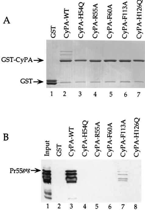
Mutations were engineered in the human CyPA cDNA and subcloned into a vector for expression as GST fusion proteins in bacteria (Fig. 1). Crude preparations of each of the GST-CyPA fusion proteins were incubated with bacterially
pro-duced HIV-1 protein Pr55gag in the presence of
glutathione-agarose beads; proteins retained by the beads were subjected to SDS-PAGE (Fig. 2). A Coomassie blue-stained gel showed that for all of the CyPA point mutants equal amounts of GST fusion proteins were retained by the glutathione-agarose beads (Fig. 2A), as demonstrated by the major high-molecular-weight band in lanes 2 to 7; the minor band in each lane is most likely a proteolytic cleavage product of the GST peptide from
the fusion proteins. Pr55gagbound to wild-type CyPA,
(appar-ent from the higher-molecular-weight bands seen in lane 2) but binding to any of the mutant proteins was not detectable by Coomassie blue staining.
Western blotting was performed on the samples from the same binding experiment by using a monoclonal antibody to HIV-1 CA. In agreement with the results from the Coomassie
blue-stained gel, Pr55gagwas retained by wild-type CyPA but
not by the mutant CyPAs (Fig. 2B). The only exception re-vealed by the greater sensitivity of the Western blot was weak
binding of Pr55gagto F113A-CyPA, though by serially diluting
the wild-type CyPA-Gag complexes (data not shown), we es-timated that even this mutation reduced CyPA’s affinity for Gag 10-fold. The results of the binding experiments with these CyPA mutants indicate that the Gag-binding site on CyPA is contained within the hydrophobic pocket.
Hydrophobic pocket mutations abolish CyPA packaging into virions.We next determined if the in vitro binding results could be extended to a more physiologically relevant condition. Previous analysis of total protein from purified HIV-1 virions demonstrated that CyPA is specifically packaged into virion
on November 9, 2019 by guest
http://jvi.asm.org/
particles via the proline-rich region of the CA protein (4, 8, 15, 24, 31). Since the engineered mutations in the hydrophobic pocket of CyPA disrupt the Gag-CyPA association in vitro, we hypothesized that virions produced in tissue culture cells
ex-pressing the mutant CyPs would likewise fail to package the mutant CyPAs.
To produce virions in the presence of the mutant CyPs, 293T
cells were cotransfected with pSRa55, a Rev-independentgag
expression construct that produces immature particles (15), and one of several constructs for expression of the CyPA mu-tants as fusions with the HA epitope tag. The epitope tag allowed us to distinguish endogenous CyPA from CyPA ex-pressed from the transfected plasmids by using the anti-HA antibody (data not shown), but because the HA-tagged fusion proteins migrate more slowly than endogenous CyPA, for the experiments shown here, a polyclonal antibody to CyPA was used which recognizes both the HA-tagged and endogenous CyPA proteins as two discrete bands (Fig. 3A, lanes 2 to 7).
293T cells were lysed 48 h posttransfection and assayed for relative steady-state levels of the proteins expressed by the transfected constructs. Wild-type HA-CyPA and three of the mutants were expressed at levels comparable to that of endog-enous CyPA (Fig. 3A, lanes 2, 4, 5, and 7). Two of the mutant CyPA proteins were not expressed as well, and a rough quan-titation of the relative amounts of each HA-CyPA protein was obtained by serially diluting the samples run on SDS-PAGE and then comparing band intensities with dilution factors (data not shown): the amount of CyPA-H54Q was estimated to be decreased more than 30-fold relative to that of wild-type CyPA (compare Fig. 3A, lanes 2 and 3); CyPA-F113A (Fig. 3A, lane 6) was also decreased relative to wild-type CyPA, by about a factor of 10.
Virions were harvested from the transfection supernatants and purified by centrifugation through 25% sucrose. Total virion proteins were processed for Western analysis by using the same antibodies that were used with the cell lysates. As
judged by the intensity of the Pr55gagsignal from the virion
preparations, virion yields from all of the transfections were comparable (Fig. 3B, lanes 1 to 7), indicating that none of the mutant CyPA proteins had an inhibitory effect on virion re-lease. As a control, a CyPA expression construct was not
in-cluded in one of the pSRa55 transfections; thus, only
endog-enous CyPA was detected in these virions (Fig. 3B, lane 1).
When expressed concomitantly with Pr55gag, HA-tagged
wild-type CyPA was incorporated into virions (Fig. 3B, lane 2). In fact, of the virion-associated CyPA, there was significantly more HA-CyPA than endogenous CyPA.
The efficiency of virion incorporation for each of the CyPA mutants was then compared to that of transfected wild-type CyPA. To do this, we first determined the relative amount of transfected mutant CyPA versus endogenous CyPA that was virion associated. This value, normalized based on the relative
FIG. 2. In vitro binding of the HIV-1 Gag polyprotein to GST-CyPA fusion proteins containing mutations in the hydrophobic pocket. Pr55gagwas mixed with
either GST or the GST-CyPA fusions indicated, and complexes retained on glutathione-agarose beads were subjected to SDS-PAGE. Gels were stained with Coomassie blue (A) or processed for Western blotting with an anti-CA mono-clonal antibody (B). The total bacterial lysate of Pr55gagin lane 1 (B) represents
1/10 of the total input of Pr55gagused in the individual binding reactions. The
[image:3.612.57.295.308.651.2]positions of migration of the GST-CyPA fusion proteins, GST, and the full-length HIV-1 Gag polyprotein (Pr55gag) are indicated on the left.
FIG. 1. Amino acid sequence alignment of human CyPA and CyPB by using Clustal W. Conservation of amino acids is indicated by black shading; conservative changes are indicated by grey shading; divergent changes are indicated by no shading, and sequence gaps are indicated by dashes. Asterisks are placed over residues in the CyP hydrophobic pocket that were targeted for mutagenesis in the work presented here; the identities of the mutant amino acids are indicated below the residues which were altered.
on November 9, 2019 by guest
http://jvi.asm.org/
levels of the transfected mutant CyPAs expressed in cells, was then compared to the same value obtained for the transfected wild-type CyPA. Virion incorporation for each of the mutant CyPAs was 10 to 100 times less efficient than for the wild-type HA-CyPA (Fig. 3B, lanes 3 to 7). In agreement with the in vitro binding data, the magnitude of the reduction was the least with CyPA-F113A. The results from these experiments dem-onstrate that mutations within the hydrophobic pocket of CyPA affect its ability to be incorporated into virions. That
contaminated with cellular debris (26).
Deletion of the N-terminal leader peptide of CyPB alters its binding properties in vitro.Previous in vitro binding experi-ments have demonstrated that CyPB binds to both HIV-1
Pr55gagand the Gag polyprotein of SIV
MAC239, Pr57gag; these
data contrast with those obtained with CyPA, which does not
bind to SIVMAC239Pr57gag(8, 15, 24). We found this difference
puzzling, considering the strong sequence homology between these two CyPs and, in particular, the conservation of the hydrophobic pocket residues (Fig. 1). Aside from scattered sequence differences found between the conserved core do-mains of the two proteins, CyPB is distinguished from CyPA by its 33-amino-acid leader peptide and a small carboxy-terminal peptide of 10 amino acids (19).
To determine if the leader peptide has an effect on the binding properties of CyPB, the core domain of 510 amino acids plus the C-terminal 10 amino acids was cloned into a bacterial expression vector to produce a GST fusion protein
(GST-DCyPB). The binding properties of GST-DCyPB were
tested with respect to both the HIV-1 and SIVMAC239 Gag
polyproteins by mixing GST-DCyPB or GST-CyPA with either
bacterially produced Pr55gagof HIV or Pr57gagof SIV
MAC239
in the presence of glutathione-agarose beads; adsorbed protein complexes were purified and then subjected to SDS-PAGE
(Fig. 4). In addition, GST-CyPB was mixed with Pr57gag of
SIVMAC239. A Coomassie blue-stained SDS-polyacrylamide
gel (Fig. 4A) showed that comparable amounts of HIV-1
Pr55gagwere retained by GST-DCyPB and GST-CyPA and that
SIVMAC239 Pr57gag bound to CyPB but not to
GST-DCyPB or to GST-CyPA (Fig. 4A, lanes 5 to 8).
Western blotting with a monoclonal antibody that
recog-nizes both Pr55gag of HIV-1 and Pr57gag of SIV
MAC239 was
performed on the same complexes (Fig. 4B). The results par-allel exactly those obtained by Coomassie blue staining. HIV-1
Gag bound to CyPA and DCyPB (Fig. 4B, lanes 3 and 4,
respectively), but SIVMAC239 Gag bound only to full-length
CyPB (Fig. 4B, lane 7). These results indicate that HIV-1 Gag
interacts with the core of CyPB; SIVMAC239Gag does not bind
to the core of CyPB but to the hydrophobic leader peptide of this protein, calling into question the biological significance of this particular interaction.
It has been shown previously that HIV-1 Gag mutants G221A and P222A interfere with the CyPA-Gag association but have no effect on the CyPB-Gag association (4, 15). To
further assess the binding characteristics of DCyPB, in vitro
experiments were performed to compare the binding
proper-ties of DCyPB and CyPB with respect to these HIV-1 Gag
mutants. GST-DCyPB or GST-CyPB was mixed with
equiva-lent amounts of bacterially produced mutant Gag proteins. Protein complexes adsorbed to glutathione-agarose beads were purified and separated by SDS-PAGE. Coomassie blue
staining demonstrated that equal amounts of GST-DCyPB and
GST-CyPB bound to the beads (Fig. 5A). Western blotting on an identical gel, using the monoclonal antibody to HIV-1 CA, demonstrated that both mutant proteins bound to GST-CyPB
FIG. 3. Mutations within the hydrophobic pocket inhibit CyPA incorporation into virions. 293T cells were cotransfected with pSRa55 and each of the indicated CyPA mutant expression constructs. Total cell lysates (A) and virion proteins purified from the supernatant (B) were subjected to SDS-PAGE and Western blot analysis with both anti-HIV-1 CA and anti-CyPA monoclonal antibodies. Lane 1 shows samples from cells transfected with pSRa55 alone. The position of migration of the HIV-1 Gag polyprotein (Pr55gag) is indicated on the left. The
positions of migration of endogenous CyPA and lower-mobility HA-CyPA are indicated on the left. WT, wild type.
on November 9, 2019 by guest
http://jvi.asm.org/
(Fig. 5B, lanes 2 and 4), which is consistent with what has been seen previously with CyPB (15). In contrast, neither Gag
mu-tant bound to GST-DCyPB (Fig. 5B, lanes 1 and 3). These
results again demonstrate the similarity of the binding
proper-ties of CyPA andDCyPB.
DCyPB/Gag and CyPA/Gag interactions are inhibited by the same concentration of CsA.The ability of CsA to interfere with the CyPA-Gag interaction is well documented (2, 4, 14, 15, 21, 24, 27, 31, 32). It has also been demonstrated that the CyPB-Gag interaction is much less sensitive to disruption by CsA than is the CyPA-Gag interaction (24). Figure 6 shows a West-ern blot analysis of protein complexes obtained from a binding
experiment in which GST-DCyPB (Fig. 6A) and GST-CyPB
(Fig. 6B) were compared with respect to binding to HIV-1 Gag in the presence of increasing concentrations of CsA. The re-sults show that CyPB remained bound to HIV-1 Gag even at
over 10mM CsA (Fig. 6A, lanes 1 to 6), which is consistent with
what has been reported previously. In contrast, binding of
DCyPB to Gag was blocked at CsA concentrations between 0.3
and 1.0mM (Fig. 6B, lane 3), the same range of concentrations
at which the CyPA-Gag interaction is blocked under identical binding conditions (24). This indicates that the strength of the interaction between HIV-1 Gag and the core of either CyPA or CyPB cannot be distinguished in this assay.
DCyPB is incorporated into virions in a manner analogous to that of CyPA. Since CyPB is localized to the secretory pathway by its leader peptide, we expected that it would not be
incorporated into virions despite its ability to bind to HIV-1
Gag in vitro. Since DCyPB lacks the leader peptide—and
therefore would not be fated for localization to the secretory
pathway—we determined if DCyPB itself contains sequences
necessary and sufficient for incorporation into virions.
pSRaDCyPB, pSRaCyPB, and pSRaCyPA were each
cotrans-fected with pSRa55 into 293T cells. Figure 7 shows Western
blotting results obtained with cell lysates (Fig. 7A) and purified virion particles (Fig. 7B) produced from transfected cells; blots were probed by using both anti-HIV-1 CA and anti-HA mono-clonal antibodies. In the lysates, each of the CyPs was ex-pressed at about the same steady-state level (Fig. 7A). In the
virion preparations, CyPA andDCyPB were detected (Fig. 7B,
lanes 1 and 2) but CyPB was not (lane 3). Thus, like CyPA,
DCyPB is specifically incorporated into virions. These results,
[image:5.612.314.530.67.420.2]together with those of the in vitro binding experiments, indi-cate that the sequences necessary for incorporation into HIV-1 virions are conserved between CyPA and CyPB but that CyPB
FIG. 4.DCyPB binds to HIV-1 Gag but not to SIVMAC239Gag. Lysates of bacterially expressed HIV-1 Gag (lanes 1 to 4) or SIVMAC239Gag (lanes 5 to 8) were mixed with lysates from bacteria expressing either GST or GST fused to the indicated CyPs. Complexes retained by glutathione-agarose beads were sub-jected to SDS-PAGE and then visualized by Coomassie blue staining (A) or processed for Western blot analysis by using a monoclonal antibody which recognizes the Gag polyproteins of both HIV-1 and SIVMAC239(B). Lanes 1 and 5 show 10% of the Gag lysates used in the binding experiments. The positions of migration of GST and the HIV-1 Pr55gagand SIV
MAC239Pr57gagpolyproteins are indicated on the sides. The positions of migration of the different GST-CyP
fusion proteins are also indicated. FIG. 5.DCyPB does not bind to HIV-1 Gag mutants. Lysate of bacteria expressing HIV-1 Gag mutant G221A (lanes 1 and 2) or P222A (lanes 3 and 4) was mixed with lysate from bacteria expressing GST fused to the indicated CyPs. Complexes retained by glutathione-agarose beads were subjected to SDS-PAGE and then visualized by Coomassie blue staining (A) or processed for Western blotting by using an anti-CA monoclonal antibody (B). G221A and P222A are mutations within the proline-rich region of Gag that block the binding of CyPA. The position of migration of the full-length HIV-1 Gag polyprotein (Pr55gag) is
indicated on the left. The positions of migration of the different GST-CyP fusion proteins are also indicated.
on November 9, 2019 by guest
http://jvi.asm.org/
[image:5.612.61.293.68.346.2]is not normally incorporated into virions because it is seques-tered in the cellular secretory apparatus.
DISCUSSION
The importance of understanding the primary sequence re-quirements of CyP binding to the Gag protein of HIV-1 is evident from the fact that HIV-1 virions depleted of CyPA are not infectious (4, 15, 31). The infectivity of these virions is blocked prior to reverse transcription but after membrane fu-sion (4). Genetic information about the Gag-CyPA interaction will undoubtedly prove critical for determining the exact bio-chemical role of CyPA in HIV-1 replication. For example, it has already been determined that HIV-1 dependence upon CyPA maps to residues in CA that are distinct from, though immediately adjacent to, residues required for CyPA binding (3). This information suggests that CyPA is required for HIV-1 uncoating, the process by which CA separates from the re-mainder of the viral nucleoprotein complex following entry into a new target cell (6, 9, 16). The identification of a specific Gag peptidyl-prolyl bond (G221-P222) that is required for
CyPA binding suggests that CA undergoescis-trans
isomeriza-tion at this posiisomeriza-tion as a result of the interacisomeriza-tion; consistent with this possibility is the finding that of the 12 prolines
ob-served in the CA structure, only P222 exists in both cisand
transconformations (17). On the other hand, the stable
inter-action that exists between these two proteins suggests that, as
has been proposed forDrosophilaCyP NinaA (1), the primary
function of CyPA in HIV-1 replication may be as a chaperone of CA uncoating, which simply exhibits peptidyl-prolyl isomer-ase activity as a result of its hydrophobic proline-binding site (29).
To extend the genetic analysis further, in this study we dem-onstrated that specific mutations within the hydrophobic pocket of CyPA disrupt binding to HIV-1 Gag. The CyPA mutations selected for study here have been characterized pre-viously: structural studies have demonstrated that these resi-dues form the hydrophobic pocket where proline-containing peptides and CsA bind (10, 11, 20, 22, 23, 33), and biochemical studies have confirmed that these residues are important for binding to CsA and for peptidyl-prolyl isomerase activity (34). Our results here, indicating that HIV-1 Gag binds to the hy-drophobic pocket of CyPA, agree nicely with the previous demonstration that Gag residues G221 and P222 are required for the interaction (4, 15) and lend further support to the idea that CA is a substrate of the CyPA isomerase. The CyPA mutagenesis data presented here are also in agreement with the previous demonstration that CsA acts as a competitive inhibitor of the Gag-CyPA interaction (24), an activity due to CsA’s ability to bind to the hydrophobic pocket of CyPA.
By using a truncated form of CyPB, we have shown that the leader peptide is responsible for the observed difference be-tween the binding profiles of CyPB and CyPA with respect to Gag proteins encoded by different lentiviruses. Until now, it was possible to suggest that, because of its different binding
property with respect to Gag of SIVMAC239, CyPB’s active site
in fact had very different substrate requirements than that of CyPA. The data presented here discount this possibility. That
DCyPB binds like CyPA to all of the substrate Gag proteins
tested here strongly suggests that the same regions ofDCyPB
and CyPA are involved in the interaction; this is especially likely since the critical amino acids comprising the active site of CyPA are completely conserved in CyPB.
[image:6.612.59.296.64.410.2]Together, the results presented above provide a profile of the primary sequence requirements for CyP binding to HIV-1
FIG. 6. Inhibition ofDCyPB binding to HIV-1 Gag by CsA. Lysate of bac-teria expressing HIV-1 Gag was mixed with lysates from bacbac-teria expressing GST fused toDCyPB (A) or to CyPB (B) in the presence of the indicated concentra-tions of CsA. Complexes retained by glutathione-agarose beads were visualized by Western blotting with an anti-HIV-1 CA monoclonal antibody. The position of migration of the full-length HIV-1 Gag polyprotein (Pr55gag) is indicated on
the left.
FIG. 7. Incorporation ofDCyPB into HIV-1 virions. 293T cells were cotrans-fected with pSRa55 and either pSRaCyPA, pSRaDCyPB, or pSRaCyPB. (A) Total cell lysate from each transfection was subjected to Western blotting with both anti-HIV-1 CA and anti-HA monoclonal antibodies. (B) Virions produced from the same transfections were subjected to Western blotting with the same two antibodies as in panel A. The positions of migration of the HIV-1 Gag polyprotein (Pr55gag) and the different HA-CyPs are indicated.
on November 9, 2019 by guest
http://jvi.asm.org/
[image:6.612.318.553.66.233.2]Gag: a region homologous to the hydrophobic pocket of CyPA is necessary and sufficient for the interaction; this is true whether or not this region is in the context of CyPA itself, as evidenced by the conservation of the core sequences in CyPB. Additional genetic and structural studies are needed to deter-mine the precise biochemical requirements for the interaction between CyPs and HIV-1 Gag.
ACKNOWLEDGMENTS
We thank Hengming Ke for helpful advice.
This work was supported by grant AI 36199 from the National Institute of Allergy and Infectious Diseases and grant MSTP 5T32GM07367 from the NIH. J.L. is an Irma T. Hirschl scholar.
REFERENCES
1.Baker, E. K., N. J. Colley, and C. S. Zuker.1994. The cyclophilin homolog NinaA functions as a chaperone, forming a stable complex in vivo with its protein target rhodopsin. EMBO J.13:4886–4895.
2.Bartz, S. R., E. Hohenwalter, M.-K. Hu, D. H. Rich, and M. Malkovsky. 1995. Inhibition of human immunodeficiency virus replication by nonimmu-nosuppressive analogs of cyclosporin A. Proc. Natl. Acad. Sci. USA92:5381– 5385.
3.Braaten, D., C. Aberham, E. K. Franke, L. Yin, W. Phares, and J. Luban. 1996. Cyclosporine A-resistant human immunodeficiency virus type 1 mu-tants demonstrate that Gag encodes the functional target of cyclophilin A. J. Virol.70:5170–5176.
4.Braaten, D., E. K. Franke, and J. Luban.1996. Cyclophilin A is required for an early step in the life cycle of human immunodeficiency virus type 1 prior to the initiation of reverse transcription. J. Virol.70:3551–3560.
5.Bram, R. J., D. T. Hung, P. K. Martin, S. L. Schreiber, and G. R. Crabtree. 1993. Identification of the immunophilins capable of mediating inhibition of signal transduction by cyclosporin A and FK506: roles of calcineurin binding and cellular location. Mol. Cell. Biol.13:4760–4769.
6.Bukrinsky, M. I., N. Sharova, T. L. McDonald, T. Pushkarskaya, W. G. Tarpley, and M. Stevenson.1993. Association of integrase, matrix, and reverse transcriptase antigens of human immunodeficiency virus type 1 with viral nucleic acids following acute infection. Proc. Natl. Acad. Sci. USA 90:6125–6129.
7.Colgan, J., H. E. H. Yuan, E. K. Franke, and J. Luban.1996. Binding of the human immunodeficiency virus type 1 Gag polyprotein to cyclophilin A is mediated by the central region of capsid and requires Gag dimerization. J. Virol.70:4299–4310.
8.Dorfman, T., and H. G. Go¨ttlinger.1996. The human immunodeficiency virus type 1 capsid p2 domain confers sensitivity to the cyclophilin-binding drug SDZ NIM 811. J. Virol.70:5751–5757.
9.Farnet, C. M., and W. A. Haseltine.1991. Determination of viral proteins present in the human immunodeficiency virus type 1 preintegration complex. J. Virol.65:1910–1915.
10. Fesik, S. W., R. T. Gampe, H. L. Eaton, G. Gemmecker, E. T. Olejniczak, P. Neri, T. F. Holzzman, D. A. Egan, R. Edalji, R. Simmer, R. Helfrich, J. Hochlowski, and M. Jackson.1991. NMR studies of [U-13C] cyclosporin A bound to cyclophilin: bound conformation and portions of cyclosporin in-volved in binding. Biochemistry30:6574–6583.
11. Fesik, S. W., R. T. Gampe, T. F. Holzman, D. A. Egan, R. Edalji, J. R. Luly, R. Simmer, R. Helfrich, V. Kishore, and D. H. Rich.1990. Isotope-edited NMR of cyclosporin A bound to cyclophilin: evidence for a trans 9,10 amide bond. Science250:1406–1409.
12. Field, J., J.-I. Nikawa, D. Broek, B. MacDonald, L. Rodgers, I. A. Wilson, R. A. Lerner, and M. Wigler.1988. Purification of a RAS-responsive adenylyl cyclase complex fromSaccharomyces cerevisiaeby use of an epitope addition method. Mol. Cell. Biol.8:2159–2165.
13. Fischer, G., B. Wittmann-Liebold, K. Lang, T. Kiefhaber, and F. X. Schmid. 1989. Cyclophilin and peptidyl-prolyl cis-trans isomerase are probably iden-tical proteins. Nature337:476–478.
14. Franke, E. K., and J. Luban.1996. Inhibition of HIV-1 replication by cyclo-sporine A or related compounds correlates with the ability to disrupt the Gag-cyclophilin A interaction. Virology222:279–282.
15. Franke, E. K., H. E. H. Yuan, and J. Luban.1994. Specific incorporation of cyclophilin A into HIV-1 virions. Nature372:359–362.
16. Gallay, P., S. Swingler, C. Aiken, and D. Trono.1995. HIV-1 infection of nondividing cells: C-terminal tyrosine phosphorylation of the viral matrix protein is a key regulator. Cell80:379–388.
17. Gitti, R. K., B. M. Lee, J. Walker, M. F. Summers, S. Yoo, and W. I. Sundquist.1996. Structure of the amino-terminal core domain of the HIV-1 capsid protein. Science273:231–235.
18. Handschumacher, R., M. Harding, J. Rice, and R. Drugge.1984. Cyclophi-lin: a specific cytosolic binding protein for cyclosporin A. Science226:544– 547.
19. Hasel, K. W., J. R. Glass, M. Godbout, and J. G. Sutcliffe.1991. An endo-plasmic reticulum-specific cyclophilin. Mol. Cell. Biol.11:3484–3491. 20. Kallen, J., C. Spitzfaden, M. G. M. Zurini, G. Wider, H. Widmer, K.
Wuth-rich, and M. D. Walkinshaw.1991. Structure of human cyclophilin and its binding site for cyclosporin A determined by X-ray crystallography and NMR spectroscopy. Nature353:276–279.
21. Karpas, A., M. Lowdell, S. Jacobson, and F. Hill.1992. Inhibition of human immunodeficiency virus and growth of infected T cells by the immunosup-pressive drugs cyclosporin A and FK506. Proc. Natl. Acad. Sci. USA89: 8351–8355.
22. Ke, H.1992. Similarities and differences between human cyclophilin A and otherb-barrel structures. Structural refinements at 1.63 A resolution. J. Mol. Biol.228:539–550.
23. Ke, H., L. D. Zydowsky, J. Liu, and C. T. Walsh.1991. Crystal structure of recombinant human T-cell cyclophilin A at 2.5 A resolution. Proc. Natl. Acad. Sci. USA88:9483–9487.
24. Luban, J., K. A. Bossolt, E. K. Franke, G. V. Kalpana, and S. P. Goff.1993. Human immunodeficiency virus type 1 Gag protein binds to cyclophilins A and B. Cell73:1067–1078.
25. Luban, J., and S. Goff.1991. Binding of human immunodeficiency virus type 1 (HIV-1) RNA to recombinant HIV-1 Gag polyprotein. J. Virol.65:3203– 3212.
26. Ott, D. E., L. V. Coren, D. G. Johnson, R. C. Sowder, L. O. Arthur, and L. E. Henderson.1995. Analysis and localization of cyclophilin A found in virions of human immunodeficiency virus type 1 MN strain. AIDS Res. Hum. Ret-roviruses11:1003–1006.
27. Rosenwirth, B., A. Billich, R. Datema, P. Donatsch, F. Hammerschmid, R. Harrison, P. Hiestand, H. Jaksche, P. Mayer, P. Peichl, V. Quesniaux, F. Schatz, H.-J. Schuurman, R. Traber, R. Wenger, B. Wolff, G. Zenke, and M. Zurini.1994. Inhibition of HIV-1 replication by SDZ NIM 811, a non-immunosuppressive cyclosporin A analogue. Antimicrob. Agents Che-mother.38:1763–1772.
28. Sambrook, J., E. F. Fritsch, and T. Maniatis.1989. Molecular cloning: a laboratory manual, 2nd ed. Cold Spring Harbor Laboratory Press, Cold Spring Harbor, N.Y.
29. Schreiber, S. L., and G. R. Crabtree.1992. The mechanism of action of cyclosporin A and FK506. Immunol. Today13:136–142.
30. Takebe, Y., M. Seiki, J. Fujisawa, P. Hoy, K. Yokota, K. Arai, M. Yoshida, and N. Arai.1988. Sr alpha promoter: an efficient and versatile mammalian cDNA expression system. Mol. Cell. Biol.8:466–472.
31. Thali, M., A. A. Bukovsky, E. Kondo, B. Rosenwirth, C. T. Walsh, J. So-droski, and H. G. Go¨ttlinger.1994. Functional association of cyclophilin A with HIV-1 virions. Nature372:363–365.
32. Wainberg, M., A. Dascal, N. Blain, L. Fitz-Gibbon, F. Boulerice, K. Numazaki, and M. Tremblay.1988. The effect of cyclosporine A on infection of susceptible cells by human immunodeficiency virus type 1. Blood72:1904– 1910.
33. Weber, C., G. Wider, B. von Freyberg, R. Traber, W. Braun, H. Widmer, and K. Wuthrich.1991. The NMR structure of cyclosporin in aqueous solution. Biochemistry30:6563–6574.
34. Zydowsky, L. D., F. A. Etzkorn, H. Y. Chang, S. B. Ferguson, L. A. Stolz, S. I. Ho, and C. T. Walsh.1992. Active site mutants of human cyclophilin A separate peptidyl-prolyl isomerase activity from cyclosporin A binding and calcineurin inhibition. Protein Sci.1:1092–1099.